ELSEVIER Food and Chemical Toxicology 42 (2004) 157-185
Review
FUlIlnlllllllil>lTIIIialqy
www.elsevier.com/locate/foodchemtox
The FEMA GRAS assessment of cinnamyl derivatives used asflavor ingredients
Timothy B. Adamsa,*,2, Samuel M. Cohenb,l, John Daulle,3, Victor J. Ferond,l,
Jay 1. Goodmane,l, Lawrence J. Marnettf,l, Ian C. Munrog,4, Philip S. Portogheseh,l,
Robert L. Smithi,l, William J. WaddeW,l, Bernard M. Wagnerk,l,l
"Flavor and Extract Manufacturers Association, i620 i Street, N. w., Suite 925, Washington, DC 20006, USAhDepartment of Pathology and Microbiology, University of Nebraska Medical Center, Omaha, Nebraska, USA
CDepartment of Pharmacology and Toxicology, University of Kansas Medical Center, Kansas City, Kansas, USAdTNO Nutrition & Food Research institute, Toxicology, Utrechtseweg 48, The Netherlands
CDepartment of Pharmacology and Toxicology, Michigan State University, B440 Life Science Building, East Lansing, Michigan, USAfDepartment of Biochemistry, Vanderbilt University School of Medicine, Nashville, Tennessee, USA
gCanTox, inc., Mississauga, Ontario, CanadahDepartment of Medicinal Chemistry, University of Minnesota, Minneapolis, Minnesota, USA
iDivision of Biomedical Sciences Section of Molecular Toxicology, imperial College School of Medicine, South Kensington, London SW7 2AZ, UKiDepartment of Pharmacology and Toxicology, University of Louisville School of Medicine, Louisville, Kentucky, USA
kNew York University, School of Medicine, New York, New York, USAIBernard M. Wagner, Associates, Millburn, New Jersey, USA
Received 2 July 2003; received in revised form 21 August 2003; accepted 31 August 2003
Abbreviations: ABS, chromosomal aberration; ADH, alcohol dehydrogenase; ALD, aldehyde dehydrogenase; B. subtilis, Bacillus subtilis; CHO,Chinese hamster ovary; CoA, coenzyme A; DNA, deoxyribonucleic acid; ECETOC, European Centre for Ecotoxicology and Toxicology of Chemicals; E. coli, Escherichia coli; F. Female; FDA, United States Food and Drug Administration; FEMA. The Flavor and Extract ManufacturersAssociation; GRAS. Generally Recognized as Safe; GRASa, GRAS affirmed; GRASr, GRAS reaffirmed; IARC, International Agency for Researchon Cancer; i.p., intraperitoneal: LDso, median lethal dose; M. Male; MLA, mouse lymphoma cell assay; NAS, National Academy of Science; NCI,National Cancer Institute; NOEL. No observed effect level; NR, Not reported; NTP, National Toxicology Program; PPARex, peroxisomeproliferator-activated receptor ex; PE, polychromatic erythrocytes; ppm, parts per million; S. typhimur;um. Salmonella typhimurium; SCE, sisterchromatid exchanges; SLR, scientific literature review.
I The authors are members of the FEMA Expert Panel.2 Scientific Secretary to the FEMA Expert Panel., Emeritus member of the FEMA Expert Panel4 Consultant to the FEMA Expert Panel.
0278-6915/Si - see front maller 'f:! 2003 Elsevier Ltd. All rights reserved.doi: 1O.1016/j.fct.2003.08.021
jlevy
Typewritten Text
This article was published in Food and Chemical Toxicology, Vol 42, T.B. Adams et al, "The FEMA GRAS assessment of cinnamyl derivatives used as flavor ingredients", Pages 157-185, Copyright Elsevier 2004.
jlevy
Typewritten Text
jlevy
Typewritten Text
158
Contents
T.B. Adarns et al. / Food and Chemical Toxicology 42 (2004) 157-185
1. Chemical identity 158
2. Exposure ································································ 1582.1. Flavor use and natural occurrence 158
3. Hydrolysis, absorption, distribution, excretion and nletabolism 1643.1. Hydrolysis 1643.2. Absorption, distribution and excretion 1643.3. Metabolism 165
3.3.1. Cinnamyl alcohol and cinnamaldehyde derivatives 1653.3.2. Cinnamic acid 1663.3.3. Ring and chain substituted cinnamyl derivatives ~ 1673.3.4. Cinnamyl anthranilate 167
4. Toxicological studies 1684.1. Acute toxicity 1684.2. Short-term toxicity 1684.3. Carcinogenicity studies on cinnamyl anthranilate, cinnamaldehyde, and anthranilic acid 171
4.4. Genotoxicity studies 1754.4.1. In vitro 1754.4.2. In vivo 1784.4.3. Conclusion 180
4.5. Other relevant studies 180
5. Recognition of GRASr status 181
6. Correction 181
References 181
1. Chemical identity
This summary presents the key data relevant to thesafety evaluation of cinnamyl alcohol, cinnamaldehyde,cinnamic acid (trans-3-phenylpropenoic acid), and 53structurally related substances for their intended use asflavoring substances (Table 1). All members of thisgroup are primary alcohols, aldehydes, or carboxylicacids, or their corresponding esters and acetals. Theprimary oxygenated functional group is located on athree-carbon saturated or unsaturated (i.e~, at the 2,3position) chain with a benzene ring at the 3 position(i.e., a 3-phenylpropyl or 3-phenyl-2-propenyl group).The aromatic ring also may be substituted with alkyl,alkoxy, or hydroxy substituents.
2. Exposure
2.1. Flavor use and natural occurrence
The total annual volume of the 56 cinnamyl derivatives used as flavoring ingredients is approximately
485,050 kg in the USA. (Lucas et aI., 1999; NAS, 1970;1982; 1987) (see Table 1). Approximately 93% of thetotal annual volume in the USA is accounted for solelyby cinnamaldehyde (No. 22). Production volumes andintake values for each substance are reported in Table 1.
Cinnamyl compounds are a fundamental part of plantbiochemistry. trans-Cinnamic acid is ubiquitous in theplant kingdom and is required for lignin formation inplants. It is derived from the action of L-phenylalanineammonia lyase upon L-phenylalanine, forming ammonia and cinnamic acid (Goodwin and Mercer, 1972).Cinnamic acid is also converted to p-hydroxy cinnamicacid (p-coumaric acid) by plants. p-Coumaric acid is oneof the more important precursors of lignins as it can beconverted to polyphenolic alcohols which readily polymerize to form lignin (Goodwin and Mercer, 1972).Twenty-two of the 56 flavoring substances in this grouphave been detected as natural components of traditionalfoods (Maarse et aI., 1999) (See Table 1). Quantitativenatural occurrence data have been reported for 3-phenylpropyl acetate (No.3), ethyl 3-phenylpropionate (No.9),cinnamyl alcohol (No. 12), cinnamaldehyde (No. 22),cinnamic acid (No. 23), methyl cinnamate (No. 24), and
T.B. Adams et al.j Food and Chemical Toxicology 42 (2004) 157-185 159
Table 1Identity and exposure data for cinnamyl derivatives used as flavor ingredients
Flavoring ingredient FEMA CAS No. Most recent Daily per capita intake Annual volume in ConsumptionNo. and structure annual volume, ("eaters only") naturally ratioC
44. -Amylcinnamyl isovalerate 2067 ~u 4d 0.7 0.01 NA
939-21-9
uG°45. 3-Phenyl-4-pentenal 3318"" (H
16d 2 0.04 NA
I~
7775-00-0
46. 3-(p-Isopropylphenyl) 2957 >-Oyo 0.2 4 NA
propionaldehydeH
Table I (continued)
T.B. Adams et al./ Food and Chemical Toxicology 42 (2004) 157-185 163
Flavoring ingredient FEMANo.
CAS No.
and structure
91-87-2
Most recent
annual volume,kga
Daily per capita intake("eaters only")
~g/d ~g/kg bw/d
Annual volume innaturally
occurring foods,kgb
ConsumptionratioC
47. cx-Amylcinnamaldehyde
dimethyl acetal
48. p-Methylcinnamaldehyde
49. cr-Methylcinnamaldehyde
50. a-Butylcinnamaldehyde
51. a-Amylcinnamaldehyde
52. a-Hexylcinnamaldehyde
2062
3640
2697
2191
2061
2569
1504-75-2
o
~H101-39-3
o
v{H7492-44-6
H
~OV ~
122-40-7
H
~101-86-0
o
~1963-36-6
0.05
2926
172
82
0.006
385
0.08
23
11
0.0001
0.02
0.001
0.4
0.2
+
+
+
+
NA
NA
NA
NA
NA
NA
53. p-Methoxycinnamaldehyde 3567
54. o-Methoxycinnamaldehyde 3181
o
~H----O~
1504-74-1
o
~HV-O/
65405-67-6
540
265
71
4 +
+
NA
NA
55. p-Methoxy methYlcinnamaldehyde
56. Cinnamyl anthranilate
3182
2295
o
~H"O~ I87-29-6
o
0.06
29
0.001
0.5
NA
NA
a Intake (~g/person/day) calculated as follows: [(annual volume, kg)x(1 x 10⁹ ~g/kg)]/[populationxsurvey correction factorx365 days], where population (10%,"eaters only") = 26 x I 06 for the U.S.A.; where correction factor = 0.6 for NAS surveys and 0.8 for the Lucas et al. U.S.A. survey representing the assumption that only60% and 80% of the annual flavor volume, respectively, was reported in the poundage surveys (Lucas et aI., 1999; NAS, 1970,1982, 1987). Intake (Ilg/kg bw/d) calculated as follows: [(~gjperson per day)jbody weight], where body weight = 60 kg. Slight variations may occur from rounding.
h Quantitative data for the United States reported by Stofberg and Grundschober, 1987
C The consumption ratio is calculated as follows: (annual consumption via food, kg)/(most recent reported volume as a flavoring substance, kg); NA = data not available.d Annual volume reported in previous U.S.A. surveys (NAS, 1970, 1982, 1987).
164 T.B. Adams et al. / Food and Chemical Toxicology 42 (2004) 157-185
ethyl cinnamate (No. 25), and indicate that intake of thesesubstances are predominately from food (i.e., consumption ratio > 1) (Stofberg and Kirschman, 1985; Stofbergand Grunschober, 1987). Cinnamaldehyde has beendetected in the oils derived from natural sources such ascinnamon, cinnamomum, and cassia leaf at levels up to750,000 ppm (Maarse et aI., 1999).
Esters and acetals formed from the parent alcohol,aldehyde, or carboxylic acid are hydrolyzed prior to orduring or after absorption. Once formed, cinnamylalcohol, cinnamaldehyde and cinnamic acid have allbeen shown to be rapidly absorbed from the gut, metabolized and excreted primarily in the urine and, to aminor extent, in the feces. Results of numerous studiesindicate that cinnamyl derivatives are absorbed, metabolized and excreted as polar metabolites within 24 h.
In general, esters containing an aromatic ring systemare hydrolyzed in vivo by classes of enzymes recognized as carboxylesterases or esterases (Heymann,1980), the most important of which are the A-esterases.In mammals, A-esterases occur in most tissuesthroughout the body (Anders, 1989; Heymann, 1980)but predominate in the hepatocytes (Heymann, 1980).Acetals are rapidly hydrolyzed in acidic medium(Morgareidge, 1962).
Esters of cinnan1ic acid and structurally related aromatic esters have been shown to hydrolyze rapidly tothe component acid and alcohol. Oral administration ofa single dose of 50 mg methyl cinnamate (No. 24)/kg bwresulted in the urinary excretion, after 24 h, of hippuricacid (66%) and benzoylglurcuronide (5%). This distribution of metabolites, nearly identical to that forcinnamic acid, indicates that rapid hydrolysis of the esterin vivo precedes metabolism of the acid (Fahelbum andJames, 1977). Ethyl cinnamate (No. 25) administeredsubcutaneously to a cat also produced only cinnamicacid metabolites in the urine (Dakin, 1909). Incubation of benzyl cinnamate (No. 36) or benzyl acetatewith simulated intestinal fluid (pH 7.5; pancreatin) at37°C for 2 h resulted in 80 and 50% hydrolysis,respectively (Grundschober, 1977). in vitro incubationof the structurally related aromatic acetal, 2-phenylpropanal dimethyl acetal (1 mM) with simulated gastric juice at 37°C resulted in 97% hydrolysis in 1 h.Under the same experimental conditions, benzaldehyde propylene glycol acetal (1 mM) was 97% hydrolyzed in 5 h when compared with a blank incubationof the acetal and 0.1 N HCI under reflux (Morgareidge, 1962).
3.2. Absorption, distribution and excretion
In male Fischer 344 (F344) rats (4/group), 83%,77%, or 79% of an oral dose of 2.5 mmol/kg bw of[3- 14C-ds]-cinnamyl alcohol (335 mg/kg bw), [3_ 14C-ds]cinnamaldehyde (330 mg/kg bw), or [3- 14C-ds]-cinnamic acid (370 mg/kg bw), respectively, is excretedprimarily in the urine within 24 h. Excretion in thefeces accounted for only minor amounts of the administered alcohol (6.1%), aldehyde (15.8%), or acid(0.90/0). Greater than 90% of the administered dose ofany of the three substances is recovered in the urineand feces within 72 h. Administration of the samedoses of the parent alcohol, aldehyde, or acid to groupsof CD-1 mice by intraperitoneal injection results ina similar pattern of excretion in the urine and feces at24 (75, 80 and 930/0, respectively) and 72 h (>930/0)(Nutley, 1990).
In a study (Sapienza et aI., 1993) of tissue distributionand excretion of cinnamaldehyde, male F344 rats (8/group) were pretreated with single daily oral doses of 5,50, or 500 mg/kg bw of cinnamaldehyde by gavage for 7days. Twenty-four hours later, animals in each groupreceived a single oral dose of [3- 14C]cinnamaldehydeequivalent to the pretreatment level. Groups of rats (8/group) receiving no pretreatment were also given singleoral doses of 5, 50 or 500 mg/kg bw. Radioactivity isdistributed primarily to the gastrointestinal tract, kidneys, and liver, after single- or multiple-dose oraladministration. After 24 h, > 80% of the radioactivity isrecovered in the urine and < 7% in the feces from allgroups of rats, regardless of dose level. At all doselevels, a small amount of the dose is distributed to thefat. At 50 and 500 mg/kg bw, radioactivity could bemeasured in animals terminated 3 days after dosing.Except for the high dose pretreatment group, the majorurinary metabolite "is hippuric acid, accompanied bysmall amounts of cinnamic and benzoic acid. In the highdose pretreatment group, benzoic acid is the n1ajormetabolite, suggesting that saturation of the glycineconjugation pathway occurs at repeated high dose levelsof cinnamaldehyde.
In a study of the effect of dose, species, and sex on thedisposition of [3- 14C]cinnamaldehyde (Peters and Caldwell, 1994). A 2.0 or 250 mg/kg bw dose of cinnamaldehyde was administered to groups of male andfemale F344 rats (4/group) or CD1 mice (6/group) byintraperitoneal injection. Regardless of the dose level,species, or sex, greater than 85% of the radiolabel isrecovered in the urine and feces within 24 h. Greaterthan 90% is recovered after 72 h. When 250 mg/kg bwof [3- 14C]cinnamaldehyde is administered ·orally to F344rats, 98 % is recovered from the urine (91 %) and feces(70/0) within 24 h (Peters and Caldwell, 1994). The effectof dose on the disposition of [3- 14C-ds]-cinnamic acid inF344 rats and CD 1 mice has also _been_ -studied.. Five
T.B. Adams et al. / Food and Chemical Toxicology 42 (2004) 157-185 165
3-hydroxy-3-phenylpropionic acid
dose levels of cinnamic acid in the range from 0.0005mmol/kg bw to 2.5 mmol/kg bw were given orally togroups of F344 rats (4/group) or by intraperitonealinjection to groups of CDI mice (4/group). Aftertwenty-four hours, 73-88% of the radioactivity isrecovered in the urine of rats and 78-93% in the urineof mice. After 72 h, 85-100°A> of the radioactivity isrecovered from rats mainly in the urine (Caldwell andNutley, 1986). In mice, the recovery is 89-100% within72 h. Only trace amounts of radioactivity are presentin the carcasses, indicating that cinnamic acid is readily and quantitatively excreted at all dose levels (Nutley et aI., 1994). In summary, it appears that theparent alcohol, aldehyde, and acid undergo rapidabsorption, metabolism, and excretion independent ofdose (up to 250 mg/kg bw), species, sex, and modeof administration.
Cinnamic acid is rapidly absorbed and cleared fromthe blood in humans. Eleven adult human subjects eachreceived a single intravenous dose of cinnamic acid,equivalent to 5 mg/kg bw. Analysis of the blood revealscinnamic acid at 100°A> of the total dose within 2.5 min,declining to 00/0 after 20 min (Quarto di Palo andBertolini, 1961).
A 1.5 mmol/kg bw oral dose (243 mg/kg bw) ofmethyl cinnamate is rapidly, and almost completely(95%), absorbed from the rat gut. Methyl cinnamatewas partially hydrolyzed to cinnamic acid in the stomach (9%) and gut (40%). The rate of absorption fromthe gut was similar for cinnamic acid and methyl cinnamate. No ester was detected in the peripheral bloodof rabbits or rats dosed with methyl cinnamate. Onlytraces were detected in portal and heart blood samplestaken from dosed rats, indicating that almost completehydrolysis of methyl cinnamate occurs during intestinalabsorption (Fahelbum and James, 1977).
More sterically hindered esters are also readilyhydrolyzed in vivo. Following administration of a single250 mg/kg i.p. dose of [3- 14C]cinnamyl anthranilate toboth rats and mice, greater than 91 °/0 of the radioactivity is eliminated within 24 h for both species(Keyhanfar and Caldwell, 1996).
3.3. Metabolism
3.3.1. Cinnamyl alcohol and cinnamaldehyde derivativesThe aromatic primary alcohols and aldehydes used as
flavoring substances or formed by the hydrolysis ofesters and acetals are readily oxidized to the corresponding cinnamic acid derivative (see Fig. 1). HumanNAD + dependent alcohol dehydrogenase (ADH) catalyzes oxidation of primary alcohols to aldehydes(Pietruszko et aI., 1973). Isoenzyme mixtures of NAD +
dependent aldehyde dehydrogenase (ALD) (Weiner,1980) catalyze oxidation of aldehydes to carboxylicacids. Aromatic alcohols and aldehydes have been
reported to be excellent substrates for ADH (Sund andTheorell, 1963) and ALD (Feldman and Wiener, 1972),respectively. The urinary metabolites of cinnamyl alcohol and cinnamaldehyde are mainly those derived frommetabolism of cinnamic acid (see Fig. 1).
Fifty-two percent of a 335 mg/kg bw oral dose ofcinnamyl alcohol given to rats (4) is recovered in 0-24 hin the urine as the glycine conjugate of benzoic acid(hippuric acid). Ten minor metabolites cumulativelyaccount for about 10% of the dose (Nutley, 1990).Administered to mice by intraperitoneal injection, cinnamyl alcohol undergoes functional group oxidationfollowed by ~-oxidation and cleavage to yield benzoicacid that is subsequently excreted in the urine as theglycine conjugate, hippuric acid (Nutley, 1990).
In a study of the effect of species, route and dose onthe metabolism of cinnamaldehyde, doses of 2 and 250mg trans-[3- 14C]cinnamaldehyde/kg bw were given byi.p. injection to male and female F344 rats and CD1mice (Peters and Caldwell, 1994). Doses of 250 mg/kgbw were administered via oral gavage to male rats andmice only. In both species and via both routes ofadministration, the major urinary metabolites formfrom oxidation of cinnamaldehyde to cinnamic acid,
henzoic acid benzoyl CoA benzoylglycine(hippuric acid)
Principal Metabolite
Fig. 1. MetabolislTI of cinnamyl derivatives.
166 T.B. Adams et al. / Food and Chemical Toxicology 42 (2004) 157-185
which is subsequently oxidized in the ~-oxidation pathway. The major urinary metabolite is hippuric acid (7175% in mice and 73-87% in rats), accompanied bysmall amounts of 3-hydroxy-3-phenylpropionic acid(0.4-4%), benzoic acid (0.4-3%), and benzoyl glucuronide (0.8-7.0%). The glycine conjugate of cinnamicacid is formed to a considerable extent only in themouse (4-13%). To a small extent, glutathione conjugation of cinnamaldehyde competes with the oxidationpathway. Approximately 6-9% of either dose is excretedin 24 h as glutathione conjugates of cinnamaldehyde.The authors concluded that the excretion pattern andmetabolic profile of cinnamaldehyde in rats and mice arenot systematically affected by sex, dose size, or route ofadnlinistration (Peters and Caldwell, 1994).
The toxicokinetic profile of cinnamaldehyde has beeninvestigated in male F344 rats (Yuan et aI., 1992).Plasma levels of cinnamaldehyde ( < 0.1 ~g/ml) and cinnamic acid « 1 Jlg/ml) are not measurable when rats(3-6/group) are administered a single oral dose of 50nlg/kg bw of cinnamaldehyde by gavage in corn oil. Atdose levels of 250 and 500 mg/kg bw, plasma levels ofcinnamaldehyde and cinnamic acid are approximately 1and greater than 10 ~g/ml, respectively. The bioavailability of cinnanlaldehyde was calculated to be less than20% at both dose levels. A dose-dependent increase inhippuric acid, the major urinary metabolite, occurs 6 hafter gavage and continues over the next 18 h. Onlysmall amounts of cinnamic acid are excreted in the urineeither free or as the glucuronic acid conjugate. Urinaryhippuric acid recovered over 50 h accounted for 7281 % over the dose range from 50 to 500 mg/kg bw.
Data from different studies suggest that conjugationof cinnamaldehyde with glutathione is dose-dependent.Approximately 15% of an oral dose of 250 mg cinnamaldehyde/kg bw administered to rats by gavage isexcreted in the urine as two mercapturic acid derivatives, N-acetyl-S-(1-phenyl-3-hydroxypropyl)cysteine andN-acetyl-S-( 1-phenyl-2-carboxyethyl)cysteine, in a ratio offour to one. At a dose of2 mg/kg bw, rats excrete only 6%of cinnamaldehyde as glutathione conjugates. Approximately 9% of an oral dose of 125 mg cinnamy1 alcohol/kgbw is excreted in the urine as N-acetyl-S-(1-phenyl-3hydroxypropyl)cysteine (Delbressine et aI., 1981).
3.3.2. Cinnamic acidIntracellular cinnamic acid is converted to acylCoA
esters (Nutley et aI., 1994). CinnamoylCoA either conjugates with glycine, a reaction catalyzed by N-acyltransferase, or undergoes ~-oxidation eventually leadingto the fornlation of benzoylCoA. The reactions thatform benzoic acid from cinnamic acid are reversible butthe equilibrium favors formation of the benzoic acidCoA ester (Nutley et aI., 1994). The equilibrium in thereaction of cinnamylCoA to yield benzoylCoA andacetylCoA represents a high capacity pathway for the
metabolism of cinnamic acid. BenzoylCoA is in turnconjugated with glycine, yielding hippuric acid, or theCoA thioester is hydrolyzed to yield free benzoic acidwhich is then excreted (Nutley et aI., 1994). CoA thioesters of carboxylic acids are obligatory intermediates inamino acid conjugation reactions (Hutt and Caldwell,1990). The reactions in this sequence are of historicalsignificance in biochemistry, since it was studies on cinnamic acid and fatty acids that revealed the ~-oxidation
pathway of fatty acid catabolism (Nutley et aI., 1994).Regardless of dose or species, the ~-oxidation pathwayis the predominant pathway of metabolic detoxicationof cinnamic acid in animals.
In an extensive study of the effect of dose on the conversion of cinnamic acid to benzoic acid, six dose levelsin the range of 0.0005-2.5 mmol/kg (ca. 0.08--400 mg/kgbw) p4C]_ or P4C/2H s]-cinnamic acid were administeredorally to male F344 rats or by intraperitoneal injectionto male CD-l mice. In both species, 84-101°tlc> wasrecovered within 72 h with the majority (73-93%)recovered from the urine within 24 h. The nletabolitesidentified at all dose levels included hippuric acid, benzoyl glucuronide, 3-hydroxy-3-phenyl-propionic acid,benzoic acid, and unchanged cinnamic acid. The majormetabolite was hippuric acid at all dose levels (4477%). At the highest dose given, (2.5 mmol/kg bw) thepercentage of hippuric acid decreased while the percentages of benzoyl glucuronide and benzoic acidincreased. Increased formation of benzoyl glucuronide(0.5-5%) and free benzoic acid (0.4-2%) at dose levelsabove 0.5 mmol/kg bw provide evidence that saturationof the glycine conjugation pathway occurs at thesehigher dose levels. The fact that 3-hydroxy-3-phenylpropionic acid was only slightly changed over the doserange (0.2-0.9%) supports the conclusion that the ~
oxidation pathway is not capacity-limited up to 2.5mmoljkg bw cinnamic acid in the male rat (Nutley et aI.,1994). The increasing role of glucuronic acid conjugation relative to glycine conjugation as dose size increases is a general trend observed in the metabolism ofcarboxylic acids (Caldwell et aI., 1980).
In mice, glycine conjugation of cinnamic acid competes with the ~-oxidation pathway, but only at lowdose levels. However, as dose levels increase from0.0005 to 2.5 mmol/kg bw, urinary hippuric acidincreases from 44 to 67 %, while cinnamoylglycine levelsdecrease from 29 to 2.4%. These results suggest thatglycine N-acetyl transferase has high affinity but lowcapacity for cinnamic acid compared with benzoic acid.At the highest dose (2.5 mmol/kg bw), an increase inexcreted free benzoic acid (0.8-8.6%) suggests that glycine conjugation of benzoylCoA is also capacity limitedin mice. At all dose levels, the mouse excretes a smallproportion of benzoyl glucuronide, which suggests thatthis conjugation reaction is of minimal importance inthis species (Nutley et aI., 1994).
T.B. Adams et al. / Food and Chen1ical Toxicology 42 (2004) 157-185 167
Like cinnamic acid, the saturated analog, 3-phenyl-1propanol, participates in the same metabolic pathway.When ring deuterated 3-phenylpropionic acid is administered orally to a human as a single dose (57 mg),deuterobenzoic acid corresponding to 110% of the doseis isolated from the alkaline hydrolyzed urine collectedwithin 100 min of dosing (Pollitt, 1974).
Eleven adult volunteers received single intravenousdoses of cinnamic acid, equivalent to 5 mg/kg bw.Analysis of the blood plasma revealed cinnamic acid at100% of the total dose within 2.5 n1in declining to oo~
after 20 min. Ninety minutes after dosing, urinalysisrevealed hippuric acid, cinnamoylglucuronide, and benzoylglucuronide present in a ratio of 74:24.5: 1.5 (Quartodi Palo and Bertolini, 1961). These data demonstratethat cinnamic acid is rapidly oxidized to benzoic acidmetabolites, and excreted in the urine of humans.
3.3.3. Ring and chain substituted cinnamyl derivativesThe position and size of ring substituents playa role
in the metabolism of cinnamyl derivatives. Cinnamylderivatives containing cr-methyl substituents (e.g. crmethylcinnamaldehyde, No. 49) participate in the ~
oxidation and cleavage to yield mainly the corresponding hippuric acid derivative. A benzoic acid metaboliteis isolated from the urine of dogs given either crmethylcinnamic acid or cr-methylphenylpropionic acid(Kay and Raper, 1924). Substituents greater than C1
located at the alpha- or beta-position, to son1e extent,inhibit ~-oxidation (Kassahun et aI., 1991; Deuel, 1957).In these cases, there may be direct conjugation of the carboxylic acid with glucuronic acid followed by excretion.While cr-methylcinnamic acid undergoes oxidation to benzoic acid, cr-ethyl- and cr-propylcinnamic acids are excretedunchanged (Carter, 1941). cr-Ethylcinnamic alcohol and crethylcinnamaldehyde administered orally to rabbits resultin the urinary excretion of cr-ethylcinnamic acid, in addition to small amounts of benzoic acid (Fischer and Bielig,1940). These observations suggest that cx-methylcinnamaldehyde undergoes oxidation to benzoic acid whilehigher homo10gues primarily are excreted unchanged or asthe conjugated form of the cinnamic acid derivative.
Ortho (0) ring substituents (e.g. .o-methoxycinnamaldehyde, No. 54) selectively inhibit oxidationof CoA esters of ~-hydroxyacidswithin the ~-oxidation
pathway. In these cases, the hydroxyacid derivative isexcreted unchanged as a glycine conjugate. The ~
hydroxy derivative is a principal metabolite of o-methoxycinnamaldehyde (Samuelsen et aI., 1986).
The glycine conjugates of o-methoxycinnamic and 0
methoxyphenylpropionic acids are principal urinarymetabolites of o-methoxycinnamaldehyde in rats. Relatively large amounts of the ~-hydroxylated phenylpropionic acid derivatives are also detected, but only tracesof benzoic and hippuric acid derivatives (i.e., productsof further p-oxidation) are excreted. The detection of
relatively large amounts of a ~-hydroxylated derivativesuggests that this metabolite is not readily oxidized,possibly due to steric hinderance of the ortho substituent (Solheim and Scheline, 1973).
In contrast, para (P-) ring. substituents (e.g. 3-(p-isopropylphenyl)propionaldehyde, No. 46, and p-methylcinnamaldehyde, No. 48) may not significantly impactmetabolism via ~-oxidation. In male albino rats, pmethoxycinnamic acid has been shown to metabolizemainly to p-methoxybenzoic acid and its correspondingglycine conjugate (Solheim and Scheline, 1973). Similarresults are reported with 3,4-dimethoxycinnamic acid,which is meta and para substituted (Solheim and Scheline,1976). The structurally related substance p-tolualdehydemetabolizes to p-methylbenzoic acid without any apparent oxidation of the methyl group (Williams, 1959).Based on these observations, it may be concluded that thepresence of side-chain alkyl substituents greater than C 1
and ortho-ring substituents inhibit the ~-oxidation pathway. In these cases, the parent acid (cinnamic acid derivative) or an intermediary ~-oxidationmetabolite (e.g., ~
hydroxy-3-phenylpropanoic acid derivative) is efficientlyexcreted as the glycine or glucuronic acid conjugate.
3.3.4. Cinnamyl anthranilateResults of a 2-year bioassay with cinnamyl anthrani
late stimulated numerous metabolic studies that aredescribed below (NCI, 1980) (see Carcinogenicity Studies in SectilDn 4.3.1). The results of these studiesdemonstrate the presence of the intact ester in the liverof mice given high dose levels of cinnamyl anthranilate.
At low dose levels in rodents, cinnamyl anthranilate ishydrolyzed to cinnan1yl alcohol and anthranilic acid.However, at high dose levels (> 500 mg/kg bw/day) inmice, ester hydrolysis is incomplete, resulting in the in vivopresence of the intact ester (Keyhanfar and Caldwell,1996). Saturation of the hydrolysis pathway has only beenobserved at high dose levels in mice (Keyhanfar andCaldwell, 1996; Caldwell and Viswalingam, 1989). A singledose of 250 mg cinnamy1 anthranilate/kg administered byi.p. injection to both rats and mice. In the rat, 95 and 4%of the dose are recovered in the 24-h urine as hippuric acidand benzoic acid, respectively. No unchanged cinnamylanthranilate is recovered. In mice, 77% of the dose isrecovered as hippuric acid, 19% as benzoic acid and 2% asunchanged cinnamyl anthranilate (Keyhanfar and Caldwell, 1996). In a multiple dose study, male mice receivedintraperitoneal injections of 5, 10, 20, 50, 100 or 250 mgcinnamyl anthranilate/kg bw. Over all dose levels, therelative amounts of hippuric acid and benzoic acid presentin the urine as metabolites is essentially unchanged. However, at dose levels greater than or equal to 10 mg/kg bw,unhydrolyzed cinnamyl anthranilate is detected in theurine. The relative amount of cinnamyl anthranilateincreases with increasing dose levels of greater than 10mg/kg bw (Keyhanfar and Caldwell, 1996).
168 T.R. Adams et al.j Food and Chemical Toxicology 42 (2004) 157-185
In a dietary study, concentrations of 0, . 100, 1000,5000, 15,000 or 30,000 ppm, which corresponds to estimated daily intakes of 15, 150,750,2250 or 4500 mg/kgbw, respectively (FDA, 1993) of cinnamyl anthranilatewere administered in feed to mice for 21 days. The twohighest concentrations correspond to the same doselevels used in the NTP 2-year bioassay (NCI, 1980). Inboth the male and female mice, unchanged cinnamylanthranilate is detected in the urine at dietary levels ofgreater than or equal to 5000 ppm (ca. 750 mg/kg bw/day) (Keyhanfar and Caldwell, 1996). There is no evidence of unhydrolyzed ester in the urine of humansadministered a single i.p. injection of 250 mg cinnamylanthranilate/kg bw (Keyhanfar and Caldwell, 1996).
Large doses of cinnamyl anthranilate administered tomice, resulting in saturation of the hydrolysis pathway,have also been associated with hepatic enzyme induction (Caldwell, 1992). The enzymic basis for the speciesdifferences in metabolism has been studied in hepaticmicrosomes of rats, mice, and humans. The results showthat while cinnamyl anthranilate is hydrolyzed relativelyslowly by hepatic microsomes of rat and human, theester is essentially unreactive in mouse liver microsomes,with less than 10% hydrolysis occurring over a 24-hperiod (Caldwell, 1992). In mice, cinnamy1 anthranilatewas shown to cause a pattern of enzyme induction thatis characteristic of peroxisome proliferation, includingincreases in cytochrome P450, lauric acid omega-hydroxylation and peroxisomal fatty-acid oxidation (Viswalingam et aI., 1988). Peroxisome proliferation would notbe expected in humans given the absence of the intactester in human urine (Keyhanfar and Caldwell, 1996).
Although the lack of hydrolysis exhibited by cinnamylanthranilate is not observed for other cinnamyl esters(Fahelbum and James, 1977; Grundschober, 1977;Dakin, 1909; Morgareidge, 1962), it resembles thehydrolytic behavior of other anthranilate esters.Hydrolysis studies performed in a number of in vitrosystems including simulated intestinal fluid, simulatedstomach juice, and freshly prepared rat liver homogenate(Gangolli and Shilling, 1968; Longland et aI., 1977), inhomogenates of pig liver and jejenum (Grundschober,1977), and in vivo in the blood of guinea pigs (Pelling etaI., 1980) indicated that methyl anthranilate and methylN-methylanthranilate are resistant to ester hydrolysis. Itis anticipated that the anthranilate moiety inhibits esterhydrolysis leading, in the case of cinnamyl anthranilate,to elevated in vivo concentrations of ester.
4. Toxicological studies
4.1. Acute toxicity
Oral LDso values have been reported for 39 of the 55substances in this group. In rats, LDso values are in therange of 1520 to greater than 5000 mg/kg bw, demon-
strating that the oral acute toxicity of these cinnamylderivatives is extremely low (Denine and Palanker,1973; Jenner et aI., 1964; Keating, 1972; Levenstein,1972,1974, 1975, 1976; Moreno, 1971, 1972,1973,1974,1975, 1976, 1977, 1981, 1982; Opdyke, 1974; Russell,1973; Schafer et aI., 1983; Weir and Wong, 1971; Wohl,1974; Zaitsev and Rakhmanina, 1974). LDso values arein the range of 913 to greater than 5000 mg/kg bw inmice (Colaianni, 1967; Draize et aI., 1948; Harada andOzaki, 1972; Levenstein, 1975; Schafer and Bowles,1985; Zaitsev and Rakhmanina, 1974), and 3130 togreater than 5000 mg/kg bw in guinea pigs (Draize etaI., 1948; Zaitsev and Rakhmanina, 1974) (see Table 2).
4.2. Short-term toxicity
Studies performed for cinnamyl alcohol, the corresponding aldehyde, two cinnamate esters, two ex-alky1substituted cinnamaldehyde derivatives, two alkoxysubstituted cinnamaldehyde derivatives, and a mixtureof five cinnamyl derivatives show no evidence of anytoxicity at dose levels exceeding the estimated daily percapita intake of the respective cinnamyl derivative by atleast three orders of magnitude (see discussion belowand Table 2). Data on the structurally related ester cinnamyl anthranilate is also included, even though it is nolonger used as a flavoring substance (voluntarily discontinued in 1986).
Daily doses of 53.5 mg/kg bw of cinnamyl alcohol(No. 12), 68 mg/kg bw of cinnamaldehyde (No. 22), or80 mg/kg bw of ethyl cinnamate (No. 25), each equivalent to 2% of the LDso for the respective substance,were each administered in a sunflower oil solution (0.2ml/100 g bw) to white rats (12 males/group, strain notidentified) by oral intubation once daily for 4 months.Liver function tests were performed on animals at days40 and 140. Increased (26%) blood serum fructose diphosphate aldolase activity was observed in the cinnamyI alcohol and ethyl cinnamate group at day 140.Activity of serum cholinesterase and alanine aminotransferase, as well as levels of blood serum SH groups,exhibited no change compared to controls. The authorsconcluded that none of the three cinnamyl derivativescaused any significant pathological change in the liver ofrats (Zaitsev and Rakhmanina, 1974).
Groups (10/sex/group) of male and female OsborneMendel rats were maintained on a diet containing eithero (control), 1000, 2500 or 10,000 ppm cinnamaldehyde(No. 22) for a total of 16 weeks. These dietary concentrations correspond to average daily intakes of 50,125, or 500 mg/kg bw/day, respectively (FDA, 1993).Measurement of body weight and food intake recordedweekly showed no significant difference between testand control animals at any dose level. At termination,hematological exan1inatiol1s revealed normal values. Atnecropsy, no differences were reported between major
T.B. Adams et al. / Food and Chemical Toxicology 42 (2004) 157-185 169
Table 2Acute and short-term toxicity studies for cinnamyl derivatives used as flavor ingredients
a M = Male; F = Female. If not listed, sex was not specified in the report.
h This study was performed at either a single dose or multiple dose levels that produced no adverse effects. Therefore, this dose level is not a true NOEL, but is thehighest dose tested that produced no adverse effects. The actual NOEL would be higher.
C The test substance was administered as a component of a mixture.d Calculated, based on a reported LD50 of 3.57 ml/kg (Levenstein, 1976) and a density of 1.2475 (CRC, 1989).C Calculated, based on a reported LD50 of 7 ml/kg (Draize et aI., 1948) and a density of 1.0435 (CRC, 1989).r Calculated, based on a reported LD50 of 3 ml/kg (Draize et aI., 1948) and a density of 1.0435 (CRC, 1989).
170 T.B. Adams et al.j Food and Chemical Toxicology 42 (2004) 157-185
organ weights of test and control animals. Grossexamination of the tissue of all animals was unren1arkable. Histopathological examination of 6-8 animals,equally represented by gender, in the high-dose grouprevealed a slight hepatocyte swelling and a slight hyperkeratosis of the stomach (Hagan et aI., 1967).
Groups of male and female rats (20/sex/group) weremaintained on a diet containing cinnamaldehyde atlevels calculated to result in the approximate dailyintake of either °(control), 58, 114, or 227 mg/kg bw for12 weeks. Observations of general condition and behavior, as well as measurements of bodyweight, foodintake, and efficiency of food utilization were recordedregularly. No statistically significant differences betweentest and control animals were noted. At week 12 ofexperimentation, hematological examination revealednormal blood hemoglobin levels, and urine analysisrevealed the absence of glucose in either sex and onlytrace levels of albumin in male urines (attributed to thepossible presence of semen). At necropsy, measurementof Iiver and kidney weights revealed no significant difference between test and control groups. Gross examination revealed occasional occurrence of respiratoryinfections in animals from all groups. Histopathologicalexamination revealed no evidence of adverse effects thatcould be related to administration of the test substance(Trubeck Laboratories, 1958a).
In a 13-week study, groups of 10 male and 10 fen1aleF344/N rats were administered 0, 1.25, 2.5, 5.0, or10.0% microencapsulated cinnamaldehyde in the diet.These dietary levels correspond to estimated dailyintakes of 0, 625, 1250, 2500 or 5000 mg/kg bw, respectively (FDA, 1993). Necropsies were performed on allsurvivors and histopathological examinations were performed on the two highest dose groups and the controlgroup. There were no early deaths and no cinnamaldehyde-related clinical observations of toxicology. Groupmean terminal body weight values were similar tountreated controls for the male and the female vehiclecontrol group. However, the group mean body weightvalues decreased for males and females in the 2.5, 5.0, and10.0% dose groups. Food consumption for treated maleand female rats was depressed during the first study weekand was attributed to taste aversion. Hematological evaluations did not show any overt cinnamaldehyde-relatedtoxicity. Clinical chemistry paran1eters that wereincreased by treatment included bile salts and alaninetransan1inase levels (male and female 10.0% dose group),suggesting mild cholestasis. There were no morphologicalalterations to the liver based on n1icroscopic examination.Gross necropsy findings were limited to the stomach ofthe 2.5,5.0, and 10.0% dose groups (NTP, 1995).
Charles River CD rats (10-16/group) were maintainedfor 90 days on diets containing either o-methoxycinnamaldehyde (No. 54) at levels calculated to result inthe approximate daily intake of°(control), or 47.1 mg/kg
bw for males and 52.5 mg/kg bw for females or p-methoxy-cx-methylcinnamaldehyde (No. 55) at levels calculatedto result in the approximate daily intake of 2.43 mg/kg bwfor males and 2.74 mg/kg bw for females. Control groupsreceived basal diets only. Control and test groups, eachconsisting of 10-16 male and female Charles River CDrats, were housed in pairs of the same sex and given adlibitum access to water and food. The concentration of thetest material in the diet was adjusted during the study tomaintain constant levels of dietary intake. Clinicalobservations recorded daily and food consumption andbody weights determined weekly failed to show any differences between test and control animals. Hematological examinations and blood urea determinationsperformed on 50% of the animals at week 7 and againon all animals at week 13 reveal normal values. Atnecropsy, measurement of liver and kidney weightsshowed no difference in absolute or relative organ weightsbetween test and control groups. Histopathologicalexamination on a wide range of tissues and organs failedto reveal any lesions that could be associated with administration of the test substances (posternak et al., 1969).
Rats (5/sex/dose) were maintained on a diet containing cx-methylcinnamaldehyde (No. 49) at levels calculated to result in an average daily intake of 0, 58, 115 or221 mg/kg bw for 90 days. Observations of growth andfood intake volume were recorded weekly with results ofregular examinations of physical appearance, behavior,and efficiency of food utilization. At week 12 of experimentation, urine samples were collected from both maleand females and analyzed for presence of sugar and albumin, and blood samples were taken for determination ofhemoglobin level. Neither measurements of bodyweight,general observations, hematology, clinical chemistry, urinalysis, nor histopathology revealed any statistically significant differences between test and control animals atany dietary level (Trubeck Laboratories, 1958c).
Groups of male and female rats (CFE strain; 15/sex/group) were maintained on a diet containing°(control),80, 400 or 4000 ppm cx-an1ylcinnamaldehyde (No. 51)for 14 weeks. Additional groups of five male and fivefemale rats were maintained on diets containing 400 and4000 ppm a-amylcinnamaldehyde for 2 and 6 weeks.The respective mean dietary intakes over the 14-weekperiod were reported to be 0,6.1,29.9, and 287.3 mg/kgbw/day for males and 0,6.7,34.9, and 320.3 mg/kg bw/day for females (Carpanini et aI., 1973). Measurementof bodyweight, food and water consumption revealedno significant differences between treated and controlgroups. Hematological examinations (hemoglobin content, hematocrit, erythrocyte and leucocyte counts, andindividual leucocyte counts) and blood chemistry determinations conducted at 2, 6, and 14 weeks revealednormal values. Reticulocyte counts performed only oncontrol and the high dose groups showed no significantdifferences. ~rine ~nalys~ perforI?e~ _d~ri~g_ the final
T.B. Adams et al.j Food and Chemical Toxicology 42 (2004) 157-185 171
week of treatment revealed no difference in cell contentand renal concentration tests for test and controlgroups. Measurement of organ weights at autopsyrevealed a statistically significant increase in relativeliver weight in males (P<O.OI) and females (P<0.05) atthe 4000 ppm dietary level after 14 weeks, increasedstomach weights in males at the 400 ppm level after 6weeks, and increased relative kidney weight in males(P<O.OI) at 4000 ppm after 14 weeks. The relativeorgan weight increases were not associated with anyevidence of histopathology. Microscopic examination ofprepared tissues from all major organs revealed no evidence of histopathological changes that could be associated with administration of the test material in the diet(Carpanini et aI., 1973).
In a study on the same substance, groups of male andfemale rats (15/sex) were maintained on a diet containing ex-amylcinnamaldehyde (No. 51) at levels calculatedto result in the approximate daily intake of 6.1 mg/kgbw for males and 6.6 mg/kg bw for females for a total of90 days. Bodyweight measurements, food consumption,and observations of general condition were recordedregularly. Hematological and clinical chemistry examinations were conducted on 8 rats of each sex at week 6and again on all animals at week 12 of experimentation.Neither measurements of growth, hematology, clinicalchemistry, nor histopathology at necropsy revealed anyevidence of toxic effects (Oser et aI., 1965).
A mixture of flavorings containing 897 ppm cinnamaldehyde (No. 22) and 25 ppm each of methyl cinnamate (No. 24), ethyl cinnamate (No. 25), cinnamylcinnamate (No. 39), and ex-methylcinnamaldehyde (No.49) was added to the diet of rats (12/sex/group) for 12weeks, resulting in the approximate daily intake of 110mg/kg bw (male) and 119 mg/kg bw (female) [approximately equivalent to 103 mg/kg bw of cinnan1aldehydeand 3 mg/kg bw of each of the other components (FDA,1993)]. Weekly measurement of body weight and foodintake revealed a decreased weight gain in treated malescompared to controls animals. The decrease was not statistically significant. There was a statistically significantdecrease in efficiency of food utilization for male(P<O.OI) and female (P<0.05) test groups compared totheir respective control group. At week 12, measurementof blood hemoglobin, urinary sugar, and urinary albuminlevels in three animals of each sex revealed normal values.At necropsy, liver, kidney, and brain weights were withinnormal limits for both sexes. Gross examination revealedno observable differences between test and control groups(Trubeck Laboratories, 1958b).
Groups (10/sex/group) of male and female OsborneMendel rats were provided a diet containing either 0(control), 1000, 2500 or 10,000 ppm linalyl cinnan1ate(No. 34) for 17 weeks or 0 (control), 1000 or 10,000 ppmbenzyl cinnamate (No. 36) for 19 weeks. These dietarylevels correspond to estimated daily intakes of 0, 50, 125
or 500 mg/kg bw per day of linalyl cinnamate or 0, 50 or500 mg/kg bw per day of benzyl cinnamate, respectively(FDA, 1993). Diets were prepared weekly. Analysis ofold diet preparations revealed a 4°~ weekly loss of linalyl cinnamate. Dietary loss of benzyl cinnamate was notdetermined. Measurement of body weight and foodintake recorded weekly showed no significant differences between test and control animals at any intakelevel. At termination, hematological exan1inationsrevealed no significant differences between test andcontrol animals. At necropsy, no differences werereported between major organ weights of test and control animals. Gross examination of tissue of all animalswas unremarkable and histopathological examinationof 6-8 animals, equally represented by gender, from thehigh-dose group and the control group revealed notreatment-related lesions (Hagan et aI., 1967).
4.3. Carcinogenicity studies on cinnamyl anthranilate,cinnamaldehyde, and anthranilic acid
4.3.1. Cinnamyl anthranilateGroups of 50 F344 rats or 50 B6C3Fl mice of each
sex were fed cinnamyl anthranilate in diets containing 0,15,000 or 30,000 ppm for 103 weeks and then observedfor an additional 2-3 weeks (NCI, 1980). The dietarylevels of 15,000 and 30,000 ppm are calculated to provide an average daily intake of 2250 and 4500 mg/kg bwper day, respectively (FDA, 1993). Control groups consisted of 50 untreated rats and 50 untreated mice of eachsex. All surviving animals were terminated and necropsied at 105-107 weeks. Dose-related reductions in meanbody weight gain occurred in all groups of dosed maleand female rats and mice. Mean body weight gains forhigh dose groups of both sexes of mice were as much as30o~ lower than those for respective control groups(NCI, 1980).
Pathological ,findings. Renal non-neoplastic and neoplastic lesions. An increased incidence of chronic renalinflammation was observed in control (35/48), low- (47/50) and high-dose (44/49) groups of n1ale rats. Anincreased incidence of renal mineralization in the low(17/50) and high-dose group (30/49) was observed inmale rats when compared to controls (0/48). The lowerincidence of renal mineralization (controls, 2/48; lowdose 0/50; high dose, 3/50) and chronic inflammation(controls, 9/48; low dose 9/50; high dose, 16/50) in allgroups of female rats suggest that renal toxicity is lesspronounced in the female rat than in the male rat. Noincreased incidences of renal toxicity or renal neoplasmswere reported for dosed groups of male or female mice.
Tubular adenomas (2/50) and adenocarcinomas (2/50) of the renal cortex were reported in the high-dosegroup of male rats but were not statistically significantas compared with controls (0/48). No renal tumors were
172 T.B. Adams et al. / Food and Chemical Toxicology 42 (2004) 157-185
observed in control or low-dose groups of male rats orin any group of female rats or mice. Based on the historical incidence among male controls at the laboratory(0/634) and the incidence in all laboratories in the NTPTesting Program (8/1538, 0.37%), the NTP report concluded the following: "Under th~ conditions of these 2year dietary studies, there was evidence of carcinogenicity of cinnamyl anthranilate in male F344 rats based onthe increased incidence of renal tubule adenomas andadenocarcinomas." (NCI, 1980).
Chronic renal nephropathy (i.e., inflammation andmineralization) and renal tubule neoplasms were reported when cinnamyl anthranilate was administered tomale rats in the diet for 2 years. Although treatedfemale rats also exhibited a slight increase in the incidence of renal inflammation, they did not show anyrenal tubular neoplasms. The data indicate that renaltoxicity and subsequent neoplasms are sex and speciesspecific effects that occur only at chronic high levels ofintake (>2000 mg/kg bw/d). The sensitivity of the malerat to this type of kidney toxicity is apparently due tospontaneous nephropathy during aging, which may beexacerbated by administration of high dose levels of thetest material. Similar findings have been observed athigh intake levels of other substances (NTP, 1992,1993a, 1993b). When species and sex sensitivity arecombined with the facts that dosed groups of male ratsshowed significantly lower growth rates (30 % lower),and that the increase in the incidence of neoplasms wasnot statistically significant, there is no clear evidencethat the incidence of these neoplasms is related toadministration of cinnamyl anthranilate in the diet. Therenal effects of cinnamyl anthranilate in the male rat area speci'es- and sex-specific phenomena and reflect thesensitivity of the male rat kidney to chronic progressivenephropathy, focal hyperplasia, and specific tumorigenic responses (Adams et aI., 1996, 1998). The relationship of age to the induction of kidney tumors byvarious chemical agents in laboratory rodents in now awell recognized phenomenon (Hard, 1998).
Pancreatic acinar-cell neoplasms in male rats. The incidence of pancreatic acinar-cell adenomas (2/45) andcarcinomas (1/45) was increased in the high-dose males(3/45; 7%) compared with controls (0/42). The difference was not statistically significant. However, according to the NTP, the incidence of this type of neoplasm inaging F344 control rats is extremely low [historicalincidence for controls in participating NTP laboratories(6/1538; 0.28%)]. Therefore, the NTP consideredoccurrence of these neoplasms to be related to administration of the test material.
Since completion of the 2-year bioassay with cinnamy1 anthranilate, other carcinogenicity studies haveestablished a relationship between peroxisome proliferation and the appearance of pancreatic acinar-cell
neoplasms in the male F344 rat. The sex-specific phenomenon also has been observed when F344 male ratswere exposed to high dose levels of other peroxisomeproliferators (e.g. butyl benzyl phthalate and hypolipidemic drugs, clofibrate and nafenopin) (Malley et aI.,1995; NTP, 1997a; Reddy and Qureshi, 1979; Svobodaand Azanoff, 1979). It appears that the effect on the ratpancreas is secondary to the effect of these substanceson the liver.
The sequence of pancreatic acinar cell hypertrophy,hyperplasia, and adenomas in male rats is affected byseveral factors including steroids, growth factors such ascholecystokinin (CCK), growth factor receptor, anddiet. Studies show that testosterone stimulates, andestrogen inhibits, the growth of pancreatic acinar-cellneoplasms in rats (Lhotse et aI., 1987a,b; Sumi et aI.,1989; Longnecker, 1987; Longnecker and Sumi, 1990).Cholecystokinin has been shown to stimulate adaptiveand neoplastic changes of pancreatic acinar cells(Longnecker, 1987). The impact of diet on stimulationof CCK and the subsequent appearance of acinar cellneoplasms in male rats has also been reported (Longnecker, 1987; NTP, 1997b). In rat bioassays, the corn oilvehicle has been shown to increase the incidence ofpancreatic acinar call neoplasms (Longnecker, 1987).Also, the incidence of pancreatic acinar-cell neoplasmsinduced by benzyl phthalate was 10/50 for male rats fedad libitum, but 0/10 for rats placed on a restricted feedprotocol for 2 years. The latter study clearly demonstrated the effect of excess caloric intake on the incidence of pancreatic acinar cell neoplasms. In summary,the appearance of these neoplasms is sex, species, dose,and even diet specific.
Apparently, prolonged peroxisome proliferation inhibits bile flow leading to cholestasis (Lu et aI., 2000;Marrapodi and Chiang, 2000). The cholestasis, in turn,leads to a decrease in trypsin activity and an increase inmonitor protein in the gut lun1en which stimulates cholecystokinin (CCK) (Obourn, 1997a,b). CCK then actson CCK receptors on pancreatic acinar cells leading tohyperplasia and eventually adenomas. This is a highdose phenomenon in rats and is unlikely to occur inhumans. Several human studies of hypolipidemic drugsthat are recognized peroxisome proliferators in rodentshave failed to show any significant difference in cancerdeaths between treated patients and placebo-treatedgroup (IARC, 1996). Also, acinar cell neoplasms areextremely rare in humans. These results are expected,since humans and rodents show quantitative difference intheir response to peroxisome proliferators. Apparently,increased CCK levels in humans do not stimulate acinarcell proliferation, because humans possess a relativelysmall number of CCK receptors compared with the rat.
Given this more recent data and the lack of any correspondence between bioassay results and human studies with peroxisome proliferators, it is concluded that
T.B. Adams et al. / Food and Chemical Toxicology 42 (2004) 157-185 173
Table 3Incidences of hepatocellular neoplasms associated with administrationof cinnamyl anthranilate to mice in the diet for 2 years
a Historical incidence for 2-year dietary studies with control groups(lnean±std. dev.): 112/257(47%).
h Historical incidence: 37/273 (14%).
the peroxisome proliferator-activated receptor (PPARcx)in· the hepatocyte nucleus. The role of PPARcx in theinduction of hepatocarcinogenicity in the mouse hasbeen clearly established (Peters et aI., 1997). Carcinogenicity studies with mice genetically modified toremove PPARa. show no evidence of either peroxisomeproliferation or carcinogenicity. Given that levels ofexpression of PPARcx in humans is 1-10% of levels foundin the rat or mouse (Palmer et aI., 1994, 1998), it is notunexpected that humans are refractory to peroxisomeproliferation following chronic exposure to potent rodentperoxisome proliferators. No significant evidence of peroxisome proliferation has been observed in human studieswith several potent hypolipidemic drugs that are peroxisome proliferators (reviewed in Doull et aI., 1999; Ashby etaI., 1994). Based on these observations, it is concluded thatthe hepatocarcinogenic response in rodents is not relevantto the human health assessment of cinnamyl anthranilate.
Summary. When the above information is combinedwith data on metabolism and enzyme induction, it maybe concluded that hepatic peroxisome proliferation isboth a rodent-specific and dose-dependent phenomenoninduced by the intact ester cinnamyl anthranilate (Viswalingam et aI., 1988; Keyhanfar and Caldwell, 1996;Caldwell, 1992). Specifically, repeated-dose metabolismstudies have shown that above a threshold dose greaterthan 500 mg/kg bw/day, intact cinnamyl anthranilategiven i.p. or in the diet to mice shows a dose-dependentincrease in liver weight, total cytochrome P-450, microsomal lauric acid hydroxylation and cyanide (CN-)insensitive palmitoyl-CoA activity, and peroxisome/mitochondria ratio in hepatic cells (Caldwell, 1992; Viswalingam et aI., 1988). These markers for peroxisomeproliferation correspond to dose levels at which saturation of the hydrolysis pathway leads to the presence ofthe intact ester in vivo. Therefore, peroxisome proliferation caused by cinnamyl anthranilate is a dosedependent effect. In addition, the results of chronic
1. Male MiceHepatocellular adenomaHepatocellular carcinomaCOlnbined Ratesa
Hepatocellular neoplasms in mice. Neoplastic and nonneoplastic lesions associated with administration of cinnamyl anthranilate to mice developed principally in theliver (Table 3). Treated groups of male and female miceshowed evidence of lipoidosis, hemosiderosis, andhyperplasia of hepatocytes. There was a statisticallysignificant increase in the incidence of combined hepatocellular adenomas and carcinomas [control, 14/48;15,000 ppm or 2250 mg/kg bw, 30/50 (P = 0.003); 30,000ppm or 4500 mg/kg bw, 37/47 (P<O.OOl)] in male micecompared with that of the control group (Table 3).However, the increase in the incidence of hepatocellularcarcinomas (control, 6/48; 15,000 ppm or 2250 mg/kg,7/50; 30,000 ppm or 4500 mg/kg, 12/47) was not statistically significant. There was a statistically significantincrease in the incidence of hepatocellular carcinomas[control, 1/50; 15,000 ppm or 2250 mg/kg bw, 8/49(P= 0.014); 30,000 ppm or 4500 mg/kg bw, 14/49(P<O.OOl)] and combined adenomas and carcinomas[control, 3/50; 15,000 ppm or 2250 mg/kg bw, 20/49(P<O.OOl); 30,000 ppm or 4500 mg/kg bw, 33/49(P<O.OOI)] in dosed groups of female mice. Four highdose and two low-dose females were diagnosed as having both adenomas and carcinomas.
The NTP report concluded the following: "Based onincreased incidences of hepatocellular adenomas, andhepatocellular adenomas and carcinomas, cinnamylanthranilate was considered carcinogenic for male andfemale B6C3F1 mice receiving 15,000 or 30,000 ppmcinnamyl anthranilate in the diet" (NCI, 1980).
Since performance of the original bioassay (NCI, 1980),additional studies on over 70 substances have establisheda direct correlation between the increased incidence ofhepatocarcinogenicity and the induction of peroxisomeproliferation in rodent livers (Ashby et aI., 1994). Studiesperformed by the European Centre for Ecotoxicity andToxicology of Chemicals (ECETOC) (1992) show thatperoxisome proliferators form a discrete category ofrodent liver carcinogens, the carcinogenicity of whichdoes not involve direct genotoxic mechanisms.
Histological evidence of peroxisome proliferation inrodents is reflected by an increased peroxisome/mitochondrial ratio which is correlated with increases intarget organ weights, total cytochrome P-450 content,and activities in microsoI11al lauric acid hydroxylation,carnitine acetyl transferase, and cyanide (CN-) insensitive palmitoyl-CoA (Reddy et aI., 1980, 1986~ Reddyand Lalwai, 1983; Barber et aI., 1987). Peroxisome proliferation is a transcription-mediated process involving
the increased incidence of acinar-cell neoplasms in theF344 male rat are associated with peroxisomal proliferation induced by high dose levels of cinnamylanthranilate. This effect is specific to the male F344 ratand, therefore, is not relevant to the human healthassessment of cinnamyl anthranilate.
174 T.B. Adams et al. / Food and Chemical Toxicology 42 (2004) 157-185
studies on the hydrolysis product, anthranilic acid, andon the intermediary metabolite cinnamyl alcohol, provide additional evidence for this mechanism of action.
4.3.2. trans-CinnamaldehydeIn a 2-year bioassay on trans-cinnamaldehyde (NTP,
2002), groups of 50 F344/N rats and B6C3F1 mice ofboth sexes were administered diets containing 0, 1000,2100, or 4100 ppm of trans-cinnamaldehyde in modifiedcorn starch and sucrose microcapsules. The microcapsuleswere coated with modified corn starch. The dietary load ofmicroencapsulated trans-cinnamaldehyde was maintainedat 1.25%. A vehicle control group (SO/sex) received placebo microcapsules (1.25%) in the diet and an untreatedcontrol (SO/sex) was maintained on the stardard NTP2000 feed. Analysis of the diet every 9-12 weeks demonstrated that the diet was homogeneous throughout thestudy. The dietary levels were estimated to provide anaverage daily intake of 0, 50, 100 or 200 mg/kg bw oftrans-cinnamaldehyde in rats and 125,270 or 540 mg/kgbw of trans-cinnamaldehyde in mice.
Food and water was made available ad libitum toanimals housed either individually (male mice), 2-3 percage (male rats) of 5 per cage (female rats and mice). Allanimals were observed twice daily and body weightswere recorded initially, on days 8 and 36, and then every4 weeks to completion of the study. Complete necropsies and histopathological examinations were performedon all animals at the conclusion of the study. The urineof randomly selected male and female rats (10/sex/group) from each treated group was collected and analyzed for hippuric acid, the principal metabolite oftrans-cinnamaldehyde.
Survival in male rats at the highest feeding level (4100ppm) was greater than that for the vehicle controlgroup. Mean body weight in males in the 4100 ppmgroup and in the 2100 ppm group after week 94 wereless than that of the vehicle control group. Throughoutthe study, the rate of hippuric acid excretion reported asthe hippuric acid/creatinine ratio was proportional todose, supporting the conclusion that the primary metabolic pathway was not saturated over the 2 years ofexposure in rats. There was no increase in the incidenceof either non-neoplastic or neoplastic lesions in anygroup of treated male or female rats.
In mice, there was no dose-related decrease in survivalfor either sex of B6C3F1 mice. Mean body weight of the2100 and 4100 ppm groups was generally less than thatfor the vehicle control group. Although squamous cellpapillomas [1 (M) and 3(F)] and carcinoma [1 (M) and1(F)] were reported in the 2100 ppm group (4% in malesand 8% in females), the incidence of these lesions waswithin the historical control range (0-6 %
) for animalsmaintained on an NTP 2000 diet. Also there was nosignificant increase in this type of lesion in the higherdose group (4100 ppm). Although there was no evidence
of a statistically significant increase in the incidence ofneoplasms in any group treated with trans-cinnamaldehyde, there was a statistically significant decrease in theincidence of hepatocellular adenomas and carcinomasin male mice in the 2100 and 4100 ppm groups and anegative trend in female mice compared with the vehiclecontrol group. NTP researchers had previously correlated (Haseman et aI., 1997) the decreased incidence ofliver neoplasms with decreased body weights in previousNTP studies using the NTP 2000 diet. The NTP Boardof Scientific Counselors Technical Report Review Subcommittee met for a peer review of the recently issueddraft NTP Technical Report on trans-cinnamaldehyde(NTP, 2002). The Subcommittee concluded: "Under theconditions of these 2-year feed studies there was no evidence of carcinogenic activity of trans-cinnamaldehydein male or female F344/N rats exposed to 1000, 2100, or4100 ppm. There was no evidence of carcinogenicactivity of trans-cinnama1dehyde in male or femaleB6C3F1 mice exposed to 1000,2100, or 4100 ppm."
4.3.3. ConclusionThe lack of any evidence of carcinogenicity in either
rats or mice at levels exceeding 4000 ppm of the diet isconsistent with the results of other bioassays in whichaldehydes (e.g. citral) (NTP, 2002) or reactive substances (e.g. benzyl acetate) (NTP, 1993b) were provided in microencapsulated form adn1inistered in thediet. A comparison of the 2-year bioassay results fordietary administration of microencapsulated cinnamaldehyde to the gavage administration of a structurally related aromatic aldehyde, benzaldehyde (NTP,1993a), provides a basis for evaluating the effect ofroute of administration on selected carcinogenic endpoints, specifically the increased incidence of forestomach papillomas and squamous cell carcinomas inrodent species. The increased incidence of forestomachhyperplasia, papillomas and eventually the appearanceof squamous cell carcinomas in gavage studies usinghigh concentrations of an irritating aldehyde confirmthe impact of the mode of administration on the toxicologieal sequelae in the rodent forestomach. Futuredesign of 2-year bioassays studies with low molecularweight, irritant substances should avoid the use ofgavage as a mode of administration.
The lack of any evidence of carcinogenicity in the 2year bioassay for trans-cinnamaldehyde provides further clarification for the mechanism by which hepaticneoplasms are induced in B6C3F1 mice exposed to highdose levels of a related cinnamyl ester, cinnamylanthranilate (NCI, 1980). The toxicology data are alsoconsistent with previously reported dose-dependentmetabolic data on cinnamyl anthranilate.
At low dose levels, cinnamyl anthranilate is adequately hydrolyzed to cinnan1yl alcohol and anthranilicacid (Keyhanfar and Caldwell, 1996). Cinnamyl alcohol
T.B. Adarns et al. / Food and Chemical Toxicology 42 (2004) 157-185 175
is then readily oxidized in the liver to yield cinnamaldehyde, then cinnamic acid, and eventually hippuric acid(Keyhanfar and Caldwell, 1996; Nutley, 1990; Teuchy etaI., 1971). However, at elevated dietary levels, thoseexceeding 15,000 ppm in mice, the hydrolysis of cinnamy1 anthranilate approaches saturation leading toaccumulation of unhydrolyzed ester in the liver compartment. This phenomenon is accompanied by a pattern of hepatic enzyme induction that is characteristic ofperoxisome proliferation (Caldwell, 1992; Caldwell andViswalingam, 1989; Keyhanfar and Caldwell, 1996;Viswalingam et aI., 1998).
In an earlier GRAS article (Newberne et aI., 2000), itwas concluded that the hepatic neoplasms in theB6C3Fl mouse in the NTP bioassay are secondaryresponses to peroxisome proliferation, a rodent-specificand dose-dependent phenomenon induced by the intactester cinnamyl anthranilate (Caldwell, 1992; Caldwelland Viswalingam, 1989; Keyhanfar and Caldwell, 1996;Viswalingam et aI., 1988). If the intact ester is responsible for induction of peroxisome proliferation andsubsequent appearance of liver neoplasms, then thehydrolysis products (anthranilic acid and cinnamylalcohol) or their liver metabolites (cinnamaldehyde orcinnamic acid) should show no evidence of hepatocarcinogenicity in bioassay studies in the san1e speciesand strain at similar or higher levels of exposure. Theresults of the bioassay studies for trans-cinnamaldehydeand anthranilic acid support this hypothesis.
An intake of 15,000 ppm (i.e., the LOAEL for peroxisome proliferation in the cinnamyl anthranilatestudy) corresponds to a potential production of 7945ppm of cinnamyl alcohol and 8240 ppm of anthranilicacid. 5 There was no evidence of carcinogenicity reportedwhen B6C3F1 mice were maintained on diets of 1)25,000 or 50,000 ppm anthranilic acid 5 days per weekfor 78 weeks and then observed for an additional 26-27weeks (NCI, 1980) or 2) 1000, 2100 or 4100 ppmmicroencapsulated trans-cinnamaldehyde for 2 years(NTP, 2002). The lack of any evidence of hepatocarcinogenicity for the hydrolysis products supports a mechanism of action in which high concentrations of the intactester are responsible for the onset of peroxisome proliferation and the eventual appearance of liver tumors.
The FEMA Expert Panel considers that the lack ofany carcinogenic effect in either species of rodent in 2year chronic studies supports the current recognition ofGRAS for trans-cinnamaldehyde for its intended use asa flavoring substance. The Panel concludes that thesedata also support the conclusion that cinnamyl anthranilate is GRAS for its intended use as a flavoring substance given its historically low level of use by the flavorindustry (NAS~ 1970). This material was voluntarily
5 Molecular weight alcohol or acid/Molecular weight ester X dietary level (pPlTI).
withdrawn fronl use as a flavoring substance more thana decade ago.
4.4. Genotoxicity studies
4.4.1. In vitroThe results of in vitro studies are summarized in
Table 4. Incubation of cinnamaldehyde (trans andunspecified regiochemistry), cinnamyl alcohol (trans andunspecified regiochemistry), cinnamic acid, cx-methylcinnamaldehyde, cinnamyl acetate, benzyl cinnamate,cyclohexyl cinnamate, cx-amylcinnamaldehyde, cx-hexylcinnamaldehyde, p-methoxy-cx-methylcinnamaldehyde, 3-phenylpropionaldehyde, or cinnamylanthranilate in Salmonella typhimurium, includingstrains TA92, TA94, TA97, TA98, TAl 00, TAl 02,TA104, TA1535, TA1537, TA1538, and TA2637 produced no evidence of mutagenicity with a few exceptions. Assays were performed at concentrations rangingup to 10,000 J.!g/plate and in some instances the level ofcytotoxicity, both in the absence and presence of metabolic activation (S9 fraction) obtained from the livers ofAroclor 1254 or methylcholanthrene-induced SpragueDawley rats or Syrian hamsters (Azizan and Blevins,1995; Dillon et aI., 1992; Dunkel and Simon, 1980; Ederet aI., 1980; 1982a, b; 1991; Florin et aI., 1980; Fujitaand Sasaki, 1987; Huang et aI., 1985; Ishidate et aI.,1984; Kasamaki et aI., 1982; Kato et aI., 1989; Lijinskyand Andrews, 1980; Lutz et aI., 1980; 1982; Marnett etaI., 1985; Mortelmans et aI., 1986; Neudecker et aI.,1983; NTP, 2002; Prival et aI., 1982; Sekizawa and Shibamoto, 1982; Tennant et aI., 1987; Wild et aI., 1983).
A few weakly positive to positive results were reported for cinnamaldehyde in Salmonella typhimuriumstrain TAl 00 using the pre-incubation method (Dillonet aI., 1992; Ishidate et aI., 1984; NTP, 2002). However,the majority of similar studies in strain TAI00, including a recent study using a prolonged pre-incubationtime (120 min), and others using the standard plateincorporation method, did not find any evidence ofmutagenicity in the TA 100 strain (Azizan and Blevins,1995; Eder et aI., 1982a, b; 1991; Kasamaki et aI., 1982;Kato et aI., 1989; Lijinsky and Andrews, 1980; Lutz etaI., 1982; Neudecker et aI., 1983; Prival et aI., 1982;Sasaki and Endo, 1978; Sekizawa and Shibamoto, 1982).
Ames/Salmonella typhimurium assays using a preincubation method with o-methoxycinnamaldehydeproduced negative to weak positive results (Eder et aI.,1991; Mortelmans et aI., 1986). Of these two studies, theweak evidence of mutagenicity was reported in strainTAI00 with metabolic activation (Mortelmans et aI.,1986) using two different activation systems, whereasnegative results were obtained in strains TA 1535,TA1537, and TA98 both with and without metabolicactivation. In a second study using tester strain TAI00,negative results were reported without metabolic acti-
Table 4 -..l
in vitro genotoxicity studies for cinnamyl derivatives used as flavoring ingredients0\
Agent Test system Test object Concentration of agent Results Reference
10. 3-Phenylpropionaldehyde Ames test S. typhimurium TA98, TA100, TA1535, TA153? 3 Jlmo1/p1ate (402 Jlg/p1ate) Negativea Florin et al. (1980)iO. 3-pheny1propiona1dehyde Sister chromatid exchange Chinese hamster ovary cells 33.3 JlM (4468 Jlg) Negativeb Sasaki et al. (1989)i2. Cinnamy1 alcohol Ames teste S. typhimurium TA1537, TA1538, TA98, TAI00, TA1535 3000 Jlg/p1ate Negativea Sekizawa and Shibimoto (1982)i2. Cinnamy1 alcohol Rec-assay B. subtilis M45 (rec-) & H 17 (rec ~) 21 Jlg/disk Negativeb Oda et al. (1979)i2. Cinnamy1 alcohol Rec-assay B. subtilis, H17 or M45 1.0 mg/disk (1000 Jlg/disk) Positiveb Sekizawa and Shibimoto (1982)i2. Cinnamyl alcohol Rec-assay B. subtilis M45 (rec-) & H 17 (rec ~) 10 JlI/disk (10,400 Jlg/disk) Positiveb Yoo (1986)i2. Cinnamyl alcohol Mutation E. coli WP2 uvrA 3000 Jlg/plate Negativeb Sekizawa and Shibimoto (1982)i2. Cinnamyl alcohol Mutation E. coli WP2 uvrA 4.0 mg/plate (4000 Jlg/plate) Negativeb Yoo (1986)12. Cinnamyl alcohol Sister chromatid exchange Chinese hamster ovary cells 33.3 JlM (4468 Ilg) Negativeb Sasaki et al. (19B9)15. Cinnamyl acetate Sister chromatid exchange Chinese hamster ovary cells 33.3 JlM (5868 Jlg) Negativeb Sasaki et al. (1989)
22. Cinnamaldehyde Ames teste S. typhimurium TA1537, TA1538, TA98, TAI00, TA1535 600 Jlg/plate Negativea Sekizawa and Shibamoto (1982) ~
22. trans-Cinnamaldehyde Ames test S. typhi117urium TA1537, TA98, TAlOO, TAl535 10 mg/plate (10,000 Jlg/plate) Negativea Prival et al. (1982) ~
22. Cinnamaldehyde Ames test (preincubation method) S. typhimurium TAl 04 0.8 Jlmoles (105 Ilg) Negativea Marnett et al. (1985) ~
§--22. Cinnama1dehyde Ames test (preincubation method) S. typhimurium TA153?, TA92, TA94, TA98, TAIOO, TA1535 0.5 mg/plate (500 Jlg/plate) Positivea ,d Ishidate et al. (1984)
~22. trans-Cinnamaldehyde Ames test (plate incorporation S. typlzimurium TA1537, TA1538, TA98, TA100, TA1535 500 Jlg/plate Negativea Lijinsky and Andrews (1980) ~
~and preincubation methods)~22. trans-Cinnamaldehyde Ames test S. typhimurium TA98, TA100 500 Jlg/plate Negativea Kasamaki et al. (1982) ---22. Cinnamaldehyde Ames test (preincubation method) S. typhimurium TA9?, TA98, TA100 1 mg/ml (1000 Jlg/ml) Negativea Azizan and Blevins (1995) ~
22. trans-Cinnamaldehyde Ames test (preincubation method) S. typlzimurium TA98, TAIOO, TAl04 Not reported Negativea Kato et al. (1989) 0~
22. trans-Cinnamaldehyde Ames test (preincubation method) S. typlzimurium TA1537, TA98, TA100, TA1535 100 Jlg/plate Negativea Mortelmans et al. (1986) $:::l~
22. trans-Cinnamaldehyde Ames test (preincubation method) S. typlzimurium TAl 00 5 ~lmoles/plate (661 Jlg/plate) Negativea Neudecker et al. (1983) $:::l...
22. trans-Cinnamaldehyde Ames test (preincubation method) S. typlzimurium TA100, TA1535, TA153?, TA98 333 Jlg/plate Negativea NTP (2002) Q22. trans-Cinnamaldehyde Ames test (preincubation method) S. typhimurium TA100, TA102, TA104 300 Jlg/plate Negativea NTP (2002) ~
Weakly Positivee r:;'$:::l
22. Cinnamaldehyde Mutation E. coli WP2 uvrA 600 Jlg/plate Negativeb Sekizawa and Shibimoto (1982) '""-
22. Cinnamaldehyde Rec-assay B. subtilis, H17 or M45 0.2 mg/disk (200 Jlg/disk) Positiveb Sekizawa and Shibimoto (1982) r:;'0
~~2. Cinnamaldehyde Rec-assay B. subtilis M45 (rec-) & H17 (rec-+-) 10 Jll/disk (10,500 Jlg/disk) Positiveb Yoo (1986) C~~2. Cinnamaldehyde Rec-assay B. subtilis M45 (rec-) & H 17 (rec -+-) 10 Jll/disk (10,500 Ilg/disk) Positivea Kuroda et al. (1984) ~~2. Cinnamaldehyde Rec-assay B. subtilis M45 (rec-) & H 17 (rec +) 21 Ilg/disk Negativeb ada et al. (1979) ~
N
~2. Cinnama1dehyde Sister chromatid exchange Chinese hamster ovary cells 33.3 JlM (4401 ~lg) Negativeb Sasaki et al. (1987) --.....N
~2. Cinnama1dehyde Chromosome aberration assay Chinese hamster fibroblasts 0.015 mg/ml (15 Ilg/ml) Positiveb Ishidate et al. (1984) cc~2. Cinnamaldehyde Chromosome aberration assay Chinese hamster B241 cells 20 nM (2.6 ~lg) Positiveb Kasamaki and Urasawa (1985) ~
'--
~2. Cinnamaldehyde Chromosome aberration assay Chinese hamster B241 cells 10 nM (1.3 Jlg) Positive Kasamaki et al. (1982) .......v.
22. trans-Cinnamaldehyde Chromosome aberration assay Chinese hamster ovary cells 18.3 Jlg/ml Negativeb Galloway et al. (1987) j'J100 Jlg/ml Negative f .......
00
22. trans-Cinnamaldehyde Sister chromatid exchange Chinese hamster ovary cells 6.8 Jlg/ml Weak Positiveb Galloway et al. (1987)v.
22. Cinnama1dehyde DNA strand breaks Mouse L1210 lymphoma cells 500 Ilmol (66,080 Jlg) Positiveb Eder et al. (1993)22. Cinnama1dehyde Cytotoxicity Mouse Ll210 lymphoma cells 10 Jlg/m1 Positiveb Moon and Pack (1983)22. Cinnama1dehyde Mutation Chinese hamster V79 cells 100 JlM (13,216 Jlg) Negativeb Fiorio and Bronzetti (1994)22. Cinnamaldehyde Micronucleus assay Hep-G2 cells 500 Jlg/ml Weak Positiveh Sanyal et al. (1997)23. Cinnamic acid Ames test (plate incorporation S. typlzimurium TA1537, TA1538, TA98, TA100, TA1535 1000 Jlg Negative Lijinsky and Andrews (1980)
and preincubation methods)23. Cinnamic acid Rec-assay B. subtilis M45 (rec-) & H 17 (rec -+-) 25 Jlg/disk Negativeb ada et al. (1979)23. Cinnamic acid Rec-assay B. subtilis M45 (rec-) & H17 (rec~) 2.0 mg/disk (2000 Jlg/disk) Negativeb Yoo (1986)
23. Cinnamic acid Sister chromatid exchange Chinese hamster ovary cells 33.3 ~lM (4934 Jlg) Positiveb Sasaki et al. (1989)24. Methyl cinnamate Rec assay B. subtilis M45 (rec-) & H17 (rec-+-) 20 J.lg/disk Negativeb ada et al. (1979)24. Methyl cinnamate Sister chromatid exchange Chinese hamster ovary cells 33.3 JlM (5401 Jlg) Positiveb Sasaki et al. (1989)
25. Ethyl cinnamate Ames test (preincubation method) S. typlzimurium TA1537, TA92, TA94, TA98, TA100, TA1535 5.0 mg/p1ate (5000 Jlg/plate) Negative Ishidate et al. (1984)
2? Ethyl cinnamate Chromosome aberration Chinese hamster fibroblasts 0.063 mg/] (63 Jlg/ml) Equivocal b Ishidate et al. (1984)25. Ethyl cinnamate Rec-assay B. subtilis M45 (rec-) & HI7 (rec-+-) 20 Jlg/disk Negativeb Oda et al. (1979)
(continued on next page)
T.B. Adams et al. / Food and Chemical Toxicology 42 (2004) 157-185 177
00 00 00 00 00 00
~~ r"i r"i r"i ~ ~V) V) V) V) V) V)
« < « < ~(-(- (- (-(- (- t-r-:~r-
~r--~~ ~r--~
~ r"i r"i ~ ~ ~ ~ ~ ~V) V) V) V) V) V) V) V) V)
«< < «< ~ ~~(-(-(- (- (-(-(- t- t- t-
vation (Eder et aI., 1991). No standard plate incorporation Ames test data were available for o-methoxycinnamaldehyde, which may be expected to behavesimilarly to the other cinnamyl compounds based onstructural and metabolic similarities.
There was no evidence of mutagenicity In assays(several using the pre-incubation method) in whichEscherichia coli strains WP2 uvrA, PQ37, and Sd-4-73were incubated with cinnamaldehyde, cinnamyl alcohol,cinnamic acid, rt-methylcinnamaldehdye, and rt?amylcinnamaldehyde (Eder et aI., 1991; 1993; Kato et aI.,1989; Ohta et aI., 1986; Sekizawa and Shibamoto, 1982;Szybalski, 1958; Yoo, 1986).
In the Rec assay in Bacillus subtilis, overall positiveresults were reported for cinnan1aldehyde and cinnamylalcohol, whereas cinnamic acid, ethyl cinnamate, methylcinnamate, and benzyl cinnan1ate gave negative resultsin all tests using this assay (Kuroda et aI., 1984; Oda etaI., 1979; Sekizawa and Shibamoto, 1982; Y00, 1986).Assays in isolated mammalian cells produced mixed butpositive results for cinnamyl esters overall. Cinnamaldehyde produced equivocal to positive results in theforward mutation assay in L5178Y mouse lymphomacells both with and without metabolic activation, butthe reports describing these tests did not provide sufficient details on the methodology, test concentrations, orcytotoxic effects to adequately evaluate the results (Palmer, 1984; Rudd et aI., 1983). In L1210 mouse lymphoma cells, DNA strand breaks were observed, butonly at cytotoxic concentrations of cinnamaldehyde(Eder et aI., 1993).
Tests for the induction of sister chromatid exchange(SCE) in Chinese hamster ovary (CHO) cells exposed tocinnamaldehyde produced negative results at low concentrations and weakly positive results at concentrations approaching cytotoxic levels, suggesting only weakSCE activity (Galloway et aI., 1987; Sasaki et aI., 1987).A dose-dependent increase in SCE was reported onlywhen cultures were pre-treated with mitomycin C(Sasaki et aI., 1987); however, in the absence of SCEactivity by cinnamaldehyde alone, the activity in conjunction with n1iton1ycin contributes little to the evaluation of the potential SCE activity. Cinnamaldehydewas reported to induce chromosome aberrations at lowconcentrations (i.e., < 15 /lg/ml) in Chinese hamsterfibroblasts and B241 cells tested with and withoutmetabolic activation (Ishidate et aI., 1984; Kasamaki etaI., 1982; Kasamaki and Urasawa, 1985). However,higher concentrations were negative in CHO cells, bothwith and without metabolic activation in a well-conducted, repeated assay (Galloway et aI., 1987). Transformation assays showed mixed activity forcinnamaldehyde, with positive results obtained at nearcytotoxic concentrations or after multiple generations ofgrowth, and with negative results obtained in humanHAIN-55 cells (Kasamaki et aI., 1987; Matthews et aI.,
178 T.B. Adams et al. / Food and Chemical Toxicology 42 (2004) 157-185
1993). Subcutaneous injection of these transformed cellsinto nude mice led to the formation of nodules at thesite of injection and neoplastic growth in the spleen(Kasamaki et aI., 1987). Negative results were obtainedwith cinnamaldehyde in the mutation assay in Chinesehamster V79 cells (Fiorio and Bronzetti, 1994), while aweakly positive increase in the incidence of n1icronucleated Hep-G2 cells was reported (Sanyal et aI.,1997).
Cinnamyl anthranilate did not increase chromosomalaberrations (ABS) or the frequency of chromatidbreaks and exchanges (SCE) in Chinese hamster ovarycells with or without metabolic activation at concentrations of 40 or 30 Jig/ml, respectively (Tennant etaI., 1987).
The results with the other cinnamyl compounds inisolated mammalian cells were, in general, comparableto those obtained with cinnamaldehyde. SCE was. notobserved in CHO cells exposed to cinnamyl alcohol,cinnamic acid, ethyl cinnamate, methyl cinnamate, cinnamyl acetate, or 3-phenylpropionaldehyde. Pretreatment with mitomycin C resulted in increased SCE inassays with cinnamic acid, methyl cinnamate, and ethylcinnamate but not cinnamyl alcohol, cinnamyl acetate,or 3-phenylpropionaldehyde (Sasaki et aI., 1989). Cinnamyl alcohol, cinnamic acid, cinnamyl cinnamate, ando-methoxycinnamaldehyde have been reported toproduce a dose related increase in the incidence ofreversions in L5178Y mouse lymphoma cells with andwithout metabolic activation (Palmer, 1984).
Results of the L5178Ytk±mouse lymphoma cells(MLA) assay have yielded equivocal results. Cinnamylanthranilate induced an increase in trifluorothymidineresistance when incubated at a concentration of 10 Jigjml with metabolic activation, but showed no mutagenicactivity without metabolic activation (Tennant et aI.,1987). No mutagenic activity was detected in a MLAassay performed at 40 Jig/ml without S-9 activation.With S-9 activation, mutational frequency increased butonly at concentrations approaching those causing cytolethality (18-31 Jlg/ml) (Myhr and Caspary, 1991).Other reports (palmer, 1984; Rudd et aI., 1983) of positive responses in the MLA assay failed to report concentration and cytolethality data.
The positive results obtained in MLA assays were atnear-lethal concentrations in studies reporting cell lethality. The results of the MLA for simple aliphatic andaromatic substances have been shown to be inconsistentwith the results of other standardized genotoxicityassays (Heck et aI., 1989; Tennant et aI., 1987). Cultureconditions of low pH and high osmolality, which mayoccur upon incubation with substances (aldehydes, carboxylic acids, lactones, hydrolyzed esters) having apotentially acidifying influence on the culture medium,have been shown to produce false-positive results in thisand other assays (Heck et aI., 1989).
4.4.2. In vivoThe results of in vivo studies are summarized in Table 5.
The majority of information relating to in vivo administration of cinnamyl compounds pertains to cinnamaldehyde. An increase in the frequency of sex-linked recessivelethal mutations was reported when Drosophila melanogaster was injected with 20,000 ppm cinnamaldehyde.However, no increase in the frequency of n1utationsoccurred when Drosophila melanogaster were fed 800 ppmcinnamaldehyde for 3 days. Reciprocal translocationswere not observed in either assay (Woodruff et aI., 1985).In mammalian test systems, there was no evidence of anincrease in unscheduled DNA synthesis in hepatocyteswhen rats or mice were administered 1000 mg cinnamaldehyde/kg bw by oral gavage (Mirsalis et aI., 1989). Inthe rodent micronucleus assay, the frequency of micronuclei was not increased when rats or mice were given1700 mg/kg bw or 1100 mg/kg bw, respectively, of cinnamaldehyde by oral gavage (Mereto et aI., 1994) or whenmice were administered 500 mg/kg bw by intraperitonealinjection (Hayashi et aI. 1984, 1988). The frequency ofmicronucleated bone marrow cells in mice that had beenexposed to X-rays decreased after 500 mg cinnamaldehyde was administered by injection (Sasaki et aI., 1990).
In one study (Mereto et aI., 1994), an increase inmicronucleated cells was reported in rat and mousehepatocytes, and in rat (but not in mouse) forestomachcells after oral gavage dosing with up to 1100 (rats) or1700 (mice) mg cinnamaldehyde/kg bw. No increase inliver or forestomach micronuclei were observed at doselevels less than or equal to 850 mg/kg bw. No DNAfragmentation was observed in the rat hepatocytes orgastric mucosa cells. An increase in the incidence andsize of GGT-positive foci in hepatocytes of rats pretreated with N-nitrosodiethylamine and then administered 500 mg cinnamaldehyde/kg bw/day by oral gavagefor 14 days was observed (Mereto et aI., 1994).
The positive in vivo findings with cinnamaldehyde inthe rat forestomach and in the liver of both rats andmice are inconsistent with negative results observed inthe standard bone marrow assays or peripheral bloodassays and are observed at dose levels that far exceedthose resulting from intake of cinnamaldehyde in foods.It has been reported that cinnamaldehyde given at oraldoses of greater than or equal to 500 mg/kg bw resultsin the depletion of hepatocellular glutathione levels(Swales and Caldwell, 1991, 1992, 1993). Therefore,increases in micronuclei were reported at dose levels(1100 and 1700 mg/kg bw) that appear to affect cellulardefense mechanisms (i.e., glutathione depletion). Basedon the fact the micronuclei formation is dose-dependent, it appears that induction of micronuclei is athreshold phenomenon, which occurs at intake levelsorders of magnitude greater than intake of cinnamaldehyde as a flavoring ingredient. Also, the bolus dosesresulting from gavage administration likely produce
Table 5In vivo genotoxicity studies for cinnamyl derivatives used as flavoring substances
Agent Test system Test object Concentration of agent Results Reference
22. trans- cinnamaldehyde DNA fragmentation Rat hepatocytes and gastric 1,100,000 'tlgjkg Negative Mereto et al. (1994) jJ.........
mucosa cells 00v..22. Cinnamaldehyde Induction of Rat hepatocytes 500,000 'tlgjkgjdayb Positive Mereto et al. (1994)
hyperplastic foci28. Allyl cinnamate Micronucleus assay Mouse bone marrow cells 282,000 Ilgjkg Negative Wild et al. (1983)40. e<-Alnylcinnamyl alcohol Micronucleus assay Mouse bone marrow cells 510,000 'tlgjkg Negative Wild et al. (1983)49. cx- Methylcinnalnaldehyde Micronucleus assay Mouse bone marrow cells 438,000 Ilgjkg Negative Wild et al. (1983)51. cx-Amylcinnamaldehyde Micronucleus assay Mouse bone marrow cells 1,213,000Ilgjkg Negative Wild et al. (1983)52. cr-Hexylcinnalnaldehyde Micronucleus assay Mouse bone marrow cells 756,000 Ilgjkg Negative Wild et al. (1983)
a Includes%micronuclei, pyknosis, and karyorrhexis.h Rats were initiated with N-nitrosodiethylamine then administered cinnamaldehyde by oral gavage for 14 consecutive days.
-J\0
180 T.B. Adams et al. / Food and Chemical Toxicology 42 (2004) 157-185
much greater exposures to both the forestomach andliver, as compared with dietary admixture administration. The authors (Mereto et aI., 1994) acknowledgedthese facts and concluded that the data did not justifythe conclusion that cinnamaldehyde was clastogenic. Asa result of the apparent threshold for micronucleiinduction and the lack of activity in the remainder ofthe in vivo studies, the results obtained with bolus, highdose exposures occurring in the liver and forestomachare not considered relevant to the safety of cinnamaldehyde from use as a flavoring ingredient. In a morerecent study (NTP, 2002) no increase in micronucleatedperipheral blood cells was observed when B6C3F1 mice(5/dose/sex) were maintained on diets supplementedwith 0 (control), 4100,8200, 16,500 or 33,000 ppm dailyfor 3 months. These dietary levels correspond to averagedaily intakes of 0, 615, 1230,2475 or 4950 mg/kg bw perday (FDA, 1993).
In other submamma1ian and mammalian in vivo tests,Wild et aI. (1983) reported negative results in the sexlinked recessive lethal mutation assay in Drosophilamelanogaster and in the micronucleus assay in mousebone marrow cells, each after the administration ofrt-methylcinnamaldehyde, allyl cinnamate, rt-amylcinnamyl alcohol, rt-amylcinnamaldehyde, or rt-hexylcinnamaldehyde.
Cinnamyl anthranilate did not induce sex-linkedrecessive lethal mutations or reciprocal translocations inmale Drosophila melanogaster when incorporated intothe diet at 5 mM for three days (Wild et aI., 1983). Nosex-linked recessive lethal mutations were observedwhen male Drosophila melanogaster were maintained on5000 ppm cinnamyl anthranilate for three days or weregiven 2000 ppm cinnamyl anthranilate by intraperitoneal injection daily for three days (Foureman et aI.,1994).
Cinnamyl anthranilate was administered to maleF344/N rats at a dose level of 1000 mg/kg bw. Pancreatic cells failed to exhibit any evidence of unscheduled DNA synthesis (Steinmetz and Mirsalis, 1984). Noincrease in micronucleated polychromatic erythrocytes(PE) was observed 30 hours after groups male andfemale NMRI mice (5/dose/sex) were given single intraperitoneal injections of 2533, 1901, or 761 mg cinnamylanthranilate/kg bw (Wild et aI., 1983). No increase inmicronucleated PE was reported when male B6C3F1mice were given 500, 1000 or 2000 mg cinnamylanthranilate/ kg bw daily by intraperitoneal injectionfor three consecutive days (Shelby et aI., 1993).
4.4.3. ConclusionCinnamyl alcohol and related compounds lack direct
mutagenic or genotoxic activity, as indicated by thenegative results obtained in bacterial test systems. Themixed results in the Rec assay and in the various antimutagenicity studies are associated with cytotoxicity, as
noted by Sekizawa and Shibamoto (1982). Evidence ofgenotoxic activity was observed in isolated mammaliancells, with the cinnamyl compounds producing chromosome aberrations and/or mutations in the respective testsystems regardless of the presence or absence of metabolic activation; however, the reported in vitro activitydid not translate into mutagenic, clastogenic, or genotoxic activity in vivo.
4.5. Other relevant studies
Female rats were orally administered a 53.5 mg/kg bwdose of cinnamyl alcohol (No. 12) on either day 4(implantation) or on days 10-12 (organogenesis) ofgestation. On day 20 of gestation, all animals were terminated and fetuses removed for examination. Neithermeasurements of fetal bodyweight, length, nor survivalnumber revealed any significant differences between testand control animals. Histopathological examinationsrevealed a slight reduction in skeletal ossification of theextremities. Examination of the sagital sections revealedno anomalies in relation to palatal structure, eyes,brain, or other internal organs (Maganova and Saitsev,1973).
In a second study, female rats were orally administered a 53.5 mg/kg bw dose of cinnamyl alcohol onceper day for the entire course ofpregnaI].cy. On qay 20 ofgestation, 50% of animals from both test and controlgroups were terminated and the fetuses removed forexamination. Neither measurements of fetal bodyweight, liver nucleic acids, number of survivors, norexamination of bone development revealed any significant differences between test and control animals.The remaining females from both groups delivered norn1ally. Neither measurements of offspring bodyweight,survival number, nor size and general development atbirth or at one month revealed significant differencesbetween test and controls (Zaitsev and Maganova,1975).
Rats were administered 5, 25 or 250 mg/kg bw/daycinnamaldehyde (No. 22) by gavage in olive oil on days7-17 of gestation. A control group was included; however, it was not stated whether or not the controlsreceived the olive oil vehicle. The number of dams treated per group was 15, 14, 16, and 15 for the control,low-, mid-, and high-dose groups, respectively. Fetalabnormalities observed included: poor cranial ossification in all dose groups; increased incidences of dilatedpelvis/reduced papilla in the kidney as well as dilatedureters in the low- and mid-dose groups; and anincrease in the number of fetuses with two or moreabnormal sternebrae in the mid-dose group. However,these effects were not dose related and may be attributed to a decrease in maternal weight gain that wasnoted in the mid- and high-dose groups (Mantovani etaI., 1989).
T.B. Adams et ale / Food and Chemical Toxicology 42 (2004) 157-185 181
Female rats were orally administered 0, 5 or 50 mg cinnamic acid (No. 23)/kg bw once daily for the entire courseof pregnancy. On day 20 of gestation, 50% of the femalesfrom all groups were terminated and the fetuses removedfor examination. Fetal body weight measurements, number of survivors, bone development, and hepatic nucleicacids were determined and no significant differencesbetween test and control animals were noted. The remaining females from both treated and control groups deliverednormally on days 22-23 of gestation. Neither measurements of offspring bodyweight, size, survival number, norgeneral development at birth or one month followingrevealed any significant differences between test and control animals (Zaitsev and Maganova, 1975).
5. Recognition of GRASr status
The group of cinnamyl derivatives discussed here wasdetermined to be generally recognized as safe (GRAS)under conditions of intended use as flavor ingredientsby the FEMA Expert Panel in 1965. In 1978, the Panelevaluated the available data and affirmed the GRASstatus of these flavor ingredients (GRASa). In 1993, thePanel initiated a comprehensive program to reevaluatethe status of all FEMA GRAS flavor ingredients concurrent with a systematic revision of the FEMA Scientific Literature Reviews (SLRs). The group of cinnamylderivatives was reaffirmed as GRAS (GRASr) based, inpart, on their self-limiting properties as flavoring substances in food; their rapid absorption, metabolic detoxication, and excretion in humans and other animals; theirlow level of flavor use; the wide margins of safety betweenthe conservative estimates of intake and the no adverseeffect levels determined from subchronic and chronicstudies and the lack of significant genotoxic and mutagenic potential. This evidence of safety is supported bythe fact that the intake of cinnamyl derivatives as naturalcomponents of traditional foods is much greater thantheir intake as intentionally added flavoring substances.
6. Correction
In the Safety Assessment of allylalkoxybenzene derivatives used as flavor ingredients-methyl eugenol andestragole published by FeT in 2002, the publication waserroneously referred to as the seventh in the series. Thatpublication was actually the sixth publication in theseries of safety evaluations performed by FEMA'sExpert Panel.
B.M., Weil, C.S., Woods, L.A., Ford, R.A., 1996. The FEMAGRAS assessment of alicyclic substances used as flavour ingredients. Food and Chemical Toxicology 34, 763-828.
Adams, T.B., Greer, D.B., Doull, J., Munro, I.C., Newberne, P., Portoghese, P.S., Smith, R.L., Wagner, B.M., Weil, C.S., Woods, L.A.,Ford, R.A., 1998. The FEMA GRAS assessment of lactones used asflavour ingredients. Food and Chemical Toxicology 36,249-278.
Anders, M.W., 1989. Biotransformation and bioactivation of xenobiotics by the kidney. In: Hutson, D.H., Caldwell, J., Paulson, G.D.(Eds.), Intermediary Xenobiotic Metabolism in Animals. Taylorand Francis, New York, pp. 81-97.
Ashby, J., Brady, A., Elcombe, C.R., Elliot, B.M., Ismael, J., Odum,J., Tugwood, J.D., Kettle, S., Purchase, I.F.H., 1994. Mechanisitically-based human hazard assessment of peroxisome proliferator-induced hepatocarcinogenesis. Human and ExperimentalToxicology 13 (Suppl. 2), Sl-SI18.
Azizan, A., Blevins, R.D., 1995. Mutagenicity and antimutagenicity testing of six chemicals associated with the pungent properties of specificspices as revealed by the Ames Salmonella/microsomal assay. Archivesof Environmental Contaminants and Toxicology 28, 248-258.
Caldwell, J., 1992. Problems and opportunities in toxicity testing arising from species differences in xenobiotic metabolism. In: Chambers, P.L., Chambers, C.M., Bolt, H.M., Preziosi, P. (Eds.),Toxicology from Discovery to Experimentation to the Human Perspective. Elsevier, New York, pp. 651-659.
Caldwell, J., Idle, J., Smith, R.L., 1980. The amino acid conjugations.In: Gram, T.E. (Ed.), The Extrahepatic Metabolism of Drugs andOther Foreign Compounds. SP Medical and Scientific Books, NewYork, pp. 453-492.
Caldwell, J., Nutley, B., 1986. Comparative metabolism of cinnamicacid in rats and mice and its variation with dose. British Journal ofPharmacology 88 (Suppl), 423.
Caldwell, J., Viswalingam, A., 1989. A comparative study of thehepatic effects of cinnamyl esters in mice. In: Fifth InternationalCongress of Toxicology.
Carpanini, F.M.B., Gaunt, I.F., Wright, M.G., Grasso, P., Gangolli,S.D., 1973. Short-term toxicity of amyl cinnamic aldehyde in rats.Food and Cosmetics Toxicology 11,725-734.
Carter, H.E., 1941. The oxidation of branched-chain fatty acids. Biological Symposia 5, 47-63.
Colaianni, L.J., 1967. Acute toxicity, eye and skin irritation tests onaromatic compounds. Private communication to Research Institutefor Fragrance Materials.
CRC. In: Lide, D.R. (Ed.), CRC Handbook of Chemistry and PhysicsSeventieth ed. CRC Press, Boca Raton, FL.
Dakin, H.D., 1909. The mode of oxidation in the animal organism ofphenyl derivatives of fatty acids. Part IV. Journal of BiologicalChemistry 6, 203-209.
Delbressine, L., Klippert, P., Reuvers, J., Seutter-Berlage, F., 1981.Isolation and identification of mercapturic acids of cinnamic aldehyde and cinnamyl alcohol from urine of female rats. Archives ofToxicology 49, 57-64.
Denine, E.P., Palanker, A., 1973. Acute Oral and Dermal ToxicityStudies. Unpublished Report to Research Institute for FragranceMaterials.
Deuel, H.J., 1957. The oxidation and metabolisln of triglycerides, fattyacids, and glycerol in the animal body. In: The Lipids, TheirChelnistry and Biochemistry, vol. III, Wiley Interscience, NewYork, pp. 71-99,291-301.
Dillon, D.M., McGregor, D.B., COlnbes, R.D., Zeiger, E., 1992.Detection of mutagenicity in Salmonella of some aldehydes andperoxides. Environmental and Molecular Mutagenesis 19, 15.
Doull, J., CaUley, R., Elcombe, C., Lake, B.G., Swenberg, J., Wilkinson, C., Williams, G., van Gernert, M., 1999. A cancer risk
182 T.B. Adams et al. / Food and Chemical Toxicology 42 (2004) 157-185
assessment of di(2-ethyhexyl)phthalate: Application of the new U.S.EPA risk assessment guidelines. Regulatory Toxicology and Pharmacology 29, 327-357.
Draize, J.H., Alvarez, E., Whitesell, M.F., Woodward, G., Hagen,E.C., Nelson, A.A., 1948. Toxicological investigations of compounds proposed for use as insect repellents. Journal of Pharmacology and Experimental Therapeutics 93, 2Cr-39.
Dunkel, V., Simon, V., 1980. Mutagenic activity of chemicalspreviously tested for carcinogenicity in the National CancerInstitute Bioassay program. IARC Scientific Publications 27,283-302.
ECETOC, 1992. Hepatic Peroxisome Proliferation. Monograph 17.European Centre for Ecotoxicity and Toxicology of Chemicals,Brussels. (Self-published).
Eder, E., Neudecker, T., Lutz, D., Henschler, D., 1980. Mutagenicpotential of allyl and allylic compounds: Structure-activity relationship as determined by alkylating and direct in vitro mutagenicproperties. Biochemical Pharmacology 29, 993-998.
Eder, E., Neudecker, T., Lutz, D., Henschler, D., 1982a. Correlationof alkylating and mutagenic activities of allyl and allylic compounds: Standard alkylation test vs. kinetic investigation. ChemicoBiological Interactions 38, 303-315.
Eder, E., Henschler, D., Neudecker, T., 1982b. Mutagenic propertiesof allylic and Cl~-unsaturated carbonylic compounds: considerationof alkylating mechanisms. Xenobiotica 12, 831-848.
Eder, E., Deininger, C., Muth, D., 1991. Genotoxicity of p-nitrocinnamaldehyde and related Cl~-unsaturatedcarbonyl compounds intwo bacterial assays. Mutagenesis 6,261-269.
Eder, E., Scheckenbach, S., Deininger, C., Hoffman, C., 1993. Thepossible role of Cl~-unsaturatedcarbonyl compounds in mutagenesisand carcinogenesis. Toxicology. Letters 67,87-103.
Fahelbum, 1., James, S., 1977. The absorption and metabolism ofmethyl cinnamate. Toxicology 7, 123-132.
Feldman, R.1., Weiner, H., 1972. Horse liver aldehyde dehydrogenase.I. Purification and characterization. Journal of Biological Chemistry247,260-266.
Fiorio, R., Bronzetti, G., 1994. Effects of cinnamaldehyde on survivaland formation ofHGPRT- mutants in V79 cells treated with methylmethanesulfonate, N-nitroso-N-methylurea, ethyl methanesulfonateand UV light. Mutation Research 324,51-57.
Fischer, F.G., Bielig, H.J., 1940. On the hydrogenation of unsaturatedmaterials in the animal body. Hoppe-Seylers Zeitschrift fur Physiologische Chemie 266,73-98.
Florin, 1., Rutberg, L., Curvall, M., Enzell, C.R., 1980. Screening oftobacco smoke constituents for mutagenicity using the Ames' test.Toxicology 18, 219-232.
FDA (Food and Drug Administration) 1993. Priority-based assessment of food additives (PAFA) database. Center for Food Safetyand Applied Nutrition.
Foureman, P., Mason, J., Valencia, R., Zimmering, S., 1994. Chemicalmutagenesis testing in Drosophila. Results of 70 coded chemicalstested for the National Toxicity Program. Environmental andMolecular Mutagenesis 23,208-227.
Fujita, H., Sasaki, M., 1987. Mutagenicity test of food additives withSalmonella typhimurium TA 97 and TA 102(11). Ann Rep TokyoMetr Res Lab P.H 38, 423-430.
Galloway, S.M., Armstrong, M.J., Reuben, C., Colman, S., Brown,B., Cannon, C., Bloom, A.D., Nakamura, F., Ahmed, M., Duk, S.,Rimpo, J., Margolin, B.H., Resnick, M.A., Anderson, B., Zeiger,E., 1987. Chromosome aberrations and sister chromatid exchangesin Chinese hamster ovary cells: Evaluations of 108 chemicals.Environmental and Molecular Mutagenesis 10 (Supp. 10), 1-175.
Gangolli, S.D., Shilling, W.H., 1968. Hydrolysis of Esters by ArtificialGastric and Pancreatic Juices. Research Report No. 11/1968. Private communication to FEMA.
Goodwin, T.W., Mercer, E.!., 1972. The Plant Cell Wall. In Introduction to Plant Biochemistry. Pergamon Press, Oxford.
Grundschober, F., 1977. Toxicological assessment of flavoring esters.Toxicology 8,387-390.
Hagan, E.C., Hansen, W.H., Fitzhugh, O.G., Jenner, P.M., Jones,W.I., Taylor, J.M., Long, E.L., Nelson, A.A., Brouwer, J.B., 1967.Food flavorings and compounds of related structure. II. Subacuteand chronic toxicity. Food and Cosmetics Toxicology 5,141-157.
Harada, M., Ozaki, Y., 1972. Pharmacological studies on Chinesecinnamon. I. Central effects of cinnamaldehyde. Journal of thePharmaceutical Society, Japan 92, 135-140.
Hard, G.L., 1998. Mechanisms of chemically induced renal carcinogenesis in laboratory rodents. Toxicologic Pathology 26, 104-112.
Hayashi, M., Sofuni, T., Ishidate Jr., M., 1984. A pilot experiment forthe micronucleus test. The multi-sampling at multi-dose levelsmethod. Mutation Research 141, 165-169.
Hayashi, M., Kishi, M., Sofuni, T., Ishidate Jr., M., 1988. Micronucleus tests in mice on 39 food additives and eight miscellaneouschemicals. Food and Chemical Toxicology 26, 487-500.
Heck, J., Vollmuth, T., Cifone, M., Jagannath, D., Myrh, B., Curren,R., 1989. An evaluation of food flavoring ingredients in a genetictoxicity screening battery. The Toxicologist 9, 257.
Heymann, E., 1980. Carboxylesterases and amidases. In: Jakoby,W.B. (Ed.), Enzymatic Basis of Detoxication. Academic Press, NewYork, pp. 291-323.
Huang, M., Chang, R.L., Wood, A.W., Newmark, H.L., Sayer, J.M.,Yagi, H., Jerina, D.M., Coney, A.H., 1985. Inhibition of themutagenicity of bay-region diol-epoxides of polycyclic aromatichydrocarbons by tannic acid, hydroxylated anthraquinones andhydroxylated cinnamic acid derivatives. Carcinogenesis 6, 237242.
Hutt, A.J., Caldwell, J., 1990. Amino acid conjugations. In: Mulder,G.J. (Ed.), Conjugation Reactions in Drug Metabolism. Taylor andFrancis, Ltd, London, pp. 273-305.
International Agency for Research in Cancer (IARC), 1990. Hypolipidaemic Drugs 66, 390-485.
Ishidate Jr., M., Sofuni, T., Yoshikawa, K., Hayashi, M., Nohmi, T.,Sawada, M., Matsuoka, A., 1984. Primary mutagenicity screeningof food additives currently used in Japan. Food and ChemicalToxicology 22, 623-636.
Jenner, P.M., Hagan, E.C., Taylor, J.M., Cook, E., Fitzhugh, O.G.,1964. Food flavorings and compounds of related structure. I. Acuteoral toxicity. Food and Cosmetics Toxicology 2, 327-343.
Kasamaki, A., Urasawa, S., 1985. Transforming potency of flavoringagents in Chinese hamster cells. Journal of Toxicolical Sciences 10,177-185.
Kasamaki, A., Yasuhara, T., Urasawa, S., 1987. Neoplastic transformation of Chinese hamster cells in vitro after treatment with flavoring agents. Journal of Toxicological Sciences 12, 383-396.
Kasamaki, A., Takahashi, H., Tsumura, N., Niwa, J., Fujita, T.,Urasawa, S., 1982. Genotoxicity of flavoring agents. MutationResearch 105, 387-392.
Kassahun, K., Farrell, K., Abbott, F., 1991. Identification and characterization of the glutathione and N-acetylcysteine conjugates of(E)-2-propyl-2,4-pentadienoic acid, a toxic metabolite of valproicacid, in rats and humans. Drug Metabolism and Disposition 19,525-535.
Kato, F., Araki, A., Nozaki, K., Matsushima, T., 1989. Mutagenicityof aldehydes and diketones. Mutation Research 216, 36Cr-367.
Kay, H., Raper, H., 1924. The mode of oxidation of fatty acids withbranched chains III. Biochemical Journal 18, 153-160.
Keating, J.W., 1972. Acute Toxicity Study in Rats and Rabbits.Unpublished Report to Research Institute for Fragrance Materials.
Keyhanfar, F., Caldwell, J., 1996: Factors affecting the metabolism ofcinnamyl anthranilate in the rat and mouse. Food and ChemicalToxicology 34,241-249.
T.B. Adams et al./ Food and Chemical Toxicology 42 (2004) 157-185 183
Levenstein, I., 1974. Acute Toxicity Study in Rats and Rabbits.Unpublished Report to Research Institute for Fragrance Materials.
Levenstein, I., 1975. Acute Toxicity Studies in Rats, Mice, and Rabbits. Unpublished Report to Research Institute for FragranceMaterials.
Levenstein, I., 1976. Acute aral Toxicity Study in Rats and AcuteDermal Toxicity Study in Rabbits. Unpublished Report to ResearchInstitute for Fragrance Materials.
Levenstein, I. ] 972. Acute Toxicity Studies in Rats and in Rabbits.Unpublished Report to Research Institute for Fragrance Materials.
Lhoste, E.F., Roebuck, B.D., Brinck-Johnsen, T., Longnecker, D.S.,1987a. Effect of castration and hormone replacement on azaserineinduced pancreatic carcinogenesis in male and female Fischer rats.Carcinogenesis 8, 699-703.
Lhoste, E.F., Roebuck, B.D., Stern, J.E., Longnecker, D.S., 1987b.Effect of orchiectomy and testosterone on the early stages of azaserine-induced pancreatic carcinogenesis in the rat. Pancreas 2, 38-43.
Lijinsky, W., Andrews, A.W., 1980. Mutagenicity of vinyl compoundsin Salmonella typhimurium. Teratogenesis Carcinogenesis andMutagenesis 1,259-267.
Longland, R.C., Shilling, W.H., Gangolli, S.D., 1977. The hydrolysisof flavouring esters by artificial gastrointestinal juices and rat tissuepreparations. Toxicology 8, 197-204.
Longnecker, D.S., 1987. Interface between adaptive and neoplasticgrowth in the pancreas. Gut 28, 253-258.
Longnecker, D.S., Sumi, C., 1990. Effects of sex steroid hormones onpancreatic cancer in the rat. Internationa] Journal of Pancreatology7, 159-165.
Lu, Y., anda, M., Uchida, E., Yamamura, S., Yanagi, K., Matsushita, A., Kobayashi, T., Fukuhara, M., Tajiri, T., 2000. The cytotoxiceffects of bile acids in crude bile on human pancreatic cancer celllines. Surg Today 30, 903-909.
Lucas, C.D., Putnam, J.M., Hallagan, J.B., 1999. Flavor and ExtractManufacturers Association (FEMA) of the United States 1995Poundage and Technical Effects Update Survey. (Self-published),Washington DC.
Lutz, D., Neudecker, T., Eder, E., 1980. Mutagenic effects of allylicalcohols and their corresponding aldehydes. Naunyn-SchlTIiedebergsArchives of Pharmacology 311 (Suppl.), R25. (Abstr. No. 97).
Lutz, D., Eder, E., Neudecker, T., Henschler, D., 1982. Structuremutagenicity relationship in cx~-unsaturated carbonylic compoundsand their corresponding allylic alcohols. Mutation Research 93,305-315.
Maarse, H., Visscher, C.A., Willemsens, L.C., Boelens, M.H., 1999.Volatile Components in Food-Qualitative and Quantitative Data.Centraal Instituut Voor Voedingsonderzioek TNa, Zeist, TheNetherlands.
Maganova, N., Saitsev, A., 1973. Study of the embryotoxic action ofsome synthetic food flavorings. Voprosy Pitaniia 32 (4),50-54.
Malley, L.A., Carakostas, M., Hansen, J.F., Rusch, G.M., Kelly,D.P., Trochimowicz, H.J., ]995. Two-year inhalation toxicity studyin rats with hydrochlorofluorocarbon 123. Fundamental andApplied Toxicology 25, 101-114.
Mantovani, A., Stazi, A., Macri, C., Ricciardi, C., Piccioni, A.,Badellino, E., 1989. Pre-natal toxicity study of cinnamic aldehyde inthe Sprague-Dawley rat. Food and Chemical Toxicology 27,781-786.
Marrapodi, M., Chiang, J., 2000. Peroxisome proliferator-activatedreceptor .alpha. (PPAR.alpha.) and agonist inhibit cholesterol7.alpha.-hydroxylase gene (CYP7A]) transcription. Journal of LipidResearch 41, 514-520.
Matthews, EJ., Spalding, J.W., Tennant, R.W., 1993. Transformationof BALB/c-3T3 cells: IV. rank-ordered potency of 24 chemicalresponses detected in a sensitive new assay procedure. Environmental Health Perspectives Supplement 101 (Suppl. 2), 319-345.
Mereto, E., Brambilla-Campart, G., Ghia, M., Martelli, A., Brambilla, G., 1994. Cinnamaldehyde-induced micronuclei in rodentliver. Mutation Research 322, 1-8.
Mirsalis, J.C., Tyson, C.K., Steinmetz, K.L., Loh, E.K., Hamilton,C.M., Bakke, J.P., Spalding, J.W., 1989. Measurement of unscheduled DNA synthesis and s-phase synthesis in rodent hepatocytesfollowing in vivo treatment: testing of 24 compounds. Environmental and Molecular Mutagenesis 14, 155-164.
Moon, K.H., Pack, M.Y., 1983. Cytotoxicity of cinnamic aldehyde onleuken1ia L1210 cells. Drug and Chemical Toxicology 6,521-535.
Moreno, a.M., 1971. Acute Toxicity Studies in Rats, Mice, Rabbitsand Guinea Pigs. Unpublished Report to RIFM.
Moreno, a.M., 1972. Acute Toxicity Studies in Rats, Mice, Rabbitsand Guinea Pigs. Unpublished Report to RIFM.
Moreno, a.M., 1973. Acute Toxicity Studies in Rats, Mice, Rabbitsand Guinea Pigs. Unpublished Report to RIFM.
Moreno, a.M., 1974. Acute Toxicity Studies in Rats, Mice, Rabbitsand Guinea Pigs. Unpublished Report to RIFM.
Moreno, a.M., 1975. Acute Toxicity Studies in Rats, Mice, Rabbitsand Guinea Pigs. Unpublished Report to RIFM.
Moreno, a.M., 1976. Acute Toxicity Studies in Rats, Mice, Rabbitsand Guinea Pigs. Unpublished Report to RIFM.
Moreno, a.M., 1977. Acute Toxicity Studies in Rats, Mice, Rabbitsand Guinea Pigs. Unpublished Report to RIFM.
Moreno, a.M., 1981. Acute Toxicity Studies in Rats, Mice, Rabbitsand Guinea Pigs. Unpublished Report to RIFM.
Moreno, a.M., 1982. Acute Toxicity Studies in Rats, Mice, Rabbitsand Guinea Pigs. Unpublished Report to RIFM.
Morgareidge, K., 1962. In vitro digestion of four acetals. PrivateCommunication to FEMA.
Mortelmans, K., Haworth, S., Lawlor, T., Speck, W., Tainer, B., Zeiger, E., 1986. Salmonella mut.agenicity tests: II. Results from thetesting of 270 chemicals. Environmental and Molecular Mutagenesis8,1-119.
Myhr, B., Caspary, W., 1991. Chemical mutagenesis at the thymidinekinase locus L5178Y mouse lymphoma cells: results for 31 codedcompounds in the National Toxicology Program. Environmentaland Molecular Mutagenesis 18,5]-83.
National Academy of Sciences (NAS), 1970. Evaluating the Safety ofFood Chemicals. NAS Washington, DC.
National Academy of Sciences (NAS), 1970. Evaluating the Safety ofFood Chemicals. NAS Washington, DC.
National Academy of Sciences (NAS), 1970. Evaluating the Safety ofFood Chemicals. NAS Washington, DC.
National Cancer Institute (NCI), 1980. Bioassay of CinnamylAnthranilate for Possible Carcinogenicity (NCI-CG-TR-]96; NTP80-10).
National Toxicology Program (NTP), 1992. Toxicology and Carcinogenesis Studies of Quercetin in F344/N Rats (NTP-TR-409, NIHPublication No. 92-3140). NTP, Research Triangle Park, NC.
National Toxicology Program (NTP), ]993a. Toxicology and Carcinogenesis Studies of Coumarin in F344/N Rats and B6C3F Mice(Gavage Studies) (NTP-TR-422; NIH Publication No. 93-3]53).NYP, Research Triangle Park, NC.
National Toxicology Program (NTP), 1993b. Toxicology and carcinogenesis studies of 3,4-dihydrocoumarin in F344/N rats andB6C3Fl mice (gavage studies) (NTP-TR-423; NIH Publication No.93-3154). NYP, Research Triangle Park, NC.
National Toxicology Program (NTP), 1995. 13-Week Sub-chronic Studyof trans-Cinnamaldehyde in Fisher F344 Rats. Unpublished Report.
National Toxicology Program (NTP), 1997a. Toxicology and Carcinogenesis Studies of Butyl Benzyl Phthalate in F344/N Rats (FeedStudies) (NTP-TR- 458; NIH Publication No. 97-3374).
184 T.E. Adams et al. / Food and Chemical Toxicology 42 (2004) 157-185
National Toxicology Program (NTP), 1997b. Effect of DietaryRestriction on Toxicology and Carcinogenesis Studies in F344/NRats and B6C3F 1 Mice (NTP-TR -460: NIH Publication No. 973376).
National Toxicology Program (NTP), 2002. Toxicology and Carcinogenesis Studies of trans-Cinnamaldehyde (Microencapsulated)(Draft NTP TR-514; NIH Publication No. 02-4448).
Neudecker, T., Ohrlein, K, Eder, E., Henschler, D, 1983. Effect ofmethyl and halogen substitutions in the cxC position on the mutagenicity of cinnameldehyde. Mutation Research 110, 1-8.
Nutley, B.P., 1990. Investigations into the Metabolism of CinnamicAcid, Cinnamyl Alcohol, and Cinnamaldehyde in Relation to theirSafety Evaluation. A Dissertation submitted for the Degree ofDoctor of Philosophy in the University of London, Department ofPharmacology.
Nutley, B., Farmer, P., Caldwell, J., 1994. Metabolism of trans-cinnamic acid in the rat and mouS'e and its variation with dose. Foodand Chemical Toxicology 32, 877-886.
Obourn, J.D., Frame, S.R., Chiu, T., Solomon, T.E., Cook, J.C.,1997a. Evidence that A8947 enhances pancreas growth via a trypsininhibitor mechanism. Toxicology and Applied Pharmacology 146,116-126.
Obourn, J.D., Frame, S.R., Bell Jr., R.H., Longnecker, D.S., Elliott,G.S., Cook, J.C., 1997b. Mechanisms for the pancreatic oncogeniceffects of the peroxisome proliferator Wyeth-14,643. Toxicology andApplied Pharmacology 145, 425--436.
,Oda, Y., Hamano, Y., Inoue, K., Yamamoto, H., Niihara, T., Kunita,N., 1979. Mutagenicity of food flavors in bacteria. Osaka_FuritsuKoshu Eisei Kenkyu Hokoku. Shokuhin Eisei Hen 91,325.
Ohta, T., Watanabe, M., Watanabe, K., Shirasu, Y., Kada, T., 1986.Inhibitory effects of flavourings on mutagenesis induced by chemicals in bacteria. Food and Chemical Toxicology 24, 51-54.
Opdyke, D.LJ., 1974. Monographs on fragrance raw materials. Cinnamic alcohol. Food Cosmetics Toxicology 12, 855-856.
Oser, B.L., Carson, S., Oser, M., 1965. Toxicological tests on flavoringmatters. Food and Cosmetics Toxicology 3, 563-569.
Palmer, K.A., 1984. L5178Y TK + /-assay of cinnamaldehyde andseveral structurally related compounds. Environmental Mutagenesis6,423-424.
Palmer, C.A.N., Hsu, M.-H., Muerhoff, A.S., Griffith, K.J., Johnson,E.F., 1994. Interaction of the peroxisome proliferator-activatedreceptor A with the retinoid X receptor A unmasks a cryptic peroxisome proliferator response element that overlaps an ARP-1binding site in the CYP4A6 promoter. Journal of Biological Chemistry 269,18083-18089.
Pelling, D., Longland, R., Dulley, M., Gangolli, S.D., 1980. A studyof the intestinal absorption of four flavouring esters in the guineapig. BIBRA, Surrey, Great Britain. Private communication.
Peters, M., Caldwell, J., 1994. Studies on trans-cinnamaldehyde. Theinfluence of dose size and sex on its disposition in the mouse and rat.Food and Chemical Toxicology 32, 869-876.
Peters, J.M., Cattley, R.C., Gonzalez, FJ., 1997. Role of PPARcx inthe mechanism of action of the nongenotoxic carcinogen and peroxisome proliferator WY-14, 643. Carcinogenesis 18, 2029-2033.
Pietruszko, R., Crawford, K., Lester, D., 1973. Comparison of substrate specificity of alcohol dehydrogenase from human liver, horseliver, and yeast towards saturated and 2-enoic alcohols and aldehydes. Archives of Biochemistry and Biophysics 159, 50-60.
Pollitt, RJ., 1974. Phenylpropionic acid in the urine of patients withphenylketonuria and normals. Clinica Chimica Acta 55, 317-322.
Posternak, J.M., Linder, A., Yodoz, C.A., 1969. Summaries of toxicological data. Toxicological tests on flavoring matters. Food andCosmetics Toxicology 7, 405--407.
Prival" MJ., Sheldon Jr., A.T., Popkin, D., 1982. Evaluation, usingSalmonella typhimurium, of the mutagenicity of seven chemicalsfound in cosmetics. Food and Chemical Toxicology 20,427--432.
Quarto di Palo, F.M., Bertolini, A.M., 1961. Cinnamic acid administration to renal patients. Atti Accad. Med. Lombarada 16, 18~183.
Reddy, J.K., Azarnoff, D.L., Hignite, C.E., 1980. Hypolipidaemichepatic peroxisome proliferators form a novel class of chemicalcarcinogens. Nature 283, 397-398.
Reddy, J.K., Lalwai, N.D., 1983. Carcinogenesis by hepatic peroxisome proliferators: evaluation of the risk of hypolipidemic drugsand industrial plasticizers to humans. CRC Critical Reviews inToxicology 12, 1-58.
Reddy, J.K., Qureshi, S.A., 1979. Tumorigenicity of the hypolipidaen1ic peroxisome proliferator ethyl-alpha-chlorophenoxyisobutyrate (clofibrate) in rats. British Journal of Cancer 40,476.
Reddy, J.K., Reddy, M.K., Usman, M.I., Lalwani, N.D., Rao, M.S.,1986. Comparison of hepatic peroxisome proliferative effect and itsimplication of hepatocarcinogenicity of phthalate esters, di(2-ethylhexyl) phthalate, and di(2-ethylhexyl) adipate with a hypolipidemicdrug. Environmental Health Perspectives 65, 317-327.
Rudd, CJ., Mitchell, A.D., Spalding, J., 1983. L5178Y n10use lymphoma cell mutagenesis assay of coded chemicals incorporatinganalyses of the colony size distributions. Environmental Mutagenesis 5,419. (Abstract No. Cd_19)..
Russell, T., 1973. Acute Oral and Dermal Toxicity Studies. Unpublished report to RIFM.
Samuelsen, 0., Brenna, J., Solheim, E., Scheline, R., 1986. Metabolism of the cinnamon constituent o-methoxycinnamaldehyde in therat. Xenobiotica 16 (9), 845-852.
Sanyal, R., Darroudi, F., Parzefall, W., Nagao, M., Knasmuller, S.,1997. Inhibition of the genotoxic effects of heterocyclic amines inhuman derived hepatoma cells by dietary bioantimutagens. Mutagenesis 12, 297-303.
Sapienza, P., Ikeda, GJ., Warr, P.I., Plummer, S.L., Dailey, R.E.,Lin, C.S., 1993. Tissue distribution and excretion of 14C-labelledcinnamic aldehyde following single and multiple oral administrationin male Fischer 344 rats. Food and Chemical Toxicology 31, 253261.
Sasaki, Y., Endo, R., 1978. Mutagenicity of aldehydes in Salmonella.Mutation Research 54, 251-252.
Sasaki, Y.F., Imanishi, H., Ohta, T., Shirasu, Y., 1987. Effects ofantin1utagenic flavourings of SCEs induced by chemical mutagens incultured Chinese hamster cells. Mutation Research 189,313-318.
Sasaki, Y.F., Imanishi, H., Ohta, T., Shirasu, Y., 1989. Modifyingeffects of components of plant essence on the induction of sisterchromatid exchanges in cultured Chinese hamster ovary cells.Mutation Research 226, 103-110.
Sasaki, Y.F., Ohta, T., Imanishi, H., Watanabe, M., Matsumoto, K.,Kato, T., Shirasu, Y., 1990. Suppressing effects of vanillin, cinnamaldehyde, and anisaldehyde on chromosome aberrations inducedby X-rays in mice. Mutation Research 243,299-302.
Schafer, E.W., Bowles, W.A., 1985. Acute oral toxicity and repellencyof 933 chemicals to house and deer mice. Archives of EnvironmentalContamination and Toxicology 14, 111-129.
Schafer, E.W., Bowles, W.A., Hurlbut, J., 1983. The acute oral toxicity, repellency, and hazard potential of 998 chemicals to one ormore species of wild and domestic birds. Archives of EnvironmentalContamination and Toxicology 12, 355-382.
Sekizawa, J., Shibamoto, T., 1982. Genotoxicity of safrole-relatedchemicals in microbial test systems. Mutation Research 101, 127140.
Shelby, M., Erexson, G., Hook, G., Tice, R., 1993. Evaluation of athree-exposure mouse bone marrow micronucleus protocol: results
T.B. Adams et al. / Food and Chenzical Toxicology 42 (2004) 157-185 185
with 49 chemicals. Environmental and Molecular Mutagenesis 21,160-179.
Solheim, E., Scheline, R., 1973. Metabolism of alkenebenzene derivatives in the rat. 1. p-Methoxyallybenzene (Estragole) and p-Methoxypropenylbenzene (Anethole). Xenobiotica 3,493-510.
Solheim, E., Scheline, R., 1976. Metabolism of alkenylbenzene derivatives in the rat. II. Eugenol and Isoeugenol methyl esters. Xenobiotica 6,137-150.
Steinmetz, K., Mirsalis, J., 1984. Measurement of DNA repair in primary cultures of rat pancreatic cells following in vivo treatment.Environmental Mutagenesis 6, 446.
Stofberg, J., Kirschman, J.C., 1985. The consumption ratio of flavoring materials: a mechanism for setting priorities for safety evaluation. Food and Chemical Toxicology 23,857-860.
Stofberg, J., Grundschober, F., 1987. Consumption ratio and foodpredominance of flavoring materials. Perfumer and Flavorist 12,27.
Sumi, C., Longnecker, D.S., Roebuck, B.D., Brinck-Johnsen, T.,1989. Inhibitory effects of estrogen and castration on the early stageof pancreatic carcinogenesis in Fisher rats treated with azaserine.Cancer Research 49, 2332-2336.
Sund, H., Theorell, H., 1963. Alcohol dehydrogenases. In: Boyer,P.D., Lardy, H., Myrback, K. (Eds.), The Enzymes, second ed., vol.7. Academic Press, New York, pp. 25-83.
Svoboda, D.J., Azarnoff, D.L., 1979. Tumors in male rats fed ethylchlorophenoxyisobutyrate, a hypolipidemic drug. Cancer Research39,3419-3428.
Swales, N.J., Caldwell, J., 1991. Cytotoxicity and depletion of glutathione (GSH) by cinnamaldehyde in rat hepatocytes. HumanExperimental Toxicology 10, 488-489.
Swales, N.J., Caldwell, J., 1992. Cytotoxicity and glutathione depletion in hepatocytes by esters of cinnan1ic acid. Human ExperimentalToxicology 6, 589-590.
Swales, N.J., Caldwell, J., 1993. The Depletion of Hepatic ReducedGlutathione (GSH), Cysteine and Protein Sulphydryls by Cinnamaldehyde in F344 Rats. In: ISSX Proceedings, vol. 3, Fifth European ISSX Meeting, 26-29 September, Tours, France.International Society for the Study of Xenobiotics, USA.
Szybalski, W., 1958. Special microbiological systems. II. Observationson chemical mutagenesis in microorganisms. Annals of the NewYork Acaden1Y of Sciences 76, 475-489.
Tennant, R., Margolin, B., Shelby, M., Zeiger, E., Haseman, J.,Spalding, J., Caspary, W., Resnick, M., Stasiewicz, S., Anderson,B., Minor, R., 1987. Prediction of chemical carcinogenicity inrodents from in vitro genetic toxicity assays. Science 236, 933-941.
Teuchy, H., Quatacker, J., Wolf, G., van Sumere, G.F., 1971. Quantitative investigations of hipurric acid formation in the rat after
administration of some possible aromatic and hydroaromatic precursors. Archives of International Physiology and Biochemistry 79,573-587.
Trubeck Laboratories, 1958a. Toxicological examination of cinnamicaldehyde in rats. (Class IV, Part 2). Private communication toFEMA. Unpublished Report.
Trubeck Laboratories, 1958b. Toxicological screening of components·of food flavors. Class IV. Cinnamates. Private communication toFEMA. Unpublished Report.
Trubeck Laboratories, 1958c. Toxicological examination of methylcinnamic aldehyde (ClassIV, Part 3). Private communication toFEMA. Unpublished Report.
Viswalingam, A., Caldwell, J., Fournel, S., Schladt, L., Oesch, F.,1988. Hepatic effects of cinnamyl anthranilate resemble those of aperoxisome proliferator in mouse, but not in rat. British Journal ofPharmacology 93, 32.
Weiner, H., 1980. Aldehyde oxidizing enzymes. In: Jakoby, W. (Ed.),Enzymatic basis of Detoxication, vol. 1. Academic Press, NewYork.
Weir, R.J., Wong, L.C.K., 1971. Acute Oral and Dermal ToxicityStudies in Rabbits. Unpublished Report to RIFM.
Wild, D., King, M.T., Gocke, E, Eckhardt, K., 1983. Study of artificial flavouring substances for mutagenicity in the salmonella/microsome, BASC and micronucleus tests. Food and ChemicalToxicology 21,707-719.
Williams, R.T., 1959. Aliphatic Alcohols, Glycols and Polyols; TheMetabolism of some Aliphatic Aldehydes, Ketones and Acids. InDetoxication Mechanisms: The Metabolism and Detoxication ofDrugs, Toxic Substances, and Other Organic Compounds. Seconded. Chapman and Hall, London. pp. 46-97.
Wohl, A., 1974. Acute Oral Dermal Toxicity Studies. UnpublishedReport to RIFM.
Woodruff, R.C., Mason, J.M., Valencia, R., Zimmering, S., 1985.Chemical mutagenesis testing in Drosophila. V. Results of 53 codedcompounds tested for the National Toxicology Program. Environmental Mutagenesis 7, 677-702.
Yuan, J.H., Dieter, M.P., Bucher, J.R., Jameson, C.W., 1992. Toxicokinetics of cinnamaldehyde in F344 rats. Food and ChemicalToxicology 30, 997-1004.
Yoo, Y.S., 1986. Mutagenic and antimutagenic activities of flavoringagents used in foodstuffs. Journal Osaka City Medical Center 4,267-288.
Zaitsev, A.N., Maganova, N.B., 1975. Embryotoxic effects of somearomatizers for food products. Voprosy. Pitaniia 3, 64-68.
Zaitsev, A.N., Rakhmanina, N.L., 1974. Toxic properties of phenylethanol and cinnamic alcohol derivatives. Voprosy Pitaniia 5, 4853.


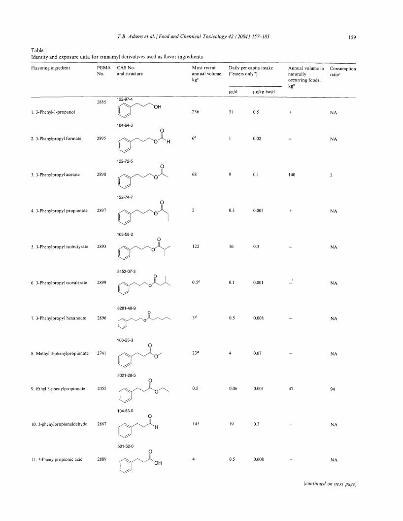


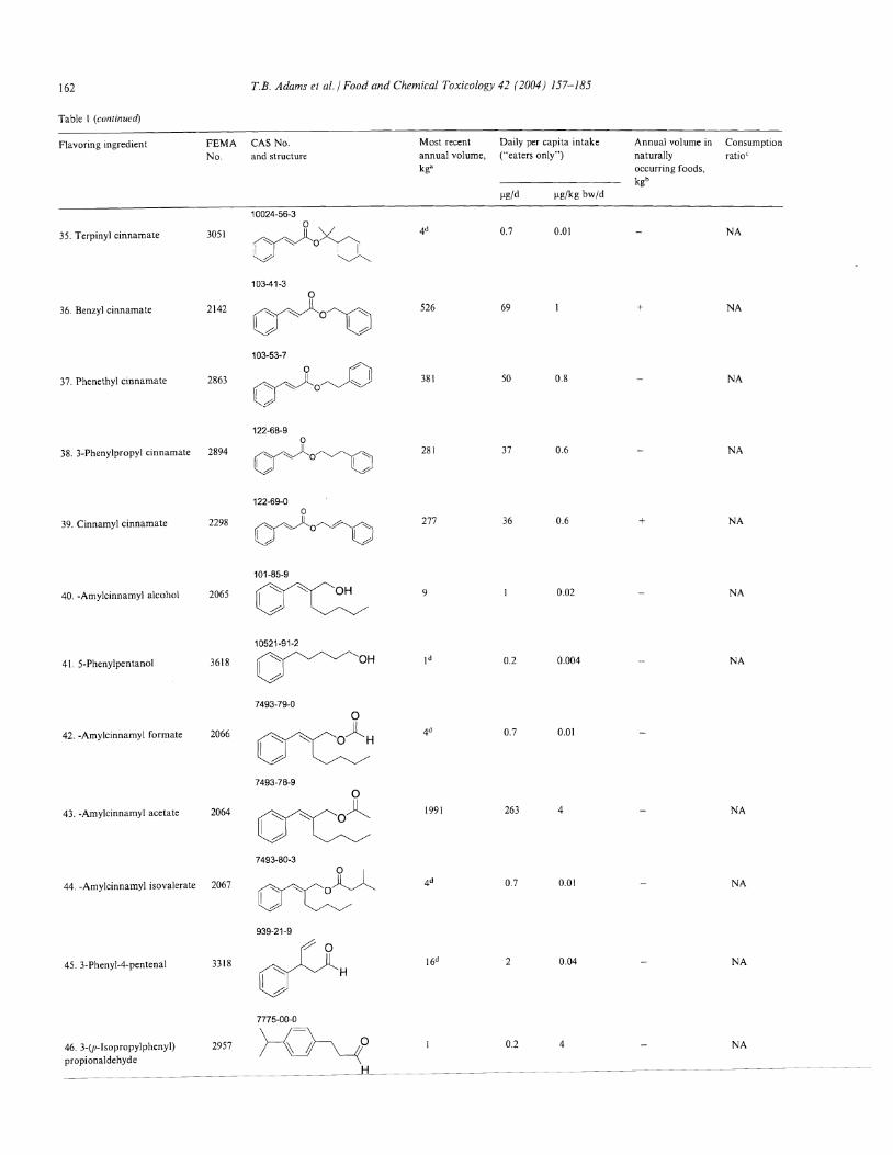

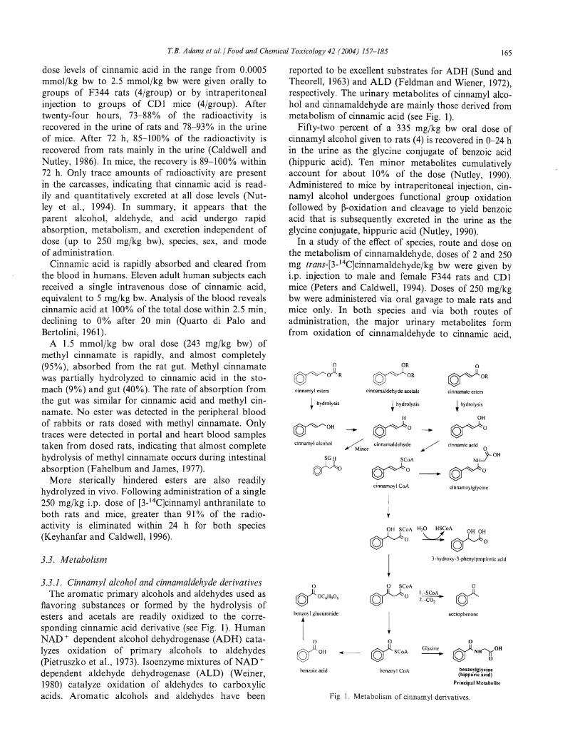


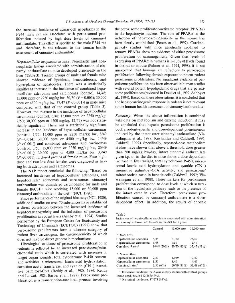


![Genome-wide analysis of GRAS transcription factor gene ... · GRAS members in G. arboreum, 82 GRAS members in G. raimondii, 33 GRAS members in A. thaliana [9], 42 GRAS members in](https://static.documents.pub/doc/80x56/5e395335b8357078586104d1/genome-wide-analysis-of-gras-transcription-factor-gene-gras-members-in-g-arboreum.jpg)


