DISEASES OF AQUATIC ORGANISMS Dis Aquat Org Published December 30 Random Amplified Polymorphic DNA analysis provides rapid differentiation among isolates of the fish pathogen Flavobacterium psychrophilum and among Flavobacterium species Chehid Chakrounl, Maria C. urdaci2, Didier ~ a u r e ~ , Francine ~ r i r n o n t ~ , Jean-Franqois Bernardetl** 'Unite de Virologie et d'Immunologie Moleculaires, Institut National de la Recherche Agronomique, F-78352 Jouy-en-Josas Cedex, France '~aboratoire de Microbiologie, Ecole Nationale des Ingenieurs des Travaux Agricoles de Bordeaux, 1 cours du General de Gaulle, BP 201, F-33175 Gradignan Cedex, France 3Unite des Enterobacteries, Unite 389 de I'Institut National de La Sante et de la Recherche Medicale, Institut Pasteur, F-75724 Paris Cedex 15. France ABSTRACT Flavobacterium psychrophilum is the agent of cold-water disease and rainbow trout fry syndrome in salmonid fish. Originally isolated in North Amenca only, the bacterium is now also respon- sible for severe mortalities in many salmonid hatcheries in Europe, as well as in Japan, Chile and Tas- mania. The random amplified polymorphic DNA (RAPD) technique was used to analyze the genetic diversity among a collection of 177 F. psychrophilum strains isolated from different fish species and in different geographical areas. Forty 10-mer primers were tested and 5 of them were selected for further analysis of the bacterial DNAs. The primers OPH 06, OPH 08, OPG 08, OPG 14, and OPG 16 generated several reproducible profiles during a preliminary screening of the whole collection of stralns. Based on these results, the polyrnerase chain reaction (PCR) products of a selection of 60 bacterial DNAs were submitted to slow agarose gel electrophoresis for numerical analysis of the DNA fingerprintings. No correlation occurred between the combined RAPD profiles of the primers and the geographical origin of the strains, while some profiles were clearly associated with the fish species from which the strains were isolated. Another primer, OPG 10, yielded a unique RAPD profile common to all F. psychrophilum strains whereas the 9 other valld Flavobactenum species, several of which coexist in freshwater envi- ronments and may also be isolated from fish, displayed other profiles. Thus, depending on the primer used, both the typing of F. psychrophilum strains for epidemiology studies as well as the identification of this fish pathogen and its differentiation from related bacterial species could be achieved by using RAPD. KEY WORDS: Flavobacterium psychrophilum . Flavobacterium spp. . Cold-water disease . Rainbow trout fry syndrome . RAPD . Epidemiology INTRODUCTION Flavobactenum psychrophilum (Bernardet et al. 1996) (syn. Cytophaga psychrophila, Flexibacter psychro- philus), the causative agent of cold-water disease and rainbow trout fry syndrome, was originally isolated from coho salmon Oncorhynchus kisutch in Washing- 'Addressee for correspondence. E-mail: [email protected]ton state, USA, in 1948 (Borg 1960),and later found in chinook salmon Oncorhynchus tshawytscha from the same area (Anderson & Conroy 1969).In recent years, cold-water disease has also caused serious problems among rainbow trout juveniles reared in the Pacific Northwest area (R. A. Holt pers, comm.). Since the mid-eighties, the same bacterium has been responsi- ble for high losses in rainbow trout Oncorhynchus mykiss fry in many European hatcheries (Weis 1987, O Inter-Research 1997 Resale of full article not permitted
Transcript
DISEASES OF AQUATIC ORGANISMS Dis Aquat Org Published December 30
Random Amplified Polymorphic DNA analysis provides rapid differentiation among isolates of
the fish pathogen Flavobacterium psychrophilum and among Flavobacterium species
Chehid Chakrounl, Maria C. urdaci2, Didier ~ a u r e ~ , Francine ~ r i r n o n t ~ , Jean-Franqois Bernardetl**
'Unite de Virologie et d'Immunologie Moleculaires, Institut National de la Recherche Agronomique, F-78352 Jouy-en-Josas Cedex, France
'~aboratoire de Microbiologie, Ecole Nationale des Ingenieurs des Travaux Agricoles de Bordeaux, 1 cours du General de Gaulle, BP 201, F-33175 Gradignan Cedex, France
3Unite des Enterobacteries, Unite 389 de I'Institut National de La Sante et de la Recherche Medicale, Institut Pasteur, F-75724 Paris Cedex 15. France
ABSTRACT Flavobacterium psychrophilum is the agent of cold-water disease and rainbow trout fry syndrome in salmonid fish. Originally isolated in North Amenca only, the bacterium is now also respon- sible for severe mortalities in many salmonid hatcheries in Europe, as well as in Japan, Chile and Tas- mania. The random amplified polymorphic DNA (RAPD) technique was used to analyze the genetic diversity among a collection of 177 F. psychrophilum strains isolated from different fish species and in different geographical areas. Forty 10-mer primers were tested and 5 of them were selected for further analysis of the bacterial DNAs. The primers OPH 06, OPH 08, OPG 08, OPG 14, and OPG 16 generated several reproducible profiles during a preliminary screening of the whole collection of stralns. Based on these results, the polyrnerase chain reaction (PCR) products of a selection of 60 bacterial DNAs were submitted to slow agarose gel electrophoresis for numerical analysis of the DNA fingerprintings. No correlation occurred between the combined RAPD profiles of the primers and the geographical origin of the strains, while some profiles were clearly associated with the fish species from which the strains were isolated. Another primer, OPG 10, yielded a unique RAPD profile common to all F. psychrophilum strains whereas the 9 other valld Flavobactenum species, several of which coexist in freshwater envi- ronments and may also be isolated from fish, displayed other profiles. Thus, depending on the primer used, both the typing of F. psychrophilum strains for epidemiology studies as well as the identification of this fish pathogen and its differentiation from related bacterial species could be achieved by using RAPD.
Flavobactenum psychrophilum (Bernardet et al. 1996) (syn. Cytophaga psychrophila, Flexibacter psychro- philus), the causative agent of cold-water disease and rainbow trout fry syndrome, was originally isolated from coho salmon Oncorhynchus kisutch in Washing-
ton state, USA, in 1948 (Borg 1960), and later found in chinook salmon Oncorhynchus tshawytscha from the same area (Anderson & Conroy 1969). In recent years, cold-water disease has also caused serious problems among rainbow trout juveniles reared in the Pacific Northwest area (R. A. Holt pers, comm.). Since the mid-eighties, the same bacterium has been responsi- ble for high losses in rainbow trout Oncorhynchus mykiss fry in many European hatcheries (Weis 1987,
O Inter-Research 1997 Resale of full article not permitted
188 Dis Aquat Org
Bernardet et al. 1988, Lorenzen et al. 1991, Austin 1992, Sarti et al. 1992, Toranzo & Barja 1993, Wiklund et al. 1994). In Europe, it was also isolated from Euro- pean eel Anguilla anguilla, carp Cyprinus carpio, tench Tinca tinca and crucian carp Carassius carassius (Lehmann et al. 1991). In Japan, the disease has become a concern in rainbow trout, coho salmon and ayu Plecoglossus altivelis farms (Wakabayashi et al. 1994). Recently this bacterium was also isolated in Chlle from rainbow trout fry (Bustos et al. 1995) and in Tasmania, Australia, from Atlantic salmon (Schmidtke & Carson 1995). An efficient typing method would allow an accurate comparison of strains retrieved from widely dfferent origins and would benefit epidemiol- ogy studies of the dsease.
Several other Flavobacterium species may be iso- lated from diseased freshwater fish (Bernardet et al. 19953. Some of thein jF~avobacl~e~~iur~~ columnare, F. branchiophilum, F. johnsoniae) have been demon- strated to be pathogenic, at least in certain circum- stances, while the pathogenicity of other species [E hydatis (syn. Cytophaga aquatilis), E succinicans] has not been established. As most Flavobacterium species are relatively fastidious and inert in many biochemical tests, the identification of F. psychrophilum is time- consuming and confusion with other Flavobacterium species may occur. A rapid and specific identification method would greatly improve the diagnosis of the conditions caused by F. psychrophilum.
In this study, we demonstrate that both a rapid and clear differentiation of Flavobacterium psychrophilum from related bacterial species, as well as an efficient intra-specific typing of E psychrophilum strains, can be achieved by using Random Amplified Polymorphic DNA (RAPD) (Williams et al. 1990), depending on the primer used.
MATERIALS AND METHODS
Bacterial strains and growth conditions. A total of 177 Flavobacterium psychrophilum strains isolated over the years in different geographical areas (59 strains from France, 6 from Spain, 4 from Sweden, 22 from Denmark, 9 from Germany, 19 from Switzerland, 6 from the United Kingdom, 1 from Norway, 1 from Finland, 28 from the USA, 1 from Canada, 6 from Chile, 10 from Japan and 5 from Tasmania) were included in this study. This collection of strains was screened (see details below) and 60 of them, listed in Table 1, were included in the final study. All F. psy- chrophilum strains were grown at 18'C on Anacker and Ordal's agar (tryptone, 0.05 %; yeast extract, 0.05 %; beef extract, 0.02 %; and sodium acetate, 0.02 %; pH 7.2 to 7.4) (Anacker & Ordal 1955) supplemented to 0.5%
tryptone. Representative strains of the 9 other valid Flavobactenum species were included in this study and are listed in Table 2. They were grown on the same medium at 22OC.
Preparation of DNA. Each strain was collected on the agar plate, suspended in 0.9% saline and cen- trifuged. The pellet was resuspended in an equal vol- ume (200 pl) of TES (50 mM Tris buffer, 1 mM EDTA, 8.56% saccharose) pH 8.00 containing 20 1-19 of lyso- zyme (Sochal, Levallois-Perret, France), and incubated at 37°C for 1 h. Fifty p1 of 20% sodium dodecyl sul- phate (SDS) was added to the mixture. The solution was treated 3 times with a mixture of phenol, chloro- form and isoamyl alcohol (25/24/1; v/v/v) and once with chloroform-isoamyl alcohol (24/1; .v/v). The DNA was precipitated by an equal volume of isopropanol, dried under vacuum, and dissolved in 200 p1 of stenle 1viij.h-Q water containing 10 pg of RNase (ribonuclease I-A from bovine pancreas, Sigma Chemical Co., St. Louis, MO, USA) and incubated at 37OC for 15 min. All DNA samples were stored at -20°C.
Amplification conditions. Two kits of 20 arbitrary 10- mer primers each (OPG and OPH, Bioprobe, Montrevil- sous-bois, France) were evaluated for random amplifi- cation of DNAs. The technique used in this study was adapted from Meunier & Grirnont (1993). Briefly, 25 p1 of reaction mixture containing 25 to 50 ng of genomic DNA, 5 pm01 of primer, 50 pM of each dNTP (Appli- gene, Illkirch, France), 0.6 U of Taq DNA polyrnerase (reference 120187C, Appligene) and 2.5 p1 of 10 X poly- merase chain reaction (PCR) reaction buffer was over- laid with mineral oil. The amplification reaction was performed in a DNA thermal cycler (Own E Hybaid, Teddington, England) programmed for 45 cycles of 30 s at 94°C (denaturing of DNA), 1 min at 36'C (primer an- nealing) and 1 min at 72'C (polymerization), with 5 min initial denaturation at 95°C and 10 min final extension at 72°C. In order to test the reproducibility of the tech- nique, DNA amplification experiments were repeated 3 times under the same conditions. A negative control, containing no template DNA, was included in each ex- periment in order to detect any contaminating DNA.
Electrophoresis conditions. Gel electrophoresis was carried out by loading 25 p1 samples of all 177 amplifi- cation products in submerged horizontal 1 % agarose gel (Bioprobe). Gels were run for 1 h at 10 V cm-' in Tris-borate buffer (89 mM Tris-base, 89 mM boric acid, 2 mM EDTA) without cooling. Amplification products were visualized after ethidium bromide staining and photographed on a UV light transilluminator
The amplification products of 60 selected strains (see details below) generated by the primers OPH 06 and OPH 08 were also compared using special conditions of electrophoresis (3 V cm-' for 8 h) , improving the resolution of the patterns for computer analysis.
Chakroun e t al.. RAPD analysis of Flavobacteriurn psychrophilum 189
Table 1. Flavobacterium psychrophjlum. Strains included in the computer analysis of their RAPD fingerprints generated by the primers OPH 06 and OPH 08
Strain Origin l Strain Origin I
SVA 31-88 SVA 120-89 SVS 91 1209-2
JIP 02/86 Rainbow trout Oncorhynchus mykiss, OSU SRCo5-90 Coho salmon, kidney. Oregon, USA, 1990 kidney, Picard~e, France, 1986
OSU PCPl.1 JlP P02/88 Rainbow trout, kidney, Picardie. Coho salmon, kldney, Oregon, USA, 1989
OSU RbS6-82 Steelhead trout Oncorhynchus mykiss, kidney. Oregon. USA, 1982
NCIMB 194fT Coho salmon Oncorhynchus kisutch, kidney, Washington, USA
NClMB 2282 Coho salmon, kidney, Washington, USA, 1948
OSU SH3-81 Coho salmon, kidney. Oregon, USA. 1981 OSU THC02-90 Coho salmon, kidney, Oregon, USA, 1990 OSU THC04-90 Coho salmon. kidney, Oregon, USA. 1990
Strain sources:
OSU Ch8-80 OSU TM3.1 FPC 828 FPC 829 FPC 830 FPC 831
1 DPIF 91/4043-4
I DPIF 91N043-8
1 DPIF 91N043-13
1 DPIF 9114043-14
1 DPIF 9114043-17
OSU AD10.1.1
OSU CCC6-86
FPC 839
FPC 837 1 FPC 838 FPC 840 LVDI 511
LFNW 16/90
LFNW 25/90
LVDJ XP189
LVDJ D 2172 LFNW 131/89
JIP: Culture Collection of Unite de Viral- DIFR strains provided by I. Dalsgaard, ogle et ~ ~ ~ ~ ~ l ~ ~ i ~ ~ ~ l e ~ ~ l ~ i ~ ~ ~ , lnsti- Danlsh Institute for Fisheries Research, tut National de la Recherche Aaronom- Fredenksberg, Denmark ., ique, Jouy-en-Josas, France LVDL: strains provided by P. Nougayrede, Laboratoire Veterinaire Departemental des Landes, Mont de Marsan, France ISTAB: Culture Collection of Institut Superieur de Technologie Alimentaire et Blotechnologie, Bordeaux, France
SVA: strain provided by A. Hellstrom, Statens Veterinarmedicinska Ansalt, Upp- sala, Sweden
SVS: strains provided by E. Lorenzen, Statens Veterinaere Serurnlaboratorium, Arhus, Denmark
LFNW: strains provided by D. Mock, Lan- desanstalt fiir Fischerei Nordrhein-West- falen, Kirchhundem-Albaum, Germany
WP: strains provided by T. Wahli. Institute of Veterinary Pathology, Berne, Switzer- land
OSU: strains provided by M. Whipple and R. A. Holt, Oregon State University, Cor- vallis, USA
UCD: strains provided by R. Hedrick. Uni- versity of California, Davis. USA
FPC: stralns provided by H. Wakabayashi, Laboratory of Aquaculture Biology, Uni- versity of Tokyo, Japan
NCIMB: Nat~onal Collection of Industrial and Marine Bacteria, Aberdeen, United Kingdom
DPIF: strains provided by J. Carson. De- partment of Primary Industry and Fish- eries, Kings Meadows, Tasmania. Aus- tralia
LVDI: strain provided by J.-L. Bind, Laboratoire Vkterinaire Departemental d'lndre-et-Loire, Tours, France
LVDJ: strains provided by M. Morand, Laboratoire Veterinaire Departemental du Jura. Lons-le-Saunier, France
190 Dis Aquat Org 31 187-196, 1997
Table 2. Flavobacteri~lm strains and species included in the random amplification using the primer OPG 10
Species Strain Origin
F. aquatile F. branchiophilum F. branchiophilum F. columnare F. columnare F. columnare F. flevense E hydatis F. johnsoniae F. johnsoniae l? pectinovorum E psychrophilum F. psychrophilum F. psychrophilum F. psychrophilum E psychrophilum -F psychrophzum E saccharophilum F. succinicans F. succinicans
Deep well, Kent, England Diseased gills of yamame Oncorhynchus masou, Gumma, Japan Diseased gills of sheatfish Silurus glanis, Hungary Skin lesion of black bullhead Ictalurus melas, Ile-de-France, France Gill lesions of Japanese eel Anguilla japonica, Shizuoka, Japan Gill lesions of chlnook salmon Oncorhynchus tshacvytscha, Oregon, USA Lake Ijssel, The Netherlands Gills of diseased salmon, Michigan, USA Soil or mud, Rothamsted or Cambridge, England Diseased freshwater fish, Manitoba, Canada Soil, England Kidney of diseased coho salmon Oncorhynchus kisutch. Washington, USA Kidney of rainbow trout Oncorhynchus mykiss, Picardie. France Kidney of tench Tinca tinca, Franche-Comte, France Cutthroat trout Salmo clarki, Oregon, USA Kidney of ayu Plecoglossus altivelis, Tokushima, Japan K ' A - ,,L,?); of s:ee:head :roii: Oncorhynchus 11iyiti33, Oreyun, VSA
h v e r Wey, Surrey, England Eroded fin of chinook salmon, Washington, USA Lesion of chinook salmon, Snake River, Idaho, USA
Strain sources: LMG: Culture Collection of the Laborato- JIP: Culture Collection 01 the Unite de NCIMB National Collection of Industrial riurn voor Microbiologie. Univers~ty of Virologie et Immunologie Moleculaires, and Manne Bacteria. Aberdeen, United Ghent, Ghent, Bela~urn Institut National de la Recherche Kinadorn - ATCC: American Type Culture Couec- Agronomique, Jouy-en-Josas, France tion, Rockville, Maryland 0s": strains provided by M, LVDJ: strain provided by M. Morand,
Laboratoire Veterinaire Departemental FPC EK28, FPC 839 and FL-15: strains pro- and R. A. HO1t, Oregon State University, dU jura, ~ ~ ~ ~ . l ~ - ~ ~ ~ ~ i ~ ~ , F~~~~~ vided bv H. Wakabavashi. Laboratorv of Corvallis, ~quacu i tu re ~ i o l o ~ y , b e ~ a r t m e n t of ~ k h - DSM: Deutsche Sarnmlung von Mkroor- eries, University of Tokyo, Tokyo, Japan ganismen, Braunschweig. Germany
Computer-assisted analysis of the DNA fingerprints. Each gel, consisting of 10 lanes of amplification prod- ucts and 3 lanes of molecular weight marker (123 base pairs, bp; Gibco BRL, USA), was photographed using a Polaroid film type 665 (Polaroid, Cambridge, MA, USA). The length of lanes was about 6 cm. The banding patterns were scanned with a laser densitometer (One- Scanner, Apple Computers, Cupertino, CA, USA) and analyzed by using various programs of the TaxotronB package (Taxolab, Institut Pasteur, Paris, France). The TIFF (tagged image file format) image was searched for lanes and bands using RestrictoScan yielding migration data. Fragments sizes were interpolated from migration data using RestrictoTyper implementing the algorithm of Schaffer & Sederoff (1981). RestrictoTyper compared pairs of patterns and calculated a distance coefficient which was the complement of the Dice index. We chose to set a fixed value of 4 %, indicating that 2 fragments were considered identical if their sizes did not differ by more than 4 %. A distance matrix was generated for the patterns obtained with each of the primers OPH 06 and OPH 08. These distance matrices were averaged using the program Adanson and treated by the Unweighted Pair Group Method of Averages (UPGMA) (Sneath &
Sokal 1973). The program generated a tree description
file which was used by the program Dendrograf to draw a dendrogram and produce an order file. The order file was used by RestrictoTyper to reorder the fragment sizes files and produce a schematic representation of the patterns. The dendrogram and the schematic graph were assembled in a single picture by using a drawing program.
RESULTS
An initial screening was carried out by testing the 40 primers on 7 representative Flavobacterium psy- chrophilum isolates. Twenty-eight primers produced clear and reproducible banding patterns, 11 of them yielding polymorphic fragments. Among these, we arbitrarily selected 6 primers for further analysis of isolates: 5 of them (OPH 06, OPH 08, OPG 08, OPG 14, OPG 16) generated different patterns among the 177 strains while the last one (OPG 10) produced an identi- cal pattern for the 35 strains tested. Among the 177 strains in our collection, 60 strains representing the variety in geographical origin and fish host were then selected for high-resolution electrophoresis of their amplification products and computer-assisted analysis
Chakroun et al.: KAPD analysis of Flavobactenum psychrophilum 191
of their DNA fingerprints. Of these, 36 represented all strains originating from fish species other than rainbow trout which were included in the whole collection. The remaining 24 strains were part of the 141 rainbow trout isolates, and originated from Europe, the USA, and Japan (17 strains, 6 strains, and 1 strain, respectively). RAPD profiles were highly reproducible when analy- ses were repeated, except for some variations in the intensity of a few faint PCR products.
Differentiation among Flavobacterium psychrophilum strains
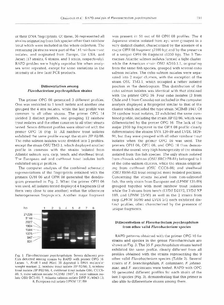
The primer OPG 08 generated 3 different profiles. One was restricted to 1 tench isolate and another one grouped the 4 ayu isolates, while the last profile was common to all other strains. The primer OPG 14 yielded 2 distinct profiles, one grouping 12 rainbow trout isolates and the other common to all other strains tested. Seven different profiles were obtained with the primer OPG 16 (Fig. 1). All rainbow trout isolates exhibited the same profile except the strain JIP 02/86. The coho salmon isolates were divided into 2 profiles, except the strain OSU TM3.1, which displayed another profile in common with the strains isolated from Atlantic salmon, ayu, carp, tench, and steelhead trout. The European eel and cutthroat trout isolates both exhibited unique profiles.
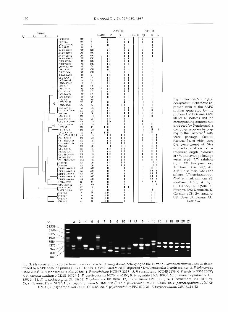
The computer analysis of the combined schematic representations of the fingerprints obtained with the primers OPH 06 and OPH 08 generated the dendro- gram presented in Fig. 2. When the primer OPH 06 was used, all isolates tested displayed 4 fragments (3 of them very close to one another) within the otherwise heterogeneous fingerprints. Another major fragment
Fig. 1 Flavobacterium psychrophilum. Seven different pro- files detected among strains by RAPD with primer OPG 16. Lanes: 1, EcoR I and Hind I11 digested h DNA molecular weight marker; 2, rainbow trout isolate JIP 02/86; 3, rainbow trout isolate JIP P02/88; 4, cutthroat trout isolate OSU CCCG- 86, 5, coho salmon isolate NCIMB 1947T; 6, coho salmon iso- late OSU BC3-81; 7, Atlantic salmon isolate DPIF 91/4043-13;
8, European eel isolate LFNW 131/89
was present in 51 out of 60 OPH 08 profiles. The 4 Japanese strains isolated from ayu were grouped in a very distinct cluster, characterized by the absence of a major OPH 08 fragment (2100 bp) and by the presence of a unique OPH 06 fragment (1350 bp). The 5 Tas- manian Atlantic salmon isolates formed a tight cluster, while the American strain OSU AD1O.l.l, originating from the same fish species, grouped with several coho salmon isolates. The coho salmon isolates were sepa- rated into 2 major clusters, with the exception of the strain OSU TM3.1, which occupied a rather isolated position in the dendrogram. This distribution of the coho salmon isolates was identical with that obtained with the primer OPG 16. Four coho isolates (3 from Chile and l from Canada) not included in the computer analysis displayed a fingerprint similar to that of the cluster which included the type strain NCIMB 1947. Of 24 rainbow trout isolates, 22 exhibited the same com- bined profile, including the strain JIP 02/86, which was differentiated by the primer OPG 16. The lack of the major 2100 bp fragment in the OPH 08 profile clearly differentiated the strains SVA 120-89 and LVDL 1829- 91, but they were grouped with all other rainbow trout isolates when the primer OPG 16 was used. The primers OPH 06, OPH 08, and OPG 16 thus demon- strated the overall very high homogeneity of the strains isolated from this fish species. The only strain isolated from chinook salmon (OSU SRChF8-81) belonged to 1 of the coho salmon clusters, while the strains originat- ing from cutthroat (OSU CCC6-86) and steelhead (OSU RbS6-82) trout occupied more isolated positions. Concerning the strains isolated from non-salmonid fish, the only strain from European eel (LFNW 131/89) grouped together with most rainbow trout isolates while the 3 strains from tench (LVDJ D2172, LVDJ XP 189, and LFNW 25/90) as well as the 2 strains from carp (LFNW 16/90 and LVDI 5/I) each exhibited dis- tinct profiles, often characterized by the presence of unique fragments.
Differentiation of Flavobacterium psychrophilurn from other valid Flavobacterium species
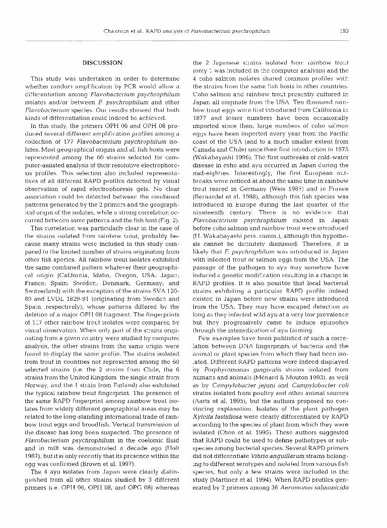
RAPD patterns obtained with the primer OPG 10 for strains and species in the genus Flavobacterium are shown in Fig. 3. The 35 F. psychrophilum strains tested exhibited the same profile, clearly different from all profiles obtained with the strains representing the 9 other valid Flavobacterium species (Table 2). Several strains of E branchiophilurn, F. columnare, F. johnso- niae, and E succinicans were tested. RAPD with OPG 10 generated different profiles for each strain of the last 3 species (Fig. 3), demonstrating that this primer is also able to differentiate strains among them.
Dlstanct 0 6 0 4 0 2 0
JP PO288 RT JP 02/86 RT LVDL 3077-91 RT SVA31-88 RT SVS 91 1209-2 RT SVS 91 1209-1 RT SVS 910614-3 RT SVS911009-3 RT DIFR 880406 R T DlFR 900406 RT LFNW 123-89 RT IVP CH7193 RT IVP CH7i94 RT ISTM AU.1 RT O S U A N A 9 1 I RT UCD 1334-95 RT LFNW 131189 El2 UCD 95-7 RT NPCHIN3 RT OSU 90-6 6A RT UCD 14(-95 RT UCD R3IW95 RT FPC X13 RT LVDJ D21 72 TE LMW I 6/90 CA
1 0 S l l A D l 0 l I AS 44 OSU NlICO2-90
LVDl 5n
LVDJ XI' I89 OSLl C026.BR.2 OSU PCPl I osu nicw-90 osu n 1~02.90 FPC 8.30 OS11 S113-RI NCIMB 1947 OSU SRCo5-90 NClMB 2282 OSU SRChFl-RI
CS CS CS CS CS CT CA CS l€
. I CS CS CS CS CS CS CS CS CS ChS
RT LVDL 1829.91 RT FPC 838 A WC R37 A WC 839 A WC 840 A
Dis Aquat Org 31. 187-196, 1997
OPH OS
F F E S DK DK DK DK DK DK D CH CH E us us D us CH us us us JP F D us US m lJS US US us F us F us us US US JP 11s 11s US us us m m AU AU AU AU AU D 11s S E m m m m
Fig. 2. Flavobacteriumpsy- chrophilum. Schematic re- presentation of the RAPD profiles generated by the priners OF: I 06 and OPH 08 for 60 ~solates and the corresponding dendrogram produced by Dendrograf, a computer program belong- ing to the ~axot ron* soft- ware package (Institut Pasteur, Paris) which uses the complement of Dice similarity coefficients. A fragment length tolerance of 4 % and average linkage were used. RT: rainbow trout; EE: European eel; TE: tench; CA: carp; AS: Atlantic salmon; CS: coho salmon; CT: cutthroat trout; ChS: chinook salmon; ST: steelhead trout; A: ayu. F: France; E: Spain; S: Sweden; DK: Denmark; D: Germany; CH: Switzerland; US: USA; JP: Japan; AU:
Australia
OPH 06
bpxlOO 10 5
I 11 I I 111 I I Ill I I 111 I I I II I I I II I I I II I I I I1 I I 111 I I 111 I I Ill I I I I I I I I II I I I II I I I II I I I II I I 111 I I 111 I I I II I I I II I I Ill I I Ill I I Ill I I Ill I Ill I I I II I I I II I I I II I I I II I 1 I I I1 I I Ill I I Ill I
1 111 I Ill I
1 111 I Ill I I Ill I I Ill I I Ill I I II I I I I II I I I II I I I II I I Ill I I I II I I I II I I I I 1 I I Ill I Ill I Ill I Ill I Ill I Ill I Ill I I II I I Ill I I
1111 I 1111 I 1111 I 1111 I
Fig. 3. Flavobactedum spp. Different profiles detected among strains belonging to the 10 valid Flavobacterium species as deter- mined by RAPD with the primer OPG 10. Lanes: 1, EcoR I and Hind I11 digested ), DNA molecular weight marker; 2, F. johnsoniae DSM 2064T; 3, F. johnsoniae ATCC 29585; 4, F. succinicans NCIMB 2277T; 5, F. succinicans NCIMB 2278; 6, F. hydatis DSM 2063T; 7, F saccharophilum NCIMB 2072T; 8, F. pectinovorum NCIMB 9059~; 9, F. aquatile LMG 4008T; 10, E branchiophilum ATCC 35035~; 11, E branchioptulum FL-15; 12, F. columnare JIP 39/87; 13, F. columnare FPC EK28; 14, F. columnare OSU DD3-69; 15, E flevense DSM 1076T; 16, F. psychrophilum NCIMB 1947~; 17. F. psychrophilum JIP P02/88; 18, F. psychrophilum LVDJ XP
189; 19, F. psychrophilum OSU CCC6-86; 20, F. psychrophilum FPC 839; 21. F. psychrophilum OSU RbS6-82
Chakroun et al . RAPD analysis of Flavobactedum psychrophjlum 193
DISCUSSION
This study was undertaken in order to determine whether random amplification by PCR would allow a differentiation among Flavobacterium psychrophilum isolates and/or between F, psychrophilum and other Flavobacterium species. Our results showed that both kinds of differentiation could indeed be achieved.
In this study, the primers OPH 06 and OPH 08 pro- duced several different amplification profiles among a collection of 177 Flavobacterium psychrophilum iso- lates. Most geographical origins and all fish hosts were represented among the 60 strains selected for com- puter-assisted analysis of their resolutive electrophore- sis profiles. This selection also included representa- tives of all different RAPD profiles detected by visual observation of rapid electrophoresis gels. No clear association could be detected between the combined patterns generated by the 2 primers and the geograph- ical origin of the isolates, while a strong correlation oc- curred between some patterns and the fish host (Fig. 2).
This correlation was particularly clear in the case of the strains isolated from rainbow trout, probably be- cause many strains were included in this study com- pared to the limited number of strains originating from other fish species. All rainbow trout isolates exhibited the same combined pattern whatever their geographi- cal origin (California, Idaho, Oregon, USA; Japan; France; Spain; Sweden; Denmark; Germany; and Switzerland) with the exception of the strains SVA 120- 89 and LVDL 1829-91 (originating from Sweden and Spain, respectively), whose patterns differed by the deletion of a major OPH 08 fragment. The fingerprints of 117 other rainbow trout isolates were compared by visual observation. When only part of the strains origi- nating from a given country were studied by computer analysis, the other strains from the same origin were found to display the same profile. The strains isolated from trout in countries not represented among the 60 selected strains (i.e. the 2 strains from Chile, the 6 strains from the United Kingdom, the single strain from Norway, and the 1 strain from Finland) also exhibited the typical rainbow trout fingerprint. The presence of the same RAPD fingerprint among rainbow trout iso- lates from widely different geographical areas may be related to the long-standing international trade of rain- bow trout eggs and broodfish. Vertical transmission of the disease has long been suspected. The presence of Flavobactenum psychrophilum in the coelomic fluid and in milt was demonstrated a decade ago (Holt 1987), but it is only recently that its presence within the egg was confirmed (Brown et al. 1997).
The 4 ayu isolates from Japan were clearly distin- guished from all other strains studied by 3 different primers (i.e. OPH 06, OPH 08, and OPG 08) whereas
the 2 Japanese strains isolated from rainbow trout (only 1 was included in the computer analysis) and the 4 coho salmon isolates shared common profiles with the strains from the same fish hosts in other countries. Coho salmon and rainbow trout presently cultured in Japan all originate from the USA. Ten thousand rain- bow trout eggs were first introduced from California in 1877 and lesser numbers have been occasionally imported since then; large numbers of coho salmon eggs have been imported every year from the Pacific coast of the USA (and to a much smaller extent from Canada and Chile) since their first introduction in 1975 (Wakabayashi 1996). The first outbreaks of cold-water disease in coho and ayu occurred in Japan during the mid-eighties. Interestingly, the first European out- breaks were noticed at about the same time in rainbow trout reared in Germany (Weis 1987) and in France (Bernardet et al. 1988), although this fish species was introduced in Europe during the last quarter of the nineteenth century. There is no evidence that Flavobacterium psychrophilum existed in Japan before coho salmon and rainbow trout were introduced (H. Wakabayashi pers. comm.), although this hypothe- sis cannot be definitely dismissed. Therefore, it is likely that F. psychrophilum was introduced in Japan with infected trout or salmon eggs from the USA. The passage of the pathogen to ayu may somehow have induced a genetic modification resulting in a change in RAPD profiles. It is also possible that local bacterial strains exhibiting a particular RAPD profile indeed existed in Japan before new strains were introduced from the USA. They may have escaped detection as long as they infected wild ayu at a very low prevalence but they progressively came to induce epizootics through the intensification of ayu farming.
Few examples have been published of such a corre- lation between DNA fingerprints of bacteria and the animal or plant species from which they had been iso- lated. Different RAPD patterns were indeed displayed by Porphyromonas gingivalis strains isolated from humans and animals (Menard & Mouton 1993), as well as by Campylobacter jejuni and Campylobacter coli strains isolated from poultry and other animal sources (Aarts et al. 1995), but the authors proposed no con- vincing explanation. Isolates of the plant pathogen Xylella fastidiosa were clearly differentiated by RAPD according to the species of plant from which they were isolated (Chen et al. 1995). These authors suggested that RAPD could be used to define pathotypes or sub- species among bacterial species. Several RAPD primers did not differentiate Vibrio anguillarum strains belong- ing to different serotypes and isolated from various fish species, but only a few strains were included in the study (Martinez et al. 1994). When RAPD profiles gen- erated by 2 primers among 36 Aeromonas salmonicida
194 Dis Aquat Org
isolates were compared, 'no geographic, mutual or fish species associated types were found' (Hdnninen et al. 1995).
Different typing methods other than RAPD have been proposed to compare Flavobacterium psychro- philum strains among themselves: serologic analysis (Lorenzen 1994, Wakabayashi et al. 1994); genetic analysis based on plasmid profile (Holt 1987, Lorenzen 1994) or rRNA gene restriction pattern (ribotype) (Cipriano et al. 1996); and electrophoretic pattern of proteases (Bertolini et al. 1994). Although no associa- tion could be found between fish host and either ribo- type or plasmid profile, some correlation between fish host and serotype did appear in the results published by Wakabayashi et al. (1994). Two serotypes were revealed by the absorption analysis with heat-stable antigens. The serotype 0 - 1 was common to all Ameri- can and Japanese coho isolates, whereas the ayu iso- lates as well as the strains isolated from rainbow trout in Japan belonged to the serotype 0 - 2 (Wakabayashi et al. 1994). These data parallel the results of our study and support the hypothesis of the American origin of the strains isolated from coho salmon in Japan. How- ever, the question of the origin of the serotype and RAPD profiles specific to the strains isolated from ayu remains unanswered.
Comparison of our data with the protease patterns is also interesting since 20 Flavobacterium psychro- philum isolates among the 29 studied by Bertolini et al. (1994) were also included in our study. According to these authors, the 29 isolates were divided into 4 groups based on the presence or absence of certain proteases visualized by substrate sodium dodecyl sul- phate polyacrylamide gel electrophoresis (SDS-PAGE). A correlation was found between protease groups and fish hosts, as all 11 isolates In protease group 1 origi- nated from coho salmon whereas all 12 isolates from other fish species belonged to protease groups 2, 3 and 4. The authors tested the virulence of representatives of each protease group in juvenile coho salmon. Some association was noticed between protease group and virulence, as most group 1 strains induced a high mor- tality and isolates belonging to other protease groups tended to be less pathogenic. In our study, all strains in Bertolini's protease group 1 belonged to the cluster which included the type strain whereas most strains in protease group 2 belonged to the other coho salmon cluster. Therefore, a correlation seems to exist be- tween fish host, proteolytic activity, RAPD profile, and virulence, suggesting that several pathotypes may exist among F. psychrophilum isolates. However, the high virulence of protease group 1 strains was also demonstrated by Bertolini et al. (1994) on steelhead trout and 6.5 to 12% mortality also occurred when 3 groups of 25 steelhead trout were each injected sub-
cutaneously with a different ayu isolate. Similarly, Holt (1987) was able to induce severe mortality in yearling coho salmon after subcutaneous injection of F. psy- chrophilum strains isolated from coho, chinook and chum salmons, but no mortality occurred when coho salmon were injected with isolates from rainbow, brook, cutthroat and steelhead trout. In the same study some isolates from coho salmon also failed to induce any mortality. These results demonstrated that the virulence of F. psychrophilum isolates is not restricted to the species or to the genus of fish from which the strains were isolated. Actually, the correlation noticed by Bertolini et al. (1994) was not absolute, as 5 highly virulent coho salmon isolates belonged to the protease group 2. The virulcncc of ,F psychrophilum may thus involve other mechanisms in addition to proteolytic activity, as already suggested by these authors (Bertolini et ai. 1994).
The only European eel isolate included in our study (LFNW 131/89) fell within the cluster grouping most rainbow trout isolates, among others the strain LFNW 123/89 isolated from the same river in Germany and during the same outbreak (Lehmann et al. 1991), sug- gesting that the eel was infected by a strain originating from a rainbow trout. However, the primer OPG 16 was able to differentiate the eel isolate from all trout iso- lates (Fig. 1). Furthermore, the strains LFNW 16/90 and LFNW 25/90, also isolated during the same out- break from a carp and a tench, respectively, each dis- played profiles different from the typical rainbow trout pattern. Hence, Flavobacterium psychrophilum strains exhibiting different RAPD profiles may coexist in the course of an epizootic, but more strains should be studied in order to verify the apparent correlation with fish host noticed during the German outbreak.
A rather similar situation was found among the strains isolated in Oregon and neighbouring states. Various RAPD profiles were found among Flavobac- terium psychrophilum strains retrieved from hatch- eries belonging to 2 main groups (i.e. the lower Colum- bia River hatcheries and the coastal hatcheries). Over the years, many transfers of fish occurred between the hatcheries within each group, as well as much less fre- quent transfers between the 2 groups. Moreover, most hatcheries raise a variety of fish species such as coho and chinook salmon as well as rainbow and steelhead trout, and straying of adult fish is known to occur ( R . A . Holt pers. comm.). The 2 strains isolated from chinook (OSU SRChF8-81) and Atlantic (OSU ADIO.l.l) sal- mon were grouped together with the coho isolates and may thus originate from coho salmon reared in the same region. Indeed, the chinook isolate was retrieved from the same fish farm at which the strain OSU SRCo5-90 was isolated from coho and both strains dis- played identical profiles. However, the 2 strains from
Chakroun et al.: RAPD analysis of Flavobacterium psychrophjlum 195
steelhead (OSU RbS6-82) and cutthroat (OSU CCC6- 86) trout, also from Oregon, exhibited very distinct profiles. Thus, different RAPD profiles have been shown to occur within a region where natural and arti- ficial transfers of fish are very frequent, and it is not clear why coho salmon isolates are divided into 2 clusters.
No conclusion could be drawn from the very homo- geneous profile displayed by the 5 Tasmanian Atlantic salmon isolates, since they were all retrieved at the same time from the same fish farm and the only other Atlantic salmon isolate included in the study grouped together with coho isolates. Moreover, no strain iso- lated from other fish species in Tasmania was avail- able.
Many Gram-negative yellow pigmented bacteria, several Flavobactenum species among them, coexist in freshwater environments and may occasionally be iso- lated from sick or healthy fish. Because most of these bacteria have a fastidious growth and are inert in many biochemical tests, contamination and erroneous identi- fication may occur. Our study demonstrated that a rapid and accurate identification of F. psychrophilum as well a s its differentiation from related species could be achieved through RAPD using the primer OPG 10 because species-specific products were amplified. Identification of F. psychrophilum with this technique is even possible using a single colony collected from a mixed culture on an agar plate (data not shown). Several bacterial species have also been differentiated from closely related species using particular RAPD primers. Examples among fish-pathogenic bacteria include primers generating fingerprints which clearly differentiated Vibrio salmonicida, V. anguillarum, and V. fisheri (Martinez et al. 1994) as well as different Aeromonas species isolated from fish (Miyata et al. 1995) or from widely different clinical and environ- mental sources (Oakey et al. 1996).
Because very few template DNA and no previous knowledge of nucleotide sequence are required, and because the whole genome is analyzed (Oakey et al. 1996), RAPD has several advantages over the other techniques that have been used alone or in various combinations for identifying Flavobactenum psychro- philun?, such as phenotypical investigations (Holt 1987), DNA/DNA hybridization (Bernardet & Grirnont 1989), serological tests revealing specific antigens (Pacha 1968, Holt 1987, Cipriano et al. 1996), whole- cell protein (Bernardet et al. 1996, Cipriano et al. 1996) or fatty acid analyses (Bernardet et al. 1996), and 16s rDNA-targeted PCR (Toyama e t al. 1994). DNA/DNA hybridization is the reference technique for determin- ing whether 2 bacterial strains belong to the same species (Wayne et al. 1987), but its use is restricted to the laboratories involved in taxonomical and phyloge-
netic studies. Phenotypic, biochemical, and serological methods of identification are usually cumbersome and time-consuming because F. psychrophilum is rela- tively fastidious and because antlsera or bacterial ex- tracts have to be prepared.
This study provides a powerful method for an effi- cient typing of Flavobacterium psychrophilum strains for epidemiological tracing. It also allows a reliable dif- ferentiation of F. psychrophilum from phylogenetically related bacterial species coexisting in the same envi- ronment. Until now, ribotyping was considered the only molecular typing method able to provide both identification and typing potentials (Brosch et al. 1996); it is now clear that RAPD profiles also carry both kinds of information.
Acknowledgements. The authors are indebted to the indi- viduals who kindly provided bacterial strains. In addition to those already cited in the footnotes of Tables 1 & 2, we thank M. vigneulle (Laboratoire de Pathologie des Anirnaux Aqua- tiques, Centre National d'Etudes Veterinaires et Alimen- taires, Plouzane, France), R . Le Goas (Laboratoire Veteri- naire Departemental de Seine Mar~time, Rouen, France), M. Schaumburg (Veterinaruntersuchungsamt Mittelhessen, Giessen, Germany), D. Bruno (Marlne Laboratory, Aberdeen. Scotland. UK), A. Toranzo and J. Barja (Departamento de Microbiologia y Parasitologia, Facultad de Biologia, Universi- dad de Santiago de Compostela, Spain), S. Haie (Central Veterinary Laboratory, Oslo, Norway), R. Ennques (Unidad de Ictiopatologia, Universidad Austral de Chile, Valdivia, Chile), J. Montana (Servicio de Ictiopatologia, Fundacion Chile, Puerto Montt, Chile), and V. Hirvela-Koski (National Veterinary and Food Research Institute, Oulu Regional Laboratory, Oulu, Finland). We are grateful to P. Tailliez (Recherches Laitieres, lnstitut National de la Recherche Agro- nomique, Jouy-en-Josas, France) as well as to E. Chaslus- Dancla, S. Leroy-Setrin, and M.-C Lesage-Descauses (Labo- ratoire d'Ecopathologie Microbienne, Institut National de la Recherche Agronomique. Nouzilly. France) for kind advice about the use of RAPD. C. Bizet and C. Barreau (Collection de Bacteries de l'lnstitut Pasteur, Paris. France) are acknowl- edged for kind help with the data analysis. Our manuscript benefited from detailed and helpful information about the local epidemiology of cold-water disease provided by H Wakabayashi (Laboratory of Aquaculture Biology, University of Tokyo. Japan) and by R . A. Holt (Department of Microbiol- ogy, Oregon State University, Corvallis, Oregon). C.C. thanks the French Ministery of Foreign Affairs for a research grant.
LITERATURE CITED
Aarts HJM, Van Lith LAJT, Jacobs-Reitsma WF (1995) Dis- crepancy between Penner serotyping and polymerase chain reaction fingerprinting of Campylobacter isolated from poultry and other animal sources. Lett Appl Micro- bio120:3?1-374
Anacker RL, Ordal EJ (1955) Study of a bacteriophage infect- ing the myxobacterium Chondrococcus columnaris. J Bac- ten01 ?0:738-741
Anderson JIW, Conroy DA (1969) The pathogenic myxobacte- na with special reference to fish diseases. J Appl Bacteriol 32:30-39
196 Dis Aquat Org 31: 187-196, 1997
Austin B (1992) The recovery of Cytophaga psychrophila from two cases of rainbow trout (Oncorhynchus mylass Wal- baum) fry syndrome in the UK. Bull Eur Assoc Fish Pathol 12:207-208
Bernardet JF, Baudin-Laurencin F, Tlxerant G (1988) First identification of Cytophaga psychrophila in France. Bull Eur Assoc Fish Pathol 8:104-105
Bernardet JF, Grimont PAD (1989). Deoxyribonucleic acid relatedness and phenotypic characterization of Flexibac- ter columnan's sp. nov., nom. rev., Flexibacter psychroph- ilus sp. nov., nom. rev., and Flexibacter maritimus Waka- bayashi, Hikida, and Masumura 1986. Int J Syst Bacteriol 39:346-354
Bernardet JF, Segers P, Vancanneyt M, Berthe F, Kersters K, Vandamme P (1996) Cutting a Gordian knot: emended classification and description of the genus Flavobac- terium, emended description of the family Flavobacteri- aceae, and proposal of FlavcSacterium hydatis nom. nov. (basonym, Cytophaga aquatilis Strohl and Tait 1978). Int J Syst Bacteriol46:128-148
Bertolini JM, Wakabayashi H, Watral VG, Whipple MJ, Roho- vec JS (1994) Electrophoretic detection of proteases from selected strains of Flexibacter psychrophilus and assess- ment of their variability. J Aquat Anim Health 6:224-233
Borg AF (1960) Stud~es on myxobacteria associated with dis- eases in salrnonid fishes. American Association for the Advancement of Science, Wildlife Disease, no. 8, Wash- ington, DC
Brosch R, Lefevre M, Grimont F, Grlmont PAD (1996) Taxo- nomic diversity of pseudomonads revealed by computer interpretation of ribotyping data. Syst Appl Microbiol 19: 54 1-555
Brown LL, Cox WT, Lev~ne RP (1997) Evidence that the causal agent of bactenal cold-water disease Flavobacterium psychrophilum is transmitted within salmonid eggs. Dis Aquat Org 29:213-218
Bustos PA, Calbuyahue J , Montana J , Opazo B, Entrala P, Solervicens R (1995) First isolation of Flexibacter psy- chrophilus as causative agent of rainbow trout fry syn- drome (RTFS), producing rainbow trout mortality in Chile. Bull Eur Assos Fish Pathol 15: 162-164
Chen J , Lamikandra 0, Chang CJ, Hopkins DL (1995) Ran- domly amplified polymorphic DNA analys~s of Xylella tastid~osa Pierce's dsease and oak leaf scorch pathotypes. Appl Environ Microbiol61:1688-1690
Cipriano RC, Schill WB, Teska JD, Ford LA (1996) Epizoo- tiological study of bacterial cold-water disease in Pacific salmon and further characterization of the etlologic agent, Flexibacter psychrophila (sic). J Aquat Anim Health 8: 28-36
Hanninen ML, Ridell J , Hirvela-Koski V (1995). Phenotypic and molecular characteristics of Aeromonas salmon~c~da subsp. salmonic1da isolated in Southern and Northern Fin- land. J Appl Bacteriol79:12-21
Holt RA (1987) Cytophaga psychrophila, the causative agent ot bacterial cold-water disease in salmonid fish. PhD the- sis. Oregon State University, Corvahs
Lehmann J , Mock D, Stdrenberg FJ, Bernardet JF (1991) First isolation of Cytophaga psychrophila from a systemic dis- ease in eel and cyprinids. Dis Aquat Org 10:217-220
Lorenzen E (1994) Studies on Flexibacter psychrophilus in relat~on to rainbow trout fry syndrome (RTFS). PhD thesis, National Vetennarian Laboratory h h u s and Royal Veteri-
Editorial responsibiLity: Larry Vaughan, Dublin, Ireland
nary and Agriculture University, Copenhagen, Denmark Lorenzen E, Dalsgaard I, From J , Hansen EM, Herrlyck V,
Korsholm H, Mellergaard S. Olsen NJ (1991) Preliminary investigations of fry mortality syndrome in rainbow trout. Bull Eur Assoc Fish Pathol 11:77-79
Martlnez I, Espelid S, Johansen A, Welsh J. McClelland M (1994) Fast identification of species and strains of Vibrio by amplification of polymorphic DNA. J Fish Dis 17: 297-302
Menard C, Mouton C (1993) Randomly amplified polymor- p h ~ c DNA analysis confirms the biotyping scheme of Por- phyromonas gingivalis. Res Microbiol 144:445-455
Meunier JR, Grimont PAD (1993) Factors affecting repro- ducibility of random amplified polymorphic DNA finger- printing. Res Microbiol 144:373-379
Miyata M, Aoki T, Inglis V, Yoshida T, Endo M (1995) RAPD analysis of Aeromonas salmonicida and Aeromonas hydro- phda. J Appl Bacteiiol 79:?81-185
Oakey HJ, Ellls JT, Gibson LF (1996) Differentiation of Aero- monas genomospecies using random amplified polymor- phic DNA polymerase chain reaction (RAPD- PCR). J Appl Bacteriol 80:402-410
Pacha RE (1968) Characteristics of Cytophaga psychrophila (Borg) isolated during outbreaks of bacterial coldwater disease. Appl ~Microbiol 16:97-101
Sarti M, Giorgetti G, Manfrin A (1992) Method for the rapid diagnosis of visceral myxobacteriosis in reared trout in Italy. Bull Eur Assoc Fish Pathol 12.53
Schaffer HE, Sederoff RR (1981) Improved estimation of DNA fragments lengths from agarose gels. Anal Biochem 115: 113-122
Schmidtke LM, Carson J (1995) Characteristics of Flexibacter psychrophllus isolated from Atlantlc salmon in Australia. Dis Aquat Org 21:157-161
Sneath PHA, Sokal RR (1973) Numerical taxonomy. WH Free- man. San Francisco
Toranzo AE, Ba rja JL (1993) Fry mortality syndrome (FMS) in Spain. Isolation of the causative bacterium Flexibacter psychrophilus. Bull Eur Assoc Fish Pathol 13:30-32
Toyama T, Kita-Tsukamoto K, Wakabayashi H (1994) Identifi- cation of Cytophaga psychrophila by PCR targeted 16s ribosomal RNA. Fish Path02 29:271-275
Wakabayashi H (1996) Importation of aquaculture seedlings to Japan. Rev Sci Tech Off Int Epizoot 15:409-422
Wakabayashi H, Toyama T, Iida T (1994) A study on serotyp- ing of Cytophaga psychrophila isolated from fishes in Japan. Fish Pathol 29: 1 0 1 104
Wayne LG, Brenner DJ, Colwell RR, Gnmont PAD, Kandler 0. Krichevsky M1, Moore LH, Moore WEC, Murray RGE, Stackebrandt E, Starr MP. TrLiper HG (1987) Report of the ad hoc committee on reconciliation of approaches to bac- terial systematics. Int J Syst Bacteriol 37:463-464
Weis von J (1987) iJber das Vorkommen einer Kaltwasser- krankheit bei Regenbogenforellen, Salmo gairdneri. Tier- arztl Umsch 42575-577
WiMund T, Kaas K, Lomstrom L, Dalsgaard I (1994) Isolation of Cytophaga psychrophila (Flexlbacter psychrophilus) from wild and farmed rainbow trout (Oncorhynchus my- kiss) in Finland. Bull Eur Assoc Fish Pathol 14:44-46
Williams JGK. Kubelik AR, Livak KJ, Rafalski JA, Tingey SV (1990) DNA polymorphisms amplified by arbitrary primers are useful as genetic markers. Nucleic Acids Res 18: 6531 -6535
Submitted: August 27, 1997; Accepted: November 10, 1997 Proofs received from author(s): December 22, 1997