1 THE FIVE-MINUTE NEUROLOGICAL EXAMINATION Ralph F. Józefowicz, MD Introduction The neurologic examination is considered by many to be daunting. It may seem tedious, time consuming, overly detailed, idiosyncratic, and even capricious. Every neurologist has his/her own version of the examination, and may appear to use “magical thinking” to come up with a diagnosis at the end. In reality, the examination is quite simple. When performing the neurological examination, it is important to keep the purpose of the examination in mind, namely to localize the lesion. A basic knowledge of neuroanatomy is necessary to interpret the examination. The key to performing an efficient neurological examination is observation. More than half of the neurological examination is performed by simply observing the patient – how he/she speaks, thinks, walks, moves, and simply interacts with the examiner. A skillful observer will already localize a lesion, based on simple observations. Formalized testing merely refines the diagnosis, and may only require several additional steps. Performing an overly detailed neurological examination without a purpose in mind is a waste of time, and often yields incidental findings that cloud the picture. The following three pages contain an outline of the components of the five-minute neurological examination, followed by a suggested order for performing this examination. I have also included a detailed handout describing the components of a comprehensive neurological examination, as well as the significance of abnormal findings. Numerous tables are included in this handout to aid in neurological diagnosis. Finally, a series of short cases are included, which illustrate how an efficient and focused neurological examination allows one to make an accurate neurological diagnosis.
Transcript
1
THE FIVE-MINUTE NEUROLOGICAL EXAMINATION
Ralph F. Józefowicz, MD
Introduction The neurologic examination is considered by many to be daunting. It may seem tedious, time consuming, overly detailed, idiosyncratic, and even capricious. Every neurologist has his/her own version of the examination, and may appear to use “magical thinking” to come up with a diagnosis at the end.
In reality, the examination is quite simple. When performing the neurological examination, it is important to keep the purpose of the examination in mind, namely to localize the lesion. A basic knowledge of neuroanatomy is necessary to interpret the examination.
The key to performing an efficient neurological examination is observation. More than half of the neurological examination is performed by simply observing the patient – how he/she speaks, thinks, walks, moves, and simply interacts with the examiner. A skillful observer will already localize a lesion, based on simple observations. Formalized testing merely refines the diagnosis, and may only require several additional steps.
Performing an overly detailed neurological examination without a purpose in mind is a waste of time, and often yields incidental findings that cloud the picture.
The following three pages contain an outline of the components of the five-minute neurological examination, followed by a suggested order for performing this examination. I have also included a detailed handout describing the components of a comprehensive neurological examination, as well as the significance of abnormal findings. Numerous tables are included in this handout to aid in neurological diagnosis.
Finally, a series of short cases are included, which illustrate how an efficient and focused neurological examination allows one to make an accurate neurological diagnosis.
2
Components of the 5-minute Neurological Examination
1. Mental Status
a. Cognition essentially tested during history taking.
b. Language also tested during history taking, except for naming.
2. Cranial Nerves
a. Don’t forget visual fields by confrontation – vision is processed by 1/3 of the cerebral hemispheres.
b. Check pupils and eye movements – don’t forget testing saccades as well as pursuits
c. Facial strength is best tested by observing the patient for asymmetries during natural speech; also observe for symmetry of eye blinks.
d. Lower cranial nerves (IX-XII) only need to be tested if dysphagia and dysarthria are present.
3. Motor Examination
a. Adventitial movements – tics, tremor and bradykinesia are best observed during history taking
b. Pronator drift – implies upper motor neuron dysfunction
c. External rotation of leg – implies upper motor neuron dysfunction
d. Muscle tone – key examination point – important for diagnosing subtle upper motor neuron lesions and Parkinson’s disease
e. Functional strength testing – more important than formal push-pull testing, more reliable, and quicker!
4. Sensory examination
a. Focus sensory testing to the patient’s symptoms
b. Sensory testing is purely subjective, so don’t over-interpret
c. Check for sensory level on the back if a spinal cord lesion is suspected
d. Touching nose with eyes closed – an excellent test of proprioception
e. The Romberg test tests proprioception (peripheral nerves and dorsal columns), and is not a test of cerebellar function!
5. Coordination
a. Many things cause ataxia – cerebellar lesions, sensory disorders and upper motor neuron lesions
b. Don’t forget truncal stability – truncal ataxia implies a lesion of the cerebellar vermis
3
6. Reflexes
a. The only purely objective part of the neurological exam
b. Look for asymmetries and sustained clonus
c. Don’t over-interpret the Babinski sign
7. Gait
a. Perhaps the most important part of the 5-minute neurological exam
b. Look at the base, stride, arm-swing, turns and symmetry
4
Order of the 5-minute Neurological Examination
1. Mental status, adventitial movements and facial symmetry (already tested during history taking)
2. Gait (casual, heel, toe, tandem)
3. Truncal stability (vermis) and Romberg test (proprioception)
4. Functional motor testing
a. Lower limbs - arise from a squat (or a chair with arms folded)
b. Upper limbs - raise arms above head
5. Visual fields, pupils and eye movements
6. Motor exam
a. Pronator drift
b. Finger-to-nose testing with eyes closed
c. Motor tone
d. Hand grips
7. Sensory exam (already performed with Romberg and finger-to-nose testing)
8. Coordination (already performed with truncal stability and finger-to-nose testing)
9. Reflexes
a. Muscle stretch reflexes
b. Babinski sign
5
THE NEUROLOGIC EXAMINATION Ralph F. Józefowicz, MD
NEUROLOGIC DIAGNOSIS
The neurologic history and physical examination are the most important tools in neurologic diagnosis. Although confirmatory laboratory data, including modern imaging techniques such as CT scanning and magnetic resonance imaging, have provided further accuracy in neurologic diagnosis, the history and physical examination remain the mainstays. Neurologic diagnosis can be divided into two types, anatomic and etiologic: The Anatomic Diagnosis localizes the lesion within a specific area of the neuraxis, i.e. cerebral hemispheres, diencephalon, brain stem, spinal cord, or the peripheral nervous system. Findings on neurologic examination are obviously most important in making an anatomic diagnosis. The Etiologic Diagnosis specifies the cause of the lesion, and is mainly obtained from information provided by the neurologic history. The time course of the illness often helps define the etiologic agent responsible for causing the anatomic lesion. Several examples follow: • Lesions of Sudden Onset are typically due to vascular accidents, such as stroke. • Slowly Progressive Lesions are typically due to expanding mass lesions, such as a tumor or
abscess. • Lesions with Exacerbating and Remitting Courses are frequently due to demyelination, such
as can be seen with multiple sclerosis. • Relentlessly Progressive Lesions Involving Diffuse Areas of the Nervous System are
typically due to nutritional deficits or to degenerative disorders of the brain and nervous system.
The Neurologic History
The neurologic history is the most important component of neurologic diagnosis. A careful history frequently determines the etiology and allows one to begin localizing the lesion(s), aiding in the determination if the disease is diffuse or focal. Symptoms of acute onset suggest a vascular etiology or seizure; symptoms that are subacute in onset suggest a mass lesion such as a tumor or abscess; symptoms that have a waxing and waning course with exacerbations and remissions suggest a demyelinating etiology; while symptoms that are chronic and progressive suggest a degenerative disorder. The history is often the only way of diagnosing neurologic illnesses that typically have normal or non-focal findings on neurologic examination. These illnesses include many seizure disorders, narcolepsy, migraine and most other headache syndromes, the various causes of dizziness, and most types of dementia. The neurologic history may often provide the first clues that a symptom is psychological in origin. Points to consider when obtaining a neurologic history:
6
• Carefully identify the chief complaint or major problem. Not only is the chief complaint important in providing the first clue to the physician as to the differential diagnosis, it is also the reason why the patient is seeking medical advice and treatment. If the chief complaint is not properly identified and addressed, the proper diagnosis may be missed and an inappropriate diagnostic work-up may be undertaken. Establishing a diagnosis that does not incorporate the chief complaint frequently focuses attention on a coincidental process irrelevant to the patient’s concerns.
• Listen carefully to the patient for as long as is necessary. A good rule of thumb is to listen initially for at least 5 minutes without interrupting the patient. The patient often volunteers the most important information at the start of the history. During this time, the examiner can also assess mental status including speech, language, fund of knowledge, and affect, and observe the patient for facial asymmetry, abnormalities of ocular movement, a paucity of spontaneous movements as seen with movement disorders.
• Steer the patient away from discussions of previous diagnostic tests and of the opinions of previous caregivers. Abnormalities on laboratory studies may be incidental to the patient’s primary problem or may simply represent a normal variant.
• Take a careful medical history, medication history, psychiatric history, family history, and social and occupational history. Many neurologic illnesses are complications of underlying medical disorders or due to adverse effects of drugs. For example, parkinsonism is a frequent complication of metoclopramide and most neuroleptic agents. A large number of neurologic disorders are hereditary, and a positive family history may establish the diagnosis in many instances. Occupation plays a major role in various neurologic disorders such as carpal tunnel syndrome (computer keyboard operators), and peripheral neuropathy (exposure to lead or other metals).
• Interview surrogate historians. Patients with dementia or altered mental status are usually unable to provide exact details of the history, and a family member may provide key details needed to make an accurate diagnosis. This is especially true for patients with dementia and certain right hemispheric lesions with various agnosias (unawareness of disease) that may interfere with their ability to provide a cogent history. Surrogate historians also provide missing historical details for patients with episodic loss of consciousness, such as syncope, epilepsy, and narcolepsy.
• Summarize the history for the patient. Summarizing the history is an effective way to insure that all details were covered in sufficient detail to make a tentative diagnosis. Summarizing will also allow the physician to fill in historical gaps that may not have been apparent when the history was initially taken. In addition, the patient or surrogate may correct any historical misinformation at this time.
• End by asking the patient what he thinks is wrong with him. This allows the physician to evaluate the patient’s insight into his condition. Some patients have a specific diagnosis in mind that brings them to seek medical attention. Multiple sclerosis, amyotrophic lateral sclerosis, Alzheimer’s disease and brain tumors are diseases that patients often suspect may be the cause of their neurologic symptoms.
The neurologic history has several components, including the history of present illness, review of systems, past medical history, medication history, family history and social history. The History of the Present Illness consists of an accurate, chronological description of the patient's presenting illness.
7
The Neurologic Review of Systems questions the patient about dysfunction affecting the various components of the nervous systems. Typical questions asked would include: • Mental Status: Changes in memory or mood, ability to care for oneself, ability to balance a
checkbook, difficulty with language, geographical orientation, etc. • Skull, Spine and Meninges: History of head trauma, neck injury, back injury, headache or
stiff neck. • Cranial Nerves: Abnormalities in vision, hearing, smell, taste, speech or swallowing. Facial
weakness or numbness. • Motor Function: History of muscular weakness, tremor, difficulty in initiating movements,
loss of muscle bulk. • Sensory Function: Numbness, tingling, or altered sensation in any limbs. • Coordination: Clumsiness, difficulty with hand writing or carrying out coordinated tasks. • Gait and Station: Abnormalities of gait, frequent falling, difficulty maintaining balance. • General Symptoms: History of seizures, vertigo, loss of consciousness, bowel or bladder
difficulty. Past Medical History: Many pre-existing medical conditions are significant risk factors for neurologic illness, including diabetes mellitus, hypertension, heart disease, systemic malignancy, immunologic or vasculitic disorders, or a history of cigarette smoking or alcohol abuse. Medication History: Numerous medications can affect the nervous system. A careful medication history should be obtained in all patients. Family History: Many neurologic disorders are hereditary. A careful family history should be taken in all patients. Social History: Many occupations predispose certain individuals to neurologic illness. Repetitive hand motion, such as that which can occur on the assembly line, in butchers or in keyboard operators, can lead to entrapment of the median nerve across the carpal tunnel at the wrist (carpal tunnel syndrome). Exposure to heavy metals or toxic fumes is a frequent cause of peripheral neuropathy. Lastly, emotional stress at work or at home can cause or significantly affect an underlying neurologic illness.
8
MENTAL STATUS TESTING The neurologic examination is typically divided into eight components: mental status; skull, spine and meninges; cranial nerves; motor examination; sensory examination; coordination; reflexes; and gait and station. The mental status is an extremely important part of the neurologic examination that is often overlooked. It should be assessed first in all patients. Mental status testing can be divided into five parts: level of alertness; focal cortical functioning; cognition; mood and affect; and thought content.
Level of Alertness (Level of Consciousness)
Level of alertness is defined as the best verbal or motor response that can be elicited from the patient in response to a specific stimulus. Many physicians label the level of alertness using such non-specific terms as "awake”, “lethargic”, “stuperous”, or “comatose". Since not all physicians agree on the exact definitions of each of these terms, it is preferable to describe the response of the patient to a specific stimulus. Structures Required for Consciousness Two neural structures are required for consciousness: the brain stem reticular activating system; and one cerebral hemisphere. Thus, a patient is unconsciousness if injury has occurred to both cerebral hemispheres or to the brain stem reticular activating system.
Focal Cortical Functioning Aphasia, apraxia and agnosia are three examples of focal cortical dysfunction. Aphasia Aphasia is an acquired disorder in the production or understanding of language due to a lesion involving the dominant cerebral hemisphere. In general, aphasias are of two types, namely expressive or receptive. An expressive aphasia (front, motor, non-fluent, Broca) is usually seen following a lesion involving Broca's area (lateral pre-motor cortex). An expressive aphasia is marked by significant difficulty producing language, but with preserved understanding. Patients with this form of aphasia typically have a right hemiparesis, due to involvement of the adjacent motor cortex. A receptive aphasia (back, sensory, fluent, Wernicke) is seen with a lesion involving the supramarginal and angular gyri in the temporal lobe (Wernicke's area). This aphasia is characterized by fluent, nonsensical speech with numerous paraphasic errors, and markedly impaired understanding. Patients with a receptive aphasia frequently have a contralateral homonymous hemianopia due to involvement of the adjacent optic radiations. There are several other types of aphasias, including conduction, isolation, anomic, and global. The characteristics of these aphasias are detailed in table 1.
9
TABLE 1 APHASIAS
BROCA WERNICKE CONDUCTION ISOLATION ANOMIC GLOBAL
Fluency ↓ OK ↓ ↓ OK ↓ Comprehension OK ↓ OK ↓ OK ↓ Repetition ↓ ↓ ↓ OK OK ↓ Naming ↓ ↓ ↓ ↓ ↓ ↓ Reading ↓ ↓ OK OK OK ↓ Writing ↓ ↓ ↓ ↓ OK ↓ Lesion location post inferior
frontal lobe post superior temporal lobe
arcuate fasciculus
border zone post inferior temporal lobe
large portion of left hemisphere
Aphasia Testing: Six language functions are routinely tested to evaluate the patient for the presence of aphasia: • Fluency: The amount and ease of speech production. • Naming: The ability to name objects and parts of objects. • Comprehension: The ability to understand simple and complex commands. • Repetition: The ability to repeat a spoken phrase, such as "no ifs, ands or buts about it”. • Reading: The ability to read and understand a written sentence. • Writing: The ability to write to dictation. Agnosia Agnosia is a defect in recognizing a complex sensory stimulus. Normal primary sensory function is assumed. Agnosias are due to lesions involving "association cortex", primarily located in parietal and temporal lobes in either the dominant or non-dominant hemispheres. Several examples of agnosia include the following: • Anosognosia: Denial of illness. • Asomatagnosia: Denial of half of one's body. • Prosopagnosia: Inability to recognize faces. • Extinction to double simultaneous stimulation. • Geographic disorientation. Apraxia Apraxia is a defect in the performance of a complex motor task. Normal primary motor function is assumed. Apraxias are also due to lesions involving "association cortex", primarily in the
10
frontal lobes of the dominant or non-dominant hemispheres. Several examples of apraxia include the following: • Ideomotor Apraxia: Inability to perform motor tasks on command ("Show me how you would
salute", etc.). • Ideational Apraxia: Inability to plan a series of complex tasks ("How would you set the table
for dinner?") • Constructional Apraxia: Inability to copy complex figures. • Dressing Apraxia: Inability to dress oneself.
Cognition Assessing cognition implies evaluating higher cortical functions. These usually reside in diffuse areas of cortex and subcortical white matter, and damage to large areas of the cerebral hemispheres is required to produce abnormalities in cognition. Five components of cognition that can easily be tested include the following: • Orientation: To person, place, time and situation. • Memory: Including immediate recall, recent and remote memory. Typically, memory is
assessed by giving the patient a learning trial: the patient is asked to remember 3 objects, and after five minutes of distraction, is asked to recall the objects.
• Intellect: This can be assessed by asking the patient to perform simple calculations, such
as serial 7's (subtracting seven serially from 100), or by asking the patient to recall historical facts, such as the recent presidents or current world events. Asking the patient to spell a five-letter word forwards and backwards is another test of intellect.
• Abstraction: This can be assessed by asking the patient to interpret a simple proverb.
Alternatively, the patient can be asked similarities. ("How are an apple and orange alike?") • Judgment: This can be assessed by describing an ambiguous situation to the patient and
asking for an appropriate response. ("What would you do if you found a stamped, addressed envelope on a sidewalk?")
Mood and Affect
Mood refers to how the patient feels; affect refers to how the patient comes across to others. Both of these should be carefully assessed. The patient should be asked specifically about depression and manic behavior.
Thought Content
Abnormal thought content should be noted, including hallucinations, paranoid behavior, loss of reality testing, and evidence for psychosis. Abnormal thought content is seen with delirium or with schizophrenia.
11
SKULL, SPINE AND MENINGES The skull, spine and meninges are the protective covering of the central nervous system. Lesions affecting any of these structures are often associated with neurologic signs and symptoms. Hence, detailed evaluation of these structures should be part of every neurologic examination.
Skull The skull is palpated to the detect defects secondary to trauma or surgery. It is important to palpate for burr holes, since these frequently indicate surgery for previous subdural or epidural hematomas. Inspection for hematomas, particularly below the eyes (raccoon eyes) and behind the ears (battle sign), is also important, since these hematomas frequently signify the presence of a basilar skull fracture. CSF otorrhea or rhinorrhea imply leakage of spinal fluid into the auditory canals and nasal cavities, respectively, and are also sequelae of skull fractures. The skull should also be auscultated for bruits over the orbits, mastoid processes, and temporal bones. Bruits in these areas are highly suggestive of arteriovenous malformations.
Spine
The spine is inspected for scoliosis, which may indicate an underlying weakness of paraspinal muscles. Palpation of the spine is performed to detect any tenderness. Range-of-motion in the six cardinal directions is evaluated in the cervical and lumbar regions. Limitations in cervical or lumbar range-of-motion may reflect osteoarthritis, increased muscle tone due to paratonic muscle rigidity (see below), or meningismus that reflects inflammation of the meninges (see below). Straight Leg Raising Test (Sciatic Stretch Test) This test allows one to evaluate for lower lumbar or sacral nerve root irritation, as can occur with herniated lumbar disks. To perform this test, the patient lies supine and the thigh is flexed at the hip, with the leg extended at the knee, and the patient is observed for the development of lumbar pain that radiates down the involved leg in a dermatomal pattern (sciatica). This maneuver stretches the sciatic nerve, including all of the nerve roots that constitute this nerve (L4-S2). Hence, a positive straight leg-raising test implies compression or irritation of any of these nerve roots. Dorsiflexion of the foot, while the thigh is flexed and lower leg extended, increases the amount of stretch on the sciatic nerve, and hence may increase the pain felt by the patient.
Meninges
The meninges completely encircle the central nervous system and protect it from infection and other injury. Meningeal inflammation can be seen with infection (meningitis) or with a subarachnoid hemorrhage due to a ruptured saccular aneurysm. Meningeal inflammation is manifested as severe neck pain that is made profoundly worse with neck flexion (meningismus). The Brudzinski sign (spontaneous flexion of the legs at the hips and knees following neck flexion) and the Kernig sign (resistance to knee extension when the hips are flexed) are two other signs indicative of meningeal inflammation, and are often helpful in evaluating for meningismus. As noted above, neck stiffness due to meningeal inflammation should be
12
differentiated from limited neck range of motion in all directions which can be seen with degenerative arthritis of the cervical spine or with increased muscular tone as is seen with paratonic muscle rigidity. This distinction is not difficult to make, since meningeal inflammation primarily limits neck range of motion with flexion only.
13
CRANIAL NERVES The cranial nerve examination allows one to examine the brain stem. Recall that cranial nerves III through XII exit the CNS at all three levels of the brain stem: midbrain (CN III and IV); pons (CN V - VIII); and medulla (CN IX - XII). The twelve cranial nerves are usually evaluated sequentially.
Olfactory Nerve
This nerve is tested by occluding one nostril and presenting a non-volatile stimulus (e.g. spices, coffee) to the other nostril. This is then repeated on the opposite side. Smell should always be evaluated after head trauma, because the olfactory nerve may be sheared off as it penetrates the cribriform plate. Basal meningiomas also cause neurologic loss of smell by invading the cribriform plate. Remember that the most common cause of loss of smell is non-neurologic, and is due to inflammation of the nasal mucosa as seen with upper respiratory infections.
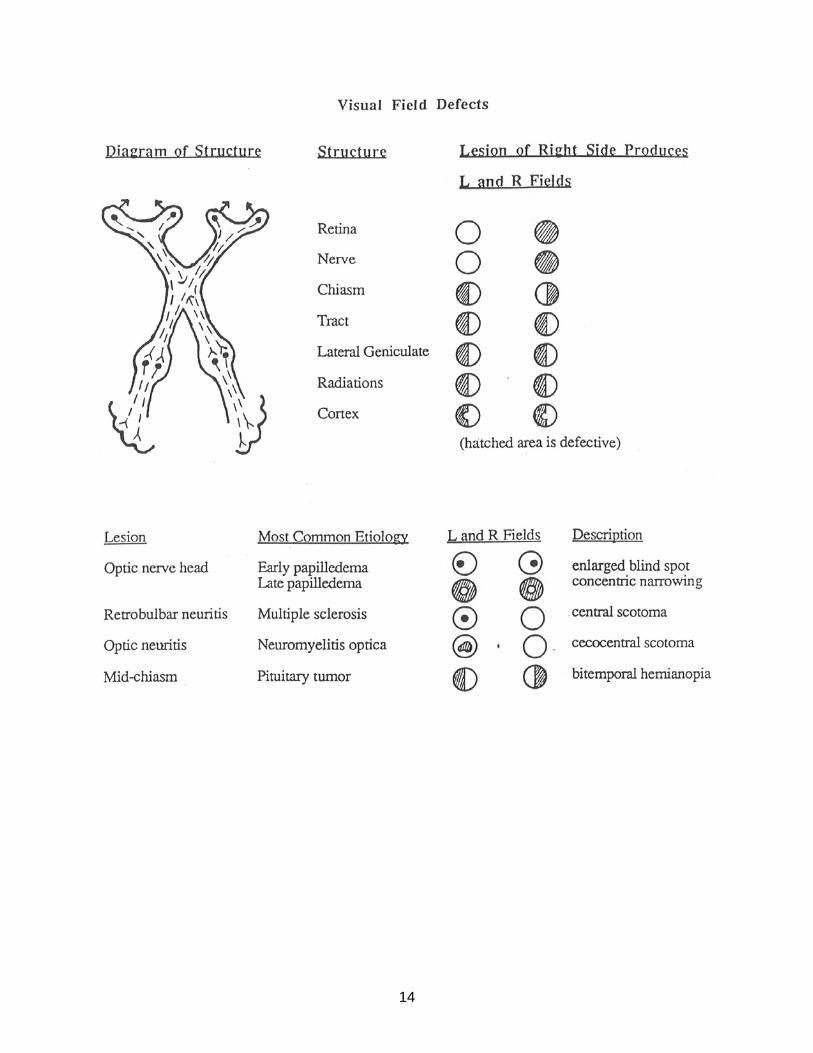
Optic Nerve Three components of the optic nerve are typically evaluated: visual acuity, visual fields and the funduscopic examination. Visual Acuity For neurologic purposes, corrected visual acuity is tested (with eyeglasses or contact lenses). Each eye is checked individually. Distance vision is checked by means of the Snellen chart, and near vision is tested by means of the Jeager chart. Visual acuity is a reflection of the integrity of the entire visual system, including the refractile components (cornea, lens, vitreous humor, retina, optic nerve, optic chiasm, optic tract, lateral geniculate nucleus, optic radiations and the occipital cortex). It is important to remember that visual acuity evaluates only macular vision, which is the central 5° of the visual field. Visual Fields These are evaluated by the confrontation method. In this method, the examiner stands directly in front of the patient, usually 2-3 feet away. The patient closes one eye and looks at the examiner's nose with the other eye. The examiner does the same. A target (usually the examiner's finger) is then introduced from the periphery of each visual quadrant and the visual field is assessed in this quadrant, using the examiner’s visual field as the control. Each eye is checked individually. To evaluate for visual neglect, the patient keeps both eyes open and looks at the examiner's nose. The examiner then presents bilateral simultaneous stimuli and the patient is asked to localize the stimuli. Visual neglect often implies parietal lobe lesions. Various visual field defects can be seen, depending on the location of the lesion within the visual pathway. These are listed in figure 1.
14
15
Funduscopic Examination The fundus is evaluated with the ophthalmoscope. The optic disk, surrounding retina, blood vessels and macula can be visualized. In addition, by changing the plane of focus on the ophthalmoscope, one can visualize the cornea and lens. With this technique, one can see evidence for optic disk swelling (papilledema), optic disk atrophy, retinal hemorrhages, retinal vascular changes of hypertension and diabetes, as well as corneal scarring and cataracts. The ophthalmoscopic examination is difficult and requires many years of practice, but, once mastered, can provide a great deal of information about the central nervous system.
Oculomotor, Trochlear, and Abducens Nerves These nerves are examined together since they have similar functions. There are three parts to the examination of these nerves: pupillary light response, ocular movements and ptosis. Pupillary Light Response Pupillary size depends on the balance between the parasympathetic nervous system which causes constriction via CN III (figure 2), and the sympathetic nervous system which causes dilatation via the sympathetic pathway originating in the hypothalamus, traversing the brain stem, cervical and upper thoracic spinal cord, and forming the peripheral sympathetic pathway passing through the superior cervical sympathetic ganglion, and traveling along the external carotid artery and with the ophthalmic division of the trigeminal nerve (figure 2).
16
Pupillary size is first observed and measured in dim light: small pupils are termed miotic, large pupils are termed mydriatic, and unequal pupils are termed anisocoric. The pupillary light response is next tested. To test this reflex, a bright light is shone on each eye individually, and the pupils are examined for direct and consensual pupillary constriction. The afferent information for this reflex is carried by the optic nerve (CN II) and the efferent response is carried by the oculomotor nerve (CN III). In addition to constricting to light, pupils also constrict when shifting from far to near gaze, and this response is known as accommodation. The stimulus for this response originates in the optic pretectum. Specific pupillary lesions are listed in table 2.
TABLE 2 PUPILLARY ABNORMALITIES
TYPE CLINICAL FINDINGS ANISOCORIA LESION
Marcus-Gunn pupil A deafferented pupil which constricts to consensual but not to direct light
Absent CN II
Hutchinson pupil A dilated pupil that does not respond to direct or consensual light
Present CN III
Horner's syndrome A small pupil with associated ipsilateral ptosis and decreased facial sweating
Present Sympathetics
Adie's tonic pupil A dilated pupil with an impaired light response and slow constriction to near vision
Present Parasympathetics
Argyll Robertson pupil
A small, irregular pupil that constricts to near vision but not to light
Absent Pretectum
Ocular Movements Voluntary and reflex eye movements are coordinated by the cortical connections (frontal eye fields and occipital cortex), vestibular apparatus, medial longitudinal fasciculus and CNs III, IV and VI. Eye movements may be dysconjugate or conjugate. Convergence is a normal dysconjugate eye movement that is part of the near response. There are three types of conjugate eye movements. Two of these fixate the image on the retina (one of these with respect to head and neck motion, and the other with respect to image motion), and one redirects the line of sight. These eye movements are detailed below.
17
Ocular movements that fixate the image on the retina: 1. Vestibulo-Ocular Reflex (VOR) (oculocephalic reflex) (“doll’s eyes” reflex): This reflex fixates the image on the retina with respect to head and neck motion. Head rotation is a form of angular acceleration that stimulates the semicircular canals in the inner ear. These canals sense and convert this angular acceleration into electrical impulses that then are conveyed to the four vestibular nuclei in the brain stem via CN VIII. The vestibular nuclei subsequently project to CNs III, IV, and VI via the medial longitudinal fasciculus (MLF), maintaining a stable visual field despite head motion. 2. Visual Pursuit: This reflex fixates the image on the retina with respect to image motion. Image motion is sensed by the occipital cortex that then relays this information in a crossed fashion to the lateral gaze center in the pons (paramedian pontine reticular formation [PPRF]), and then via the MLF to CNs III, IV, and VI. Ocular movements that re-direct the line-of-sight: 3. Visual Saccade: The stimulus for this ocular movement originates in the frontal eye fields located in the frontal lobes of the cerebral hemispheres. The information then travels in a crossed fashion to the lateral gaze center in the pons (PPRF), and then is relayed via the MLF to CNs III, IV, and VI, in an analogous fashion as for the other two eye movements noted above (figure 3).
18
19
In an awake individual, eye movements are assessed by having the patient look in the six primary directions of gaze. These directions correspond to the directions of action of the extra-ocular muscles, and are detailed in figure 4.
In a comatose individual, eye movements can be evaluated by means of the oculocephalic (Doll's eyes) reflex and by means of caloric testing. These are detailed in figure 5.
20
21
One also observes the patient for nystagmus. Nystagmus is a rhythmic, oscillatory involuntary eye movement of one or both eyes that may occur spontaneously or be evoked by a specific direction of gaze. Nystagmus may be horizontal, vertical or rotatory, and typically has a fast and a slow component. By definition, the direction of nystagmus is the direction of the fast component. Nystagmus may be physiologic or pathologic, as noted in table 3.
Gaze palsies producing dysconjugate gaze can be due to lesions involving: the extra-ocular muscles as can be seen with hyperthyroidism; the neuromuscular junction as can be seen with myasthenia gravis; individual lesions affecting CNs III, IV or VI; or a lesion involving the MLF producing an internuclear ophthalmoplegia (INO). INOs are frequently seen with multiple sclerosis in younger individuals, and with small brain stem strokes in older individuals. A gaze preference is a conjugate paresis of gaze that is frequently seen following lesions to the frontal eye fields following a large hemispheric stroke. Ptosis Ptosis refers to drooping of the eyelid. Two separate muscles, innervated by two different nerves, elevate the eyelid: The levator palpebrae muscle is a skeletal muscle innervated by CN III and is responsible for opening the eyes. The superior tarsal muscle is a smooth muscle innervated by the sympathetic nervous system and also helps elevate the eyelid. A lesion to either of these nerves can produce ptosis, although the ptosis due to a lesion of CN III is more pronounced. These two forms of ptosis are usually associated with anisocoria, and the proper diagnosis can be made by noting the pupillary size on the side of the ptotic eyelid. A sympathetic lesion causing ptosis has an accompanying small pupil (Horner's syndrome), while a CN III lesion causing ptosis has an accompanying large pupil.
22
Trigeminal Nerve
The trigeminal nerve has both sensory and motor function. Trigeminal Sensory Function The sensory component of the trigeminal nerve relays sensory information from the face via three divisions, the ophthalmic, maxillary and mandibular divisions. Testing of this component is accomplished by touching all three divisions of the face with cotton or a pin. The Corneal Reflex The corneal reflex is an excellent way to test the sensory component of the trigeminal nerve, as well as the facial nerve. To test this reflex, the cornea is touched with a wisp of cotton and the resultant direct and consensual eye blink is noted. The afferent information for this reflex is carried by the ophthalmic division of the trigeminal nerve, and the efferent information is carried by the facial nerve. Trigeminal Motor Function Trigeminal motor function includes the muscles of mastication: the temporalis, masseter, and lateral and medial pterygoid muscles. These are innervated by the mandibular division of the trigeminal nerve and can be tested by having the patient clench the jaw tightly or deviate the jaw from side to side against resistance.
Facial Nerve
Motor component This nerve supplies motor innervation to the face and has numerous divisions. To test this nerve, facial symmetry is observed at rest. The patient is then asked to wrinkle the brow, close the eyes firmly, smile and frown. Facial weakness can be due to either lower motor neuron or upper motor neuron lesions. With a lower motor neuron lesion of the facial nerve, ipsilateral weakness of the entire half of the face is observed. Bell's palsy is an example of lower motor neuron facial weakness. Facial weakness can also be seen with an upper motor neuron lesion involving the motor cortex or the corticobulbar tract. In this case, the weakness is contralateral to the lesion and involves only the lower half of the face. This pattern of weakness is seen with upper motor neuron lesions because the upper face receives bilateral cortical innervation and is therefore unaffected in unilateral upper motor neuron lesions. Taste component The facial nerve supplies taste sensation to the anterior 2/3 of the tongue via the chorda tympani nerve. Taste can be checked by applying sugar or salt solutions to the anterior tongue with a cotton applicator.
23
Vestibulo-Cochlear Nerve
This nerve has two divisions, each of which carries vestibular or auditory information respectively. Cochlear Division This can be tested rather crudely by assessing the patient's ability to hear a ticking watch or rubbing fingers held a certain distance away from the ear. The examiner is the control. This form of testing evaluates global auditory function. Hearing loss is frequently differentiated into conductive hearing loss and sensori-neural hearing loss. Conductive hearing loss implies a lesion to structures in the outer or middle ear that convert air conduction into bone conduction. Bone conduction is perceived as louder than air conduction in this form of hearing loss. Sensori-neural hearing loss is due to a lesion involving the inner ear (cochlear apparatus) or the eighth cranial nerve. Both air and bone conduction are reduced in this form of hearing loss. Sensori-neural hearing loss is sometimes further subdivided into cochlear and retro-cochlear. Cochlear hearing loss results from destructive lesions involving the labyrinth, such as Meniere's disease, occupational noise, certain ototoxins and certain infections including syphilis. Retro-cochlear hearing loss is usually due to a tumor invading the eighth cranial nerve (acoustic Schwannoma). The Weber test and Rinne test are two tests of hearing that help differentiate conductive from sensori-neural hearing loss. In the Rinne test, the base of a vibrating tuning fork (512 Hz) is placed against the mastoid process until the sound is no longer heard. The tines of the tuning fork are then moved adjacent to the external ear where sound should still be appreciated in normal individuals, since air conduction is normally better than bone conduction. If the sound is no longer heard in this second position a conductive hearing loss is suspected. In the Weber test, a vibrating tuning fork (512 Hz) is placed at the vertex of the skull and the patient is asked to localize the sound. Normally the sound should be heard equally in both ears. Lateralization of the sound to one ear is abnormal, with the sound localizing to the "bad ear" in a conductive hearing loss and to the "good ear" in a sensori-neural hearing loss. The significance of abnormalities in Weber and Rinne testing are listed in table 4.
24
TABLE 4 CLINICAL TESTS OF AIR CONDUCTION (AC) AND BONE CONDUCTION (BC)
TEST NORMAL
RESPONSE CONDUCTIVE
HEARING LOSS SENSORI-NEURAL
HEARING LOSS Rinne AC > BC BC > AC AC > BC but both ↓ Weber No lateralization Lateralized to defective ear Lateralizes to normal ear
AC Air conduction BC Bone conduction
Vestibular Division This portion of CN VIII can be evaluated by observing for nystagmus at rest, as well as following labyrinthine stimulation. Labyrinthine stimulation can be performed by means of the Nylen-Barany (Dix-Hallpike) positioning maneuver. In this test, the patient is quickly moved from the sitting position to a supine position with the head positioned 45° below the plane of the table and turned to one side (figure 6).
This position is maintained for about one minute, during which time the patient is observed for nystagmus. The test is then repeated with the head turned to the other side. If the patient reports vertigo during the maneuver, or if nystagmus develops, vestibular dysfunction may be present.
25
Caloric testing is an alternate method for stimulating the labyrinth. In this test, hot or cold water is introduced into the external auditory meatus and the patient is observed for the development of nystagmus. Both ears are irrigated sequentially and the degree of resultant nystagmus following irrigation of either ear is compared. Nystagmus and vertigo can both be seen following a peripheral lesion involving the vestibular apparatus, or following a central lesion involving the vestibular nuclei in the brain stem. The vertigo and nystagmus that result from either a central or peripheral lesion have different characteristics, and these differences are useful in localizing lesions of the vestibular system. Table 5 lists the different responses seen with central or peripheral lesions for the Nylen-Barany maneuver.
TABLE 5 NYLEN-BARANY MANEUVER FOR DIFFERENTIATING PERIPHERAL
FROM CENTRAL CAUSES OF VERTIGO
PERIPHERAL CENTRAL Latency period before onset of nystagmus
2-20 sec. None
Duration of nystagmus <1 min. >1 min. Fatigability Yes No Direction of nystagmus in one head position
Unidirectional May change direction
Vertigo intensity Severe Slight Head positions eliciting vertigo Single position More than one position Caloric and rotary tests Vestibular
These nerves are usually tested together since they have overlapping functions. The glossopharyngeal nerve primarily carries sensation from the posterior pharynx and the larynx. The vagus nerve supplies motor innervation to the soft palate, pharyngeal muscles and the vocal cords. The vagus nerve is easily tested by asking the patient to phonate and observing for a symmetric rise in the palate and uvula. Another way to test both of these nerves is to elicit the gag reflex. In this test, the examiner touches the posterior pharyngeal wall with a tongue blade and observes for a symmetric rise in the palate and uvula. The afferent arm of this reflex is carried by the glossopharyngeal nerve and the efferent arm by the vagus nerve.
Spinal Accessory Nerve The primary function of this nerve is to supply motor innervation to the sternocleidomastoid (SCM) muscle and the upper third of the trapezius muscle. Sternocleidomastoid function is assessed by asking the patient to rotate the head against resistance. Recall that contraction of the right SCM muscle allows one to turn the head to the left. Trapezius function is assessed by shoulder shrug.
26
Hypoglossal Nerve
This nerve supplies motor innervation to the tongue, and is evaluated by observing the tongue at rest, and by asking the patient to protrude the tongue in the midline or to apply lateral pressure against each cheek. A lesion of the hypoglossal nerve will eventually cause atrophy of the ipsilateral half of the tongue. Recall that the tongue will also deviate towards the side of the lesioned nerve when protruded.
Cranial Nerve Reflexes
Practically the entire brain stem can be evaluated by means of five cranial nerve reflexes. This is extremely useful in evaluating the cause of coma in an unresponsive patient. These five reflexes are detailed in table 6
TABLE 6 CRANIAL NERVE REFLEXES
REFLEX AFFERENT NERVE EFFERENT NERVE
Pupillary * II III Jaw Jerk * V V Corneal * V VII Gag * IX X Vestibulo-ocular VIII III, IV, VI (via MLF)
* has direct and consensual response
27
THE MOTOR EXAMINATION When performing the motor examination on a patient presenting with weakness, it is important to remember that weakness could be a result of a lesion at any point in the neuraxis: cerebral hemispheres, brain stem, spinal cord, anterior horn cell, nerve root (myotome), peripheral nerve, neuromuscular junction, or muscle. Another important distinction in the evaluation of weakness is whether the weakness has a characteristic "upper motor neuron pattern" or "lower motor neuron pattern". These differences will be detailed below. The motor examination consists of several parts: assessment of muscle bulk, evaluation of muscle tone, observation for spontaneous movements, and functional and formal muscle strength testing.
Muscle Bulk
In general, muscle bulk should be symmetric throughout the limbs, when comparing the right and left sides, and proximal and distal portions of the extremities. Loss of muscle bulk is known as atrophy, and is seen in two pathologic settings: • Denervation atrophy: A profound form of muscle atrophy that is seen with lower motor
neuron lesions. • Disuse atrophy: A mild form of muscle atrophy that can be seen in a variety of clinical
settings, including upper motor neuron disease, disuse, corticosteroid use, collagen-vascular disorders, and with musculoskeletal problems.
Spontaneous Movements
Several different types of abnormal spontaneous movements can be seen as follows: • Fasciculations: Worm-like contractions of muscle due to random discharge of an entire
motor unit. Although frequent fasciculations are seen with anterior horn cell disorders (i.e. amyotrophic lateral sclerosis), occasional fasciculations are commonly seen with simple muscle fatigue following exercise and are of no clinical consequence.
• Myoclonus: Sudden contractions of a muscle or group of muscles that move an entire limb
across a joint. Myoclonus is frequently seen with metabolic or hereditary neurologic disorders.
• Chorea and Athetosis: Brief, irregular, asymmetric writhing movements of basal ganglia
origin. Chorea is a quick, distal dance-like movement and athetosis is a more proximal slower movement.
• Tremor: A rhythmic, oscillatory movement of the trunk or limbs due to numerous causes,
including lesions of the cerebellum, motor system, sensory system or the basal ganglia. Tremor is frequently differentiated into resting tremor and action tremor. Resting tremor is one of the hallmarks of Parkinson's disease. Action tremor can be seen with lesions of the cerebellum or the sensory system, and may also be idiopathic (benign familial tremor or senile tremor).
28
Muscle Tone
Muscle tone is defined as the resistance of muscle to passive stretch, and is assessed by moving a relaxed limb passively through an entire range of motion. Tone may be increased or decreased in various pathologic states, and various forms of altered muscle tone are detailed in table 7.
TABLE 7 MUSCLE TONE
TYPE DESCRIPTION PATHOLOGY
Increased Spasticity (clasp knife) Has a catch which varies with position and is
velocity dependent UMN lesion
Rigidity (lead pipe) Steady resistance to movement at all speeds and positions. Superimposed tremor leads to "cog-wheeling".
Basal ganglia lesion (substantia nigra or striatum)
Paratonia Inability to relax the muscle Bihemispheric lesions Gegenhalten Opposes examiner Mitgehen Assists examiner
Decreased Flaccidity Limp LMN lesion Brain shock
Spinal shock Acutely following a stroke or spinal cord injury
Loss of check Cerebellar lesion
Muscle Strength Muscle strength is assessed by both functional testing and formal testing. Functional Testing Functional testing is a very reliable form of testing muscle strength that is easily reproducible and reflects the ability of the patient to perform certain tasks. Functional testing of the upper extremity includes the following: ability to touch the chin to the chest when lying supine; ability to raise the arms above one's head; ability to blanch the knuckles when making a fist. Functional testing of the lower extremities includes the following: ability to arise from a chair without using one's hands; ability to arise from a squat; ability to step up on a chair with one leg; ability to walk on one's toes or heels.
29
The Pronator Drift The pronator drift is another very important functional muscle test for the upper extremities. This test is performed by having the patient hold both hands outstretched with the palms up and the eyes closed. The examiner watches for subtle pronation of the arm, which sometimes is accompanied by abduction and internal rotation at the shoulder and flexion at the elbow. Pronation of the arm is a subtle sign that is strongly indicative of upper motor neuron dysfunction. Formal Testing Formal muscle strength testing involves grading muscle strength for individual muscle groups on a 0-5 scale by means of push/pull testing by the examiner. Typically, a group of muscles is tested together. Representative muscle groups that are often evaluated in a screening examination are listed in table 8.
TABLE 8 REPRESENTATIVE MUSCLE GROUPS TESTED IN A SCREENING EXAMINATION
UPPER EXTREMITY MUSCLE GROUPS LOWER EXTREMITY MUSCLE GROUPS shoulder abduction hip flexion elbow flexion hip extension elbow extension hip abduction wrist flexion hip adduction wrist extension knee flexion finger flexion knee extension finger abduction ankle plantar flexion ankle dorsiflexion
The 0-5 grading scale for muscle strength testing is based on the ability of the muscle group to oppose gravity, and was devised by the medical research council in Great Britain during the polio epidemic. Table 9 defines each of the grades.
TABLE 9 MUSCLE STRENGTH GRADING
GRADE DESCRIPTION
0 No movement 1 Flicker of contraction 2 Full range of motion with gravity eliminated 3 Full range of motion against gravity 4 Full range of motion against gravity and offers some resistance 5 Full power
30
THE SENSORY SYSTEM When evaluating a patient with sensory dysfunction, it is important to keep in mind all of the levels of the nervous system at which a lesion can produce sensory dysfunction: peripheral nerve, brachial or lumbo-sacral plexus, nerve root (dermatome), spinal cord, brain stem, thalamus, or sensory cortex. The sensory examination is largely a subjective examination that requires an alert, cooperative patient who can give reliable subjective impressions of various stimuli. In general, sensory symptoms precede sensory signs, and the sensory examination may not be revealing early on in the course of an illness that produces sensory dysfunction. When performing the sensory examination, one looks for asymmetries. In general, the examiner looks for a proximal-to-distal gradient, or for findings in the distribution of a specific nerve or nerve root. The sensory examination is divided into three parts: primary modalities, cortico-sensory modalities, and functional testing (the Romberg test).
Primary Modalities Protopathic sensation Examples of protopathic sensation include poorly localized touch, pain and temperature perception. These modalities are carried by small, unmyelinated fibers, travel contralaterally in the lateral spinothalamic tracts in the spinal cord, and are ultimately processed in the brain stem reticular formation and in the thalamus. • Pain: This is typically assessed with a pin. The "prickly" sensation may be reported by the
patient as diminished, absent or heightened in the affected areas. • Temperature: This can be assessed with a cool tuning fork, or with test tubes filled with cold
or hot water. Epicritic sensation Examples of epicritic sensation include fine, discriminative touch, vibration, and proprioception (position sense). These modalities are generally subserved by encapsulated nerve endings and are carried by large, myelinated nerve fibers, ascending ipsilaterally in the dorsal columns in the spinal cord. This information then crosses in the medulla, projects to the thalamus and is ultimately processed in the primary sensory cortex. • Vibration: This is assessed with a tuning fork (128 Hz). The vibrating tuning fork is applied
over a distal joint, such as the DIP joint of the great toe or of the index finger. The examiner places his/her finger under the patient's joint and the patient is asked to indicate when the stimulus decays. Vibratory loss is present if the examiner still feels the stimulus. If vibratory perception is absent distally, more proximal joints are assessed in a similar fashion.
31
• Proprioception: This is evaluated by assessing position sense at the interphalangeal joints with slight degrees of motion of the joint. The examiner grasps the patient's joint laterally so as not to provide digital pressure cues to the patient.
Cortico-sensory Modalities
These are more complex forms of sensation that require significant cortical processing. Four different cortico-sensory modalities are typically evaluated: stereognosis, graphesthesia, two-point discrimination, and double simultaneous stimulation. • Stereognosis: The ability to identify objects by touch alone. To evaluate this modality,
objects such as a safety pin or coin are placed in the hand of a patient for identification. • Graphesthesia: The ability to recognize numbers drawn on the palm of the hand. • Two Point Discrimination: The ability to localize and discriminate between two points that
are close together. This is typically tested on the tip of the index finger with a paper clip that is bent open. The examiner applies both ends of the clip, keeping them several millimeters apart and moving them closer and closer together. Normal subjects have a detection threshold of 2 mm at the tip of the index finger.
• Double Simultaneous Stimulation: A normal subject should be able to localize two stimuli
that are applied simultaneously to different parts of the body. Patients with a parietal lobe lesion have a phenomenon known as extinction in which they consistently fail to identify a stimulus on the side of the body contralateral to a parietal lobe lesion, when it is presented simultaneously with a stimulus on the opposite side of the body. In a broad sense, extinction to double simultaneous stimuli is a type of agnosia known as sensory neglect. To test for extinction, the right and left sides of the body are touched at the same time and the patient is asked to localize both stimuli with the eyes closed.
Functional Testing
Functional sensory testing is assessed by means of the Romberg test. This test is performed by asking the patient to stand with his/her feet together and then close the eyes. The patient is then observed to see if balance can be maintained with the eyes closed. The Romberg test is reported as positive if the patient falls to one side. Recall that three systems are routinely used to maintain balance, namely proprioception, the vestibular apparatus and vision. Only two of these systems are required at any one time. Eye closure removes visual cues for maintaining balance. If balance is maintained with the eyes closed, this implies integrity of both the vestibular apparatus and proprioception. Falling to one side implies dysfunction of one of these balance systems. The Romberg test can only be performed if the patient is able to stand well with feet together and eyes open. If the patient cannot do this well, a lesion of the cerebellum is suspected; the Romberg test cannot be performed under these circumstances.
32
COORDINATION Coordination is an integral function of the motor, sensory and cerebellar systems. Tests of coordination typically assess cerebellar function, but the contributions of the other systems, including the motor, sensory and vestibular systems, must be considered when interpreting these tests. Coordination testing is usually divided into two parts: truncal stability and limb coordination. The ability to check movements and vestibular coordination are also assessed if the clinical situation warrants.
Truncal Stability
Truncal stability is assessed by observing the patient's balance when sitting or standing with feet together and eyes open. Truncal ataxia suggests a lesion involving the midline cerebellar vermis.
Limb Coordination Limb coordination may be assessed in both the arms and legs: • Finger-to-Nose Test: The patient is asked to touch his/her nose with the index finger, then
the examiner's finger, and then his/her nose again. Speed, accuracy and any tremor are noted.
• Heel-to-Shin Test: The heel of one leg is run smoothly down the other shin, and speed,
accuracy and any tremor are noted. Rapid Alternating Movements (Diadochokinesia): The patient is asked to alternately slap the thigh with the front and back of the hand, or to touch each finger to the thumb. Each side is tested separately and compared with the other. Foot tapping is a rapid alternating movement frequently evaluated in the lower extremity. Ataxia and dysmetria are general terms used to describe unevenness in the performance of any of the above tests, and are frequently due to lesions involving the cerebellar hemispheres. Recall that cerebellar lesions produce ataxia on the side ipsilateral to the lesion. Upper motor neuron lesions or sensory lesions that result in altered proprioception can also result in ataxia and dysmetria.
Ability to Check Movements Ability to check movements is evaluated by asking the patient to maintain flexion of his/her arm at the elbow against resistance provided by the examiner. The examiner then abruptly lets go of the patient's arm and observes the ability of the patient to “check” or break the flexion movement. An inability to check movements can be seen with lesions of the ipsilateral cerebellar hemisphere, as well as with severe sensory disturbances causing altered proprioception.
33
Vestibular Coordination Past pointing and compass turning are two tests that evaluate the integrity of the vestibular system. • Past Pointing: To perform this test, the patient is asked to repeatedly elevate his/her arm
vertically and then return to the horizontal such that the index finger touches the examiner's finger that is held directly in front of the patient. This is performed with the eyes open initially, and closed later on. A drift of the patient's arm in one direction is strongly suggestive of a lesion involving the ipsilateral vestibular apparatus.
• Compass Turning: The patient is asked to march in place with the eyes closed. Rotation of
the body in one direction is suggestive of ipsilateral vestibular pathology.
34
REFLEXES Evaluation of reflexes is perhaps the most objective way to examine the nervous system. Not only are reflexes helpful in evaluating awake individuals, but they also are invaluable in examining comatose patients. The cranial nerve reflexes have already been summarized in table 6. Reflexes can be divided into non-pathologic and pathologic. The non-pathologic reflexes include muscle stretch reflexes (deep tendon reflexes) and superficial (cutaneous) reflexes. The pathologic reflexes include the Babinski sign as well as the frontal release signs.
Muscle Stretch Reflexes Muscle stretch reflexes are monosynaptic spinal cord reflexes that are elicited by striking the muscle tendon with a percussion hammer and evaluating the subsequent contraction of that muscle. Striking the muscle tendon stretches the muscle spindle and this afferent information is carried by the Ia afferent sensory nerve fibers through the dorsal root and dorsal horn of the spinal cord, eventually synapsing on a corresponding anterior horn cell in the ventral horn of the spinal cord. The efferent arm of this reflex originates in the anterior horn cell, exits the spinal cord in the ventral root, and is carried by the alpha motor neuron, eventually synapsing on the same muscle. Five muscle stretch reflexes are usually elicited in a routine neurologic examination, and these are listed in table 10.
Muscle stretch reflexes are usually graded on a 0-4 scale, as shown in table 11.
TABLE 11 GRADING OF MUSCLE STRETCH REFLEXES GRADE DESCRIPTION
0 Absent 1 Hypoactive 2 Normal 3 Hyperactive with spread across a joint 4 Hyperactive with clonus
Clonus is a rhythmic series of involuntary muscle contractions induced by a sudden passive stretch to a muscle, and is indicative of hyperreflexia. Ankle clonus is easy to obtain in a
35
hyperreflexic patient, and can be elicited by sudden dorsiflexion of the foot. Patellar clonus can be obtained by a quick, downward motion on the patella, holding the knee slightly flexed.
Superficial (Cutaneous) Reflexes Superficial (cutaneous) reflexes are polysynaptic, nociceptive reflexes that are elicited by stimulating the skin and observing for contraction of the corresponding muscle. Four superficial reflexes can be easily obtained: the abdominal reflexes, the anal wink reflex, the cremasteric reflex and the bulbo-cavernosus reflex. • Abdominal Reflexes are obtained by stroking the skin lightly on the abdomen from the
umbilicus towards any abdominal quadrant and observing for deviation of the umbilicus towards the quadrant that is stroked. The upper quadrants reflect T6-9 innervation and the lower quadrants T10-12 innervation. Abdominal reflexes may be diminished or absent in many circumstances, including a lesion to the corresponding nerve roots or with upper motor neuron lesions. In addition, obesity, previous pregnancy or abdominal surgery can result in a loss of abdominal reflexes.
• The anal wink reflex is elicited by stroking the perianal skin and observing for a contraction
of the external anal sphincter on the stroked side. Presence of this reflex indicates integrity of the S3-5 nerve roots.
• The cremasteric reflex is elicited by stroking the skin on the inner thigh in a male and
observing for elevation of the testicle on the stroked side. Presence of this reflex reflects integrity of the L1-2 nerve roots.
• The bulbo-cavernosus reflex is elicited by squeezing the glans penis and observing for
contraction of the external anal sphincter. Presence of this reflex indicates integrity of the S3-4 nerve roots.
The Babinski Sign
The Babinski sign is perhaps the most important reflex in neurology and is obtained by stroking the lateral aspect of the sole of the foot with a noxious stimulus, starting at the heel and then crossing the ball of the foot towards the great toe. A normal response consists of flexion of the great toe. An abnormal (positive) response consists of dorsiflexion of the great toe, and occasionally fanning of the other toes. A positive Babinski response is indicative of an upper motor neuron lesion.
Frontal Release Signs Frontal release signs are reflexes that are present in infancy, lost with maturation of the central nervous system, and regained with advanced age or with diffuse cortical or bihemispheric dysfunction, such as can be seen with Alzheimer's disease, Parkinson's disease or with bihemispheric strokes. Four frontal release signs are commonly tested: snout, palmomental, grasp and glabellar reflexes. • Snout Reflex: This is elicited by repeatedly tapping the upper lip and observing for
puckering of the lips. One way of eliciting this reflex is to place a tongue blade lightly over the upper lip and to tap the tongue blade with a percussion hammer.
36
• Palmomental Reflex: This is elicited by scratching the thenar eminence of the palm with a
blunt object and observing for an ipsilateral contraction of the mentalis muscle on the chin. • Grasp Reflex: This is obtained by having the examiner stroke the skin of the patient's palm
with his/her fingers and observing for a resultant grasping of those fingers by the patient. • Glabellar Sign: This is elicited by tapping the forehead repeatedly between the eyebrows
over the glabella and observing for persistent blinking. It is important to note that a normal individual will blink once or twice only with this maneuver.
37
GAIT AND STATION
Gait
Examination of gait is most important in neurology, since it provides invaluable information concerning integrity of the motor system, sensory system, and cerebellum. Gait is evaluated by observing the patient walk briskly and turn corners. Particular attention is placed on any asymmetry involving a side or one limb, the distance the feet are kept apart (base), the length of stride and associated arm swing. An important part of the gait examination is to observe a tandem gait, in which the patient is asked to walk heel-to-toe on a line. This narrows the base of the gait and will bring out subtle gait abnormalities that may not be otherwise evident. Inability to perform a tandem gait is frequently associated with altered proprioception or midline cerebellar lesions. When evaluating gait, the examiner first notes if the gait is symmetric or asymmetric. If symmetric, the examiner then evaluates the base of the gait. Various abnormal gaits, including asymmetric gaits and wide-based and narrow-based symmetric gaits are detailed in table 12.
TABLE 12 ABNORMAL GAITS
GAIT LESION
ASYMMETRIC Trendelenburg Hip pathology Hemiplegic Upper motor neuron disorder Steppage (foot drop) Peroneal nerve palsy Antalgic Foot or leg pain
Narrow Based Spastic (scissor) Bilateral upper motor neuron lesions Festinating Basal ganglia (substantia nigra)
38
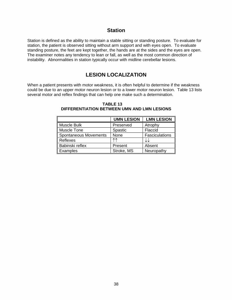
Station
Station is defined as the ability to maintain a stable sitting or standing posture. To evaluate for station, the patient is observed sitting without arm support and with eyes open. To evaluate standing posture, the feet are kept together, the hands are at the sides and the eyes are open. The examiner notes any tendency to lean or fall, as well as the most common direction of instability. Abnormalities in station typically occur with midline cerebellar lesions.
LESION LOCALIZATION
When a patient presents with motor weakness, it is often helpful to determine if the weakness could be due to an upper motor neuron lesion or to a lower motor neuron lesion. Table 13 lists several motor and reflex findings that can help one make such a determination.
TABLE 13 DIFFERENTIATION BETWEEN UMN AND LMN LESIONS
A 68-year-old woman with hypertension was brought to the emergency department by her friend because of dizziness, vertigo and difficulty walking, which she first noted when she awoke from a nap that evening.
BP=200/130 mm Hg and P=76/min. She is examined lying on a gurney in the emergency department. There is minimal nystagmus with right gaze. Facial strength and sensation are normal. Motor and sensory examinations are entirely normal. Finger-to-nose testing is normal bilaterally. Muscle stretch reflexes are normal throughout.
Questions:
1. Where would you best localize the lesion?
2. What is the most likely diagnosis?
3. What is the most appropriate next step in diagnosis?
40
Case #2
A 55-year-old man with hypertension and diabetes mellitus is admitted for cardiac catheterization because of worsening angina and an abnormal exercise tolerance test. Following the procedure, which demonstrates severe LAD disease, he is noted to be confused, and a neurological consultation is obtained.
BP=150/90 mm Hg, and P=80/min and regular. He is awake, alert and fully oriented. He appears confused when asked to describe what happened to him that day. His face is symmetrical. He has full power in all four limbs. Sensory examination is entirely normal. Muscle stretch reflexes are symmetrical.
Questions:
1. Where would you best localize the lesion?
2. What is the most likely diagnosis?
3. What is the most appropriate next step in diagnosis?
41
Case #3
A 25-year-old man with Type 1 diabetes mellitus on insulin sees his physician because of right arm weakness that he noted while working on his car last week. Several days before that, he fell while taking out the garbage, and landed on his right shoulder.
BP=120/80 mm Hg and P=84/min. He is fully awake and alert. He has difficulty raising his right arm above his head, and has a slightly weaker handgrip on the right. He has some trouble manipulating fine objects with his right hand. Muscle power in the remaining three limbs is full. There is slight vibratory loss at his toes bilaterally. Muscle stretch reflexes are reported as normal in the upper limbs and at the knees, and absent at the ankles.
Questions:
1. Where would you best localize the lesion?
2. What is the most likely diagnosis?
3. What is the most appropriate next step in diagnosis?
42
Case #4
A 70-year-old woman with hypertension, diabetes mellitus and coronary artery disease, is admitted to the CCU because of the acute onset of nausea, vomiting and a cardiac arrhythmia characterized by frequent PVC’s. Acute myocardial infarction is ruled out, and she is transferred to a regular medical floor the following day. Examination there reveals dysarthria and dysphagia, and a neurological consultation is obtained.
BP=130/90 mm Hg and P=80/min with frequent premature beats. She is fully awake and alert. Her face is symmetric. Her voice is somewhat hoarse and she is unable to swallow without coughing. Muscle power is full in all four limbs, and muscle stretch reflexes are symmetrical. Plantar responses are flexor bilaterally.
Questions:
1. Where would you best localize the lesion?
2. What is the most likely diagnosis?
3. What is the most appropriate next step in diagnosis?
43
Case #5
A 60-year-old college professor is referred for evaluation of muscle stiffness and a weak voice that has been present for the past six months. In fact, some of his students have complained that it is getting more difficult to understand him when he lectures. They also have had trouble reading his handwriting on the blackboard.
BP=160/80 mm Hg and P=68/min. Mental status is normal. His voice is soft, but cranial nerves are otherwise normal. Muscle power is full and sensation is normal in all four limbs. Muscle stretch reflexes are 2+ throughout and plantar responses are flexor bilaterally. His gait is somewhat slow but is otherwise normal.
Questions:
1. Where would you best localize the lesion?
2. What is the most likely diagnosis?
3. What is the most appropriate next step in diagnosis?