ORIGINAL RESEARCH published: 04 September 2018 doi: 10.3389/fmicb.2018.02101 Edited by: Bart Devreese, Ghent University, Belgium Reviewed by: Klas Flärdh, Lund University, Sweden Kevin D. Young, University of Arkansas for Medical Sciences, United States *Correspondence: Tanneke den Blaauwen [email protected]Specialty section: This article was submitted to Antimicrobials, Resistance and Chemotherapy, a section of the journal Frontiers in Microbiology Received: 31 May 2018 Accepted: 17 August 2018 Published: 04 September 2018 Citation: Montón Silva A, Otten C, Biboy J, Breukink E, VanNieuwenhze M, Vollmer W and den Blaauwen T (2018) The Fluorescent D-Amino Acid NADA as a Tool to Study the Conditional Activity of Transpeptidases in Escherichia coli. Front. Microbiol. 9:2101. doi: 10.3389/fmicb.2018.02101 The Fluorescent D-Amino Acid NADA as a Tool to Study the Conditional Activity of Transpeptidases in Escherichia coli Alejandro Montón Silva 1 , Christian Otten 2 , Jacob Biboy 2 , Eefjan Breukink 3 , Michael VanNieuwenhze 4 , Waldemar Vollmer 2 and Tanneke den Blaauwen 1 * 1 Bacterial Cell Biology and Physiology, Swammerdam Institute for Life Sciences, University of Amsterdam, Amsterdam, Netherlands, 2 Centre for Bacterial Cell Biology, Institute for Cell and Molecular Biosciences, Newcastle University, Newcastle upon Tyne, United Kingdom, 3 Department of Membrane Biochemistry and Biophysics, Institute of Biomembranes, Utrecht University, Utrecht, Netherlands, 4 Department of Chemistry, Indiana University Bloomington, Bloomington, IN, United States The enzymes responsible for the synthesis of the peptidoglycan (PG) layer constitute a fundamental target for a large group of antibiotics. The family of β-lactam antibiotics inhibits the DD-transpeptidase (TPase) activity of the penicillin binding proteins (PBPs), whereas its subgroup of carbapenems can also block the TPase activity of the LD- TPases. D-Ala fluorescent probes, such as NADA, are incorporated into the PG presumably by TPases in Escherichia coli and can be used to study conditions that are required for their function. Of all LD-TPases of E. coli, only LdtD was able to incorporate NADA during exponential growth. Overproduction of LdtD caused NADA to be especially inserted at mid cell in the presence of LpoB-activated PBP1b and the class C PBP5. Using the NADA assay, we could confirm that LpoB activates PBP1b at mid cell and that CpoB regulates the activity of PBP1b in vivo. Overproduction of LdtD was able to partly compensate for the inhibition of the cell division specific class B PBP3 by aztreonam. We showed that class A PBP1c and the class C PBP6b cooperated with LdtD for NADA incorporation when PBP1b and PBP5 were absent, respectively. Besides, we proved that LdtD is active at pH 7.0 whereas LdtE and LdtF are more active in cells growing at pH 5.0 and they seem to cooperate synergistically. The NADA assay proved to be a useful tool for the analysis of the in vivo activities of the proteins involved in PG synthesis and our results provide additional evidence that the LD-TPases are involved in PG maintenance at different conditions. Keywords: E. coli, LdtD, peptidoglycan, transpeptidases, NADA, aztreonam, cell division INTRODUCTION Peptidoglycan (PG) maintains the shape of bacterial cells and protects them against bursting due to the osmotic pressure. In Gram-negative bacteria, the PG layer is sandwiched between the cytoplasmic and outer membrane in the periplasm (Vollmer et al., 2008a). PG is linked to the outer membrane by covalent linkage with Braun’s lipoprotein and by non-covalent interactions with proteins such as OmpA (Magnet et al., 2008; Samsudin et al., 2017). The synthesis of PG starts Frontiers in Microbiology | www.frontiersin.org 1 September 2018 | Volume 9 | Article 2101
Transcript
fmicb-09-02101 September 3, 2018 Time: 16:40 # 1
ORIGINAL RESEARCHpublished: 04 September 2018
doi: 10.3389/fmicb.2018.02101
Edited by:Bart Devreese,
Ghent University, Belgium
Reviewed by:Klas Flärdh,
Lund University, SwedenKevin D. Young,
University of Arkansas for MedicalSciences, United States
The Fluorescent D-Amino Acid NADAas a Tool to Study the ConditionalActivity of Transpeptidases inEscherichia coliAlejandro Montón Silva1, Christian Otten2, Jacob Biboy2, Eefjan Breukink3,Michael VanNieuwenhze4, Waldemar Vollmer2 and Tanneke den Blaauwen1*
1 Bacterial Cell Biology and Physiology, Swammerdam Institute for Life Sciences, University of Amsterdam, Amsterdam,Netherlands, 2 Centre for Bacterial Cell Biology, Institute for Cell and Molecular Biosciences, Newcastle University, Newcastleupon Tyne, United Kingdom, 3 Department of Membrane Biochemistry and Biophysics, Institute of Biomembranes, UtrechtUniversity, Utrecht, Netherlands, 4 Department of Chemistry, Indiana University Bloomington, Bloomington, IN, United States
The enzymes responsible for the synthesis of the peptidoglycan (PG) layer constitutea fundamental target for a large group of antibiotics. The family of β-lactam antibioticsinhibits the DD-transpeptidase (TPase) activity of the penicillin binding proteins (PBPs),whereas its subgroup of carbapenems can also block the TPase activity of the LD-TPases. D-Ala fluorescent probes, such as NADA, are incorporated into the PGpresumably by TPases in Escherichia coli and can be used to study conditions thatare required for their function. Of all LD-TPases of E. coli, only LdtD was able toincorporate NADA during exponential growth. Overproduction of LdtD caused NADAto be especially inserted at mid cell in the presence of LpoB-activated PBP1b and theclass C PBP5. Using the NADA assay, we could confirm that LpoB activates PBP1b atmid cell and that CpoB regulates the activity of PBP1b in vivo. Overproduction of LdtDwas able to partly compensate for the inhibition of the cell division specific class B PBP3by aztreonam. We showed that class A PBP1c and the class C PBP6b cooperatedwith LdtD for NADA incorporation when PBP1b and PBP5 were absent, respectively.Besides, we proved that LdtD is active at pH 7.0 whereas LdtE and LdtF are moreactive in cells growing at pH 5.0 and they seem to cooperate synergistically. The NADAassay proved to be a useful tool for the analysis of the in vivo activities of the proteinsinvolved in PG synthesis and our results provide additional evidence that the LD-TPasesare involved in PG maintenance at different conditions.
Keywords: E. coli, LdtD, peptidoglycan, transpeptidases, NADA, aztreonam, cell division
INTRODUCTION
Peptidoglycan (PG) maintains the shape of bacterial cells and protects them against burstingdue to the osmotic pressure. In Gram-negative bacteria, the PG layer is sandwiched between thecytoplasmic and outer membrane in the periplasm (Vollmer et al., 2008a). PG is linked to theouter membrane by covalent linkage with Braun’s lipoprotein and by non-covalent interactionswith proteins such as OmpA (Magnet et al., 2008; Samsudin et al., 2017). The synthesis of PG starts
Frontiers in Microbiology | www.frontiersin.org 1 September 2018 | Volume 9 | Article 2101
Montón Silva et al. LD-Transpeptidase Activity Assay in Escherichia coli
in the cytoplasm with the formation of UDP-N-acetylmuramyl(UDP-MurNAc)-pentapetide and UDP-N-acetylglucosamine(UDP-GlcNAc) precursors (Vollmer et al., 2008a), and continuesat the cell membrane with the formation of lipid I and lipidII [GlcNAc-β-(1,4)-MurNAc(pentapeptide)-pyrophosphoryl-undecaprenol] (Rogers et al., 1980; Henrich et al., 2016). LipidII is flipped across the cytoplasmic membrane most likelyby MurJ (Ruiz, 2015), FtsW/RodA (Mohammadi et al., 2014;Meeske et al., 2016), or both, although the mechanism is notyet totally defined. Penicillin binding proteins (PBPs) use lipidII to polymerize glycan chains through their glycosyltransferase(GTase) activity (Goffin and Ghuysen, 1998; Sauvage et al.,2008). These chains are then attached to the existing PGlayer by transpeptidases (TPases) forming peptide cross-links(Vollmer and Born, 2010). Escherichia coli, transpeptidationoccurs mainly by the activity of PBPs with DD-TPase activity,which carry out the formation of the peptide cross-link betweenD-Ala4 and meso-DAP3 (4–3 or DD cross-link) (Vollmer andBorn, 2010). In E. coli, the C-terminal domain of both classA PBPs (PBP1a, PBP1b, and PBP1c) and the class B PBPs(PBP2 and PBP3) (Sauvage et al., 2008) have TPase activity.PBP1a and PBP1b are involved in both cell elongation anddivision (Bertsche et al., 2006), whereas the role of PBP1c islikely to be involved in a PG repair mechanism (Budd et al.,2004). PBP1a and PBP1b are the major bi-functional PBPsand at least one of these proteins is essential for the survivalof the cells (Yousif et al., 1985). The enzymatic activity ofPBP1a and PBP1b is stimulated by direct interaction withtheir cognate outer-membrane anchored lipoproteins LpoAand LpoB, respectively (Paradis-Bleau et al., 2010; Typaset al., 2010). Another regulatory protein, CpoB, specificallymodulates the stimulation of the TPase of PBP1b by LpoBin vitro, and together with TolA couples PG synthesis withouter-membrane constriction during cell division (Gray et al.,2015).
Class B PBPs are predicted monofunctional, essentialDD-TPases involved in cell elongation (PBP2) or cell division(PBP3) (Den Blaauwen et al., 2008). Class C PBPs performDD-carboxypeptidase (DD-CPase) activity, for hydrolysis of theD-Ala4-D-Ala5 peptide bond of the peptide stems (PBP4, PBP4b,PBP5, PBP6a, and PBP6b), and/or endopeptidase (EPase) activityfor the cleavage of the D-Ala4-meso-DAP3 bonds (PBP4, PBP7,AmpH) (Baquero et al., 1996; Broome-Smith et al., 1998; Vollmerand Holtje, 2004; Vega and Ayala, 2006; Vollmer et al., 2008b).PBP5 is the major DD-CPase in E. coli and its deletion resultsin aberrant cells and severe morphological defects that theother DD-CPases cannot compensate (Nelson and Young, 2000).Under acidic conditions, PBP6b is more active and stable in vitroand becomes the major DD-CPase (Peters et al., 2016).
In E. coli, the majority of the crosslinked stem peptides of thePG net are 4–3 cross-links, made by PBPs, but between 3 and10% connect two meso-DAP3 residues (Glauner et al., 1988). This3–3 cross-linking reaction is performed by LD-TPases. E. colihas six LD-TPases: LdtA to LdtF that were previously ErfK (A),YbiS (B), YcfS (C), YcbB (D), YnhG (E), and YafK (F) (Magnetet al., 2007, 2008; Morè et al., unpublished). The first threeenzymes (LdtA–C) transfer the meso-DAP3 residue of PG stem
peptides to the C-terminal Lys of Braun’s lipoprotein (Magnetet al., 2007), increasing the stability of the cell envelope, whileLdtD-LdtF install 3–3 cross-links (Magnet et al., 2008; Morèet al., unpublished). Whether the redundant LD-TPases performdifferently under divergent pH or other biochemical conditions,as it was observed for DD-CPases (Peters et al., 2016), remainsstill unknown.
β-Lactams antibiotics inhibit the DD-TPase activity of PBPs(Curtis et al., 1979; Kocaoglu and Carlson, 2015), althoughthe subgroup of the carbapenems can inactivate both DD- andLD-TPases (Mainardi et al., 2007). In Enterococcus faecium, LD-TPases are able to bypass DD-TPases, leading to high level ofresistance to β-lactam antibiotics (Mainardi et al., 2005). Thisresistance mechanism was recently reproduced in E. coli, wherethe expression of ldtD resulted in resistance to ampicillin afterfully bypassing the DD-TPase pathway (Hugonnet et al., 2016).This resistance relied on the overproduction of LdtD, althougha functional GTase domain of PBP1b and the DD-CPase activityof PBP5 were identified as required for growth in the presence ofampicillin (Hugonnet et al., 2016).
When the TPase activity of PBPs is blocked by β-lactams,GTases will continue to synthesize glycan chains that are notproperly cross-linked (Park, 1995; Bertsche et al., 2005; Bornet al., 2006; Banzhaf et al., 2012; Cho et al., 2014), and thestill active LD-TPases may be able to bypass the DD-TPases(Hugonnet et al., 2016). Since ampicillin does not discriminatebetween PBPs, we investigated whether LdtD would be able tocompensate for the specific activity of the essential cell divisionTPase PBP3. Interestingly, inhibition of PBP3 by aztreonam withsimultaneous expression of ldtD resulted in a specific phenotypewith bulges at the division site, which are absent in aztreonam-treated cells not overproducing LdtD, and reduced the level ofcells lysis in treated cells with an inactive PBP1b TPase domain.This indicates that LdtD is able to compensate at least partly forthe decrease in 4–3 cross-links when both PBP1b TPase domainand the essential PBP3 are blocked. To study the function ofLdtD, we used the fluorescent D-amino acid (FDAA) NADA(Kuru et al., 2012) that can be incorporated in the bacterial PGlikely by the activity of LD-TPases (Kuru et al., 2017). Throughthis method, we confirmed the role of LdtD and its partners inthe incorporation of NADA as well as the function of LpoB andCpoB in regulating PBP1b activity in vivo. These findings wouldvalidate LdtD as new antibiotic target and could encourage thedevelopment of an assay that allows the identification of newantibiotics.
MATERIALS AND METHODS
Strains and ReagentsUnless specified, all different compounds used for mediacomposition were purchased from Sigma-Aldrich. NBD-amino-D-alanine (NADA) and HCC-amino-D-alanine (HADA)were synthesized according to Kuru et al. (2012). A detaileddescription of the strains is shown in Supplementary Table S1.Wild-type (WT) E. coli BW25113 strain was described in(Datsenko and Wanner, 2000). E. coli BW251131(ldtA, ldtB,
Frontiers in Microbiology | www.frontiersin.org 2 September 2018 | Volume 9 | Article 2101
Montón Silva et al. LD-Transpeptidase Activity Assay in Escherichia coli
ldtC, ldtD, ldtE, ldtF) (BW2511316LDT) was described inKuru et al. (2017). BW251131(ldtA, ldtB, ldtC, ldtD, ldtE,ldtF, dacA) (BW2511316LDT1dacA) was constructed by P1phage transduction of E. coli BW2551316LDT as describedin Thomason et al. (2014). Donor lysate was prepared fromstrain ECK0625 (with the deletion of dacA) from the Keiocollection (Baba et al., 2006). Single colonies were pickedand checked by PCR for successful replacement of thedacA gene by the kanamycin resistance cassette. Positivetransductants were transformed with pCP20 to removethe kanamycin cassette as described in Cherepanov andWackernagel (1995). BW251131lpoA, BW251131lpoB,BW251131mrcA, BW251131mrcB, and BW251131cpoBwere described in Gray et al. (2015). BW251131pbpC isfrom the Keio collection (Baba et al., 2006). WT CS109and CS1091dacA are described in Denome et al. (1999).CS1091dacC and CS1091dacD are described in Potluri et al.(2012).
Plasmid ConstructionA detailed description of the plasmids is shown inSupplementary Table S3. pJEH12(LdtD) (Hugonnet et al., 2016)was used to construct plasmids expressing the other LD-TPasegenes. pGS121, pGS124, pAMS01(LdtE), and pAMS02(LdtF)were designed as described (Morè et al., unpublished).pAMS03(LdtA), pAMS04(LdtB), and pAMS05(LdtC) wereconstructed using the Gibson assembly method (Gibson et al.,2009) by cloning ldtA, ldtB, and ldtC into pJEH12(LdtD),respectively. ldtA, ldtB, and ldtC genes were amplified fromE. coli LMC500 (Taschner et al., 1988) chromosomal DNA usingoligonucleotides AMS-GA7k-F/AMS-GA7k-R, AMS-GA7y-F/AMS-GA7y-R, and AMS-GA7c-F/AMS-GA7c-R, respectively(Supplementary Table S2). These oligonucleotides contain 24-ntoverlapping arms for the pJEH12(LdtD) plasmid, upstreamand downstream the ldtD gene. The plasmid pJEH12(LdtD)was fully linearized, except for the ldtD cassette, by PCRamplification using oligonucleotides AMS-GA7-F and AMS-GA7-R that anneal upstream and downstream the ldtD cassette.Amplified fragments were mixed and assembled by incubatingthem for 1 h at 50◦C in Gibson assembly mix (Gibson et al.,2009).
The plasmid pSAV057 (Alexeeva et al., 2010) was used ascontrol plasmid since it lacks a cassette for the expressionof proteins involved in PG synthesis. The plasmids pWA001(Banzhaf et al., 2012), pUM1Bα (Meisel et al., 2003), andpNM039 were used to express mCherry-PBP1a, PBP1b gene,and mCherry-PBP1c, respectively. pNM039 was constructed bycloning pbpC into pNM004 (Meiresonne et al., 2017). pbpC wasamplified from chromosomal DNA with primers nm182 andnm183 containing restriction sites for NcoI and EcoRI, also usedto digest pNM004. Plasmid and insert were ligated by using a T4DNA ligase (NEB, Ipswich, MA, United States). PBP1b mutantvariants were produced from pUM1BTG∗α (PBP1bE233Q,inactive GTase; PBP1b GT∗), pUM1Bα∗ (PBP1bS510A, inactiveTPase; PBP1b TP∗), and pUM1BTG∗α∗ (PBP1bE233Q, S510A,with inactive GTase and TPase; PBP1b GT∗TP∗) (Meisel et al.,2003).
pNM009, pAM6a, and pAM6b (Meiresonne et al., 2017) wereused for the expression of the genes of PBP5, PBP6a, and PBP6b,respectively.
Peptidoglycan LabelingThe incorporation of FDAAs enables the analysis of real-timePG biosynthesis in growing cells, without causing any significanteffect on cell growth rate (Kuru et al., 2012). LD-TPases requiretetrapeptides as donor peptides, where the Ala residue at position4 will be replaced by the FDAA. Here we chose the FDAANADA to study its incorporation presumably by LdtD in vivo.Supplementary Table S4 indicates the different strains used forPG labeling experiments. For all the experiments, cells weregrown in rich medium [Lysogeny Broth (LB) or Antibiotic Broth(AB) (Sigma-Aldrich)] overnight at 37◦C. The day after, sampleswere diluted 1:500 in pre-warmed growth medium and grownat 37◦C until OD600 was equal to 0.25. Cells were then diluted(1:10) and the expression of ldtD was induced with 50 µM IPTGfor two mass doubling times when the OD600 was 0.05. Cellswere collected by centrifugation and resuspended in 100 µLpre-warmed LB or AB medium. NADA (0.5 mM) (Kuru et al.,2012) was added to the culture for 2 min at 37◦C except forexperiments shown in Figure 1 in which NADA labeling wasfor 20 min. Cells were fixed in 70% ethanol for 10 min toprevent potential cell stress resulted from the washing steps.Cells were collected by centrifugation (5,000 rpm, 5 min) andwashed three times with PBS pH 7.4 (137 mM NaCl, 2.7 mMKCl, 10 mM Na2HPO4, 1.8 mM KH2PO4) to remove the excessdye. Cells were immobilized on 1% agarose (Koppelman et al.,2004) and imaged with a Nikon Eclipse T1 microscope (NikonPlan Fluor × 100/1.30 Oil Ph3 DLL objective) coupled to aCMOS camera (Hamamatsu Flash 4.0). Quantification of thetotal signal of NADA per µm3 average cell volume (indicated astotal concentration) was performed using ImageJ with the pluginObjectJ software (Visher et al., 2015).
When antibiotics were added to the growth medium (1 µg/mLaztreonam; 1 µg/mL cefsulodin), these were added after onemass doubling time after the addition of IPTG to allow proteinproduction. BW25113 cells expressing or not ldtD were treatedwith 1 µg/mL cefsulodin for 60 min (approximately two massdoublings). BW25113, BW251131cpoB, or BW251131lpoB cellsexpressing or not ldtD were treated with 1 µg/mL aztreonamfor 20 and 60 min, respectively. BW251131mrcB cells producingPBP1b, PBP1b TP∗, PBP1b, GT∗, or PBP1b GT∗TP∗, alone or incombination with LdtD, were treated with 1 µg/mL aztreonamfor 60 min. Then cells were labeled with 0.5 mM NADA for 2 minfollowing the labeling protocol described above.
Preparation of Lysate From CellsOverexpressing LD-TPase Genesat pH 5.0BW2511316LDT was transformed with pJEH12(LdtD),pAMS01(LdtE), pAMS02(LdtF), a control plasmid (pSAV057;Alexeeva et al., 2010), or the combination of pJEH12(LdtD) andpGS124(LdtF) or pAMS02(LdtF) and pGS121(LdtE). A singletransformant was used to inoculate 5 mL of AB (Sigma-Aldrich)
Frontiers in Microbiology | www.frontiersin.org 3 September 2018 | Volume 9 | Article 2101
Montón Silva et al. LD-Transpeptidase Activity Assay in Escherichia coli
FIGURE 1 | LdtD is the main LD-TPase incorporating NADA at neutral pH during exponential growth. (A) Phase contrast and corresponding fluorescence images ofcells of BW2511316LDT (control), BW2511316LDT expressing ldtA–F (from left to right), and BW25113 in the presence of 0.5 mM NADA. Cells were grown toexponential phase in LB at 37◦C. ldtD expression was induced for two mass doubling times with 50 µM IPTG, and the NADA pulse was for 20 min. Scale bar, 2 µm.(B) Quantification of the cellular concentration of incorporated NADA in the BW2511316LDT strain not expressing (control) (n = 345) or expressing ldtA (n = 390),ldtB (n = 309), ldtC (n = 620), ldtD (n = 368), ldtE (n = 846), or ldtF (n = 423) and in BW25113 cells (n = 305). The values are mean ± SD of n number of cells.
overnight at 37◦C at pH 5.0. A 1:1000 dilution was performed infresh AB cultures (400 mL each, in duplicate) from the overnightcultures. Samples were grown at 37◦C and expression of LD-TPase genes was induced with 50 µM IPTG when the OD600was 0.2. After reaching the late exponential phase (OD600 0.8),samples were cooled in ice and harvested by centrifugation at4◦C. The cell pellet was resuspended in 6 mL ice-cold waterand dropped slowly into 6 mL boiling 8% SDS water solution.Samples were boiled for 1 h.
Analysis of Muropeptide Composition byHPLCPeptidoglycan was prepared from 300 mL of cell lysate andthe muropeptide composition was determined as described(Glauner et al., 1988; Bui et al., 2009). The PG was digestedwith cellosyl (gift from Hoechst, Frankfurt, Germany) andthe resulting muropeptides were reduced with few crystals ofsodium borohydride in 250 mM sodium borate buffer, pH 9.0.The reduced muropeptides were separated by HPLC and themuropeptide pattern was analyzed as described (Glauner et al.,1988).
Protein PurificationFor the purification of LdtD, E. coli LOBSTR-BL21(DE3)(Kerafast) cells were transformed with pETMM82, a plasmidencoding for LdtD carrying an N-terminal DsbC-His6-tagfollowed by a TEV-protease cleavage site (Hugonnet et al., 2016),and grown at 30◦C in 1 L of TB medium (Tartof and Hobbs,1987) (supplemented with 5 mM MgCl2 and 5 mM MgSO4)until OD600 0.3. LdtD overproduction was induced by addingIPTG (Generon) to a final concentration of 0.5 mM. Cells wereincubated for 19 h at 16◦C and harvested by centrifugation for15 min at 4,500 rpm and 14◦C. The resulting cell pellet wasresuspended in 60 mL buffer A (20 mM Tris pH 8.0, 1 MNaCl, 10 mM imidazole) supplemented with 1 mM phenylmethylsulfonyl fluoride (Sigma-Aldrich), 1× protease inhibitor cocktail(Sigma-Aldrich), and deoxyribonuclease I (Sigma-Aldrich).Cells were broken by sonication and centrifuged for 1 h at130,000 × g at 4◦C. The supernatant was recovered, mixed with0.5 mL Ni-NTA Superflow (Qiagen) preequilibrated in buffer A(supplemented with 10 mM imidazole), and incubated undercontinuous gentle stirring at 4◦C. After 1.5 h another 0.5 mLof Ni-NTA Superflow (Qiagen) was added and incubated for1.5 h. The suspension was poured in a gravity flow column and
Frontiers in Microbiology | www.frontiersin.org 4 September 2018 | Volume 9 | Article 2101
Montón Silva et al. LD-Transpeptidase Activity Assay in Escherichia coli
washed two times with 20 column volumes (CV) buffer B (20 mMTris/HCl pH 7.0, 150 mM NaCl) supplemented with 20 mMimidazole, 5 mM ATP, and 1 mM MgCl2 to remove tightly boundchaperone proteins. After three more washing steps with 20 CVof buffer B each (2× 40 mM imidazole, 1× 50 mM imidazole),the protein was eluted with buffer B supplemented with 300 mMimidazole and glycerol was added to the elution fractions to afinal concentration of 10%. The protein was dialyzed against2 × 2 L dialysis buffer 1 (25 mM Tris pH 7.0, 300 mM NaCl,10% glycerol) for 1 h each at 4◦C. The protein solution wassupplemented with 5 mM β-mercaptoethanol (Sigma-Aldrich),10 U/mL TEV-protease (Promega), and dialyzed against 1 L ofdialysis buffer 2 (25 mM Tris pH 7.0, 300 mM NaCl, 5 mMβ-mercaptoethanol, and 10% glycerol) for 1 h and against anadditional 1 L overnight at 4◦C.
The sample was mixed with 1 mL of Ni-NTA-agarosepreequilibrated in dialysis buffer 2 containing 50 mM ofimidazole and incubated for 2–3 h at 4◦C under gentle stirring.The suspension was poured in a gravity flow column and theDsbC-His-tag-free protein present in the flow through wasfurther purified by size exclusion chromatography on a HiLoad26/60 Supedex 200 (GE Healthcare) column using size exclusionbuffer (25 mM Tris/HCl pH 7.5, 300 mM NaCl, 10% glycerol) anda flow rate of 1 mL/min. Purity was determined by SDS–PAGEand combined fractions were concentrated and stored in aliquotsat−80◦C.
PBP1b-TP∗ was purified as described in Typas et al. (2010);LpoB was purified as described in Egan et al. (2014); PBP5 waspurified as described in Peters et al. (2016).
HADA Incorporation Assay WithMuropeptidesAssays were carried out in a final volume of 50 µL containing25 mM Tris/HCl pH 7.0, 100 mM NaCl, 10 mM MgCl2, 0.1%Triton X-100, 200 µM HADA, 2 µM LdtD, and muropeptides(∼100 µg) obtained by digesting PG from BW2511316LDT withcellosyl. The sample was incubated at 37◦C overnight and thereaction was stopped by boiling for 10 min. Muropeptides werereduced and analyzed by HPLC as described (Glauner et al.,1988).
HADA Incorporation Coupled to PGSynthesisAssays were carried out in a final volume of 50 µL containing50 mM Tris–HCl pH 7.0, 175 mM NaCl, 10 mM MgCl2,0.1% Triton X-100, 200 µM HADA, radioactively labeled lipidII (10,000 dpm) (Bertsche et al., 2005), 15 µL of PG fromBW2511316LDT, 1 µM PBP5, 1 µM PBP1b-TP∗, 2 µM LpoB,and 2 µM LdtD. The reaction mixture was incubated for 2 hat 37◦C. The reaction was stopped by boiling the samples for10 min.
Samples were centrifuged for 20 min, the supernatantrecovered, and adjusted to pH 4 with 20% phosphoric acid. HPLCanalysis was carried out as described (Bertsche et al., 2005).Muropeptides were detected by online radioactivity detector andabsorbance at 205 nm.
RESULTS
LdtD Is Required to Incorporate NADA atNeutral pHDD- and LD-TPases have been proposed as the enzymesresponsible for incorporation of FDAAs at the fourth position ofthe PG peptide stem in Gram-negative species (Kuru et al., 2015,2017). We used the green FDAA NADA (Kuru et al., 2012) tolabel the PG of WT BW25113 and a strain lacking all LD-TPasesBW2511316LDT (Kuru et al., 2017). After a 20 min labelingpulse, the strain BW25113 was labeled but BW2511316LDT didnot show any PG labeling (Figures 1A,B) despite the presenceof DD-TPases in both strains, indicating that at least one of theLD-TPases is involved in the incorporation of NADA under theseconditions.
To determine which LD-TPase is responsible for NADAincorporation, BW2511316LDT was transformed with plasmidsexpressing the different LD-TPase genes. After a 20-min pulsewith NADA, only cells expressing ldtD incorporated NADA(Figure 1A). The total concentration of incorporated NADA wascalculated and summarized in Figure 1B.
In vitro Activity of LdtD for FDAAIncorporationWe also observed that purified LdtD was able to incorporateHADA, another FDAA (Kuru et al., 2012), into muropeptides(Supplementary Figure S1). The decrease in TetraTri(3–3) inthe presence of HADA (comparing the top two chromatogramsin Supplementary Figure S1A) might be due to competitionbetween LD-TPase and FDAA exchange reactions. IncubatingLdtD with radioactive labeled lipid II, PG from BW2511316LDT,PBP5, a TPase inactive PBP1bTP∗, and LpoB lead to theincorporation of HADA in vitro (Supplementary Figure S1B),confirming the cooperation observed in vivo between LdtD andother cell division proteins.
Effect of pH on 3–3 Cross-LinkingActivityWe evaluated the incorporation of NADA in the PG ofBW2511316LDT cells expressing ldtD, ldtE, ldtF, ldtD+ldtF, orldtE+ldtF at both pH 5.0 and 7.0 to verify whether their LD-TPase activity has a pH preference. BW2511316LDT did notshow NADA incorporation in the PG of cells grown at either pH(Figures 2A,B). At pH 7.0, the signal of NADA incorporationwas restored only when ldtD was mildly overproduced, in eitherthe presence or absence of ldtF (Figure 2A). Co-expressingldtD and ldtF lead to a higher fluorescent signal (Figure 2B)compared to the expression of ldtD alone. Expressing ldtEalone or in combination with ldtF did not show NADAincorporation in the PG of BW2511316LDT grown at pH 7.0(Figure 2A).
At pH 5.0, NADA was incorporated in the PG of theBW2511316LDT when LdtE and LdtF were overproducedtogether (Figure 2A). Expressing ldtD alone or in combinationwith ldtF did not show PG labeling at pH 5.0 (Figure 2A).
Frontiers in Microbiology | www.frontiersin.org 5 September 2018 | Volume 9 | Article 2101
Montón Silva et al. LD-Transpeptidase Activity Assay in Escherichia coli
FIGURE 2 | Incorporation of NADA is pH dependent. (A) Phase contrast and corresponding fluorescence images of BW2511316LDT cells not expressing (control)or expressing ldtD, ldtE, ldtF, ldtD and ldtF, or ldtE and ldtF at pH 5.0 or 7.0, and labeled with NADA. Cells were grown to exponential phase in LB at 37◦C.LD-TPase genes expression was induced for two mass doubling with 50 µM IPTG, and the NADA pulse was for 2 min. Scale bar, 2 µm. (B) Concentration of NADAin cells of BW2511316LDT strain not expressing (control) (n = 1000 at pH 5; n = 1216 at pH 7) or expressing ldtD (n = 903 at pH 5.0; n = 1329 at pH 7.0), ldtE(n = 768 at pH 5.0; n = 2116 at pH 7.0), ldtF (n = 331 at pH 5.0; n = 1335 at pH 7.0), ldtD and ldtF (n = 294 at pH 5; n = 518 at pH 7), or ldtE and ldtF (n = 444 atpH 5.0; n = 530 at pH 7.0). The values are mean ± SD of n number of cells.
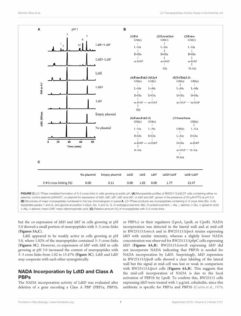
To confirm the pH preference of LdtE and LdtF observedin vivo, we analyzed the muropeptide composition of the PG ofBW2511316LDT expressing ldtD, ldtE, and/or ldtF at pH 5.0.
As expected, BW2511316LDT did not contain muropeptideswith 3–3 cross-links (Figures 3A,C). The PG of BW2511316LDTexpressing ldtD did not show any 3–3 cross-links (Figures 3A,C)
Frontiers in Microbiology | www.frontiersin.org 6 September 2018 | Volume 9 | Article 2101
Montón Silva et al. LD-Transpeptidase Activity Assay in Escherichia coli
FIGURE 3 | LD-TPase-mediated formation of 3–3 cross-links in cells growing at acidic pH. (A) Muropeptide profiles of BW2511316LDT cells containing either noplasmid, control plasmid pSAV057, or plasmid for expression of ldtD, ldtE, ldtF, ldtE and ldtF, or ldtD and ldtF, grown in the presence of 50 µM IPTG at pH 5.0.(B) Structures of major muropeptides numbered in the top chromatogram in panel A. LD-TPase products are muropeptides containing 3–3 cross-links (No. 4–6),tripeptides (peaks 1 and 5), and glycine at position 4 (Gly4, No. 2 and 4). G, N-acetylglucosamine; M(r), N-acetylmuramitol; L-Ala, L-alanine; D-Glu, D-glutamic acid;D-Ala, D-alanine; meso-DAP, meso-diaminopimelic acid. (C) Relative amount (%) of muropeptides with 3–3 cross-links.
but the co-expression of ldtD and ldtF in cells growing at pH5.0 showed a small portion of muropeptides with 3–3 cross-links(Figures 3A,C).
LdtE appeared to be weakly active in cells growing at pH5.0, where 1.02% of the muropeptides contained 3–3 cross-links(Figures 3C). However, co-expression of ldtF with ldtE in cellsgrowing at pH 5.0 increased the content of muropeptides with3–3 cross-links from 1.02 to 13.47% (Figure 3C). LdtE and LdtFmay cooperate with each other synergistically.
NADA Incorporation by LdtD and Class APBPsThe NADA incorporation activity of LdtD was evaluated afterdeletion of a gene encoding a Class A PBP (PBP1a, PBP1b,
or PBP1c) or their regulators (LpoA, LpoB, or CpoB). NADAincorporation was detected in the lateral wall and at mid-cellin BW251131mrcA and in BW251131lpoA strains expressingldtD with similar intensity, whereas a slightly lower NADAconcentration was observed for BW251131pbpC cells expressingldtD (Figures 4A,B). BW251131mrcB expressing ldtD didnot incorporate NADA indicating that PBP1b is needed forNADA incorporation by LdtD. Surprisingly, ldtD expressionin BW251131lpoB cells showed a clear labeling of the lateralwall but the signal at mid-cell was lost or weak in comparisonwith BW251131lpoA cells (Figures 4A,B). This suggests thatthe mid-cell incorporation of NADA is due to the localactivation of PBP1b by LpoB. To confirm this, BW25113 cellsexpressing ldtD were treated with 1 µg/mL cefsulodin, since thisantibiotic is specific for PBP1a and PBP1b (Curtis et al., 1979;
Frontiers in Microbiology | www.frontiersin.org 7 September 2018 | Volume 9 | Article 2101
Montón Silva et al. LD-Transpeptidase Activity Assay in Escherichia coli
FIGURE 4 | PBP1b is required for NADA incorporation by LdtD. (A) Phase contrast and corresponding fluorescence images of cells of BW25113, BW251131lpoA,BW251131mrcA (encoding PBP1a), BW251131lpoB, BW251131mrcB (PBP1b), BW251131pbpC (PBP1c), and BW251131cpoB expressing ldtD and labeledwith 0.5 mM NADA. Scale bar, 2 µm. The right panel shows the fluorescence profiles per average cell (from 0 to 1000 AU) plotted against normalized cell length(from 0 to 100%). n represents the number of cells analyzed. (B) Concentration of NADA fluorescence (fluorescence signal per µm3 average cell volume).Quantification of the incorporated NADA in the WT strain, BW251131lpoA, BW251131mrcA, BW251131lpoB, BW251131mrcB, BW251131pbpC, andBW251131cpoB strains expressing ldtD. The values are mean ± SD of n number of cells. n is indicated on the fluorescence profiles.
Sarkar et al., 2012; Kocaoglu and Carlson, 2015). NADA wasincorporated in the lateral wall but clear empty septa (non-labeled) were observed (Supplementary Figure S3), confirmingthat the inhibition of PBP1b affected the NADA-incorporationactivity of LdtD at mid-cell, as in the case of the BW251131lpoBstrain. The deletion of cpoB did not allow the incorporation ofNADA by LdtD (Figure 4A).
Because PBP1b was found to be needed for NADAincorporation by LdtD, BW251131mrcB was co-transformedwith pJEH12(LdtD) and a plasmid expressing PBP1a, PBP1b,or PBP1c genes to determine whether any of these GTasescould compensate for the absence of PBP1b. Surprisingly,co-expression of LdtD and PBP1a or PBP1c restored the
signal of PG labeling, with no differences in total NADAconcentration, but with the lack of NADA incorporation at mid-cell (Figures 5A,B). As expected, the co-expression of the genesencoding PBP1b and LdtD restored the signal not only at thelateral wall but also at mid-cell (Figure 5A), confirming thatPBP1b is required for NADA incorporation by LdtD, especiallyat mid-cell.
NADA Incorporation Requires Class CPBPsTo determine whether a DD-CPase is required for theNADA incorporation activity of LdtD, CS1091dacA (lacking
Frontiers in Microbiology | www.frontiersin.org 8 September 2018 | Volume 9 | Article 2101
Montón Silva et al. LD-Transpeptidase Activity Assay in Escherichia coli
FIGURE 5 | Effect of plasmid-expressed genes encoding class A PBPs on NADA incorporation by LdtD. (A) Phase contrast and corresponding fluorescence imagesof BW251131mrcB (without PBP1b) (control) expressing or not ldtD, mrcA, mrcB, pbpC, ldtD and mrcA, ldtD and mrcB, or ldtD and pbpC, labeled with a 0.5 mMNADA pulse of 2 min. Scale bar, 2 µm. The right panel shows the average normalized fluorescence profiles (green) and diameter (purple) per cell (from 0.0 to 1.0)plotted against normalized cell length (from 0 to 100%). n represents the number of cells analyzed. (B) Concentration is the NADA fluorescence (signal per µm3
average cell volume). Quantification of the incorporated NADA in cells of BW251131mrcB (control) or expressing ldtD, mrcA, mrcB, pbpC, ldtD and mrcA, ldtD andmrcB, and ldtD and pbpC. The values are mean ± SD of n number of cells. n is indicated on the fluorescence profiles.
PBP5), CS1091dacC (PBP6a), and CS1091dacD (PBP6b)overproducing LdtD were labeled with NADA. Deletion ofdacA did not enable LdtD to incorporate NADA in thePG (Figure 6A), whereas the deletion of dacC and dacDallowed the labeling of cells with NADA (Figures 6A,B). Thisindicates that PBP5 is required for the incorporation of NADAby LdtD.
BW2511316LDT1dacA was co-transformed withpJEH12(LdtD) and a plasmid expressing the genes of PBP5,PBP6a, or PBP6b to determine whether any of these DD-CPases
could complement for the absence of PBP5. As expected, thecombined expression of ldtD and dacA restored the signal ofNADA incorporation (Figure 6C). Interestingly, co-expressionof ldtD and the gene encoding PBP6b also complementedfor the absence of PBP5 (Figure 6C) yielding similar NADAincorporation at mid-cell compared to the co-expression ofdacA and ldtD (Figures 6C,D). Co-expressing ldtD and the geneencoding PBP6a did not allow for LdtD-mediated incorporationof NADA (Figure 6C) meaning that PBP6a, unlike PBP6b, is notable to compensate for the absence of PBP5.
Frontiers in Microbiology | www.frontiersin.org 9 September 2018 | Volume 9 | Article 2101
Montón Silva et al. LD-Transpeptidase Activity Assay in Escherichia coli
FIGURE 6 | Class C PBP activity is required for NADA incorporation by LdtD. (A) Phase contrast and fluorescence images of CS109, CS1091dacA (PBP5)expressing ldtD, CS1091dacC (PBP6a) expressing ldtD, and CS1091dacD (PBP6b) expressing ldtD labeled with a 0.5 mM NADA pulse of 2 min. Scale bar, 2 µm.The right panel shows the average fluorescence profiles (green) and diameter (purple) per cell (from 0.0 to 1.0) plotted against normalized cell length (from 0 to100%). n represents the number of cells analyzed. (B) Total concentration is the NADA fluorescence (signal per µm3 average cell volume). Quantification of theincorporated NADA in the CS109 strain, CS1091dacA expressing ldtD, CS1091dacC expressing ldtD, and CS1091dacD expressing ldtD. The values aremean ± SD of n number of cells. (C) Phase contrast and fluorescence images of BW5113D6LDTDdacA cells not expressing (control) or expressing ldtD, dacA, ldtDand dacA, dacC, ldtD, and dacC, dacD, or ldtD and dacD, labeled with 0.5 mM NADA. Scale bar, 2 µm. The right panel shows the average fluorescence profiles(green) and diameter (purple) per cell (from 0.0 to 1.0) plotted against normalized cell length (from 0 to 100%). (D) Quantification of the total concentration ofincorporated NADA in the BW2511316LDT1dacA cells not expressing (control) or expressing ldtD, dacA, ldtD and dacA, dacC, ldtD, and dacC, dacD, or ldtD anddacD. The values are mean ± SD of n number of cells.
Frontiers in Microbiology | www.frontiersin.org 10 September 2018 | Volume 9 | Article 2101
Montón Silva et al. LD-Transpeptidase Activity Assay in Escherichia coli
Effect of Aztreonam on Cells ExpressingldtDWe wanted to determine whether the overproduced LdtD couldcompensate for the block of the essential cell division class BPBP3. We treated BW25113 cells expressing or not ldtD withaztreonam for 20 and 60 min and then labeled them with NADA.We observed NADA incorporation at the lateral wall and mid-cell after the 20 min pulse (Figures 7A,B mid-panel). After60 min, cells overexpressing ldtD showed bulges at the previousdivision site (Figure 7B low-panel) while cells not expressingldtD grew as smooth filaments (Figure 7A low-panel), indicatingthat the expression of ldtD is responsible for this bulgingphenotype. Moreover, NADA incorporation was detected at one-fourth and three-fourth of the filaments (Figure 7B low-panel),presumably at preseptal sites. Similar results were observed forthe BW251131lpoB cells expressing ldtD (Figures 7C,D mid-and lower-panel). In contrast, the BW251131cpoB strain grownin the presence of aztreonam showed smooth filaments andNADA was not incorporated irrespective whether ldtD wasexpressed or not (Figures 7E,F).
Because of the importance of PBP1b for the function of LdtD,we repeated the treatment with 1 µg/mL aztreonam for 1 h inBW251131mrcB cells either expressing genes encoding differentPBP1b variants (WT PBP1b, PBP1b GT∗, PBP1b TP∗, and PBP1bGT∗TP∗) alone or co-expressed with ldtD. In the absence of ldtDexpression, cells readily lysed in all cases except for the expressionof the WT PBP1b gene, which led to filaments with bulges atmid-cell (Supplementary Figure S4). A similar level of lysis wasobserved when ldtD was co-expressed with the genes encodingPBP1b GT∗ or PBP1b GT∗TP∗. Interestingly, the level of lysiswas considerably lower when ldtDwas co-expressed with the geneencoding PBP1b TP∗ (Supplementary Figure S4).
DISCUSSION
pH Dependence for FDAAs Incorporationby LdtDPenicillin-insensitive LD-TPases were proposed to be responsiblefor the incorporation of exogenous D-Met, other D-amino acids(Tsuruoka et al., 1984; Pisabarro et al., 1985; Caparrós et al.,1992) and FDAA probes, such as NADA or HADA (Kuruet al., 2012) into the terminal position of tetrapeptide stems inE. coli. Here we aimed to identify which of the six LD-TPasesof E. coli incorporates NADA. NADA was readily incorporatedinto BW25113 cells but not those of a strain lacking all LD-TPase genes (BW2511316LDT), indicating that at least one ofthe LD-TPs is required for the PG labeling by LD-TPases. In thebackground of the BW2511316LDT strain, NADA incorporationwas only visible when LdtD was overproduced from a plasmid(Figure 1). Because cells were grown in exponential phase,LdtD might be more active in exponentially growing cells,or has a higher NADA-incorporation activity than the otherLD-TPases catalyzing 3–3 cross-links, LdtE and LdtF (Magnetet al., 2008; Morè et al., unpublished). It cannot be excludedthat PBPs can incorporate NADA, which is then immediately
removed by DD-CPases. However, since NADA incorporationwas not observed in the BW2511316LDT1dacA, the main DD-CPase PBP5 is not needed for the removal. We also observedthat LdtD was able to incorporate the FDAA HADA in vitro(Supplementary Figure S1). From both in vivo and in vitrostudies, we can conclude that FDAAs are a suitable substrate forLdtD.
A recent study found that PBP6b is more active at pH 5.0(compared to pH 7.5), whereas PBP5 and PBP6a showed a loweractivity at acidic pH, both in the cell and with the purifiedenzymes (Peters et al., 2016). Presumably, E. coli maintains sets ofPG enzymes to grow robustly at different growth conditions, suchas different pH values (Pazos et al., 2017). LdtD was not able toincorporate NADA at pH 5.0, whereas LdtE seemed to be inactiveat pH 7.0 and weakly active at pH 5.0 (Figure 2). Nevertheless,LdtE and LdtF were observed to be more active when they wereoverproduced together at acidic pH (Figures 2, 3). They maybe active under acidifying condition such as fermentation whileLdtD would be active during respiratory growth at near neutralpH. Why co-expression of both ldtE and ldtF, and not ldtD andldtF, improved the incorporation of NADA at pH 5.0 remainsstill unclear but it suggests the possibility of a potential activationsystem between these two LD-TPases.
PBP1b, LpoB, and CpoB Are Required forEfficient Incorporation of NADA by LdtDLdtD can work on old, pre-formed PG (SupplementaryFigure S1) but may also work on newly synthesized PGin the context of an ampicillin resistant mutant strain ofE. coli (Hugonnet et al., 2016). Indeed, we found here thatthe incorporation of NADA by overproduced LdtD requiresthe presence of PBP1b and its regulators LpoB and CpoB(Figure 4A). The absence of PBP1a, PBP1c, or LpoA did not affectthe NADA incorporation by LdtD, since we observed PG labelingin the BW251131lpoA, BW251131mrcA, and BW251131pbpCstrains whether (Figure 4A) or not (Supplementary Figure S2)ldtD was expressed. These data align well with previous workshowing that LdtD is functionally linked to PBP1b but not PBP1a(Hugonnet et al., 2016). In the BW251131mrcB strain, PBP1aand PBP1c did not compensate for the absence of PBP1b toenable LdtD-mediated NADA incorporation, unless they wereoverproduced from plasmid together with LdtD (Figure 5A). Co-overproducing LdtD and PBP1a or PBP1c restored the signal ofincorporated NADA only in the lateral wall but not at mid-cell.This finding supposes the first phenotype associated with PBP1cactivity. PBP1c might have a role in situations where extra GTaseactivity is required, as previously hypothesized (Budd et al., 2004).
Expression of ldtD in BW251131lpoB affected incorporationof NADA at mid-cell (Figure 4A), although the labeling atthe lateral wall was comparable to the BW251131lpoA strain(Figures 4A,B). LpoB showed enhanced localization at mid-cell(Typas et al., 2010) where it would activate both GTase and TPaseactivities of PBP1b (Egan et al., 2014). Treating BW25113 cellswith cefsulodin, which specifically inhibits PBP1a and b (Curtiset al., 1979; Sarkar et al., 2012; Kocaoglu and Carlson, 2015),provided similar results to the deletion of lpoB. Together, these
Frontiers in Microbiology | www.frontiersin.org 11 September 2018 | Volume 9 | Article 2101
Montón Silva et al. LD-Transpeptidase Activity Assay in Escherichia coli
FIGURE 7 | ldtD expression in aztreonam-treated cells creates bulges at mid-cell. Phase contrast images, corresponding fluorescent images, and fluorescenceprofiles per average cell (from 0 to 2000 AU) plotted against normalized cell length (from 0 to 100%). (A) BW25113, (B) BW25113 expressing ldtD,(C) BW251131lpoB, (D) BW251131lpoB expressing ldtD, (E) BW251131cpoB, and (F) BW251131cpoB expressing ldtD. Upper panel, no antibiotic; middlepanel, 20 min incubation with 1 µg/mL aztreonam; lower panel, 60 min of incubation with 1 µg/mL aztreonam. PG was labeled with 0.5 mM NADA with a 2 minlabeling pulse after the 20 or 60 min incubation with aztreonam. Scale bar, 2 µm. Red triangles point to mid-cell bulges. n represents the number of cells analyzed.
Frontiers in Microbiology | www.frontiersin.org 12 September 2018 | Volume 9 | Article 2101
Montón Silva et al. LD-Transpeptidase Activity Assay in Escherichia coli
results suggest that the activation of PBP1b by LpoB is requiredfor incorporation of NADA at mid-cell by LdtD.
CpoB modulates the stimulation of the TPase activity ofPBP1b (Gray et al., 2015). Deletion of cpoB renders cellsmore sensitive to cefsulodin, indicating that the TPase activityof PBP1b strongly contributes to PG synthesis (Gray et al.,2015). Consistent with the hyper TPase activity of PBP1b,NADA was presumably not incorporated in a BW251131cpoBstrain expressing ldtD because of the lack of available substrate(Figure 4A). The results confirm and extend the role of CpoB asa regulator of the TPase activity of PBP1b in vivo.
PBP6b Can Complement the Absence ofPBP5 for LdtD ActivityDeletion of dacA, encoding the major DD-CPase PBP5,did obstruct the incorporation of NADA into PG by LdtD(Figure 6A). PBP5 hydrolyzes the D-Ala4-D-Ala5 bond ofpentapeptide stems (Hugonnet et al., 2016), which results inthe production of tetrapeptides, which are the substrate for LD-TPases (Magnet et al., 2008). Deletion of dacC (encoding PBP6a)and dacD (encoding PBP6b) allowed incorporation of NADA byoverproduced LdtD and no significant differences were foundbetween the two deletion strains (Figure 6). This was expected forPBP6b considering that its gene is known to be hardly expressedduring exponential growth (Li et al., 2014; Peters et al., 2016), andalso for PBP6A for which no in vivo activity has been reported(Meiresonne et al., 2017).
To evaluate the possible complementation for the absenceof PBP5, LdtD and one of the class C PBPs (PBP5, PBP6a, orPBP6b) were co-overproduced in a strain lacking all LD-TPs andPBP5 (BW2511316LDT1dacA). PBP5 is the major DD-CPasein E. coli and the morphological defects associated with deletionof the corresponding gene cannot be compensated by the otherclass C PBPs present on the chromosome (Nelson and Young,2000). However, our studies show that overproduction of PBP6bprovided sufficient DD-CPase activity in a 1dacA background tosupport LdtD-mediated incorporation of NADA.
Expressing ldtD Causes Bulges in CellsTreated With AztreonamRecently, we proved that LdtD is able to bypass the DD-TPasepathway in cells treated with ampicillin (Hugonnet et al., 2016).We observed that NADA was strongly incorporated at mid-celland the resistant E. coli was able to divide, which suggestedthe involvement of the essential cell division class B PBP3TPase activity. We decided to evaluate whether overproducingLdtD could compensate for the decrease of 4–3 cross-linkingderived from the inhibition of the DD-TPase activity of PBP3 byaztreonam (Sykes and Bonner, 1985). These cells have completedivision machineries at potential division sites at least for twomass doublings (Pogliano et al., 1997; van der Ploeg et al.,2013). The filaments showed bulges at mid-cell after 60 minaztreonam treatment (approximately two mass doublings) in WTBW25113 cells expressing ldtD, indicating that the expressionof ldtD may be responsible for this phenotype. This phenotypewas also observed in ampicillin-resistant E. coli expressing ldtD
(Hugonnet et al., 2016). Interestingly, bulges were also present inthe BW251131lpoB strain overproducing ldtD (Figures 7C,D),whereas with active PBP3 the BW251131lpoB strain did notincorporate NADA at mid-cell (Figure 4A). Consequently, theNADA incorporation in aztreonam-inhibited BW251131lpoBcells likely corresponds to lateral wall synthesis, which wouldsupport the notion that PBP1b is not necessarily part ofthe divisome but can be active at its periphery (Cho et al.,2016).
Why does the overexpression of ldtD in aztreonam treatedcells result in bulges? After PBP3 is inhibited by β-lactams thedivision machinery remains assembled for at least two massdoublings (Pogliano et al., 1997; van der Ploeg et al., 2013). Thecells synthesize only PG for elongation, switching between phasesof pre-septal PG synthesis and dispersed, lateral PG synthesis.After blocking PBP3 with aztreonam, the incorporation oflipid-II in the PG layer is regulated by PBP1b. Perhaps theendogenous LdtD activity in E. coli is not enough to compensatefor the decrease in 4–3 cross-links and to incorporate GTase-derived glycan chains into PG when the essential DD-TPasePBP3 is blocked by aztreonam. However, overproduction ofLdtD results in extra 3–3 cross-links that may lead to animproperly orientated septal PG synthesis and subsequent bulgeformation. The presence of bulges at mid-cell is common inE. coli cells treated with other β-lactams, from low concentrationsof penicillin (Schwarz et al., 1969) to high concentrations ofampicillin (Hugonnet et al., 2016).
We also evaluated the effect of expressing ldtD when theessential DD-TPase PBP3 is blocked by aztreonam and aninactive PBP1b is overproduced, since we have detected adependence of LdtD on PBP1b. The overproduction of WTPBP1b is non-lethal for the cells in contrast to the production ofinactive forms of PBP1b that cause cells to lyse (SupplementaryFigure S4; Meisel et al., 2003). Overproducing LdtD with PBP1bTP∗ compensates for the loss in 4–3 cross-links, preventingand/or delaying cell lysis and lead to the observed survivalphenotype.
With this work, we provide additional information on the roleof LdtD in β-lactam-treated cells. These observations imply thatinhibition of LD-TPases is a possible option to keep β-lactamresistance at bay.
AUTHOR CONTRIBUTIONS
AMS and TB conceived the experiments. AMS, CO, and JBperformed the experiments and prepared figures. MV suppliedNADA and HADA. EB supplied lipid II for in vitro studies ofLdtD. AMS, TB, WV, and CO wrote the manuscript. All authorsreviewed the manuscript.
FUNDING
AMS, TB, CO, and WV received support from the NAPCLIproject within the JPIAMR program (AMS and TB from ZonMWproject 60-60900-98-207 and WV from MR/N501840/1).
Frontiers in Microbiology | www.frontiersin.org 13 September 2018 | Volume 9 | Article 2101
Montón Silva et al. LD-Transpeptidase Activity Assay in Escherichia coli
MV acknowledges the financial support from the NationalInstitutes of Health (GM 113172).
ACKNOWLEDGMENTS
We thank Nils Meiresonne (University of Amsterdam) for thecloning of the pNM039 plasmid. We thank Michel Arthur andJean Emmanuel Hugonnet (University Pierre et Marie Curie)for providing the plasmid pJEH12(LdtD). We thank Alessandra
Polissi (University of Milano) for the plasmids pGS121 andpGS124. We also thank the KEIO collection for the creation ofthe knockout strain collection and the gift of BW251131pbpC.
SUPPLEMENTARY MATERIAL
The Supplementary Material for this article can be foundonline at: https://www.frontiersin.org/articles/10.3389/fmicb.2018.02101/full#supplementary-material
REFERENCESAlexeeva, S., Gadella, T. W. Jr., Verheul, J., Verhoeven, G. S., and den Blaauwen, T.
(2010). Direct interactions of early and late assembling division proteins inEscherichia coli cells resolved by FRET. Mol. Microbiol. 77, 384–398. doi: 10.1111/j.1365-2958.2010.07211.x
Baba, T., Ara, T., Hasegawa, M., Takai, Y., Okumura, Y., Baba, M., et al. (2006).Construction of Escherichia coli K-12 in-frame, single-gene knockout mutants:the Keio collection. Mol. Syst. Biol. 2:2006.0008. doi: 10.1038/msb4100050
Banzhaf, M., van den Berg van Saparoea, B., Terrak, M., Fraipont, C., Egan, A.,Philippe, J., et al. (2012). Cooperativity of peptidoglycan synthases active inbacterial cell elongation. Mol. Microbiol. 85, 179–194. doi: 10.1111/j.1365-2958.2012.08103.x
Baquero, M. R., Bouzon, M., Quintela, J. C., Ayala, J. A., and Moreno, F. (1996).dacD, an Escherichia coli gene encoding a novel penicillin-binding. Protein(PBP6b) with DD-Carboxypeptidase activity. J. Bacteriol. 178, 7106–7111.doi: 10.1128/jb.178.24.7106-7111.1996
Bertsche, U., Breukink, E., Kast, T., and Vollmer, W. (2005). In vitro mureinpeptidoglycan synthesis by dimers of the bifunctional transglycosylase-transpeptidase PBP1B from Escherichia coli. J. Biol. Chem. 280, 38096–38101.doi: 10.1074/jbc.M508646200
Bertsche, U., Kast, T., Wolf, B., Fraipont, C., Aarsman, M. E. G., Kannenberg, K.,et al. (2006). Interaction between two murein (peptidoglycan) synthases, PBP3and PBP1b, in Escherichia coli. Mol. Microbiol. 61, 675–690. doi: 10.1111/j.1365-2958-2006.05280.x
Born, P., Breukink, E., and Vollmer, W. (2006). In vitro synthesis of cross-linkedmurein and its attachment to sacculi by PBP1A from Escherichia coli. J. Biol.Chem. 281, 26985–26993. doi: 10.1074/jbc.M604083200
Broome-Smith, J. K., Ioannidis, I., Edelman, A., and Spratt, B. G. (1998). Nucleotidesequences of the penicillin-binding protein 5 and 6 genes of Escherichia coli.Nucleic Acids Res. 16:1617. doi: 10.1093/nar/16.4.1617
Budd, A., Blandin, S., Levashina, E. A., and Gibson, T. J. (2004). Bacterial α2-macroglobulins: colonization factors acquired by horizontal gene transfer fromthe metazoan genome? Genome Biol. 5:R38.
Bui, N. K., Gray, J., Schwarz, H., Schumann, P., Blanot, D., and Vollmer, W. (2009).The peptidoglycan sacculus of Myxococcus xanthus has unusual structuralfeatures and is degraded during glycerol-induced myxospore development.J. Bacteriol. 191, 494–505. doi: 10.1128/JB.00608-08
Caparrós, M., Pisabarro, A. G., and de Pedro, M. A. (1992). Effect of D-amino acidson structure and synthesis of peptidoglycan in Escherichia coli. J. Bacteriol. 174,5549–5559. doi: 10.1128/jb.174.17.5549-5559.1992
Cherepanov, P. P., and Wackernagel, W. (1995). Gene disruption in Escherichiacoli: tcR and KmR cassettes with the option of Flp-catalyzed excision of theantibiotic-resistance determinant. Gene 158, 9–14. doi: 10.1016/0378-1119(95)00193-A
Cho, H., Uehara, T., and Bernhardt, T. G. (2014). Beta-lactam antibiotics inducea lethal malfunctioning of the bacterial cell wall synthesis machinery. Cell 159,1300–1311. doi: 10.1016/j.cell.2014.11.017
Cho, H., Wivagg, C. N., Kapoor, M., Barry, Z., Rohs, P. D. A., Suh, H., et al.(2016). Bacterial cell wall biogenesis is mediated by SEDS and PBP polymerasefamilies functioning semi-autonomously. Nat. Microbiol. 1:16172. doi: 10.1038/nmicrobiol.2016.172
Curtis, N. A., Orr, D., Ross, G. W., and Boulton, M. G. (1979). Affinities ofpenicillins and cephalosporins for the penicillin-binding proteins of Escherichia
coli K-12 and their antibacterial activity. Antimicrob. Agents Chemother. 16,533–539. doi: 10.1128/AAC.16.5.533
Datsenko, K. A., and Wanner, B. L. (2000). One-step inactivation of chromosomalgenes in Escherichia coli K-12 using PCR products. Proc. Natl. Acad. Sci. U.S.A.97, 6640–6645. doi: 10.1073/pnas.120163297
Den Blaauwen, T., De Pedro, M., Nguyen-Distèche, M., and Ayala, J. (2008).Morphogenesis of rod-shaped sacculi. FEMS Microbiol. Rev. 32, 321–344.doi: 10.1111/j.1574-6976.2007.00090.x
Denome, S. A., Elf, P. K., Henderson, T. A., Nelson, D. E., and Young, K. D. (1999).Escherichia coli mutants lacking all possible combinations of eight penicillinbinding proteins: viability, characteristics, and implications for peptidoglycansynthesis. J. Bacteriol. 181, 3981–3993.
Egan, A. J. F., Jean, N. L., Koumoutsi, A., Bougault, C. M., Biboy, J., Sassine, J., et al.(2014). Outer-membrane lipoprotein LpoB spans the periplasm to stimulate thepeptidoglycan synthase PBP1b. Proc. Natl. Acad. Sci. U.S.A. 111, 8197–8202.doi: 10.1073/pnas.1400376111
Gibson, D. G., Young, L., Chuang, R. Y., Venter, J. C., Hutchison, C. A., and Smith,H. O. (2009). Enzymatic assembly of DNA molecules up to several hundredkilobases. Nat. Methods 6, 343–345. doi: 10.1038/nmeth.1318
Glauner, B., Holtje, J. V., and Schwarz, U. (1988). The composition of the mureinof Escherichia coli. J. Biol. Chem. 263, 10088–10095.
Goffin, C., and Ghuysen, J. M. (1998). Multimodular penicillin-binding proteins:an enigmatic family of orthologs and paralogs. Microbiol. Mol. Biol. Rev. 62,1079–1093.
Gray, A. N., Egan, A. J. F., van’t Veer, I. L., Verheul, J., Colavin, A., Koumoutsi, A.,et al. (2015). Coordination of peptidoglycan synthesis and outer membraneconstriction during Escherichia coli cell division. eLife 4:e07118. doi: 10.7554/eLife.07118
Henrich, E., Ma, Y., Engels, I., Münch, D., Otten, C., Schneider, T., et al. (2016).Lipid requirements for the enzymatic activity of MraY translocases and in vitroreconstitution of the lipid II synthesis pathway. J. Biol. Chem. 291, 2535–2546.doi: 10.1074/jbc.M115.664292
Hugonnet, J. E., Mengin-Lecreulx, D., Montón, A., den Blaauwen, T.,Carbonnelle, E., Veckerlé, C., et al. (2016). Factors essential for L,D-transpeptidase mediated peptidoglycan cross-linking and β-lactam resistancein Escherichia coli. eLife 5:e19469. doi: 10.7554/eLife.19469
Kocaoglu, O., and Carlson, E. E. (2015). Profiling of β-lactam selectivity forpenicillin-binding proteins in Escherichia coli strain DC2. Antimicrob. AgentsChemother. 59, 2785–2790. doi: 10.1128/AAC.04552-14
Koppelman, C. M., Aarsman, M. E., Postmus, J., Pas, E., Muijsers, A. O.,Scheffers, D. J., et al. (2004). R174 of Escherichia coli FtsZ is involvedin membrane interaction and protofilament bundling, and is essential forcell division. Mol. Microbiol. 51, 645–657. doi: 10.1046/j.1365-2958.2003.03876.x
Kuru, E., Hughes, V. H., Brown, P. J., Hall, E., Tekkam, S., Cava, F., et al.(2012). In situ probing of newly synthesized peptidoglycan in live bacteriawith fluorescent d-amino acids. Angew. Chem. Int. Ed. Engl. 51, 12519–12523.doi: 10.1002/anie.201206749
Kuru, E., Tekkam, S., Hall, E., Brun, Y. V., and Van Nieuwenhze, M. S. (2015).Synthesis of fluorescent D-amino acids and their use for probing peptidoglycansynthesis and bacterial growth in situ. Nat. Protoc. 10, 33–52. doi: 10.1038/nprot.2014.197
Kuru, K., Lambert, C., Rittichier, J., Till, R., Ducret, A., Derouaux, A., et al. (2017).Fluorescent D-amino-acids reveal bi-cellular cell wall modifications important
Frontiers in Microbiology | www.frontiersin.org 14 September 2018 | Volume 9 | Article 2101
Montón Silva et al. LD-Transpeptidase Activity Assay in Escherichia coli
for Bdellovibrio bacteriovorus predation. Nat. Microbiol. 2, 1648–1657.doi: 10.1038/s41564-017-0029-y
Li, G. W., Burkhardt, D., Gross, C., and Weissman, J. S. (2014). Quantifyingabsolute protein synthesis rates reveals principles underlying allocation ofcellular resources. Cell 157, 624–635. doi: 10.1016/j.cell.2014.02.033
Magnet, S., Bellais, S., Dubost, L., Fourgeaud, M., Mainardi, J. L., Petit-Frère, S., et al. (2007). Identification of the L,D-Transpeptidases responsiblefor attachment of the braun lipoprotein to Escherichia coli peptidoglycan.J. Bacteriol. 189, 3927–3931. doi: 10.1128/JB.00084-07
Magnet, S., Dubost, L., Marie, A., Arthur, M., and Gutmann, L. (2008).Identification of the L,D-transpeptidases for peptidoglycan cross-linking inEscherichia coli. J. Bacteriol. 190, 4782–4785. doi: 10.1128/JB.00025-08
Mainardi, J. L., Fourgeaud, M., Hugonnet, J. E., Dubost, L., Brouard, J. P.,Ouazzani, J., et al. (2005). A novel peptidoglycan cross-linking enzyme for aβ-lactam-resistant transpeptidation pathway. J. Biol. Chem. 280, 38146–38152.doi: 10.1074/jbc.M507384200
Mainardi, J. L., Hugonnet, J. E., Rusconi, F., Fourgeaud, M., Dubost, L., Moumi,A. N., et al. (2007). Unexpected Inhibition of Peptidoglycan LD-Transpeptidasefrom Enterococcus faecium by the β-lactam imipenem. J. Biol. Chem. 282,30414–30422. doi: 10.1074/jbc.M704286200
Meeske, A. J., Riley, E. P., Robins, W. P., Uehara, T., Mekelanos, J. J., Kahne, D.,et al. (2016). SEDS proteins are a widespread family of bacterial cell wallpolymerases. Nature 537, 634–638. doi: 10.1038/nature19331
Meiresonne, N. Y., van der Ploeg, R., Hink, M. A., and den Blaauwen, T. (2017).Activity-related conformational changes in d,d-carboxypeptidases revealed byin vivo Periplasmic Förster resonance energy transfer assay in Escherichia coli.mBio 8:e01089-17. doi: 10.1128/mBio.01089-17
Meisel, U., Hoeltje, J. V., and Vollmer, W. (2003). Overproduction of inactivevariants of the murein synthase PBP1B causes lysis in Escherichia coli.J. Bacteriol. 185, 5342–5348. doi: 10.1128/JB.185.18.5342-5348.2003
Mohammadi, T., Sijbrandi, R., Lutters, M., Verheul, J., Martin, N. I., denBlaauwen, T., et al. (2014). Specificity of the transport of lipid II by FtsW inEscherichia coli. J. Biol. Chem. 289, 14707–14718. doi: 10.1074/jbc.M114.557371
Nelson, D. E., and Young, K. D. (2000). Penicillin binding protein 5 affectscell diameter, contour, and morphology of Escherichia coli. J. Bacteriol. 182,1714–1721. doi: 10.1128/JB.182.6.1714-1721.2000
Paradis-Bleau, C., Markovski, M., Uehara, T., Lupoli, T. J., Walker, S., Kahne, D. E.,et al. (2010). Lipoprotein cofactors located in the outer membrane activatebacterial cell wall polymerases. Cell 143, 1110–1120. doi: 10.1016/j.cell.2010.11.037
Park, J. T. (1995). Why does Escherichia coli recycle its cell wall peptides? Mol.Microbiol. 17, 421–426.
Pazos, M., Peters, K., and Vollmer, W. (2017). Robust peptidoglycan growthby dynamic and variablemulti-protein complexes. Curr. Opin. Microbiol. 36,55–61. doi: 10.1016/j.mib.2017.01.006
Peters, K., Kannan, S., Rao, V. A., Biboy, J., Vollmer, D., Erickson, S. W., et al.(2016). The redundancy of peptidoglycan carboxypeptidases ensures robust cellshape maintenance in Escherichia coli. mBio 7, 819–816. doi: 10.1128/mBio.00819-16
Pisabarro, A. G., de Pedro, M. A., and Vazquez, D. (1985). Structural modificationsin the peptidoglycan of Escherichia coli associated with changes in the state ofgrowth of the culture. J. Bacteriol. 161, 238–242.
Pogliano, J., Pogliano, K., Weiss, D. S., Losick, R., and Beckwith, J. (1997).Inactivation of FtsI inhibits constriction of the FtsZ cytokinetic ring and delaysthe assembly of FtsZ rings at potential division sites. Proc. Natl. Acad. Sci. U.S.A.21, 559–564. doi: 10.1073/pnas.94.2.559
Potluri, L. P., de Pedro, M. A., and Young, K. D. (2012). Escherichia coli low-molecular-weight penicillin-binding proteins help orient septal FtsZ, and theirabsence leads to asymmetric cell division and branching. Mol. Microbiol. 84,203–224. doi: 10.1111/j.1365-2958.2012.08023.x
Rogers, H. J., Perkins, H. R., and Ward, J. B. (1980). Microbial cell walls andmembranes. New York, NY: Chapman and Hall. doi: 10.1007/978-94-011-6014-8
Ruiz, N. (2015). Lipid flippases for bacterial peptidoglycan biosynthesis. LipidInsights 8, 21–31. doi: 10.4137/LPI.S31783
Samsudin, F., Boags, A., Piggot, T. J., and Khalid, S. (2017). Braun’s lipoproteinfacilitates OmpA interaction with the Escherichia coli cell wall. Biophys. J. 113,1496–1504. doi: 10.1016/j.bpj.2017.08.011
Sarkar, S. K., Dutta, M., Kumar, A., Mallik, D., and Ghosh, A. S. (2012).Sub-Inhibitory cefsulodin sensitization of E. coli to β-lactams Is mediatedby PBP1b inhibition. PLoS One 7:e48598. doi: 10.1371/journal.pone.0048598
Sauvage, E., Kerff, F., Terrak, M., Ayala, J. A., and Charlier, P. (2008).The penicillin-binding proteins: structure and role in peptidoglycanbiosynthesis. FEMSMicrobiol. Rev. 32, 234–258. doi: 10.1111/j.1574-6976.2008.00105.x
Schwarz, U., Asmus, A., and Hermann, F. (1969). Autolytic enzymes and celldivision of Escherichia coli. J. Mol. Biol. 41, 419–429. doi: 10.1016/0022-2836(69)90285-X
Sykes, R. B., and Bonner, D. P. (1985). Aztreonam: the first monobactam. Am. J.Med. 78, 2–10. doi: 10.1016/0002-9343(85)90196-2
Tartof, K. D., and Hobbs, C. A. (1987). Improved media for growing plasmid andcosmid clones. Bethesda Res. Lab. Focus 9:12.
Taschner, P. E. M., Huls, P. G., Pas, E., and Woldringh, C. L. (1988).Division behavior and shape changes in isogenic ftsZ, ftsQ, ftsA, pbpB,and ftsE cell division mutants of Escherichia coli during temperature shiftexperiments. J. Bacteriol. 170, 1533–1540. doi: 10.1128/jb.170.4.1533-1540.1988
Thomason, L. C., Costantino, N., and Court, D. J. (2014). E. coli genomemanipulation by P1 transduction. Curr. Protoc. Mol. Biol. 79, 1.17.1–1.17.8.
Tsuruoka, T. A., Tamura, A., Miyata, T., Takei, K., and Iwamatsu, S.(1984). Penicillin-insensitive incorporation of D-amino acids into cell wallpeptidoglycan influences the amount of bound lipoprotein in Escherichia coli.J. Bacteriol. 60, 889–894.
Typas, A., Banzhaf, M., van der Berg van Saparoea, B., Verheul, J., Biboy, J., Nichols,R. J., et al. (2010). Regulation of peptidoglycan synthesis by outer-membraneproteins. Cell 143, 1097–1109. doi: 10.1016/j.cell.2010.11.038
van der Ploeg, R., Verheul, J., Vischer, N. O., Alexeeva, S., Hoogendoorn, E.,Postma, M., et al. (2013). Colocalization and interaction between elongasomeand divisome during a preparative cell division phase in Escherichia coli. Mol.Microbiol. 87, 1074–1087. doi: 10.1111/mmi.12150
Vega, D., and Ayala, J. A. (2006). The DD-carboxypeptidase activity encoded bypbp4B is not essential for the cell growth of Escherichia coli. Arch. Microbiol.185, 23–27. doi: 10.1007/s00203-005-0057-5
Visher, N. O., Verheul, J., Postma, M., van den Berg, van Saparoea, B., Galli, E., et al.(2015). Cell age dependent concentration of Escherichia coli divisome proteinsanalyzed with imageJ and objectJ. Front. Microbiol. 6:586. doi: 10.3389/fmicb.2015.00586
Vollmer, W., and Born, P. (2009). “Bacterial cell envelop peptidoglycan,” inMicrobial Glycobiology, eds A. Moran, O. Holst, P. Brennan, and M. von Itzstein(London: Academic Press), 15–28.
Vollmer, W., Blanot, D., and de Pedro, M. A. (2008a). Peptidoglycan structure andarchitecture. FEMSMicrobiol. Rev. 32, 149–167. doi: 10.1111/j.1574-6976.2007.00094.x
Vollmer, W., and Holtje, J. V. (2004). The architecture of the murein(peptidoglycan) in gram-negative bacteria: vertical scaffold or horizontallayer(s)? J. Bacteriol. 186, 5978–5987. doi: 10.1128/JB.186.18.5978-5987.2004
Vollmer, W., Joris, B., Charlier, P., and Foster, S. (2008b). Bacterial peptidoglycan(murein) hydrolases. FEMS Microbiol. Rev. 32, 259–286. doi: 10.1111/j.1574-6976.2007.00099.x
Yousif, S. Y., Broome-Smith, J. K., and Spratt, B. G. (1985). Lysis of Escherichiacoli by beta-lactam antibiotics: deletion analysis of the role of penicillin-bindingproteins 1A and 1B. J. Gen. Microbiol. 131, 2839–2845.
Conflict of Interest Statement: The authors declare that the research wasconducted in the absence of any commercial or financial relationships that couldbe construed as a potential conflict of interest.