Pyrrolysine Analogues DOI: 10.1002/anie.201402595 The Formation of Pyrroline and Tetrahydropyridine Rings in Amino Acids Catalyzed by Pyrrolysine Synthase (PylD)** Felix Quitterer, Philipp Beck, Adelbert Bacher, and Michael Groll* Abstract: The dehydrogenase PylD catalyzes the ultimate step of the pyrrolysine pathway by converting the isopeptide l- lysine-Ne-3R-methyl-d-ornithine to the 22nd proteinogenic amino acid. In this study, we demonstrate how PylD can be harnessed to oxidize various isopeptides to novel amino acids by combining chemical synthesis with enzyme kinetics and X- ray crystallography. The data enable a detailed description of the PylD reaction trajectory for the biosynthesis of pyrroline and tetrahydropyridine rings as constituents of pyrrolysine analogues. Pyrrolysine (1) is the 22nd genetically encoded amino acid, which is incorporated into some proteins by means of ribosomal read-through of an amber stop codon (UAG) in certain methanogenic archaea and some eubacteria, including the human pathogen Bilophila wadsworthia. [1] Pyrrolysine comprises a 4-methylpyrroline-5-carboxylate, which is linked by an isopeptide bond to Ne of l-lysine. [1b] Recent studies revealed that the gene cluster pylBCDST [2] orchestrates the biosynthesis of 1 [3] (Scheme 1) and its insertion into the proteins MtmB, MtbB, and MttB, [4] which function in the breakdown of methylamines. [5] Crystal structures of PylD in complex with the substrate and product surrogates l-lysine-Ne-d-ornithine (0a) and pyrroline-carboxy-lysine (0) revealed that the C-terminal lysine moiety is well coordinated, whereas the N-terminal unit only partially occupies the hydrophobic active site cavity, which is filled by a cluster of water molecules. [3e] The aim of the presented work was to expand the scope of PylD substrates and harness the enzyme to oxidize isopeptides to novel amino acids (Scheme 2). Since structural and functional data of PylD with its natural product pyrrolysine (1) have not been reported so far, we synthesized the substrate l-lysine-Ne-3R-methyl-d-orni- thine (1a, Figure S1). Compound 1a was cocrystallized with PylD and the complex structure was solved to 2.2 ĸ resolution (Figure 1a, for details see the Supporting Infor- mation). Inspection of the electron density map revealed that 1 has been formed. The structure displays that the l-lysine moiety of the isopeptide is enclosed in a hydrophobic channel, which is comprised by Leu3, Ile60, Phe63, and Ala103. The Scheme 1. Biosynthesis of pyrrolysine (1) starting from two l-lysine molecules. C-terminal and N-terminal parts of 1a and 1 are depicted in gray and black, respectively. SAM = S-adenosylmethionine. Scheme 2. Isopeptide substrates (0a–6a) and their respective prod- ucts (0–3). [*] F. Quitterer, [+] P. Beck, [+] Prof. Dr. A. Bacher, Prof. Dr. M. Groll Center for Integrated Protein Science Munich (CIPSM) Lehrstuhl fɒr Biochemie, Technische UniversitȨt Mɒnchen Lichtenbergstrasse 4, 85748 Garching (Germany) E-mail: [email protected][ + ] These authors contributed equally to this work. [**] We thank the staff of the beamline X06SA at the Paul Scherrer Institute, Swiss Light Source, Villigen (Switzerland) for their help with data collection and Katrin GȨrtner for excellent technical assistance. Kinetic measurements were carried out by F.Q. at the Biological & Organometallic Catalysis (BOC) Laboratories of Prof. Dr. Jçrg Eppinger, King Abdullah University of Science and Technology (KAUST), Thuwal, Saudi Arabia. This work was sup- ported by the Hans-Fischer-Gesellschaft, by Award No. FIC/2010/07 from KAUST, and by the Deutsche Forschungsgemeinschaft (DFG, grant GR1861/7-1). Supporting information for this article is available on the WWW under http://dx.doi.org/10.1002/anie.201402595. . Angewandte Communications 8150 # 2014 Wiley-VCH Verlag GmbH & Co. KGaA, Weinheim Angew. Chem. Int. Ed. 2014, 53, 8150 –8153
Transcript
Pyrrolysine AnaloguesDOI: 10.1002/anie.201402595
The Formation of Pyrroline and Tetrahydropyridine Rings in AminoAcids Catalyzed by Pyrrolysine Synthase (PylD)**Felix Quitterer, Philipp Beck, Adelbert Bacher, and Michael Groll*
Abstract: The dehydrogenase PylD catalyzes the ultimate stepof the pyrrolysine pathway by converting the isopeptide l-lysine-Ne-3R-methyl-d-ornithine to the 22nd proteinogenicamino acid. In this study, we demonstrate how PylD can beharnessed to oxidize various isopeptides to novel amino acidsby combining chemical synthesis with enzyme kinetics and X-ray crystallography. The data enable a detailed description ofthe PylD reaction trajectory for the biosynthesis of pyrrolineand tetrahydropyridine rings as constituents of pyrrolysineanalogues.
Pyrrolysine (1) is the 22nd genetically encoded amino acid,which is incorporated into some proteins by means ofribosomal read-through of an amber stop codon (UAG) incertain methanogenic archaea and some eubacteria, includingthe human pathogen Bilophila wadsworthia.[1] Pyrrolysinecomprises a 4-methylpyrroline-5-carboxylate, which is linkedby an isopeptide bond to Ne of l-lysine.[1b] Recent studiesrevealed that the gene cluster pylBCDST[2] orchestrates thebiosynthesis of 1[3] (Scheme 1) and its insertion into theproteins MtmB, MtbB, and MttB,[4] which function in thebreakdown of methylamines.[5]
Crystal structures of PylD in complex with the substrateand product surrogates l-lysine-Ne-d-ornithine (0 a) andpyrroline-carboxy-lysine (0) revealed that the C-terminallysine moiety is well coordinated, whereas the N-terminal unitonly partially occupies the hydrophobic active site cavity,which is filled by a cluster of water molecules.[3e] The aim ofthe presented work was to expand the scope of PylDsubstrates and harness the enzyme to oxidize isopeptides tonovel amino acids (Scheme 2).
Since structural and functional data of PylD with itsnatural product pyrrolysine (1) have not been reported so far,we synthesized the substrate l-lysine-Ne-3R-methyl-d-orni-thine (1a, Figure S1). Compound 1a was cocrystallized withPylD and the complex structure was solved to 2.2 �resolution (Figure 1a, for details see the Supporting Infor-mation). Inspection of the electron density map revealed that1 has been formed. The structure displays that the l-lysinemoiety of the isopeptide is enclosed in a hydrophobic channel,which is comprised by Leu3, Ile60, Phe63, and Ala103. The
Scheme 1. Biosynthesis of pyrrolysine (1) starting from two l-lysinemolecules. C-terminal and N-terminal parts of 1a and 1 are depictedin gray and black, respectively. SAM = S-adenosylmethionine.
Scheme 2. Isopeptide substrates (0a–6a) and their respective prod-ucts (0–3).
[*] F. Quitterer,[+] P. Beck,[+] Prof. Dr. A. Bacher, Prof. Dr. M. GrollCenter for Integrated Protein Science Munich (CIPSM)Lehrstuhl f�r Biochemie, Technische Universit�t M�nchenLichtenbergstrasse 4, 85748 Garching (Germany)E-mail: [email protected]
[+] These authors contributed equally to this work.
[**] We thank the staff of the beamline X06SA at the Paul ScherrerInstitute, Swiss Light Source, Villigen (Switzerland) for their helpwith data collection and Katrin G�rtner for excellent technicalassistance. Kinetic measurements were carried out by F.Q. at theBiological & Organometallic Catalysis (BOC) Laboratories ofProf. Dr. Jçrg Eppinger, King Abdullah University of Science andTechnology (KAUST), Thuwal, Saudi Arabia. This work was sup-ported by the Hans-Fischer-Gesellschaft, by Award No. FIC/2010/07from KAUST, and by the Deutsche Forschungsgemeinschaft (DFG,grant GR1861/7-1).
Supporting information for this article is available on the WWWunder http://dx.doi.org/10.1002/anie.201402595.
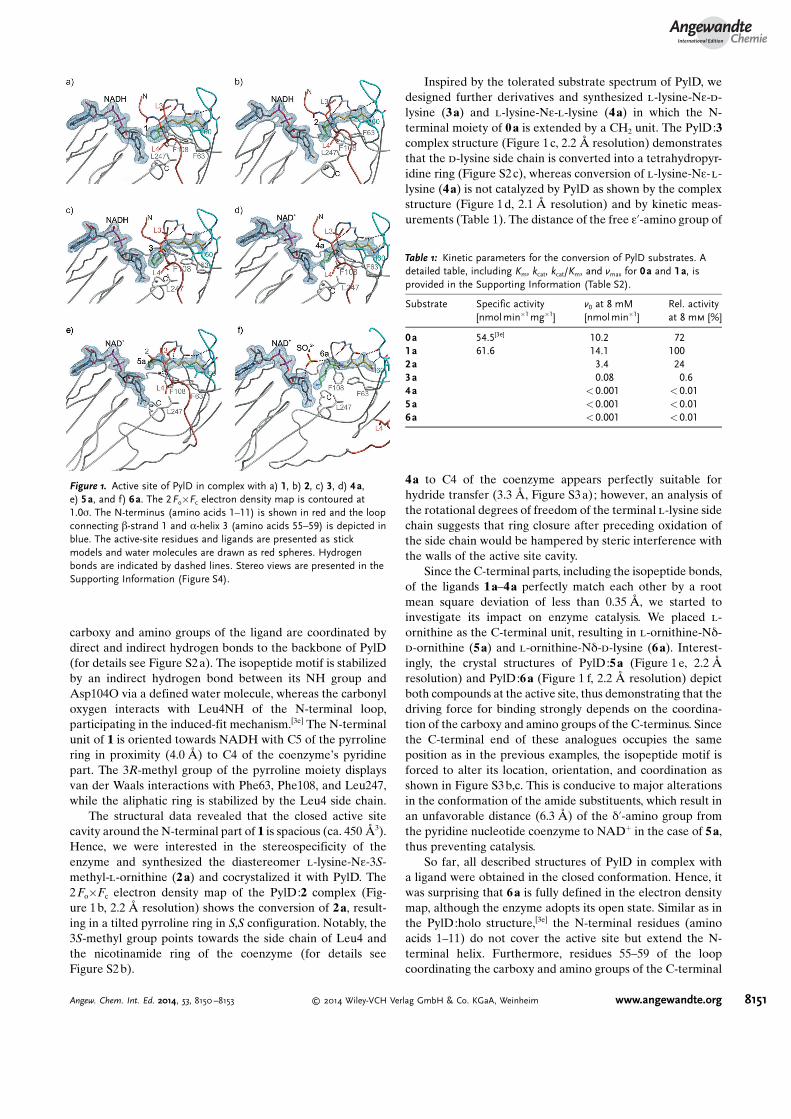
carboxy and amino groups of the ligand are coordinated bydirect and indirect hydrogen bonds to the backbone of PylD(for details see Figure S2 a). The isopeptide motif is stabilizedby an indirect hydrogen bond between its NH group andAsp104O via a defined water molecule, whereas the carbonyloxygen interacts with Leu4NH of the N-terminal loop,participating in the induced-fit mechanism.[3e] The N-terminalunit of 1 is oriented towards NADH with C5 of the pyrrolinering in proximity (4.0 �) to C4 of the coenzyme�s pyridinepart. The 3R-methyl group of the pyrroline moiety displaysvan der Waals interactions with Phe63, Phe108, and Leu247,while the aliphatic ring is stabilized by the Leu4 side chain.
The structural data revealed that the closed active sitecavity around the N-terminal part of 1 is spacious (ca. 450 �3).Hence, we were interested in the stereospecificity of theenzyme and synthesized the diastereomer l-lysine-Ne-3S-methyl-l-ornithine (2 a) and cocrystalized it with PylD. The2Fo�Fc electron density map of the PylD:2 complex (Fig-ure 1b, 2.2 � resolution) shows the conversion of 2a, result-ing in a tilted pyrroline ring in S,S configuration. Notably, the3S-methyl group points towards the side chain of Leu4 andthe nicotinamide ring of the coenzyme (for details seeFigure S2 b).
Inspired by the tolerated substrate spectrum of PylD, wedesigned further derivatives and synthesized l-lysine-Ne-d-lysine (3a) and l-lysine-Ne-l-lysine (4a) in which the N-terminal moiety of 0a is extended by a CH2 unit. The PylD:3complex structure (Figure 1c, 2.2 � resolution) demonstratesthat the d-lysine side chain is converted into a tetrahydropyr-idine ring (Figure S2c), whereas conversion of l-lysine-Ne-l-lysine (4a) is not catalyzed by PylD as shown by the complexstructure (Figure 1d, 2.1 � resolution) and by kinetic meas-urements (Table 1). The distance of the free e’-amino group of
4a to C4 of the coenzyme appears perfectly suitable forhydride transfer (3.3 �, Figure S3a); however, an analysis ofthe rotational degrees of freedom of the terminal l-lysine sidechain suggests that ring closure after preceding oxidation ofthe side chain would be hampered by steric interference withthe walls of the active site cavity.
Since the C-terminal parts, including the isopeptide bonds,of the ligands 1a–4a perfectly match each other by a rootmean square deviation of less than 0.35 �, we started toinvestigate its impact on enzyme catalysis. We placed l-ornithine as the C-terminal unit, resulting in l-ornithine-Nd-d-ornithine (5a) and l-ornithine-Nd-d-lysine (6 a). Interest-ingly, the crystal structures of PylD:5a (Figure 1 e, 2.2 �resolution) and PylD:6a (Figure 1 f, 2.2 � resolution) depictboth compounds at the active site, thus demonstrating that thedriving force for binding strongly depends on the coordina-tion of the carboxy and amino groups of the C-terminus. Sincethe C-terminal end of these analogues occupies the sameposition as in the previous examples, the isopeptide motif isforced to alter its location, orientation, and coordination asshown in Figure S3 b,c. This is conducive to major alterationsin the conformation of the amide substituents, which result inan unfavorable distance (6.3 �) of the d’-amino group fromthe pyridine nucleotide coenzyme to NAD+ in the case of 5a,thus preventing catalysis.
So far, all described structures of PylD in complex witha ligand were obtained in the closed conformation. Hence, itwas surprising that 6a is fully defined in the electron densitymap, although the enzyme adopts its open state. Similar as inthe PylD:holo structure,[3e] the N-terminal residues (aminoacids 1–11) do not cover the active site but extend the N-terminal helix. Furthermore, residues 55–59 of the loopcoordinating the carboxy and amino groups of the C-terminal
Figure 1. Active site of PylD in complex with a) 1, b) 2, c) 3, d) 4a,e) 5a, and f) 6a. The 2Fo�Fc electron density map is contoured at1.0s. The N-terminus (amino acids 1–11) is shown in red and the loopconnecting b-strand 1 and a-helix 3 (amino acids 55–59) is depicted inblue. The active-site residues and ligands are presented as stickmodels and water molecules are drawn as red spheres. Hydrogenbonds are indicated by dashed lines. Stereo views are presented in theSupporting Information (Figure S4).
Table 1: Kinetic parameters for the conversion of PylD substrates. Adetailed table, including Km, kcat, kcat/Km, and vmax for 0a and 1a, isprovided in the Supporting Information (Table S2).
moiety are structurally distorted. Though the terminal e’-amino group is correctly orientated towards the coenzyme(4.3 � to C4 of NAD+), 6 a was not transformed. Thesefindings demonstrate that the closed state, which is notfeasible in case of 6a due to clashes with the PylD side chainsof Leu3 and Leu4 (Figure S5), is crucial for catalysis. On thebasis of the structural results, Leu4 seems to play a major rolein the coordination and orientation of the N-terminalsubstrate moiety. In line with that, an L4A mutant did notshow any detectable turnover of 1a and therefore confirmedthe major impact of this amino acid in the closed state.
A closer inspection of the PylD complex structuresidentified a set of organized water molecules in proximity tothe N-terminal isopeptide part (Figure S6). Notably, thiscluster is in contact with the heterocyclic nitrogen of allproducts through a defined solvent molecule at a distance of2.6–3.1 � (Figure 2a), which is absent in the electron densitymaps of the unreacted surrogates (Figure 2 b). Due to the lack
of any activating amino acid in the active site, it appearsreasonable that the terminal steps of PylD catalysis, inparticular the addition of the a-amino group to the iminemotif followed by the release of ammonia, are facilitated bythe cluster. A related reaction, the conversion of l-ornithineto D1-pyrroline-5-carboxylate, is catalyzed by the enzymeornithine d-aminotransferase in plants. In contrast to thePylD substrates, ornithine is oxidized by transamination with2-oxoglutarate.[6]
The presented data on PylD complexes with substrateanalogues in their open and closed conformations providedetailed insights into the reaction mechanism of PylD(Scheme 3):a) Ligand binding occurs in the open conformation, mainly
driven by interactions of the C-terminal substrate partwith the protein.
b) The correct positioning of the ligand�s isopeptide bondinitiates the enzymatic induced fit, including amino acids1–11 and 55–59.
c) The substrate sensor Leu4NH forms a defined H-bondwith the isopeptidic carbonyl oxygen of the ligand,resulting in an appropriate orientation of the N-terminalsubstrate moiety within the active site cavity.
d) In addition, the Leu4 side chain restricts the active sitecavity and forces the terminal amino group of thesubstrate towards NAD+ to enable hydride transfer. Theresulting Schiff base is poised for a nucleophilic attack bythe ligand�s a amino group, resulting in a 2-aminopyrro-lidinium intermediate.
e) The closed active site encompasses a fixed network ofwater molecules that act as the proton shuttle, thusenabling proton release from the positively charged aminonitrogen. In particular, a single small molecule, H2O orNH3, interacting with the pyrroline or pyrrolidine nitro-gen, was identified only in the product structures. Thissolvent molecule is in contact with the water cluster andtherefore might initiate a proton transfer cascade, trigger-ing the formation and release of 1.
Figure 2. Superposition of PylD complex structures. The active site isshown for PylD:1 as a surface representation (gray). Hydrophobicamino acid side chains of PylD are colored in light green. The N-terminus of the protein is depicted as a transparent coil. a) Productstructures 0–3. Solvent molecules in contact with the heterocyclicnitrogen are shown as spheres; interactions are indicated by dashedlines (distances in �). b) Substrate structures 0a and 4a–6a. Stereoviews are presented in the Supporting Information (Figures S7 and S8,respectively).
Scheme 3. Schematic representation of crucial substrate interactions and the reaction trajectory of PylD catalysis. Blue spheres represent fixedwater molecules in proximity to the N-terminal substrate or product part. The purple sphere symbolizes a protonated water molecule.
The turnover rates and substrate affinities for 1a revealthat PylD only possesses moderate activity (Km = 1.6 mm
� 0.18 mm, kcat = 1.72 s�1� 0.07 s�1, Table S1). These findingsare in good agreement with the proposed mechanism, sincethe enzyme lacks activating residues in the active site as wellas elaborate specificity for the coordination of the N-terminalisopeptide moiety. Furthermore, PylD is subject to majorconformational rearrangements, which so far is a singular casein the large family of dehydrogenases.
Nowadays, unnatural amino acids are indispensable inprotein engineering as well as high-throughput technologies.[7]
We could show that primary amines with different stereo-chemical properties are well tolerated in the active site cavityof PylD. Thus, in vivo incorporation of pyrrolysine analoguesinto defined target proteins is also possible by utilizing thePylDST machinery, hereby obviating cost- and time-intensivechemical synthesis.
Received: February 19, 2014Published online: June 10, 2014
[1] a) G. Srinivasan, C. M. James, J. A. Krzycki, Science 2002, 296,1459 – 1462; b) B. Hao, W. Gong, T. K. Ferguson, C. M. James,J. A. Krzycki, M. K. Chan, Science 2002, 296, 1462 – 1466; c) S. K.Blight, R. C. Larue, A. Mahapatra, D. G. Longstaff, E. Chang, G.Zhao, P. T. Kang, K. B. Green-Church, M. K. Chan, J. A. Krzycki,Nature 2004, 431, 333 – 335; d) C. Hertweck, Angew. Chem. 2011,123, 9712 – 9714; Angew. Chem. Int. Ed. 2011, 50, 9540 – 9541;
e) M. A. Gaston, R. Jiang, J. A. Krzycki, Curr. Opin. Microbiol.2011, 14, 342 – 349.
[2] D. G. Longstaff, R. C. Larue, J. E. Faust, A. Mahapatra, L. Zhang,K. B. Green-Church, J. A. Krzycki, Proc. Natl. Acad. Sci. USA2007, 104, 1021 – 1026.
[3] a) M. A. Gaston, L. Zhang, K. B. Green-Church, J. A. Krzycki,Nature 2011, 471, 647 – 650; b) S. E. Cellitti, W. Ou, H. P. Chiu, J.Grunewald, D. H. Jones, X. Hao, Q. Fan, L. L. Quinn, K. Ng, A. T.Anfora, S. A. Lesley, T. Uno, A. Brock, B. H. Geierstanger, Nat.Chem. Biol. 2011, 7, 528 – 530; c) F. Quitterer, A. List, W.Eisenreich, A. Bacher, M. Groll, Angew. Chem. 2012, 124,1367 – 1370; Angew. Chem. Int. Ed. 2012, 51, 1339 – 1342; d) F.Quitterer, A. List, P. Beck, A. Bacher, M. Groll, J. Mol. Biol. 2012,424, 270 – 282; e) F. Quitterer, P. Beck, A. Bacher, M. Groll,Angew. Chem. 2013, 125, 7171 – 7175; Angew. Chem. Int. Ed.2013, 52, 7033 – 7037; f) J. A. Krzycki, Curr. Opin. Chem. Biol.2013, 17, 619 – 625.
[4] J. A. Soares, L. Zhang, R. L. Pitsch, N. M. Kleinholz, R. B. Jones,J. J. Wolff, J. Amster, K. B. Green-Church, J. A. Krzycki, J. Biol.Chem. 2005, 280, 36962 – 36969.
[5] a) S. A. Burke, J. A. Krzycki, J. Biol. Chem. 1997, 272, 16570 –16577; b) D. J. Ferguson, N. Gorlatova, D. A. Grahame, J. A.Krzycki, J. Biol. Chem. 2000, 275, 29053 – 29060; c) D. J. Ferguson,J. A. Krzycki, J. Bacteriol. 1997, 179, 846 – 852.
[6] J. Str�nsk�, D. Kopecny, M. Tylichov�, J. Sn�garoff, M. Sebela,Plant Signaling Behav. 2008, 3, 929 – 935.
[7] a) B. H. Geierstanger, W. Ou, S. Cellitti, T. Uno, T. Crossgrove,H. P. Chiu, J. Gr�newald, X. Hao, International patent applica-tion WO/2010/048582, 2010 ; b) S. Lepthien, L. Merkel, N. Budisa,Angew. Chem. 2010, 122, 5576 – 5581; Angew. Chem. Int. Ed. 2010,49, 5446 – 5450; c) E. Kaya, M. Vrabel, C. Deiml, S. Prill, V. S.Fluxa, T. Carell, Angew. Chem. 2012, 124, 4542 – 4545; Angew.Chem. Int. Ed. 2012, 51, 4466 – 4469; d) C. H. Kim, M. Kang, H. J.Kim, A. Chatterjee, P. G. Schultz, Angew. Chem. 2012, 124, 7358 –7361; Angew. Chem. Int. Ed. 2012, 51, 7246 – 7249.


![Tetrahydropyridine: a promising heterocycle for ...antiinflammatory activity.46 4,5,6,7-Tetrahydrothieno[2,3-c]pyridine derivatives were synthesized by Fujita et al. and evaluated](https://static.documents.pub/doc/80x56/60d5bc575f680b4fdb5a8441/tetrahydropyridine-a-promising-heterocycle-for-antiinflammatory-activity46.jpg)


