The Fornix and Limbic System Karl-Olof Lövblad, MD, * Karl Schaller, MD, † and Maria Isabel Vargas, MD* The limbic system is predominantly involved in memory and emotional output. Its 2 principle components are the hippocampus (involved in memory as part of the Papez circuit) and the amygdala (involved in emotional responses, memories and drives). The principle clinical manifestations of limbic disease are epilepsy, confusional states, and cognitive impairment. The connections of the limbic system are widespread and are now becoming visible on diffusion tensor imaging. Many different diseases may affect the limbic system. An appreciation of its functional anatomy along with its white matter tract connections improves assessment of infiltrative disease in particular. Small lesions in the Papez circuit may have devastating neuropsychological consequences. An active search strategy based on the knowledge presented in this paper will increase the likelihood of making an accurate diagnosis for patients affected by these conditions. Semin Ultrasound CT MRI 35:459-473 C 2014 Elsevier Inc. All rights reserved. Introduction Anatomy T he limbic system is involved in emotion, drives, and memory. Although the list of structures typically included in the limbic system varies between authors, the 2 major systems are centered around the hippocampus and the amygdala. 1 The hippocampal system is predominantly involved in the formation of new memories via a circuit of connections with many other parts of the brain. This circuit, referred to as the Papez circuit, consists of the following structures: the fornix, the hippocampus, the mammillary bodies and the cingulum. 2 Sensory information from the various parietal, temporal, and occipital association cortices converge on the cingulate gyrus, which, via the cingulum, passes information in a “C”-shaped loop around the corpus callosum down into the temporal lobes. The destination is the entorhinal cortex —the principle input to the hippocampus. The output of the hippocampus starts as the alveus, a thin band of white matter between the hippocampus and the overlying ependymal margin of the temporal horn of the lateral ventricle. This gradually thickens posteriorly initially into the fimbriae and then into the crus of the fornix on each side. The crura unite in the midline just anterior to the splenium of the corpus callosum and pass anteriorly in the inferior free edge of the septum pellucidum. At the level of the foramen of Monro, they turn inferiorly and posteriorly to form the columns of the fornix, which divide around the anterior commissure, ending in the septal nuclei (precommissural fibers) and predominantly the mammillary bodies (postcommissural fibers). The mam- millothalamic tract passes posterosuperiorly from the mam- millary bodies to the anterior nucleus of the thalamus. From here, the loop is completed via projections back to the cingulum or cingulate cortex ( Fig. 1). This loop of structures needs to be intact for new memories to be “laid down. ” Disruption of the whole system, per se, is more important than damage to individual components. Many of the individual components of this system can be identi fied on routine neuroimaging such as computed tomography (CT) or magnetic resonance imaging (MRI) (Figs. 2-4) but the application of diffusion tensor imaging (DTI) and subsequent tractography allows exquisite identi fication of orientation of white matter tracts and their integrity (Fig. 5). The amygdala is predominantly involved in emotional responses to sensory stimuli. Connections of the amygdale run in a bidirectional fashion, with both inputs and outputs traveling along the same pathways. 3 Many cortical areas project to the amygdala—particularly the insula, oribitofrontal, ante- rior cingulate cortex, and temporal lobes, but the most prominent input is from the olfactory cortex, along with http://dx.doi.org/10.1053/j.sult.2014.06.005 459 0887-2171/& 2014 Elsevier Inc. All rights reserved. *Division of Diagnostic and Interventional Neuroradiology, University Hospi- tals and Geneva University Medical School, Geneva Switzerland. †Division of Neurosurgery, University Hospitals and Geneva University Medical School, Geneva, Switzerland. Address reprint requests to Karl-Olof Lövblad, MD, Service Neuro- diagnostique et Neuro-interventionnel, Hôpitaux Universitaires de Genève, 4 rue Gabrielle-Perret-Gentil, 1211 Genève 4, Switzerland. E-mail: [email protected]
Transcript
http://d0887-21
*Divisiotals
†DivisioMed
Addressdiag4 rkarl
The Fornix and Limbic SystemKarl-Olof Lövblad, MD,* Karl Schaller, MD,† and Maria Isabel Vargas, MD*
x.doi.org/10.1071/& 2014 El
n of Diagnosticand Geneva Unn of Neurosuical School, Gereprint requ
nostique etNeuue Gabrielle-P-olof.lovblad@h
The limbic system is predominantly involved in memory and emotional output. Its 2 principlecomponents are the hippocampus (involved in memory as part of the Papez circuit) and theamygdala (involved in emotional responses, memories and drives). The principle clinicalmanifestations of limbic disease are epilepsy, confusional states, and cognitive impairment.The connections of the limbic system are widespread and are now becoming visible ondiffusion tensor imaging. Many different diseases may affect the limbic system. Anappreciation of its functional anatomy along with its white matter tract connections improvesassessment of infiltrative disease in particular. Small lesions in the Papez circuit may havedevastating neuropsychological consequences. An active search strategy based on theknowledge presented in this paperwill increase the likelihood ofmaking an accurate diagnosisfor patients affected by these conditions.Semin Ultrasound CT MRI 35:459-473 C 2014 Elsevier Inc. All rights reserved.
IntroductionAnatomy
The limbic system is involved in emotion, drives, andmemory. Although the list of structures typically included
in the limbic system varies between authors, the 2 majorsystems are centered around the hippocampus and theamygdala.1
The hippocampal system is predominantly involved in theformation of new memories via a circuit of connections withmany other parts of the brain. This circuit, referred to as thePapez circuit, consists of the following structures: the fornix,the hippocampus, the mammillary bodies and the cingulum.2
Sensory information from the various parietal, temporal,and occipital association cortices converge on the cingulategyrus, which, via the cingulum, passes information in a“C”-shaped loop around the corpus callosum down intothe temporal lobes. The destination is the entorhinal cortex—the principle input to the hippocampus. The output of thehippocampus starts as the alveus, a thin band of white matterbetween the hippocampus and the overlying ependymal
53/j.sult.2014.06.005sevier Inc. All rights reserved.
and Interventional Neuroradiology, University Hospi-iversity Medical School, Geneva Switzerland.rgery, University Hospitals and Geneva Universityneva, Switzerland.ests to Karl-Olof Lövblad, MD, Service Neuro-ro-interventionnel,HôpitauxUniversitaires deGenève,erret-Gentil, 1211 Genève 4, Switzerland. E-mail:cuge.ch
margin of the temporal horn of the lateral ventricle. Thisgradually thickens posteriorly initially into the fimbriae andthen into the crus of the fornix on each side. The crura unite inthe midline just anterior to the splenium of the corpuscallosum and pass anteriorly in the inferior free edge of theseptum pellucidum. At the level of the foramen ofMonro, theyturn inferiorly and posteriorly to form the columns of thefornix, which divide around the anterior commissure, endingin the septal nuclei (precommissuralfibers) andpredominantlythe mammillary bodies (postcommissural fibers). The mam-millothalamic tract passes posterosuperiorly from the mam-millary bodies to the anterior nucleus of the thalamus. Fromhere, the loop is completed via projections back to thecingulum or cingulate cortex (Fig. 1).This loop of structures needs to be intact for new memories
to be “laid down.” Disruption of the whole system, per se, ismore important than damage to individual components.Manyof the individual components of this system can be identifiedon routine neuroimaging such as computed tomography (CT)or magnetic resonance imaging (MRI) (Figs. 2-4) but theapplication of diffusion tensor imaging (DTI) and subsequenttractography allows exquisite identification of orientation ofwhite matter tracts and their integrity (Fig. 5).The amygdala is predominantly involved in emotional
responses to sensory stimuli. Connections of the amygdalerun in a bidirectional fashion, with both inputs and outputstraveling along the same pathways.3Many cortical areas projectto the amygdala—particularly the insula, oribitofrontal, ante-rior cingulate cortex, and temporal lobes, but the mostprominent input is from the olfactory cortex, along with
Figure 1 Anatomy of the limbic system or the Papez circuit: (A) coronal T1-inversion recovery image showing fimbriae (redarrows) and crura (blue arrows) of the fornix; (B) sagittal magnetization-prepared 180 degrees radio-frequency pulses andrapid gradient-echo (MPRAGE) image showing the body of the fornix (blue arrow) and mammillary body (purple arrow);(C) sagittal MPRAGE image showing retrocommisural posterior column of the fornix (blue arrow) and anteriorcommissure (green arrow); (D) coronal T1-inversion recovery image showing the hippocampus (yellow arrow), dentategyrus (orange arrow), and parahippocampal gyrus (blue arrow); (E) axial MPRAGE image showing anterior commissure(green arrows), posterior columns of the fornix (blue arrow, left), and mammillothalamic tract (black arrows); and(F) sagittal MPRAGE image showing cingulated gyrus (blue arrows). (Image courtesy of Dr Adam G. Thomas.)
Figure 2 Methods of imaging the limbic system: (A) plain radiograph showing increased density projected over the left orbit;(B) coronal reformat of a CT scan showing a calcified left medial temporal lobe mass; and (C) coronal T1 MPRAGE imageobtained after contrast administration showing a nonenhancing left medial temporal lobe ganglioglioma. PH = posteriorhippocampus; AH = anterior hippocampus.
K.-O. Lövblad et al.460
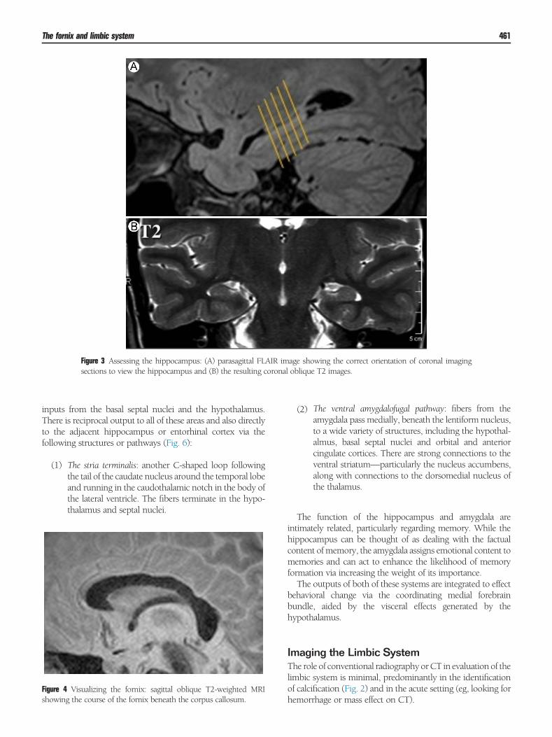
Figure 3 Assessing the hippocampus: (A) parasagittal FLAIR image showing the correct orientation of coronal imagingsections to view the hippocampus and (B) the resulting coronal oblique T2 images.
The fornix and limbic system 461
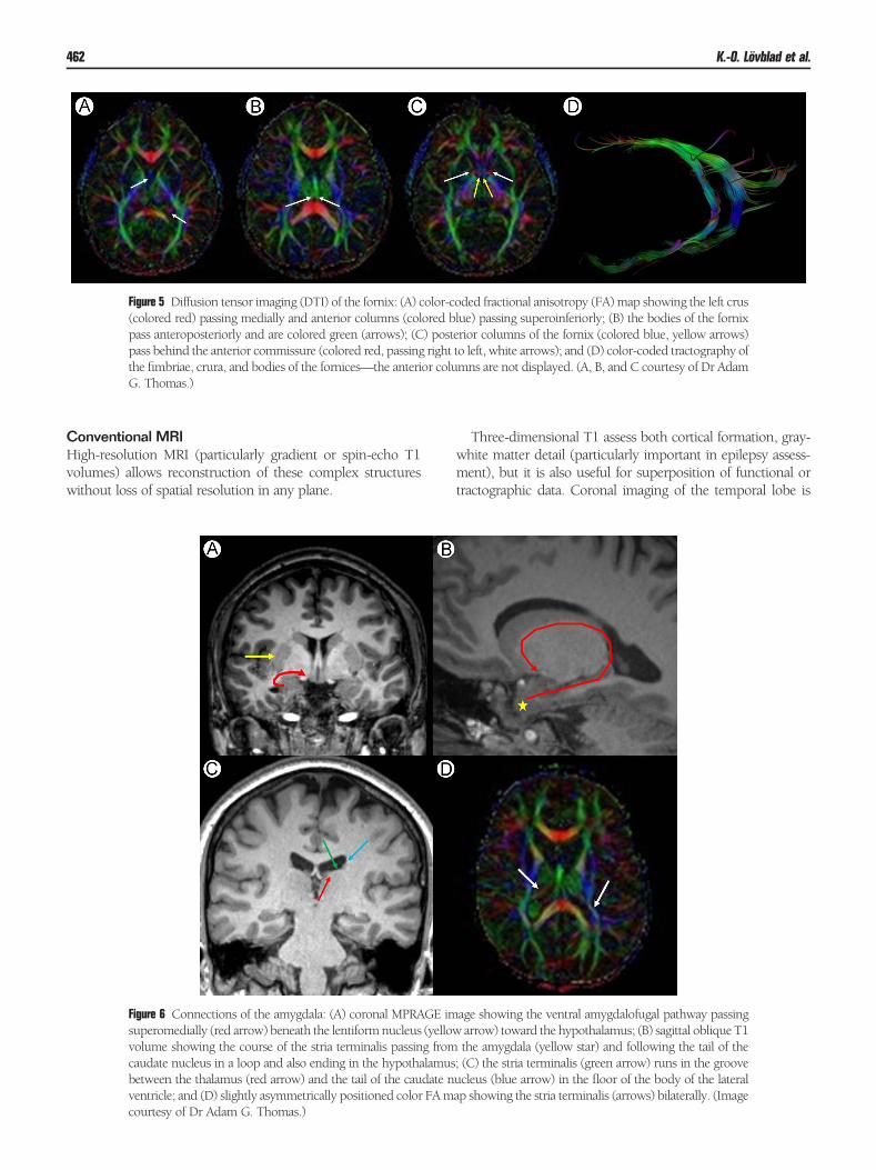
inputs from the basal septal nuclei and the hypothalamus.There is reciprocal output to all of these areas and also directlyto the adjacent hippocampus or entorhinal cortex via thefollowing structures or pathways (Fig. 6):
Figsh
(1)
ure 4owin
The stria terminalis: another C-shaped loop followingthe tail of the caudate nucleus around the temporal lobeand running in the caudothalamic notch in the body ofthe lateral ventricle. The fibers terminate in the hypo-thalamus and septal nuclei.
Visualizing the fornix: sagittal oblique T2-weighted MRIg the course of the fornix beneath the corpus callosum.
(2)
The ventral amygdalofugal pathway: fibers from theamygdala pass medially, beneath the lentiform nucleus,to a wide variety of structures, including the hypothal-almus, basal septal nuclei and orbital and anteriorcingulate cortices. There are strong connections to theventral striatum—particularly the nucleus accumbens,along with connections to the dorsomedial nucleus ofthe thalamus.
The function of the hippocampus and amygdala areintimately related, particularly regarding memory. While thehippocampus can be thought of as dealing with the factualcontent ofmemory, the amygdala assigns emotional content tomemories and can act to enhance the likelihood of memoryformation via increasing the weight of its importance.The outputs of both of these systems are integrated to effect
behavioral change via the coordinating medial forebrainbundle, aided by the visceral effects generated by thehypothalamus.
Imaging the Limbic SystemThe role of conventional radiography orCT in evaluation of thelimbic system is minimal, predominantly in the identificationof calcification (Fig. 2) and in the acute setting (eg, looking forhemorrhage or mass effect on CT).
Figure 5 Diffusion tensor imaging (DTI) of the fornix: (A) color-coded fractional anisotropy (FA)map showing the left crus(colored red) passing medially and anterior columns (colored blue) passing superoinferiorly; (B) the bodies of the fornixpass anteroposteriorly and are colored green (arrows); (C) posterior columns of the fornix (colored blue, yellow arrows)pass behind the anterior commissure (colored red, passing right to left, white arrows); and (D) color-coded tractography ofthe fimbriae, crura, and bodies of the fornices—the anterior columns are not displayed. (A, B, and C courtesy of Dr AdamG. Thomas.)
K.-O. Lövblad et al.462
Conventional MRIHigh-resolution MRI (particularly gradient or spin-echo T1volumes) allows reconstruction of these complex structureswithout loss of spatial resolution in any plane.
Figure 6 Connections of the amygdala: (A) coronal MPRAGE imsuperomedially (red arrow) beneath the lentiformnucleus (yellovolume showing the course of the stria terminalis passing fromcaudate nucleus in a loop and also ending in the hypothalamusbetween the thalamus (red arrow) and the tail of the caudate nventricle; and (D) slightly asymmetrically positioned color FAmcourtesy of Dr Adam G. Thomas.)
Three-dimensional T1 assess both cortical formation, gray-white matter detail (particularly important in epilepsy assess-ment), but it is also useful for superposition of functional ortractographic data. Coronal imaging of the temporal lobe is
age showing the ventral amygdalofugal pathway passingw arrow) toward the hypothalamus; (B) sagittal oblique T1the amygdala (yellow star) and following the tail of the; (C) the stria terminalis (green arrow) runs in the grooveucleus (blue arrow) in the floor of the body of the lateralap showing the stria terminalis (arrows) bilaterally. (Image
The fornix and limbic system 463
important in both epilepsy assessment and in evaluation ofcognitive impairment. The coronal plane is actually obliquelyorientated to be directly perpendicular to the body or head ofthe hippocampus to ensure accurate evaluation (Fig. 3).4
Sequences commonly employed in this plane are thin-section coronal T2 and fluid attenuation inversion recovery(FLAIR) imaging (particularly useful in mesial temporalsclerosis) and T1-inversion recovery (excellent gray-whitematter separation). Calcification or hemorrhage is frequentlya source of seizure generation, hence, a susceptibility-weightedsequence (gradient echo T2* or susceptibility weighted imag-ing) is also important.5 The superior signal-to-noise ratioobtained at 3 T can enhance diagnostic detection, particularlyin the case of epileptogenic lesions.6,7
Diffusion and Advanced MRI TechniquesDiffusion-weighted imaging (DWI) is now clinically routineand has a role to play inmany pathologies that affect the limbicsystem (described later).8 DTI allows visualization of the whitematter connections of the limbic system and their integrity incases of disease.9 Perfusion MRI and MR spectroscopy arevaluable in the evaluation of tumors and tumorlike condi-tions.10,11 Functional MRI is particularly useful in presurgicalplanning, particularly for language localization in patients whoare about to undergo temporal lobe surgery.12
Clinical PresentationOwing to the widespread connections of the limbic system,pathology that affects it may present in varied, and often
Figure 7 Pediatric mesial temporal sclerosis: (A) sagittal MPRAmicrocephaly; (B) axial FLAIR image showing a right middle cshowing loss of volume and hyperintensity of the right hippocamleft (arrow).
nonspecific, ways. However, given the functions outlinedpreviously, there are 3 particular circumstances in whichlimbic system pathology should be suspected. The clinicalpresentations may overlap considerably in any of the con-ditions discussed.
EpilepsyTemporal lobe epilepsy is the most common focal epilepsyin adults and the hippocampus may be involved in eithersecondary to seizure activity or as a seizure-generatingfocus itself. The most common hippocampal pathologyassociated with seizures is mesial temporal sclerosis.7
Although a causative relationship has not been estab-lished, increased rates of childhood febrile convulsions areseen in retrospective studies of patients with hippocampalsclerosis.13 The imaging findings are characteristic withreduced volume and T2 and FLAIR hyperintense signal(easily detected because of overlying cerebrospinal fluidsuppression) of the affected hippocampus, best appreci-ated in the coronal plane. The condition may be encoun-tered in childhood (Fig. 7) or adulthood (Fig. 8). Diffusionand perfusion changes encountered depend on whetherthe study is acquired in the ictal (hyperperfusion, diffu-sion restriction) or interictal period (hypoperfusion,increased diffusivity).14,15 The output tract of the hippo-campus, the fornix, and even the mammillary body maybe affected, with ipsilateral atrophy encountered in casesof severe hippocampal volume loss. This can be easilyassessed using DTI.16
GE image in a 3-year-old patient with epilepsy showingranial fossa arachnoid cyst; and (C) coronal FLAIR imagepal body (arrow) but relative preservation on the patient 's
Figure 8 Adult mesial temporal sclerosis—patient with complex partial seizures. (A and B) Coronal FLAIR images showingloss of volume and hyperintensity of the left hippocampus (arrows). (C) Axial color FAmap showing reduced FA, which isbetter appreciated on coronal reconstructions (D and E—arrows). There is associated hypoperfusion (F, arrow).
K.-O. Lövblad et al.464
Any mass lesion in the temporal lobe may present withepilepsy. These range from benign vascular lesions to aggres-sive high-grade astroglial series tumors. Cavernomatous mal-formations (cavernomas) may be highly epileptogenic in any
Figure 9 Hippocampal cavernoma: (A) heterogenous left mehyperintensity and (B) gradient echo T2*-weighted image shconsistent with a cavernoma.
area of cortex but particularly within the temporal lobe (Fig. 9).Benign or low-grade lesions that particularly affect the tempo-ral lobes include gangliogliomas and developmental neuro-ectodermal tumors (Fig. 2).
dial temporal lobe lesion (arrow) with regions of T1owing hypointense signal (arrow) and blooming artifact
Figure 10 Grade III hippocampal astroglial series tumor: (A) axial T1 image showing low signal mass (arrow); (B) the lesionshows patchy enhancement after contrast (arrow); (C) it is T2 hyperintense; and (D) there is evidence of infiltration oftemporal white matter on the color FA map with reduced visualization of the occipitotemporal fasciculus (arrow).
Figure 11 Multimodal imaging of a limbic glioblastoma: (A) axial T2-weighted image showing right temporal lobe tumorand extensive edema (arrow); (B) color-coded relative cerebral blood volume map showing elevated perfusion within thelesion (arrow); (C) multivoxel MR spectroscopy acquired over the lesion, the green voxel is enlarged in (D) and showselevated Cho level, reduced NAA level, and a lipid or macromolecule peak; and (E) functional MRI localizing languagefunction to the left hemisphere. Cho, choline; NAA, N-acetyl aspartate.
The fornix and limbic system 465
Figure 12 Inferior right MCA infarction in a 23-year-old patient with acute confusion: (A) axial T2-weightedimage showing cortical and subcortical signal hyperintensity; (B) DWI image showing restricted diffusion inthe right insula and frontal and temporal opercula; (C) follow-up axial T2-weighted image showing maturedamage in the regions of diffusion restriction; and (D) coronal FLAIR image showing severe loss ofhippocampal or medial temporal lobe tissue. The patient had severe anterograde amnesia. MCA, middlecerebral artery.
Figure 13 Bilateral ACA territory infarction: (A) volume-rendered catheter angiogram image of anteriorcommunicating artery aneurysm (arrow); (B) axial T2-weighted image showing bilateral superior frontal andcingulated cortical infarction (arrows); (C) axial T2-weighted image showing T2-hypointense hematoma in theanterior interhemispheric fissure (arrow); and (D) maximum intensity projection of time-of-flight MRangiography of the circle of Willis showing absence of flow in the ACAs bilaterally (arrow)—there is subtle“T1 shine through” from the interhemispheric hematoma. ACA, anterior cerebral artery.
K.-O. Lövblad et al.466
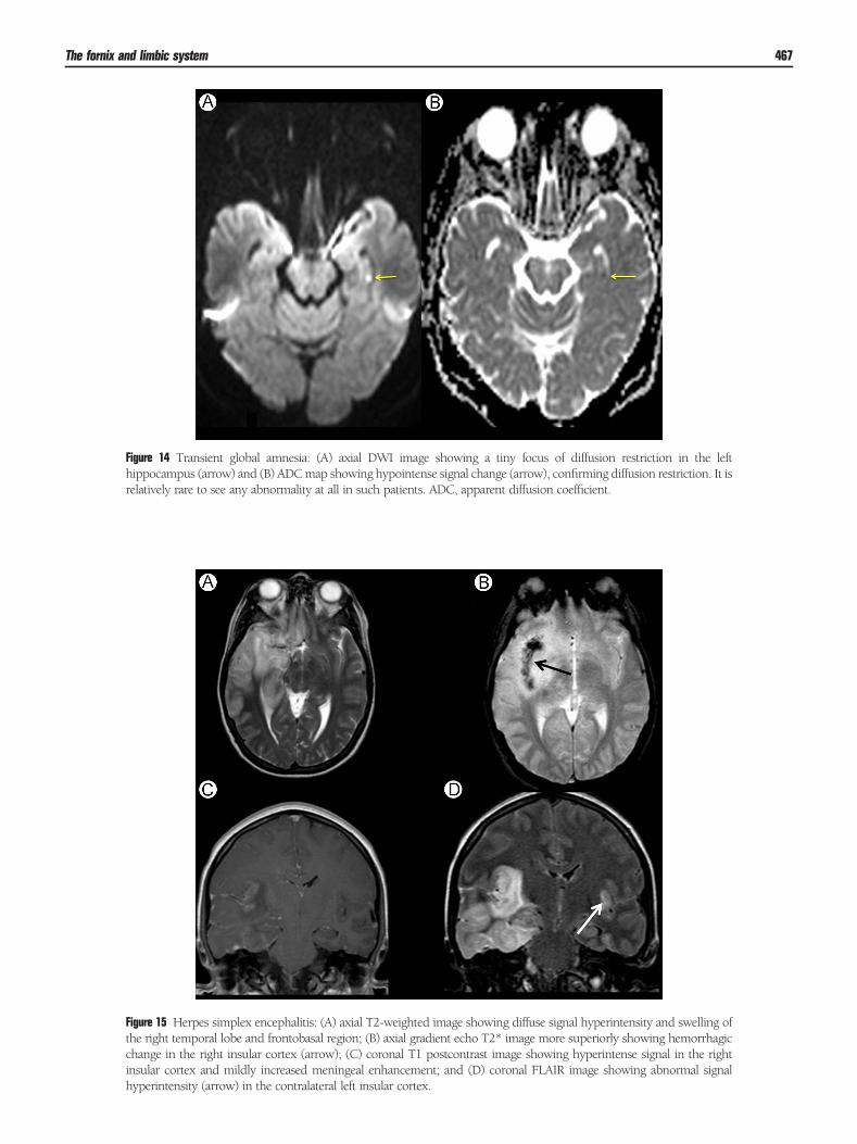
Figure 14 Transient global amnesia: (A) axial DWI image showing a tiny focus of diffusion restriction in the lefthippocampus (arrow) and (B) ADCmap showing hypointense signal change (arrow), confirming diffusion restriction. It isrelatively rare to see any abnormality at all in such patients. ADC, apparent diffusion coefficient.
Figure 15 Herpes simplex encephalitis: (A) axial T2-weighted image showing diffuse signal hyperintensity and swelling ofthe right temporal lobe and frontobasal region; (B) axial gradient echo T2* image more superiorly showing hemorrhagicchange in the right insular cortex (arrow); (C) coronal T1 postcontrast image showing hyperintense signal in the rightinsular cortex and mildly increased meningeal enhancement; and (D) coronal FLAIR image showing abnormal signalhyperintensity (arrow) in the contralateral left insular cortex.
The fornix and limbic system 467
Figure 16 Limbic encephalitis: (A) axial T2-weighted image showing hyperintense signal abnormality in the hippocampibilaterally (arrows); (B) axial DWI image showingmild signal hyperintensity (owing to T2 shine through, ADCnot shown);(C) axial T1 postcontrast image—there is no pathological enhancement; (D) coronal FLAIR image showing bilateralhippocampal signal hyperintensity (arrows). This patient had proven anti-Hu limbic encephalitis secondary to lung cancer.ADC, apparent diffusion coefficient.
K.-O. Lövblad et al.468
Both low- and high-grade astroglial tumors haveinfiltrative patterns of spread. Knowledge of the pre-existing anatomical connections outlined earlier shouldallow focused assessment of the MRI study to assess thefull extent of disease. As with any infiltrative lesion,complete resection is not surgically feasible. The role ofthe radiologist in the preoperative setting is to try andidentify the highest grade or most aggressive componentfor biopsy. Gliomas have typically low signal onT1-weighted imaging and high signal on T2 and FLAIRand may show intralesional hemorrhage or calcification(Fig. 10). Regions of higher grade are associatedwith contrast enhancement and increased relativecerebral blood volume on perfusion-weighted imaging(usually owing to neovascular proliferation withouteffective blood-brain barrier formation), diffusion restric-tion (owing to increased cellularity), and evidence ofnecrosis (elevated lipid/lactate) and neuronal destruction(reduced N-acetyl aspartate) on MR spectroscopy(Fig. 11).17,18 A multivoxel spectroscopic approach
may help in identifying the most aggressive part of alarge lesion.
Acute and Subacute Confusional StateAcuteSudden onset of any neurologic dysfunction should alwaysprompt consideration of a vascular etiology. However, suddenconfusion is a relatively rare isolated presenting feature ofstroke. There are 2 vascular territories (the middle andposterior cerebral artery) involving elements of the limbicsystem that may be accountable.Inferior middle cerebral artery division occlusion leading to
anterior temporal lobe and possible hippocampal infarction(Fig. 12). This mode of presentation appears more commonwhen involving the left temporal lobe.19 Anterior cerebralartery infarction—this is particularly severe when bilateralinvolvement is demonstrated, as may be encountered inpatients with an azygos anterior cerebral artery. Damage tothe anterior cingulate cortex, the medial and orbitofrontal
Figure 17 Alzheimer disease: (A) series of 18FDG-PET images showing bilateral temporal lobe hypoperfusion (arrows);(B) axial T2-weighted image of the brain showing diffuse cortical atrophy; (C) coronal T1 image showing advanced bilateralhippocampal atrophy (arrows); (D) arterial spin-labeling (ASL) perfusion imaging also shows the temporal lobehypoperfusion (arrows). 18FDG-PET, fludeoxyglucose-positron emission tomography.
The fornix and limbic system 469
lobes, and critically the columns of the fornix may result indevastating behavioral alteration with features of abulia (pov-erty of speech and emotional apathy), severe anterogradeamnesia and, particularly in the case of cingulate involvement,hypersexuality and agitation20 (Fig. 13). DWI is clearly criticalin stroke evaluation, allowing for detection of infarction withinminutes of onset and providing reasonable estimation of age ofinfarct in patients with delayed presentation.21
Transient Global AmnesiaThis distinct, short-lived amnestic syndrome remains ofunknown etiology, although occasionally, DWI-hyperintenselesions are encountered in the hippocampus (Fig. 14), leadingmany to conclude a probable vascular etiology.22
SubacuteSubacute presentations should prompt consideration ofinflammatory or infectious causes. The infection thatappears to show a particular tropism for the limbic systemis herpes simplex (types 1 and 2). The typical presentationis with fever, confusion, and seizures. CT may show lowdensity in the temporal lobes, but MRI is the mostsensitive way of detecting subtle or early changes. Thereis T2 and FLAIR cortical or gray matter hyperintensity inthe amygdala and hippocampus (often bilaterally)(Fig. 15). Detecting involvement of the insula and anteriorcingulate cortex is helpful in unilateral cases to helpsuggest infection rather than tumor. Lesions may oftenbe hemorrhagic and show contrast enhancement. DWIis sensitive at showing regions of involvement beforeT2-FLAIR signal alteration develops.23
A more prolonged presentation tends to occur with auto-immune limbic encephalitis. This may be idiopathic but isfrequently associated with an underlying systemic malignancy.The antibodies detectedmay help in directing the search for anunderlying lesion: anti-Hu antibodies are associated with smallcell lung cancer,24 anti-N-methyl-D-aspartate receptor anti-bodies with ovarian teratomas, and anti-Ma2 antibodies withtesticular germ cell tumors. Anti–voltage-gated potassiumchannel antibodies are associatedwith thymic tumors. Imagingfindings in limbic encephalitis may overlap with herpeticinfection with bilateral T2-FLAIR-hyperintense swelling of thehippocampi and amygdala (Fig. 16). A more symmetricalpattern of involvement and lack of meningeal enhancement,along with clinical features, may act as a clue to autoimmunerather than infectious etiology.25
Cognitive ImpairmentGiven its role in memory formation, any disease thataffects the hippocampus may present with problems withshort-term memory in particular. Alzheimer diseaseaffects the hippocampi preferentially, and although dis-proportionate hippocampal atrophy may be demon-strated on standard structural imaging, these featuresmay not be apparent until late in the course of disease.In addition to focal atrophy, functional studies such aspositron emission tomography perfusion scanning mayshow temporal hypoperfusion (Fig. 17) and newer ligandssuch as Pittsburgh Compound B allow detection ofabnormal amyloid deposition.26 A more readily available,
Figure 18 Artery of Percheron infarcts: (A) axial ADC map showing bilateral medial thalamic and midbrain hypointensity;(B) axial DWI showing corresponding hyperintense signal; and (C) axial T2-weighted images showing matchingT2-hyperintense signal. The patient presented with acute drowsiness and reduced conscious state. There is disruption ofthe mammillothalamic tracts and anterior thalamic radiations bilaterally. ADC, apparent diffusion coefficient.
K.-O. Lövblad et al.470
Figure 19 Acute Wernicke encephalopathy: (A) axial T2-weighted image showing bilateral paramedian thalamic signalhyperintensity; (B) axial DWI image showing corresponding diffusion restriction, confirmed by the absence ofhyperintensity on the ADC map (C); and (D) sagittal FLAIR image showing atrophy of the mammillary bodies (arrow)and hyperintensity of the massa intermedia (dashed arrow). ADC, apparent diffusion coefficient. (Case courtesy ofDr Adam G. Thomas.)
The fornix and limbic system 471
noninvasive means of assessing hypoperfusion may beapplication of arterial spin labeling on MRI.27
Vascular dementia is highly prevalent in developedcountries. Imaging usually reveals widespread small vesselischemic change but focal ischemic involvement of thePapez circuit structures can account for severe memoryimpairment. Bilateral anteromedial thalamic infarcts(Fig. 18) damage the anterior thalamic nuclei (junctionbetween the mammillothalamic tract and cingulum) andalone may suffice to diagnose vascular dementia (accordingto the National Institute of Neurological Disorders andStroke and Association–Internationale pour la Recherché etl 'Enseignement enNeurosciences criteria).28 Other vascularcauses of thalamic damage, such as internal cerebral venousinfarction, can have similarly devastating consequences.Thiamine deficiency is most commonly encountered in
cases of chronicmalnutrition (eg, commonly in alcoholics) andthe Wernicke-Korsakoff (involving anterograde amnesia)
shows involvement of the Papez circuit structures. There isoften T2-hyperintense mammillary body atrophy, and for-niceal involvement may be encountered (Fig. 19). Contrastenhancement may be encountered in the acute setting.29
Neurodegenerative disease in children may showinvolvement of the limbic structures with pathologicenhancement of the mammillary bodies and amygdalaencountered in Alexander disease (Fig. 20).30 T1 hyper-intensity of the amygdala is encountered in neurocuta-neous melanosis.
Other Behavioral DisordersThe anterior temporal lobes are susceptible to traumatic injuryand a specific behavioral syndrome may be encountered insevere bilateral injury with limbic neuroanatomical basis.Klüver-Bucy syndrome consists of fearlessness and placidity(owing to amygdaloid damage), hypersexuality (owing to
Figure 20 Alexander disease: (A and B) axial postcontrast T1-weighted images showing enhancement of the mammillarybodies (arrow, A) and amygdala (arrow, B).
K.-O. Lövblad et al.472
damage to the piriform cortex, a strong functional connectionof the amygdala), and visual agnosia owing to damage ofinferior temporal lobe structures31 (see Visual Pathways articlein this issue).
ConclusionThe limbic system is critical in human memory, emotionalexperience, learning, and behavior. A wide variety of con-ditionsmay affect these structures, butmost conditions presentwith epilepsy, confusion, memory impairment, or a combina-tion or all of them. Knowledge of the connections of the limbicsystem allows detailed assessment of temporal lobe pathologybut also allows one to detect possible limbic effects from moresubtle lesions in distant locations. Standard structural imagingsuffices in most cases but new information provided by evenbetter DTI may provide new insights into the functionalneuroanatomical consequences of disease.
References1. Lövblad KO, Schaller K: Surgical anatomy and functional connectivity of
the limbic system. Neurosurg Focus 27:E3, 20092. Papez JW: A proposed mechanism of emotion. 1937. J Neuropsychiatry
Clin Neurosci 7:103-112, 19953. CataniM,Della 'acqua F, Thiebaut de SchottenM: A revised limbic system
model for memory, emotion and behavior. Neurosci Biobehav Rev 37:1724-1737, 2013
4. Jeukens CR, Vlooswijk MC, Majoie HJ, et al: Hippocampal MRIvolumetry at 3 Tesla: Reliability and practical guidance. Invest Radiol44:509-517, 2009
5. Haacke EM, Mittal S, Wu Z, et al: Susceptibility-weighted imaging:Technical aspects and clinical applications, part 1. Am J Neuroradiol 30:19-30, 2009
6. Vargas MI, Delavelle J, Kohler R, et al: Brain and spine MRI artifacts at3 Tesla. J Neuroradiol 36:74-81, 2009
7. Craven I, Griffiths PD, Hoggard N: Magnetic resonance imaging ofepilepsy at 3 Tesla. Clin Radiol 66:278-286, 2011
9. Le Bihan D, Mangin JF, Poupon C, et al: Diffusion tensor imaging:Concepts and applications. J Magn Reson Imaging 13:534-546, 2001
10. Le Bihan D, Breton E, Lallemand D, et al: MR imaging of intravoxelincoherent motions: Application to diffusion and perfusion in neurologicdisorders. Radiology 161:401-407, 1986
11. Rosen BR, Belliveau JW, Vevea JM, et al: Perfusion imaging with NMRcontrast agents. Magn Reson Med 14:249-265, 1990
12. Binder JR, Swanson SJ, Sabsevitz DS, et al: A comparison of two fMRImethods for predicting verbal memory decline after left temporallobectomy: Language lateralization versus hippocampal activation asym-metry. Epilepsia 51:618-626, 2010
13. Kasperaviciute D, Catarino CB, Matarin M, et al: Epilepsy, hippocampalsclerosis and febrile seizures linked by common genetic variation aroundSCN1A. Brain 136:3140-3150, 2013
14. El-KoussyM,Mathis J, Lovblad KO, et al: Focal status epilepticus: Follow-up by perfusion- and diffusion MRI. Eur Radiol 12:568-574, 2002
15. Pendse N,Wissmeyer M, Altrichter S, et al: Interictal arterial spin-labelingMRI perfusion in intractable epilepsy. J Neuroradiol 37:60-63, 2010
16. Assaf BA, Mohamed FB, Abou-Khaled KJ, et al: Diffusion tensor imagingof the hippocampal formation in temporal lobe epilepsy. Am J Neuro-radiol 24:1857-1862, 2003
17. Romano A, Rossi Espagnet MC, Calabria LF, et al: Clinical applications ofdynamic susceptibility contrast perfusion-weighted MR imaging in braintumours. Radiol Med 117:445-460, 2012
18. Alimenti A, Delavelle J, Lazeyras F, et al: Monovoxel 1H magneticresonance spectroscopy in the progression of gliomas. Eur Neurol 58:198-209, 2007
19. Ott BR, Saver JL: Unilateral amnesic stroke. Six new cases and a review ofthe literature. Stroke 24:1033-1042, 1993
20. Kumral E, Bayulkern G, Evyapan D, et al: Spectrum of anterior cerebralartery territory infarction: Clinical and MRI findings. Eur J Neurol 9:615-624, 2002
21. Lövblad KO, Laubach HJ, Baird AE, et al: Clinical experience withdiffusion-weighted MR in patients with acute stroke. Am J Neuroradiol19:1061-1066, 1998
22. Strupp M, Brüning R, Wu RH, et al: Diffusion-weighted MRI in transientglobal amnesia: Elevated signal intensity in the left mesial temporal lobe in7 of 10 patients. Ann Neurol 43:164-170, 1998
25. Sureka J, Jakkani RK: Clinico-radiological spectrum of bilateral temporallobe hyperintensity: A retrospective review. Br J Radiol 85:e782-e792,2012
26. Cohen AD, Klunk WE: Early detection of Alzheimer 's disease using PiBand FDG PET. Neurobiol Dis [Epub ahead of print, doi: 0.1016/j.nbd.2014.05.001]
27. Detre JA, Rao H, Wang DJ, et al: Applications of arterial spin labeled MRIin the brain. J Magn Reson Imaging 35:1026-1037, 2012
28. van Straaten EC, Scheltens P, Knol DL, et al: Operational definitions forthe NINDS-AIREN criteria for vascular dementia: An interobserver study.Stroke 34:1907-1912, 2003
29. Zuccoli G, Pipitone N: Neuroimaging findings in acute Wernicke 'sencephalopathy: Review of the literature. Am J Roentgenol 192:501-508, 2009
30. Poloni CB, Ferey S, Haenggeli CA, et al: Alexander disease: Early presenceof cerebral MRI criteria. Eur J Paediatr Neurol 13:556-558, 2009
31. Faria Jr Miguel A: Violence, mental illness, and the brain—A brief historyof psychosurgery: Part 2—From the limbic system and cingulotomy todeep brain stimulation. Surg Neurol Int 4:75, 2013