REVIEW The fracture mechanics of human bone: influence of disease and treatment Elizabeth A Zimmermann 1 , Bjo ¨ rn Busse 1 and Robert O Ritchie 2,3 1 Department of Osteology and Biomechanics, University Medical Center Hamburg-Eppendorf, Hamburg, Germany. 2 Materials Sciences Division, Lawrence Berkeley National Laboratory, Berkeley, CA, USA. 3 Department of Materials Science & Engineering, University of California, Berkeley, CA, USA. Aging and bone diseases are associated with increased fracture risk. It is therefore pertinent to seek an understanding of the origins of such disease-related deterioration in bone’s mechanical properties. The mechanical integrity of bone derives from its hierarchical structure, which in healthy tissue is able to resist complex physiological loading patterns and tolerate damage. Indeed, the mechanisms through which bone derives its mechanical properties make fracture mechanics an ideal framework to study bone’s mechanical resistance, where crack-growth resistance curves give a measure of the intrinsic resistance to the initiation of cracks and the extrinsic resistance to the growth of cracks. Recent research on healthy cortical bone has demonstrated how this hierarchical structure can develop intrinsic toughness at the collagen fibril scale mainly through sliding and sacrificial bonding mechanisms that promote plasticity. Furthermore, the bone-matrix structure develops extrinsic toughness at much larger micrometer length-scales, where the structural features are large enough to resist crack growth through crack-tip shielding mechanisms. Although healthy bone tissue can generally resist physiological loading environments, certain conditions such as aging and disease can significantly increase fracture risk. In simple terms, the reduced mechanical integrity originates from alterations to the hierarchical structure. Here, we review how human cortical bone resists fracture in healthy bone and how changes to the bone structure due to aging, osteoporosis, vitamin D deficiency and Paget’s disease can affect the mechanical integrity of bone tissue. BoneKEy Reports 4, Article number: 743 (2015) | doi:10.1038/bonekey.2015.112 Introduction The greater goals of bone research are to assess the risk of fractures in bone and to prevent their occurrence. Fracture events encompass the initiation and growth of cracks as well as the resulting complete fracture of the tissue. This makes the fracture toughness (that is, resistance to fracture) a critical aspect of bone’s mechanical integrity. In healthy tissue, bone’s complex structure has evolved to resist physiological forces and can further tune its mechanical resistance through remodeling. However, bone tissue is vulnerable to skeletal aging, environmental conditions and various genetic or metabolic bone diseases that increase fracture risk or affect the mechanical integrity. These diseases include osteoporosis, vitamin D deficiency and Paget’s disease of bone (PDB), to name but a few. Thus, it is necessary to understand how such disease alters the bone-matrix structure leading to a higher risk of fracture and how corresponding therapeutic treatments may affect bone’s mechanical properties. Akin to most natural and engineered materials, the mechanical performance of bone derives directly from its structure. Similar to many natural materials, its structure is hierarchical, meaning that there are discrete characteristic structural features at differing length scales (Figure 1). The hierarchical structure of human cortical bone spans numerous architectural levels from the individual collagen molecules and mineral platelets at the nanoscale to the whole bone at the macroscale. 1 At the smallest length scales, the key feature is the mineralized collagen fibril. Here, collagen molecules form arrays with a 67-nm offset, where nanoplatelets of hydroxyapatite mineral are embedded within the collagen array and on the surface of the fibril. 2,3 At this length-scale, the intra- and extra-fibrillar matrix contains non-collagenous proteins (for example, bone sialoprotein, osteopontin, osteonectin), as well as enzymatic and non-enzymatic cross-links. 4–6 The non-enzymatic cross-links are particularly influential to bone’s mechanical integrity in disease states. 7 These cross-links also known as advanced glycation end-products form through a glucose-mediated reaction and may form both intra- and interfibrillar connections. 8,9 The mineralized collagen fibrils hierarchically assemble into lamellae to form osteons, which are the most prominent motif on Correspondence: Dr EA Zimmermann, Department of Osteology and Biomechanics, University Medical Center Hamburg-Eppendorf, 22529 Hamburg, Germany. E-mail: [email protected]Received 29 March 2015; revised 30 June 2015; accepted 3 July 2015; published online 2 September 2015 Citation: BoneKEy Reports 4, Article number: 743 (2015) | doi:10.1038/bonekey.2015.112 & 2015 International Bone & Mineral Society All rights reserved 2047-6396/15 www.nature.com/bonekey BoneKEy Reports | SEPTEMBER 2015 1
Transcript
REVIEW
The fracture mechanics of human bone: influenceof disease and treatmentElizabeth A Zimmermann1, Bjorn Busse1 and Robert O Ritchie2,3
1Department of Osteology and Biomechanics, University Medical Center Hamburg-Eppendorf, Hamburg, Germany.2Materials Sciences Division, Lawrence Berkeley National Laboratory, Berkeley, CA, USA. 3Department of MaterialsScience & Engineering, University of California, Berkeley, CA, USA.
Aging and bone diseases are associated with increased fracture risk. It is therefore pertinent to seek an understanding of
the origins of such disease-related deterioration in bone’s mechanical properties. The mechanical integrity of bone derives
from its hierarchical structure, which in healthy tissue is able to resist complex physiological loading patterns and tolerate
damage. Indeed, the mechanisms through which bonederives its mechanical properties make fracture mechanics an ideal
framework to study bone’s mechanical resistance, where crack-growth resistance curves give a measure of the intrinsic
resistance to the initiation of cracks and the extrinsic resistance to the growth of cracks. Recent research on healthy
cortical bone has demonstrated how this hierarchical structure can develop intrinsic toughness at the collagen fibril scale
mainly through sliding and sacrificial bonding mechanisms that promote plasticity. Furthermore, the bone-matrix structure
develops extrinsic toughness at much larger micrometer length-scales, where the structural features are large enough to
resist crack growth through crack-tip shielding mechanisms. Although healthy bone tissue can generally resist
physiological loading environments, certain conditions such as aging and disease can significantly increase fracture risk.
In simple terms, the reduced mechanical integrity originates from alterations to the hierarchical structure. Here, we review
how human cortical bone resists fracture in healthy bone and how changes to the bone structure due to aging,
osteoporosis, vitamin D deficiency and Paget’s disease can affect the mechanical integrity of bone tissue.
The greater goals of bone research are to assess the risk offractures in bone and to prevent their occurrence. Fractureevents encompass the initiation and growth of cracks aswell as the resulting complete fracture of the tissue. Thismakes the fracture toughness (that is, resistance to fracture) acritical aspect of bone’s mechanical integrity. In healthy tissue,bone’s complex structure has evolved to resist physiologicalforces and can further tune its mechanical resistancethrough remodeling. However, bone tissue is vulnerable toskeletal aging, environmental conditions and various geneticor metabolic bone diseases that increase fracture risk or affectthe mechanical integrity. These diseases include osteoporosis,vitamin D deficiency and Paget’s disease of bone (PDB), toname but a few. Thus, it is necessary to understand how suchdisease alters the bone-matrix structure leading to a higher riskof fracture and how corresponding therapeutic treatments mayaffect bone’s mechanical properties.
Akin to most natural and engineered materials, themechanical performance of bone derives directly from itsstructure. Similar to many natural materials, its structure is
hierarchical, meaning that there are discrete characteristicstructural features at differing length scales (Figure 1). Thehierarchical structure of human cortical bone spans numerousarchitectural levels from the individual collagen molecules andmineral platelets at the nanoscale to the whole bone atthe macroscale.1 At the smallest length scales, the key feature isthe mineralized collagen fibril. Here, collagen moleculesform arrays with a 67-nm offset, where nanoplatelets ofhydroxyapatite mineral are embedded within the collagen arrayand on the surface of the fibril.2,3 At this length-scale, theintra- and extra-fibrillar matrix contains non-collagenousproteins (for example, bone sialoprotein, osteopontin,osteonectin), as well as enzymatic and non-enzymaticcross-links.4–6 The non-enzymatic cross-links are particularlyinfluential to bone’s mechanical integrity in disease states.7
These cross-links also known as advanced glycationend-products form through a glucose-mediated reactionand may form both intra- and interfibrillar connections.8,9
The mineralized collagen fibrils hierarchically assemble intolamellae to form osteons, which are the most prominent motif on
Correspondence: Dr EA Zimmermann, Department of Osteology and Biomechanics, University Medical Center Hamburg-Eppendorf, 22529 Hamburg, Germany.E-mail: [email protected]
Received 29 March 2015; revised 30 June 2015; accepted 3 July 2015; published online 2 September 2015
the microstructural scale (Figure 1). The osteons arecylindrical with a nominal circular cross-section and a centralvascular channel called the Haversian canal. At the outerlimits of the osteon is a thin border called the cement line,which is considered to be either collagen deficient or highlymineralized relative to the surrounding tissue.10
Deformation and fracture mechanisms at each level of bone’shierarchical structure contribute to its mechanical integrity,most significantly its strength and toughness. A prime benefitto the hierarchical arrangement is that strength (that is,the resistance to inelastic deformation) and toughness (that is,the resistance to crack growth or fracture) can be generatedindependently.11 Indeed, strength originates from intrinsicmechanisms at small length-scales that promote plasticity anddetermine the inherent resistance of the material to deformationor crack initiation. Intrinsic toughening mechanisms operateahead of the crack tip to generate resistance to microstructuraldamage. The most prominent mechanism is that of plasticdeformation, which provides a means of blunting the crack tipthrough the formation of ‘plastic’ zones. Toughness originatesfrom extrinsic mechanisms at micron length-scales that arelarge enough to shield the growth of cracks (Figure 2).12
Extrinsic toughening mechanisms, conversely, operate pri-marily in the wake of the crack tip to inhibit cracking by‘shielding’ the crack from the applied driving force, whereasintrinsic toughening mechanisms are effective in inhibiting boththe initiation and the growth of cracks; extrinsic mechanisms,for example, crack bridging, are only effective in inhibiting crackgrowth.
Human cortical bone generates intrinsic resistance tofracture, similar to other materials at small length-scales wherethe bonds between the smallest components stretch, deform,slip and/or break. Thus, the intrinsic fracture resistance of boneis generated through plasticity at the level of the mineralizedcollagen fibril (see Figure 2). Here, fibrils stretch during elasticdeformation and generate plasticity through intra- and
extrafibrillar deformation mechanisms, such as dilatationalband formation and/or fibrillar sliding. These fibrillar defor-mation mechanisms largely involve sacrificial bonds, such ascross-links and non-collagenous proteins that are critical to thegeneration of mechanical properties.5,13–19 Indeed, studiesusing animal models with deficiencies in non-collagenousproteins have found significant decreases in fracture toughnessand nanoscale damage.16,17 An important distinction hereis that plasticity, which is the source of bone’s ductility, isgenerated primarily at the fibrillar level rather than solely at thecollagen molecule level.20,21 The damaged caused bymechanical loading increases fracture resistance but may alsotrigger bone repair, where micron-scale damage (that is, linearmicrocracks) severing cellular connections is repairedthrough normal bone remodeling, and nanoscale damage(that is, diffuse damage) is repaired through a separate, asyet unknown, process.22–24 Further studies are required tobetter understand how each aspect of the complexnanostructure contributes to bone deformation, repair andoverall fracture risk in health and disease.
Apart from the generation of plasticity and intrinsictoughness, extrinsic mechanisms can develop the material’sresistance to the growth of cracks (Figure 2). Human corticalbone generates extrinsic resistance at the microstructurallength-scale where micron-scale mechanisms are activated toresist propagation of micron-scale cracks.25 As such, theextrinsic resistance of bone shields the crack from the fullmechanical driving force. In bone, crack-tip shielding isdeveloped at the level of the osteon primarily through crackbridging and crack deflection mechanisms. Here, as a crackgrows through the bone structure, collagen fibers or uncrackedregions of bone matrix can remain intact in the crack wake toform ‘bridges’ that span the crack opening (Figures 3a and b).These bridges can carry part of the load that would otherwise beused to further extend the crack, thereby lowering the drivingforce for crack propagation.26 Crack deflection and twist is
Figure 1 Hierarchical structure of human cortical bone. The structure of bone spans numerous length-scales from the macroscopic whole-bone structure to the nanoscalecollagen and mineral components. At the microscale, the bone is composed of osteons that are 170–250mm in diameter.60 The osteons have a central vascular channel (60–90mmdiameter60), called the Haversian canal, and a highly mineralized outer boundary, called the cement line (o5mm thickness).10 In the osteons, each vascular channel isconcentrically surrounded by lamellae (2–9mm thickness).80 The lamellae, which are composed of bundles of collagen fibrils, have a twisted plywood arrangement, whereneighboring lamellae have different fibril orientations.80 Certain lamellae may be less organized,81 and it is here that the osteocytes bone cells reside in lacunae (5–10mm diameter)and interconnect through canaliculi (100–400 nm in diameter).82,83 The fibrils (80–100 nm diameter1) are surrounded by polycrystalline extrafibrillar mineral platelets (5 nmthickness, 50–80 nm width and 40–200 nm length3); the extrafibrillar as well as the intrafibrillar matrix may also contain molecular components, such as non-collagenous proteins orcross-links, promoting the formation of sacrificial bonds.5,9,15,16,84 In the fibrils, type I collagen molecules (1.5 nm diameter, 300 nm length) and hydroxyapatite nanocrystals (50 nmwidth, 25 nm height, 1.5–4 nm thickness) form a composite structure, where arrays of collagen molecules staggered at 67 nm are embedded with nanoplatelets of hydroxyapatitemineral.1 Adapted from Zimmermann et al. and Milovanovic et al.45,82
The fracture mechanics of human boneEA Zimmermann et al
anotherextrinsic mechanismoccurring whena crack encountersan interface in the bone, such as the highly mineralized cementlines at the outer boundary of the osteons or the modulating
mechanical properties of the lamellae (Figures 3c and d).27,28 Ifthe crack deflects and/or twists away from the original crackplane, the driving force can be significantly reduced.
Figure 2 Toughening mechanisms in bone. Toughness is a competition between intrinsic and extrinsic toughening mechanisms. The intrinsic mechanisms primarily work aheadof the crack tip to develop plasticity, whereas the extrinsic mechanisms act in the crack wake (that is, after crack extension commences) to resist crack propagation through crack-tipshielding mechanisms. In human cortical bone, the extrinsic toughening mechanisms are primarily developed at large length-scales on the order of 1–100’s of microns. Here, themost potent extrinsic toughening mechanisms are crack deflection and twist at cement lines and uncracked ligament bridging; however, collagen fiber bridging and microcrackingcould also have a role. Intrinsic toughness is developed at small length-scales, less than B1 mm. Here, plasticity is generated through sliding mechanisms between fibrils that arefacilitated by sacrificial bonds (for example, non-collagenous proteins, cross-links) within the extrafibrillar matrix. Similarly, plasticity could also be generated within the fibril throughdeformation mechanisms, such as the formation of dilatational bands, which are also facilitated by sacrificial bonding in the intrafibrillar matrix. Adapted from Launey et al.85
Figure 3 Crack-briding and crack deflection promote extrinsic toughness. Human cortical bone develops extrinsic toughness on the microstructural scale through (a,b)uncracked ligament bridging and (c,d) crack deflection/twist mechanisms. Using (a) 3-D synchrotron tomography and (b) scanning electron microscopy, uncracked segments ofbone (orange arrows) can be observed in the wake of the crack. These uncracked regions of bone tissue, commonly generated by microcracking ahead of the crack tip (red arrows),carry part of the load that would otherwise be used to extend the crack. Similarly, evidence of crack deflection can be viewed (c) on the fracture surface after testing and (d) during atest using the scanning electron microscope. Here, the crack grows from the notch (red arrows). As the crack encounters the interfaces within the bone tissue aligned with theosteons, primarily cement lines, the crack will often deflect or twist (black arrows). Adapted from Zimmermann et al.20,45
The fracture mechanics of human boneEA Zimmermann et al
BoneKEy Reports | SEPTEMBER 2015 3
Another aspect of the bone structure that should beaddressed is the porosity, which in cortical bone exists inthe form of vascular canals (that is, Haversian canals) andcellular pores (that is, osteocytes lacunae and canaliculi).From a mechanics perspective, porosity can impact themechanical properties29,30 because it reduces the load bearingarea, which effectively causes a higher stress on the material.However, porosity is not the sole determinant of bone fracturerisk.31–34 Indeed, compositional factors, such as the cross-linking profile, mineralization distribution, or the collagencontent, also have a strong correlation with fracture risk.35,36 Interms of fracture mechanics, the precise effects of porosity oncrack propagation in bone have not been isolated from otheraspects of the structure that influence toughness. However,cracks are commonly observed to follow cement lines duringfracture mechanics experiments in the presence of normalamounts of bone porosity.20 The effects of porosity can becorrected by normalizing the applied stress by the bone volumefracture, which is especially critical when comparing themechanical properties of groups with significantly differentporosities.19,37 Thus, although bone porosity is not the soledeterminant of fracture risk in bone, it should be consideredwhen addressing the mechanical properties of cortical bone.
Because of bone’s hierarchical structure, its mechanicalbehavior originates from plasticity-related intrinsic mechanismsat small length-scales and crack-shielding extrinsic mechan-isms at larger length-scales to create strength and toughness,respectively. Bone’s intrinsic resistance can be measured bytraditional characterization methods through simply a strengthtest or investigated more deeply through a suite of char-acterization methods (for example, small-angle X-ray scat-tering, nanoindentation, or atomic force microscopy) to assessfibrillar-level mechanical properties. Alternatively, corticalbone’s intrinsic and extrinsic resistance can be quantified usingfracture mechanics principles. In fracture mechanics, thetoughness of a material can be measured in terms of the criticalvalue of a parameter, such as the stress intensity K, whichcharacterizes the stress and displacements fields in the vicinityof a crack tip; under nominally linear elastic (small-scaleyielding, plane strain) conditions, one such characterizingparameter is a critical value of K known as the plane-strainfracture toughness, KIc . The fracture toughness, KIc, is definedas the critical stress intensity at fracture instability; if measuredcorrectly, it strictly characterizes the crack-initiation toughness.However, as bone allows stable crack propagation, a single-value toughness, such as KIc, does not always fully characterizethe material behavior. In this case, a crack-growth resistancecurve or a R-curve can be used to more faithfully quantifymaterial behavior during crack propagation. With an R-curve,the crack-driving force, defined in terms of K, G or J, ismeasured as a function of crack extension, Da. Here the stressintensity K is a measure of the amplitude of the elastic stress anddisplacement fields at the crack tip. Alternatively, the toughnesscan be measured as a critical value of the strain-energy releaserate, Gc, defined as the change in potential energy per unitincrease in crack area in an elastic solid. If the presence of localplasticity is no longer small enough to be ignored, bothapproaches can also be expressed in terms of the J-integral,defined as the amplitude of the nonlinear elastic stress anddisplacement fields at the crack tip and/or as the change inpotential energy per unit increase in crack area in a nonlinear
elastic solid. Where nominally elastic conditions prevail, J¼G¼K2/E (where E is the appropriate Young’s modulus). Fromthe R-curve, the crack-initiation toughness can be measured byextrapolating the toughness to Da-0, whereas the slope of thecurve can be taken as a measure of the crack-growthtoughness. An example of an R-curve is shown in Figure 4 forbone tested where cracking is perpendicular (that is, transverseorientation) and parallel (that is, longitudinal orientation) to theosteonal bone structure. The positive R-curve slope in bothorientations demonstrates rising R-curve behavior stemmingfrom the fact that the extrinsic toughening mechanisms (that is,crack deflection and bridging) increase the bone’s toughness asthe crack extends. However, the slope of the R-curve in thetransverse orientation is significantly larger than in the long-itudinal orientation, that is, the bone is easier to split than tobreak in two. The different R-curve slopes result from thediffering extrinsic toughening mechanisms that are active inthese two orientations. In the transverse orientation, themechanism of crack deflection is paramount because once acrack deflects at the cement lines, it is extending in a direction,that is parallel to the osteons and nominally perpendicular to thepath of the maximum driving force (for example, the Gmax orKII¼ 0 path). Conversely, in the longitudinal orientation, thedriving force is nominally parallel to the osteonal orientation aswell as the lamellar and cement line interfaces. The preferentialformation of microcracks along these interfaces activates theformation of uncracked ligaments across the crack faces, whichin turn results in significant crack bridging. As is clear from theR-curves in Figure 4, crack deflection is a much more potentshielding mechanism than crack bridging in enhancing thetoughness of bone, which is why bone is much tougher in thetransverse orientation than in the longitudinal direction.27
Figure 4 Crack-growth resistance behavior in bone. Bone is a damage tolerantmaterial that resists the growth of cracks through its structure. As such, a single value ofthe toughness at instability does not completely characterize bone’s behavior. Instead,the toughness must be assessed as a function of crack extension, which is called acrack-growth resistance-curve or a R-curve. Here, the amplitude of the stress anddisplacement fields around the crack tip (that is, K, J or G) is measured as the crackstabily extends. With R-curves, the value of the driving force as Da-0 is a measure ofthe crack-initiation toughness, whereas the slope of the R-curve is a measure of thecrack-growth toughness. The R-curves of human cortical bone have a positive slope,which indicates rising R-curve behavior. Essentially, the slope is able to rise becausethe toughness of the material increases with crack extension. Here, the microstructuralextrinsic toughening mechanisms, such as crack deflection and uncracked ligamentbridging, become activated with crack extension and toughen the material. In thepresence of such extrinsic (shielding) mechanisms, a higher driving force is required forfurther crack extension. Adapted from Koester et al. and Zimmermann et al.27,39,86
The fracture mechanics of human boneEA Zimmermann et al
As cortical bone is a damage tolerant material,11 fracturemechanics is an ideal framework to study bone’s resistance tocrack initiation and growth. Although our understanding of howbone resists fracture is based on experimental evidence fromquasi-static loading conditions, physiological fractures occurmost often in high strain rate or fatigue conditions. Recentstudies have shown that fracture toughness decreases duringhigh strain rate loading because the intrinsic viscous defor-mation mechanisms at the nanoscale (that is, fibrillar sliding,sacrificial bonding) are restricted. The lower plasticity at thenanoscale also influences the extrinsic crack growth behavioras the heterogeneous structure, which normally interferes withcrack growth, appears more mechanically homogeneous.20,38
Although more studies are required to understand the fractureresistance of bone under physiological loading conditions, thecurrent understanding of bone’s resistance to fracture providesa starting framework to study the effects of aging and disease,where fractures can occur with minimal trauma.
Human cortical bone’s complex hierarchical structuregenerates strength and toughness, respectively, throughintrinsic plasticity-based mechanisms at small length-scalesand extrinsic crack-shielding mechanisms at larger length-scales. In healthy bone, the structure is generally able to resistphysiological loads and adapt to new loading environmentsthrough remodeling.20,39 However, disease states put bone at ahigher risk of fracture, bending or bowing, all of which can leadto a loss in mechanical integrity. Here, we review how certainprominent bone diseases affect the structural quality of bone,specifically by examining the salient mechanisms that cancause increased fracture risk due to aging, osteoporosis,vitamin D deficiency and Paget’s disease.
Aging and Osteoporosis
After reaching peak bone mass in the third or fourth decade inlife, both the quantity and quality of the bone tissue in the humanskeleton begin to deteriorate.31,40 Here bone quality encom-passes the composition and distribution of mineral andcollagen, the characteristics and structural integrity of eachhierarchical length scale (that is, osteon size and distribution),as well as the amount of microdamage present. This aging-related deterioration in the bone structure has a profoundimpact on the overall mechanical properties and fracture risk.Currently, fracture risk is mainly measured through dual-energyX-ray absorptiometry (DXA), where the degree of X-raysabsorbed by the bone mineral provides a prediction of fracturerisk. As the degree of bone mineralization shows a rather narrowrange in health and disease,37,41 standard DXA diagnosticsprovide a helpful measure of areal bone mineral density, which isbased on the present porosity and volume fraction of the bone(that is, bone quantity). In addition, DXA provides valuableinformation due to its widespread global availability and theexistence of large reference populations; however, it is notnecessarily an optimal indicator for predicting fracture risk.Indeed, as DXA predominantly assesses the quantity of bonemineral, many unanswered issues remain with respect to thedeterioration in the quality of the tissue. Here, we briefly reviewexperimental measurements of the structural and mechanicalproperties of human cortical bone due to biological aging andrecent research concerning the effectiveness of bisphosphonatesas a treatment for osteoporosis in human cortical bone.
AgingAging is associated with changes to the bone structure at smalland large length-scales that deteriorate its strength (that is,intrinsic resistance) and toughness (that is, extrinsic resistance),respectively.42–46 The major impact to bone’s intrinsic tough-ness is the marked aging-related increase in non-enzymaticcross-links, which has been correlated to increased fracturerisk.47–50 Models and experiments measuring fibril deformationhave found that cross-links restrict fibrillar sliding.45,51,52
Specifically, using small-angle X-ray scattering experiments,lower levels of fibrillar deformation in aged tissue and threefoldhigher levels of non-enzymatic cross-linking in comparisonwith young tissue suggests that the advanced glycation end-product cross-links are constraining fibrillar deformation(Figures 5a and b).45 In synchrotron X-ray scattering experi-ments, bone’s periodic nanoscale structure scatters X-rays,with the fibril’s structure scattering X-rays at small angles(SAXS) and the mineral’s structure diffracting X-rays at wideangles (WAXD). Mechanical testing of the bone samples duringthe SAXS/WAXD experiments changes the characteristicperiodicity of the nanoscale structure and thus provides ameasurement of the strain within the fibril and mineral, ascompared with the overall strain in the bone tissue.
Microdamage in the form of diffuse damage or linearmicrocracks has also been found to accumulate with age, withimplications for both bone’s intrinsic and extrinsic mechanicalresistance.53,54 The lower fibrillar deformation in aged bonecould account for the higher levels of damage; if the fibril isconstrained by cross-links, the deformation must still occur butat a different (generally higher) length-scale.45 Mechanistically,microdamage will decrease the stiffness and strength ofmaterials. In terms of extrinsic toughness, microcracks havealso been reported to result in increased fracture toughness.26
Indeed, microcracking (that is, damage formation) ahead of thecrack tip allows bridge formation and toughens the material.Therefore, an increase in microdamage in aging hasconsequences for the intrinsic and extrinsic mechanicalresistance of bone.
The fracture toughness of bone generally exhibits a steadydeterioration with age.42–44 Specifically, for crack extensions onthe order of millimeters, the crack-growth toughness of youngbone is some five times higher compared with aged bone(Figure 5c).43,45 Indeed, aged bone samples have a higherosteonal density compared with young bone, which can beobserved using three-dimensional synchrotron computedmicrotomography on fracture toughness samples (Figures 5dand e). The aging-related microstructural changes correlate tolower crack-growth toughness because the size of the uncrackedligament bridges scales with the osteonal spacing.43,45 In thedevelopment of bridges, microcracks primarily form at, or near,the highly mineralized cement lines; in turn, when the osteons arecloser together, the size of the bridges will decrease along with thepotency of the bridging toughening mechanism. The fact that theosteonal density scales inversely with the crack-growthtoughness has a large role in the fragility of aged bone.43,45
Osteoporosis treatmentsOsteoporosis is associated with increased fracture risk due tochanges in bone quality and bone quantity. The literature onosteoporosis-related changes in bone quality lacks studiescontaining large cohorts of individuals with clinically
The fracture mechanics of human boneEA Zimmermann et al
BoneKEy Reports | SEPTEMBER 2015 5
diagnosed osteoporosis and age-matched controls. Therefore,the specific changes in bone quality that differentiateosteoporosis from aging or other factors are not well knownand require further investigation. However, the other sideof osteoporosis, that is the loss in bone quantity, has beenextensively studied and results from a remodelingimbalance where more bone is resorbed than deposited.A common treatment for osteoporosis is a class of drugscalled bisphosphonates, which increase bone quantity byinhibiting bone resorption and are associated with reducedfracture risk in osteoporotic patients. Although the vast majorityof bisphosphonate-related research concerns trabecular bone,the effect of bisphosphonates on cortical bone is now of interestas well due to the site-specific effects of bisphosphonates andthe advent of atypical femoral fractures.55–57
Recent studies have highlighted changes to the multi-length-scale cortical bone structure associated with treatment. Here,we focus on reporting the results from studies investigatinghuman cortical bone, where osteoporosis was diagnosed byosteodensitometry and measurement of the areal bone mineraldensity with a T-score of o2.5 s.d.s below the referencepopulation signifying osteoporosis as measured by DXA. Atsmall length-scales, bisphosphonates have been shown toincrease collagen maturity and lower mineral crystallinity incomparison with osteoporotic cortical tissue.58 In addition,studies on bisphosphonate-treated and osteoporotic corticalbone have revealed a lower degree of mineralization in femoralmid-cortical cross-sections in comparison with young tissue;57
however, other researchers found in cylindrical cortico-can-cellous bone biopsies obtained during fracture repair thatbisphosphonate treatment may lead to more homogeneity inthe mineralization pattern of the bone structure.59 Thesedeviances from young tissue and treatment-naive osteoporosiscases indicate that the quality of the cortical bone structure atsmall length-scales is altered during bisphosphonate treatmentin comparison with the osteoporotic tissue.
At larger length-scales, the reduced cortical porosity, lowernumber of Haversian systems, larger osteon sizes and fewermineralized osteocyte lacunae in bisphosphonate-treatedcases in comparison with osteoporosis cases may indicatetrends toward counterbalancing remodeling defects withbisphosphonate treatment.57,60–62 The larger osteon sizes andlower amount of Haversian systems, in particular, may haveimplications for crack propagation and the generation ofextrinsic toughness in bone.63 In the future, studies assessingthe mechanical implications of bisphosphonate treatment inhuman cortical bone are needed, as the atypical fracturesassociated with long-term use have been hypothesized to berelated to the accumulation of fatigue damage.55 Indeed, arecent study by Bajaj et al.64 with a canine model directly relatesbisphosphonates to a deterioration in fatigue resistance.
SummaryWe can conclude that, in addition to a loss in bone mass, theincrease in fracture risk with aging can be related to adeterioration in the bone structure over multiple length-scales.The combined effects of a higher relative amount of non-enzymatic cross-links at the fibril level and a higher osteondensity at the microstructural level reduce the mechanicalintegrity of aged bone: the cross-links constrain fibrillarplasticity, and the higher osteon density reduces extrinsic
toughness during crack extension.45 Other aspects of thestructure, such as the content of non-collagenous proteins,need to be further investigated to understand their changes withaging and osteoporosis, as well as their contribution to bone’smechanical properties.5,6
The treatment of osteoporosis with bisphosphonates(for example, zoledronic acid, alendronate, risedronate andibandronate) may serve as an effective treatment strategy toreduce vertebral and non-vertebral fractures in post-meno-pausal osteoporosis.65 However, the specific effect ofbisphosphonates on cortical bone’s mechanical quality is still atopic of debate. Here, bisphosphonates’ ability to suppress boneturnover puts the tissue at risk of developing potentiallymechanistically unfavorable bone structure characteristics(for example, premature aging of the bone tissue, accumulationof damage and homogenization of mineralization63). However,experimental studies characterizing the structure ofbisphosphonate-treated human cortical bone have foundimprovements underpinning the general effectiveness of anti-resorptive treatments.57,60 A major limitation of the studies in theliterature addressing cortical bone quality in aging and osteo-porosis though is the low sample sizes. Future studies char-acterizing the structure and mechanical properties of clinicallydiagnosed osteoporosis tissue using age-matched controls arestill required to understand the effects of osteoporosis andbisphosphonates on the structure and mechanical properties ofcortical bone.
Vitamin D Deficiency
Vitamin D has an essential role in bone mineral homeostasis,where it aids the absorption of calcium and phosphate. It issynthesized in the skin as a result of sun exposure but can alsobe absorbed through food or vitamin supplements. Vitamin Ddeficiency has become a concern in certain countries, as asevere lack of vitamin D can lead to a clinical diagnosis of ricketsor osteomalacia resulting in bone pain and a higher risk offracture.66,67 Clinically, the disease is assessed through lowserum 25(OH)D3 concentrations and/or high levels of unmi-neralized bone mass assessed through laboratory tests or abone biopsy, respectively. Here, we briefly review the effects ofvitamin D deficiency on the structure and mechanical propertiesof human cortical bone identified through the use of high spatialresolution techniques.
The typical characteristic of vitamin D-deficient boneis a higher amount of unmineralized bone matrix, calledosteoid (Figures 6a and b). However, the quality of theremaining mineralized bone mass, which is mechanicallyrelevant, is also of interest. In the mineralized bone ofvitamin D-deficient individuals, a higher degree ofmineralization, cross-link ratio (that is, mature to immatureenzymatic cross-links) and carbonate-to-phosphateratio were measured, all of which indicate an apparentlyhigher tissue age in the vitamin D-deficient samples(Figure 6c–f).68 As a significantly greater amount of bonesurfaces is covered with osteoid in vitamin D-deficient indi-viduals, the age of the mineralized bone tissue increasesbecause the osteoid-covered bone surfaces prevent tissueresorption.69
Mechanistically, the changes in bone quality associated withvitamin D deficiency have a measurable effect on the toughness
The fracture mechanics of human boneEA Zimmermann et al
Figure 5 Aging-related changes to bone toughness. Aging of human cortical bone affects its mechanical properties at small and large length-scales. (a) In situ small-angle X-rayscattering measurements have shown that the fibrils in aged bone deform less during mechanical tensile tests, which indicates changes in the intrinsic toughness. (b) Indeed, theaged bone has a significantly higher amount of non-enzymatic cross-links. These cross-links, which form between collagen molecules and between fibrils, could constraindeformation in the aged fibrils and reduce plasticity through fibrillar sliding. (c) The aging-related deterioration in mechanical properties is also clear in fracture toughness crack-growth resistance curves. Here, decreases in the crack initiation toughness as well as the crack-growth toughness are evident with aging. Extrinsically, the decrease in the crack-growth toughness stems from the higher osteon density in aged tissue. Using 3-D synchrotron micro-computed tomography on fracture toughness samples after testing of (d) young(34–41 years) and (e) aged (85–99 years) human cortical bone, the higher osteon density and lack of uncracked ligament bridges are evident in the aged tissue. Haversian canals(green) are visible as well as the growing crack (yellow) extending from the notch (white arrow). Uncracked regions spanning the crack wake are found in young bone (orange arrows)but are less apparent and smaller in aged bone. As the osteon density is higher in the aged tissue, the crack bridges will be smaller and closer together causing a lower resistance tocrack growth. Adapted from Nalla et al. and Zimmermann et al.43,45
The fracture mechanics of human boneEA Zimmermann et al
BoneKEy Reports | SEPTEMBER 2015 7
(Figure 7a). In our study, the vitamin D-deficient tissue had a22% decrease in initiation toughness in comparison with thehealthy bone, which can be linked to the increase in tissue ageof the bone matrix. The vitamin D-deficient tissue also exhibiteda 31% decrease in growth toughness in comparison withhealthy tissue.68 This reduction in extrinsic toughness in thevitamin D-deficient tissue was associated with a straightercrack path with fewer crack bridges (Figures 7b and c).
SummaryVitamin D deficiency causes an imbalance in bone calciumhomeostasis, which creates a scenario where mineralizedtissue is trapped within an osteoid frame. The trapped tissuecannot be resorbed and takes on the characteristics of agedtissue with a decrease in bone quality. Therefore, theunmineralized osteoid traps the mineralized tissue, leading to
premature aging and a decreased fracture resistance, which inturn can reduce toughness, in part by inhibiting deflectedcrack paths.68 Vitamin D deficiency can clearly have asignificant impact on the mechanical integrity and toughness ofhuman cortical bone.
Paget’s Disease of Bone (PDB)
PDB is the second most common bone disease after osteo-porosis, generally occurring in individuals over the age of 50years.70,71 Paget’s disease affects localized skeletal sites, mostoften the pelvis, spine, femur or tibia.70,71 The disease is mostoften diagnosed through X-rays, by the bone’s strikingly highincrease in bone volume due to high levels of bone turnover.71
Individuals with Paget’s disease commonly suffer from boneenlargement, bowing/deformity and pain. Incomplete fractures,
Figure 6 Characteristics of vitamin D-deficient bone at small length-scales. In von Kossa stained histological sections of (a) healthy control (Nor) and (b) vitamin D-deficienthuman bone (D-), the vitamin D-deficient bone has a greater amount of osteoid (red) covering the mineralized tissue (black), scale bar equals 600mm. The mineralized tissue trappedwithin the osteoid frame has a higher mineralization, as measured through 3-D synchrotron X-ray computed microtomography. (c) The histogram shows the distribution ofmineralization values in the 3-D volume for each sample. (d) The mean calcium weight % in the histogram is significantly higher in the vitamin D-deficient samples. Fourier transforminfrared spectroscopy measurements of the (e) cross-link ratio and the (f) carbonate-to-phosphate ratio were higher in the vitamin D-deficient tissue, which indicates a greater tissueage. Adapted from Busse et al.68
The fracture mechanics of human boneEA Zimmermann et al
termed pseudo or fissure fractures, can be found at sites withsevere bowing and deformity.72 Thus, Paget’s disease clearlycompromises the mechanical integrity of bone tissue. However,despite the bowing and cracking, the overall fracture risk topatients is low, and fracture events at pathological skeletal sitesare uncommon (occurring in B2% of patients).73,74
For Paget’s disease to produce the marked changes inbone’s mechanical integrity (that is, bowing and deformities) atthe macroscale, the disease must be changing the bonestructure and in turn the mechanical mechanisms throughwhich the bone resists deformation and fracture. Indeed, the
overall mineral content is significantly lower in Paget’s disease,which has also a greater heterogeneity of mineralized bonepackets (Figure 8a–c). These compositional changes in thetissue are related to bone’s intrinsic resistance, which governsthe overall stiffness and hardness. The diseased tissue has asignificantly lower stiffness and hardness, that is, moreplasticity (Figures 8d and e), as measured through nanoin-dentation. Thus, the compositional changes in the diseasedtissue resulting in a lower mineralization affect both the elastic(that is, stretching of bonds generating stiffness) and the plastic(that is, permanent deformation promoting ductility and energy
Figure 7 Toughness of bone with vitamin D deficiency. (a) Fracture toughness measurements in the form of crack-growth resistance curves were measured in a set of healthy/control (Nor) and vitamin D-deficient (D-) human bone biopsies. The toughness of the vitamin D-deficient samples was lower compared with controls; specifically, a 22% decrease inthe crack-initiation toughness and a 31% decrease in the crack-growth toughness were measured. The samples used for toughness testing also exhibited different trends in crackextension observable with 3-D synchrotron computed microtomography. (b) The control samples had a deflected crack path consistent with the behavior of healthy tissue, whereas(c) the vitamin D-deficient samples had a significantly straighter crack path with a 30% smaller deflection angle than the control samples. These changes accounted for the decreasein the fracture toughness in the vitamin D-deficient bone. Adapted from Busse et al.68
The fracture mechanics of human boneEA Zimmermann et al
BoneKEy Reports | SEPTEMBER 2015 9
absorption) mechanical properties, resulting in a lower stiffnessand more plasticity.37
Paget’s disease also affects the higher level structuralorganization in bone. In cortical regions, instead of the structural
patterns characteristic of healthy tissue (that is, parallel alignedHaversian canals), the pathological tissue is a patchwork of
lamellar and woven bone, with less organized collagen fiberorientation.75 Interestingly, fracture toughness measurements
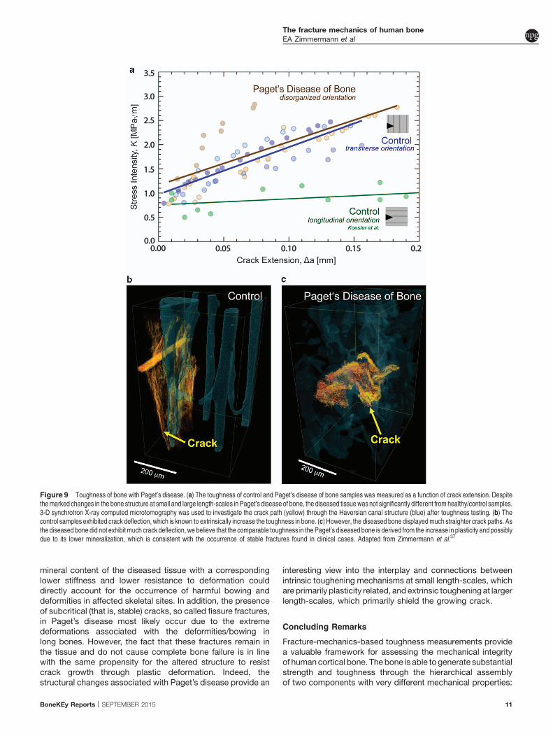
on healthy and diseased tissue from the iliac crest show littledifference in toughness (Figure 9a); however, the mechanism
for resisting crack propagation changes.37 Despite the limitednumber of toughness samples restricting statistical compar-isons, there was still clear evidence of higher fracture toughness
in the transversely oriented control and PDB bone samples, incomparison with the longitudinally oriented bone, which has a
comparatively weak resistance to crack growth.27 Indeed, thehealthy (control) bone resists crack propagation and derives
toughness in the usual way through crack deflectionmechanisms (Figure 9b).27 Conversely, human iliac crest bone
from patients with Paget’s disease exhibits a straighter crackpath (Figure 9c). In these diseased cases, the straighter crackpath arises due to the disorganized microstructure, which
cannot effectively deflect cracks. Although the ability for thediseased tissue to generate extrinsic toughening is severely
degraded, the bone structure compensates through a morepronounced mechanical resistance through plastic deforma-
tion, that is, Paget’s diseased bone shows higher ductility(intrinsic toughness).37 Indeed, the Paget’s tissue has a lower
mineralization and a correspondingly lower hardness, whichboth indicate that the tissue has the capacity for higher levels ofplasticity similar to other tissues with low mineralization levelsand significant plastic deformation.76,77 Thus, althoughtoughness is generated through crack deflection in healthybone (extrinsic toughening), the diseased tissue may generatetoughness primarily through intrinsic plasticity mechanismsthat absorb energy during deformation and fracture. However,further studies on the fracture toughness in bowed/deformedlong bones would add further detail on the mechanismsaffecting the resistance to fracture in PDB.
Treatment of PDBPaget’s disease is primarily treated with anti-resorptive agents,such as bisphosphonates. Anti-resorptive treatments act bysuppressing bone remodeling. In individuals treated for PDB,anti-resorptive agents have been shown to normalizethe serum alkaline phosphatase levels, which is a marker ofbone formation.78 In addition, in many cases significantimprovements in osteolytic lesions are visible radiologically, andhistomorphometric values return to normal with the formation oflamellar bone.79
SummaryCharacterizing the multi-scale structure and mechanicalproperties in Paget’s disease provides a basis for explainingsome of the clinical symptoms and the overall mechanicalintegrity of the diseased bone tissue. In particular, the lower
Figure 8 Characteristics of Paget’s disease at small length-scales. Paget’s disease of bone affected the composition of the bone structure at small length-scales. The mineraldistribution in (a) control and (b) Paget’s disease samples was measured with quantitative back-scattered electron imaging. (c) The Paget’s disease of bone samples had a loweraverage Ca-Wt% value and a higher degree of low mineralized bone, as seen from the histogram, which shows the distribution of Ca-Wt% values in the images. These changes incomposition at the nanoscale have a direct result on the mechanical properties. Nanoindentation was used to measure the (d) Young’s modulus and (e) hardness of the control anddiseased cases. Here, the disease samples have a lower stiffness, due to the lower mineralization, as well as a lower hardness, which measures the resistance to plastic deformation.Adapted from Zimmermann et al.37
The fracture mechanics of human boneEA Zimmermann et al
mineral content of the diseased tissue with a correspondinglower stiffness and lower resistance to deformation coulddirectly account for the occurrence of harmful bowing anddeformities in affected skeletal sites. In addition, the presenceof subcritical (that is, stable) cracks, so called fissure fractures,in Paget’s disease most likely occur due to the extremedeformations associated with the deformities/bowing inlong bones. However, the fact that these fractures remain inthe tissue and do not cause complete bone failure is in linewith the same propensity for the altered structure to resistcrack growth through plastic deformation. Indeed, thestructural changes associated with Paget’s disease provide an
interesting view into the interplay and connections betweenintrinsic toughening mechanisms at small length-scales, whichare primarily plasticity related, and extrinsic toughening at largerlength-scales, which primarily shield the growing crack.
Concluding Remarks
Fracture-mechanics-based toughness measurements providea valuable framework for assessing the mechanical integrityof human cortical bone. The bone is able to generate substantialstrength and toughness through the hierarchical assemblyof two components with very different mechanical properties:
Figure 9 Toughness of bone with Paget’s disease. (a) The toughness of control and Paget’s disease of bone samples was measured as a function of crack extension. Despitethe marked changes in the bone structure at small and large length-scales in Paget’s disease of bone, the diseased tissue was not significantly different from healthy/control samples.3-D synchrotron X-ray computed microtomography was used to investigate the crack path (yellow) through the Haversian canal structure (blue) after toughness testing. (b) Thecontrol samples exhibited crack deflection, which is known to extrinsically increase the toughness in bone. (c) However, the diseased bone displayed much straighter crack paths. Asthe diseased bone did not exhibit much crack deflection, we believe that the comparable toughness in the Paget’s diseased bone is derived from the increase in plasticity and possiblydue to its lower mineralization, which is consistent with the occurrence of stable fractures found in clinical cases. Adapted from Zimmermann et al.37
The fracture mechanics of human boneEA Zimmermann et al
BoneKEy Reports | SEPTEMBER 2015 11
ductile collagen molecules and brittle nanoplatelets ofhydroxyapatite mineral. Here, deformation mechanisms at thesmallest length-scales generate bone’s intrinsic resistance(that is, strength and crack-initiation toughness) mainlythrough plasticity provided by sliding mechanisms at the fibrillarlevel. At larger length-scales, the bone structure generatesextrinsic toughness (that is, crack-growth toughness) throughcrack-tip shielding mechanisms, such as crack deflection,principally at cement lines, and uncracked ligament bridging.The intrinsic and extrinsic toughening mechanisms workin concert to resist the initiation and propagation of cracks inhealthy cortical tissue.
Assessing fracture risk and preventing fractures are criticalissues in bone health. Many factors affect the fracturetoughness of human cortical bone, which can become moreprone to fracture due to aging, disease or other environmentalconditions (for example, vitamin deficiencies or irradiation). Thefracture risk or mechanical integrity of bone changes becauseaging, disease or other factors affect the bone structure atmultiple length-scales, which in turn affects the potency of theintrinsic and extrinsic toughening mechanisms. We havereviewed here recent studies to investigate the effects of aging,vitamin D deficiency and PDB on the fracture toughnessof human cortical bone. In many cases, changes to themineralization or cross-linking profile at small length-scalesdirectly affect the level of plasticity (that is, intrinsic toughness),whereas changes to the distribution or character of the osteonsat the microstructural scale affect the bone’s ability to resist thegrowth of cracks (that is, extrinsic toughness). Studying thesedisease conditions aids further progress in the diagnosis,prevention and treatment of diseases that increase the fracturerisk or affect the mechanical integrity of human cortical bone.
Conflict of Interest
The authors declare no conflict of interest.
Acknowledgements
We thank numerous individuals who have provided research orinput to this review, including Drs Claire Acevedo, Joel Ager,Tamara Alliston, Michael Amling, Hrishikesh Bale, Holly Barth,Neil Dave, Bernd Gludovatz, Michael Hahn, SophiIonova-Martin, Till Kohne, Kurt Koester, Jay Kruzic, NancyLane, Max Launey, Ravi Nalla, Diana Olvera, Brian Panganiban,Klaus Puschel, Eric Schaible, Simon Tang, Tony Tomsia,Wei Yao and Jozef Zustin. Several interactions/collaborationswith Drs Markus Buehler, David Burr, Paul Hansmaand Deepak Vashishth are also gratefully acknowledged.Elizabeth A. Zimmermann is supported by the Alexander vonHumboldt Foundation, Robert O. Ritchie by the Air Force Officeof Scientific Research, Multi-University Research Initiativegrant AFOSR-FA9550-15-1-0009, and Bjorn Busse by theEmmy Noether program under grant no. BU 2562-2/1 from theGerman Research Foundation (DFG).
References
1. Weiner S, Wagner HD. The material bone: structure mechanical function relations. Annu Rev
Mater Sci 1998; 28: 271–298.2. Arsenault A. Image-analysis of collagen-associated mineral distribution in cryogenically
prepared turkey leg tendons. Calcif Tissue Int 1991; 48: 56–62.
3. McNally EA, Schwarcz HP, Botton GA, Arsenault AL. A model for the ultrastructure of bone
based on electron microscopy of ion-milled sections. PLoS One 2012; 7: e29258.4. Robins SP. Biochemistry and functional significance of collagen cross-linking. Biochem Soc
Trans 2007; 35: 849–852.5. Thurner PJ, Katsamenis OL. The role of nanoscale toughening mechanisms in osteoporosis.
Curr Osteoporos Rep 2014; 12: 351–356.6. Grynpas MD, Tupy JH, Sodek J. The distribution of soluble, mineral-bound, and matrix-bound
proteins in osteoporotic and normal bones. Bone 1994; 15: 505–513.7. Bailey AJ. Molecular mechanisms of ageing in connective tissues. Mech Ageing Dev 2001;
122: 735–755.8. Knott L, Bailey AJ. Collagen cross-links in mineralizing tissues: a review of their chemistry,
function, and clinical relevance. Bone 1998; 22: 181–187.9. Avery NC, Bailey AJ. Enzymic and non-enzymic cross-linking mechanisms in relation
to turnover of collagen: relevance to aging and exercise. Scand J Med Sci Sports 2005; 15:231–240.
10. Skedros JG, Holmes JL, Vajda EG, Bloebaum RD. Cement lines of secondary osteons inhuman bone are not mineral-deficient: new data in a historical perspective. Anat Rec 2005;
286A: 781–803.11. Ritchie RO. The conflicts between strength and toughness. Nat Mater 2011; 10: 817–822.12. Zimmermann EA, Barth HD, Ritchie RO. The multiscale origins of fracture resistance in human
bone and its biological degradation. JOM 2012; 64: 486–493.13. Gupta HS, Wagermaier W, Zickler GA, Raz-Ben Aroush D, Funari SS, Roschger P et al.
Nanoscale deformation mechanisms in bone. Nano Lett 2005; 5: 2108–2111.14. Nair AK, Gautieri A, Chang S-W, Buehler MJ. Molecular mechanics of mineralized collagen
fibrils in bone. Nat Commun 2013; 4: 1724.15. Fantner GE, Hassenkam T, Kindt JH, Weaver JC, Birkedal H, Pechenik L et al. Sacrificial
bonds and hidden length dissipate energy as mineralized fibrils separate during bone fracture.Nat Mater 2005; 4: 612–616.
16. Poundarik AA, Diab T, Sroga GE, Ural A, Boskey AL, Gundberg CM et al. Dilatational band
formation in bone. Proc Natl Acad Sci USA 2012; 109: 19178–19183.17. Thurner PJ, Chen CG, Ionova-Martin S, Sun L, Harman A, Porter A et al. Osteopontin
deficiency increases bone fragility but preserves bone mass. Bone 2010; 46: 1564–1573.18. Gupta HS, Krauss S, Kerschnitzki M, Karunaratne A, Dunlop JWC, Barber AH et al. Intrafibrillar
plasticity through mineral/collagen sliding is the dominant mechanism for the extremetoughness of antler bone. J Mech Behav Biomed Mater 2013; 28: 366–382.
19. Karunaratne A, Esapa CR, Hiller J, Boyde A, Head R, Bassett JD et al. Significant deterioration
in nanomechanical quality occurs through incomplete extrafibrillar mineralization in rachiticbone: evidence from in-situ synchrotron X-ray scattering and backscattered electron imaging. J
Bone Miner Res 2012; 27: 876–890.20. Zimmermann EA, Gludovatz B, Schaible E, Busse B, Ritchie RO. Fracture resistance of human
cortical bone across multiple length-scales at physiological strain rates. Biomaterials 2014;35: 5472–5481.
21. Gautieri A, Vesentini S, Redaelli A, Buehler MJ. Viscoelastic properties of model segments ofcollagen molecules. Matrix Biol 2012; 31: 141–149.
22. Herman BC, Cardoso L, Majeska RJ, Jepsen KJ, Schaffler M. Activation of bone remodeling
after fatigue: Differential response to linear microcracks and diffuse damage. Bone 2010; 47:766–772.
23. Dooley C, Tisbo P, Lee TC, Taylor D. Rupture of osteocyte processes across microcracks:the effect of crack length and stress. Biomech Model Mechanobiol 2012; 11:
and mechanical repair of diffuse damage in cortical bone in vivo. J Bone Miner Res 2014; 29:
2537–2544.25. Ritchie RO. Mechanisms of fatigue-crack propagation in metals, ceramics and composites: role
of crack tip shielding. Mater Sci Eng A 1988; 103: 15–28.26. Nalla RK, Kruzic JJ, Ritchie RO. On the origin of the toughness of mineralized tissue:
microcracking or crack bridging? Bone 2004; 34: 790–798.27. Koester KJ, Ager JW, Ritchie RO. The true toughness of human cortical bone measured with
realistically short cracks. Nat Mater 2008; 7: 672–677.28. Katsamenis OL, Jenkins T, Thurner PJ. Toughness and damage susceptibility in human
cortical bone is proportional to mechanical inhomogeneity at the osteonal-level. Bone 2015; 76:
158–168.29. Currey JD. The effect of porosity and mineral content on the Young’s modulus of elasticity of
compact bone. J Biomech 1988; 21: 131–139.30. Schaffler MB, Burr DB. Stiffness of compact bone: effects of porosity and density. J Biomech
1988; 21: 13–16.31. Hui S, Slemenda C, Johnston C. Age and bone mass as predictors of fracture in a prospective-
study. J Clin Invest 1988; 81: 1804–1809.32. Aspray TJ, Prentice A, Cole TJ, Sawo Y, Reeve J, Francis RM. Low bone mineral content is
common but osteoporotic fractures are rare in elderly rural Gambian women. J Bone Miner Res
1996; 11: 1019–1025.33. Xiaoge D, Eryuan L, Xianping W, Zhiguang Z, Gan H, Zaijing J et al. Bone mineral
density differences at the femoral neck and Ward’s triangle: a comparison studyon the reference data between Chinese and Caucasian women. Calcif Tissue Int 2000; 67:195–198.
34. Kanis JA, Johnell O, Oden A, Dawson A, De Laet C, Jonsson B. Ten year probabilities ofosteoporotic fractures according to BMD and diagnostic thresholds. Osteoporo Int 2001; 12:
989–995.
The fracture mechanics of human boneEA Zimmermann et al
35. Yeni YN, Brown CU, Norman TL. Influence of bone composition and apparent density onfracture toughness of the human femur and tibia. Bone 1998; 22: 79–84.
36. Nyman JS, Roy A, Tyler JH, Acuna RL, Gayle HJ, Wang X. Age-related factorsaffecting the postyield energy dissipation of human cortical bone. J Orthop Res 2007; 25:646–655.
37. Zimmermann EA, Kohne T, Bale HA, Panganiban B, Gludovatz B, Zustin J et al. Modificationsto nano- and microstructural quality and the effects on mechanical integrity in Paget’s disease ofbone. J Bone Miner Res 2015; 30: 264–273.
38. Kulin RM, Jiang F, Vecchio KS. Loading rate effects on the R-curve behavior of cortical bone.Acta Biomater 2011; 7: 724–732.
39. Zimmermann EA, Launey ME, Barth HD, Ritchie RO. Mixed-mode fracture of human corticalbone. Biomaterials 2009; 30: 5877–5884.
40. Heaney RP, Abrams S, Dawson-Hughes B, Looker A, Marcus R, Matkovic V et al. Peak bonemass. Osteoporosis Int 2000; 11: 985–1009.
41. Koehne T, Vettorazzi E, Kusters N, Luneburg R, Kahl-Nieke B, Puschel K et al. Trends intrabecular architecture and bone mineral density distribution in 152 individuals aged 30–90years. Bone 2014; 66: 31–38.
42. Zioupos P, Currey JD. Changes in the stiffness, strength, and toughness of human cortical bonewith age. Bone 1998; 22: 57–66.
43. Nalla RK, Kruzic JJ, Kinney JH, Balooch M, Ager JW, Ritchie RO. Role of microstructure in theaging-related deterioration of the toughness of human cortical bone. Mater Sci Eng C 2006; 26:1251–1260.
44. Wang X, Shen X, Li X, Mauli Agrawal C. Age-related changes in the collagen network andtoughness of bone. Bone 2002; 31: 1–7.
45. Zimmermann EA, Schaible E, Bale H, Barth HD, Tang SY, Reichert P et al. Age-relatedchanges in the plasticity and toughness of human cortical bone at multiple length scales.Proc Natl Acad Sci USA 2011; 108: 14416–14421.
46. Granke M, Makowski AJ, Uppuganti S, Does MD, Nyman JS. Identifying novelclinical surrogates to assess human bone fracture toughness. J Bone Miner Res 2015; 30:1290–1300. doi:10.1002/jbmr.2452.
47. Odetti P, Rossi S, Monacelli F, Poggi A, Cirnigliaro M, Federici M et al. Advanced glycation endproducts and bone loss during aging. In: Baynes JW, Monnier VM, Ames JM, Thorpe SR (eds)Maillard Reaction: Chemistry at the Interface of Nutrition, Aging, and Disease. New YorkAcademy of Sciences: NY, USA, 2005, pp 710–717.
48. Saito M, Marumo K, Fujii K, Ishioka N. Single-column high-performance liquid chromatographicfluorescence detection of immature, mature, and senescent cross-links of collagen. AnalBiochem 1997; 253: 26–32.
49. Sell D, Monnier V. Structure elucidation of a senescence cross-link from humanextracellular-matrix - Implication of pentoses in the aging process. J Biol Chem 1989; 264:21597–21602.
50. Vashishth D, Gibson GJ, Khoury JI, Schaffler MB, Kimura J, Fyhrie DP. Influenceof nonenzymatic glycation on biomechanical properties of cortical bone. Bone 2001; 28:195–201.
51. Siegmund T, Allen MR, Burr DB. Failure of mineralized collagen fibrils: Modeling the role ofcollagen cross-linking. J Biomech 2008; 41: 1427–1435.
52. Buehler MJ. Nanomechanics of collagen fibrils under varying cross-link densities: atomistic andcontinuum studies. J Mech Behav Biomed Mater 2008; 1: 59–67.
53. Schaffler MB, Choi K, Milgrom C. Aging and matrix microdamage accumulation in humancompact bone. Bone 1995; 17: 521–525.
54. Diab T, Vashishth D. Morphology, localization and accumulation of in vivo microdamage inhuman cortical bone. Bone 2007; 40: 612–618.
55. Shane E, Burr D, Ebeling PR, Abrahamsen B, Adler RA, Brown TD et al. Atypicalsubtrochanteric and diaphyseal femoral fractures: report of a task force of the American Societyfor Bone and Mineral Research. J Bone Miner Res 2010; 25: 2267–2294.
56. Allen MR, Kubek DJ, Burr DB. Cancer treatment dosing regimens of zoledronic acid result innear-complete suppression of mandible intracortical bone remodeling in beagle dogs. J BoneMiner Res 2010; 25: 98–105.
57. Milovanovic P, Zimmermann EA, Riedel C, vom Scheidt A, Herzog L, Krause M et al.Multi-level characterization of human femoral cortices and their underlying osteocyte networkreveal trends in quality of young, aged, osteoporotic, and antiresorptive-treated bone.Biomaterials 2015; 45: 46–55.
58. Bala Y, Depalle B, Farlay D, Douillard T, Meille S, Follet H et al. Bone micromechanicalproperties are compromised during long-term alendronate therapy independently ofmineralization. J Bone Miner Res 2012; 27: 825–834.
59. Donnelly E, Meredith DS, Nguyen JT, Gladnick BP, Rebolledo BJ, Shaffer AD et al. Reducedcortical bone compositional heterogeneity with bisphosphonate treatment in postmenopausalwomen with intertrochanteric and subtrochanteric fractures. J Bone Miner Res 2012; 27:672–678.
60. Bernhard A, Milovanovic P, Zimmermann EA, Hahn M, Djonic D, Krause M et al.Micro-morphological properties of osteons reveal changes in cortical bone stabilityduring aging, osteoporosis, and bisphosphonate treatment in women. Osteoporos Int 2013;24: 2671–2680.
61. Roschger P, Rinnerthaler S, Yates J, Rodan GA, Fratzl P, Klaushofer K. Alendronate increasesdegree and uniformity of mineralization in cancellous bone and decreases the porosity incortical bone of osteoporotic women. Bone 2001; 29: 185–191.
62. Borah B, Dufresne T, Nurre J, Phipps R, Chmielewski P, Wagner L et al. Risedronatereduces intracortical porosity in women with osteoporosis. J Bone Miner Res 2010; 25:41–47.
63. Ettinger B, Burr DB, Ritchie RO. Proposed pathogenesis for atypical femoral fractures: lessonsfrom materials research. Bone 2013; 55: 495–500.
64. Bajaj D, Geissler JR, Allen MR, Burr DB, Fritton JC. The resistance of cortical bone tissue tofailure under cyclic loading is reduced with alendronate. Bone 2014; 64: 57–64.
65. Pazianas M, Epstein S, Zaidi M. Evaluating the antifracture efficacy of bisphosphonates. RevRecent Clin Trials 2009; 4: 122–130.
66. Lips P. Vitamin D deficiency and secondary hyperparathyroidism in the elderly:consequences for bone loss and fractures and therapeutic implications. Endocr Rev 2001;22: 477–501.
67. Whyte MP, Thakker RV. Rickets and osteomalacia. Medicine (Baltimore) 2005; 33: 70–74.68. Busse B, Bale HA, Zimmermann EA, Panganiban B, Barth HD, Carriero A et al. Vitamin D
deficiency induces early signs of aging in human bone, increasing the risk of fracture. Sci TranslMed 2013; 5: 193ra88.
69. Teitelbaum SL. Renal osteodystrophy. Hum Pathol 1984; 15: 306–323.70. Davie M, Davies M, Francis R, Fraser W, Hosking D, Tansley R. Paget’s disease of bone: a
review of 889 patients. Bone 1999; 24: 11S–12S.71. Seitz S, Priemel M, Zustin J, Beil FT, Semler J, Minne H et al. Paget’s disease of bone:
histologic analysis of 754 patients. J Bone Miner Res 2009; 24: 62–69.72. Cushing FR, Bone HG. Radiographic diagnosis and laboratory evaluation of Paget’s disease of
with Paget’s disease: a population-based cohort study. J Bone Miner Res 2000; 15:2123–2128.
74. Van Staa TP, Selby P, Leufkens HGM, Lyles K, Sprafka JM, Cooper C. Incidence andnatural history of Paget’s disease of bone in England and Wales. J Bone Miner Res 2002; 17:465–471.
75. Meunier PJ, Coindre JM, Edouard CM, Arlot ME. Bone histomorphometry in Paget’s disease.Quantitative and dynamic analysis of pagetic and nonpagetic bone tissue. Arthritis Rheum1980; 23: 1095–1103.
77. Krauss S, Fratzl P, Seto J, Currey JD, Estevez JA, Funari SS et al. Inhomogeneous fibrilstretching in antler starts after macroscopic yielding: indication for a nanoscale tougheningmechanism. Bone 2009; 44: 1105–1110.
78. Miller PD, Brown JP, Siris ES, Hoseyni MS, Axelrod DW, Bekker PJ. A randomized, double-blind comparison of risedronate and etidronate in the treatment of Paget’s disease of bone. AmJ Med 1999; 106: 513–520.
79. Reid IR, Nicholson GC, Weinstein RS, Hosking DJ, Cundy T, Kotowicz MA et al. Biochemicaland radiologic improvement in Paget’s disease of bone treated with alendronate: A ran-domized, placebo-controlled trial. Am J Med 1996; 101: 341–348.
80. Ascenzi M-G, Ascenzi A, Benvenuti A, Burghammer M, Panzavolta S, Bigi A. Structuraldifferences between ‘dark’ and ‘bright’ isolated human osteonic lamellae. J Struct Biol 2003;141: 22–33.
81. Reznikov N, Shahar R, Weiner S. Three-dimensional structure of human lamellar bone: thepresence of two different materials and new insights into the hierarchical organization. Bone2014; 59: 93–104.
82. Milovanovic P, Zimmermann EA, Hahn M, Djonic D, Puschel K, Djuric M et al. Osteocyticcanalicular networks: morphological implications for altered mechanosensitivity. ACS Nano2013; 7: 7542–7551.
83. You L-D, Weinbaum S, Cowin SC, Schaffler MB. Ultrastructure of the osteocyte process and itspericellular matrix. Anat Rec 2004; 278: 505–513.
84. Knott L, Whitehead CC, Fleming RH, Bailey AJ. Biochemical changes in the collagenous matrixof osteoporotic avian bone. Biochem J 1995; 310: 1045–1051.
85. Launey ME, Buehler MJ, Ritchie RO. On the mechanistic origins of toughness in bone. AnnuRev Mater Res 2010; 40: 25–53.
86. Koester KJ, Barth HD, Ritchie RO. Effect of aging on the transverse toughness ofhuman cortical bone: evaluation by R-curves. J Mech Behav Biomed Mater 2011; 4:1504–1513.
The fracture mechanics of human boneEA Zimmermann et al