130
The FRCPath Exam How to pass the short surgicals on the first go By someone who didn’t Plus some tips on frozen sections! Dr Paul Bennett
The FRCPath Exam
How to pass the short surgicals on the
first go By someone who didn’t
Plus some tips on frozen sections!
Dr Paul Bennett
Overview
• Reminder of the exam format
• How to format your answer for the short
surgicals
• Discussion of cases
• Brief discussion of frozen sections
• Mandatory viewing of holiday photos
throughout talk
Exam Overview
• Short Surgicals
– 20 cases
– Given in pairs, 20 minutes per pair
– When they’re gone, they’re gone
– Can be anything
• Long Cases
– 4 cases, 20 minutes per case
– Special stains / immuno / FISH / EM
Exam Overview
• Frozens
– 6 cases, 20 minutes per three cases
– Scribble notes in the answer book
– Viva at the end
• OSPEs
– Two stations, one manned
– A) Management issue
– B) Data set and short answers
Exam Overview
• Non-gynae cytology
- 8 cases, 20 minutes per pair
- Often benign vs. malignant cases
- Macros
- 4 cases, each with a photograph
- Draw blocks on photograph
- Answer short questions in Viva

Exam Overview
• Archaic marking system
– Scored out of 5 • 1 or 1.5 = fail
• 2 = bare fail
• 2.5 = pass
• 3 = good answer
• 3.5 = excellent answer
• 4+ = ? Possible
Overall pass mark 50%
Why focus on the short surgicals?
• Obviously, each component of the exam
requires preparation
• However, surgicals often cause problems
– Largest single component of exam
– Large variety of cases
– Lots of writing
• A well-practised approach works
Approaching the Questions
• Use a tried and tested format that you are
familiar with
• Answer every question the same way
• I emailed my preferred format to you – a 6
point approach (credit to Dr D Scott)
• This format will allow you to maximise
marks for each question
Approaching the Questions
1. Description
2. Interpretation
3. Differential diagnosis
4. Extra investigations
5. Clinico-pathological correlation
6. Bottom line diagnosis
Approach to a Case
• Description – Keep it brief
– Include description of type of lesion (eg neoplasm, inflammatory process)
– Brief description of architecture and morphology if it’s a lesion
– Mention dataset items if a tumour (margin involvement, necrosis, vascular invasion, etc)
– Don’t forget to grade any neoplasms
– May be appropriate to comment on the background tissue briefly
Approach to a Case
• Interpretation
– Should immediately follow description and
summarise your thoughts on the H+E slide
– State diagnosis if you’re sure
• “This is a malignant melanoma”
- If you’re not sure, go as far as you can
• “This is a malignant epithelioid neoplasm”
– If you’re really struggling
• “This is an epithelioid neoplasm”
Approach to a Case
• Differential Diagnosis
– One or two questions may require a
differential
– Will be unnecessary in most questions
– For example, a malignant epithelioid
neoplasm:
• Carcinoma
• Melanoma
• Large cell lymphoma
• Mesothelioma
Approach to a Case
• Extra investigations (if appropriate)
- Start from the bottom
- Levels (usually unnecessary but demonstrates
safety if uncertain)
- Special stains
- Immunocytochemistry
- Useful to incorporate immunostains into differential
diagnosis
- IMF
- Cytogenetics
- EM

Approach to a Case
• If the diagnosis is already established on morphology, mentioning extra investigations still adds extra value to your answer, eg: - Signet ring carcinoma: PAS +ve inclusions
- Glomus tumour: SMA +ve
- Pemphigus vulgaris: Reticular IgG/C3 on IMF
- Well differentiated liposarcoma: MDM-2/CDK4 amplification on cytogenetics
- Langerhan’s cell histiocytosis: Birbeck granules on EM
Approach to a Case
• Clinicopathological correlation – Chance to earn extra credit
– All malignant cases to relevant MDT
– Classical histories • COCP use in liver adenoma
• Epithelioid sarcoma – peripheral sites, young patients
– Mention any classical symptoms • Painful ANGEL skin lesions
– Typical imaging features • DCIS: Calcification on plain X-Ray
Approach to a Case
• Clinico-pathological correlation (continued)
– Treatment • Melanomas – wider excisions +/- sentinel nodes
• Adenocarcinoma of lung – EGFR inhibitors
• GISTs – Tyrosine kinase inhibitors
– Prognosis • Good – Nodular lymphocyte predominant Hodgkin’s
• Poor – Anaplastic large T cell lymphoma
– Stage • If you can remember! Mention it regardless
Approach to a Case
• Bottom line diagnosis
– One line only
This may be the first bit of your answer the
examiner looks at. If it’s right, you’re off to a
good start and the examiner can look for extra
marks in the rest of your answer

Case 1
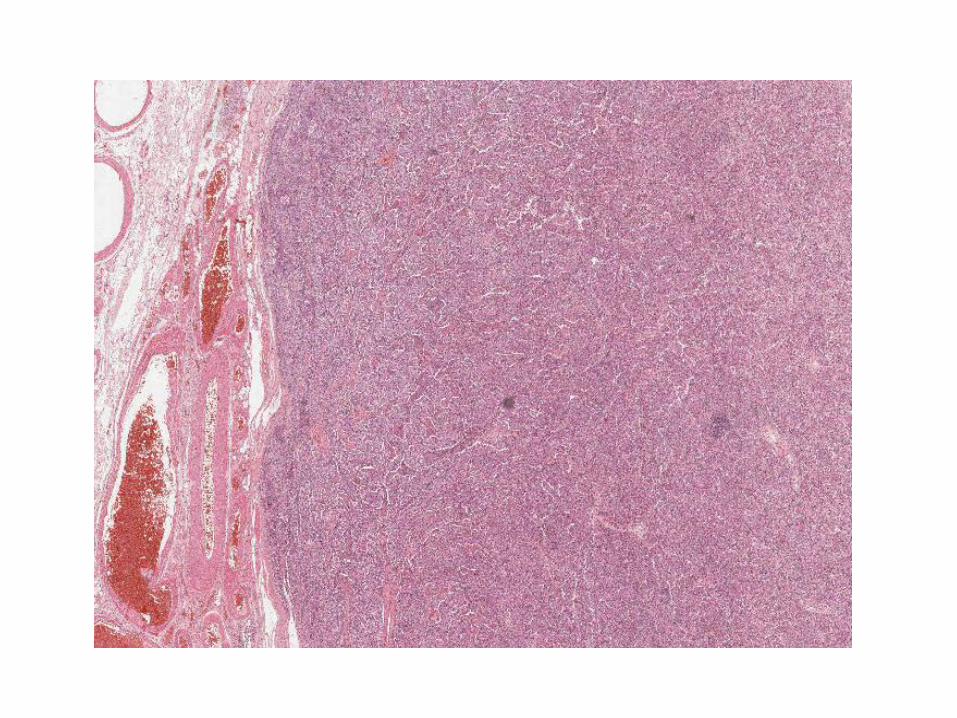
29M, testicular mass
Case 1
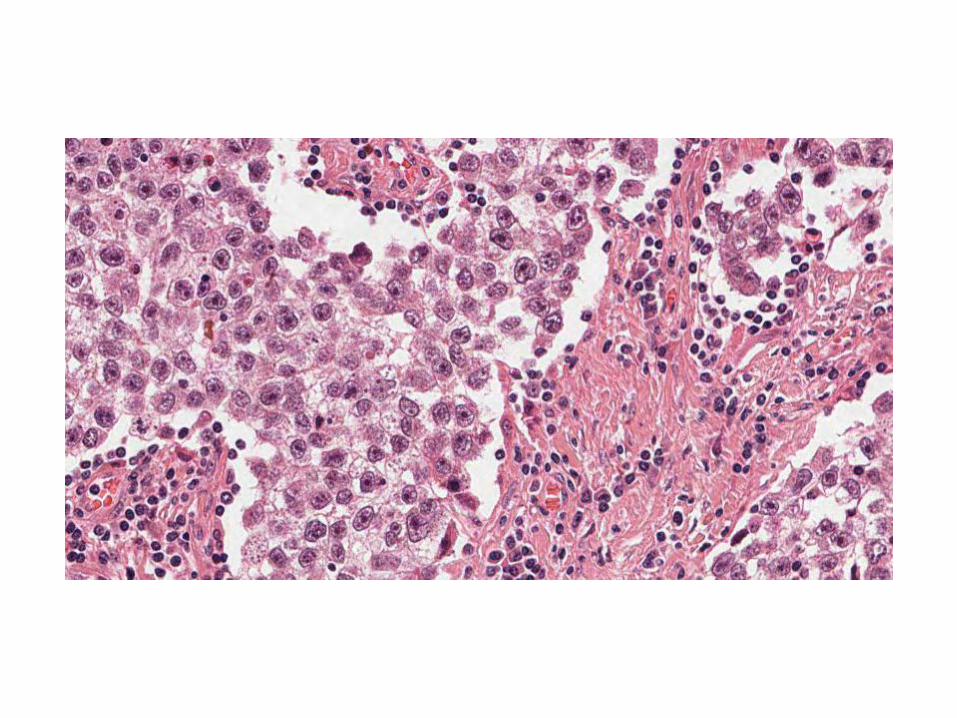
• Description
– Nested tumour separated by broad,
lymphocyte-rich fibrous bands. The tumour
cells show large nuclei, prominent nucleoli
and delicate cytoplasm.
– There is focal rete testis involvement
– There is no evidence of vascular invasion or
intratubular germ cell neoplasia
Case 1
• Interpretation
– This is a classical seminoma
– This is a malignant germ cell tumour
– This is a malignant epithelioid neoplasm
The further down the list, the less likely you
are to pick up any marks

Case 1
• Differential
– Shouldn’t really need one – H+E spot diagnosis
– Some other germ cell tumours (spermatocytic seminoma) lymphoma and melanoma are perhaps reasonable to suggest if you don’t know
– Either way…..
To the immuno!
Case 1
• Extra investigations
– Not necessary if you’ve got the diagnosis on H+E
– BUT, you can still mention classic staining pattern for
possible extra points:
• Classical seminoma: C-Kit, PLAP, Oct 3/4 positive
– If working on differentials:
• Spermatocytic Seminoma: C-kit & OCT 3/4 +ve; PLAP -ve
• Lymphoma: CD45 +ve; germ cell markers -ve
• Melanoma: Melan-A & S100 +ve; germ cell markers –ve
Case 1
• Clinico-pathological correlation
– Needs staging and discussion at MDT
– Commoner in young men
– Associated with history of undescended
testes
– Treatment – orchidectomy
– Chemo if recurrent or advanced disease
– Good prognosis in many cases
– pT1 on this slide
Case 1
• Testicular mass - Classical seminoma
Case 2
67F, retroperitoneal mass
Case 2
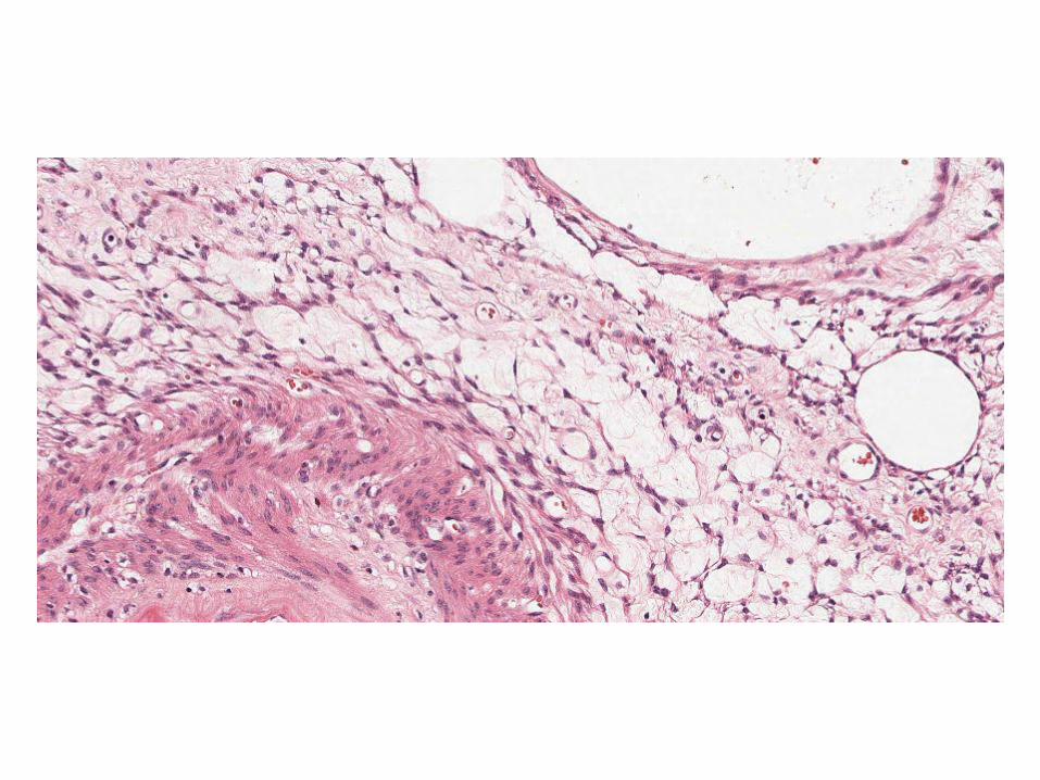
• Description
– Diffuse tumour composed of admixture of fat,
blood vessels and smooth muscle. The
smooth muscle component emanates from
the vessel walls
– No obvious renal tissue in sections
– No evidence of atypia, necrosis or increased
mitotic activity
Case 2
• Interpretation
– This is an angiomyolipoma (PEComa)
– This is a mesenchymal tumour of uncertain
malignant potential
– This is a neoplasm
Case 2
• Differential
– Very few… no real features of any other
tumours expected at this site (RCC, adrenal
tumours, well-diff liposarcomas)
– Fat predominant or muscle predominant
variants occur – in these cases a differential is
reasonable
– I wouldn’t include a differential in this case –
it’s classical
Case 2
• Extra investigations
– Positive immunoreactivity for HMB-45 +
Melan-A

Case 2
• Clinico-pathological correlation
– Discuss at sarcoma MDT
– Variable malignant potential
– Excision usually curative, though necrosis, increased mitoses and atypia increase risk of metastasis
– Many (up to a third) associated with tuberous sclerosis
– May co-exist with other PEComas • Clear cell tumours of the lung
• Lymphangiomyomatosis
Case 2
• Retroperitoneal mass -
Angiomyolipoma
Case 3
38M, lesion on arm
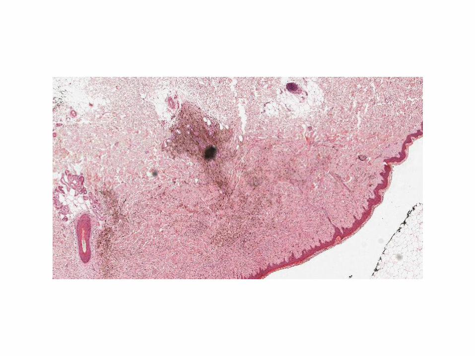
Case 3
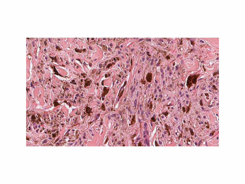
• Description
– This is skin with a normal epidermis. The
dermis contains a symmetrical proliferation of
pigmented, spindled melanocytes. There is
no evidence of atypia or mitotic activity.
There is no evidence of a junctional
component
– This lesion appears completely excised
Case 3
• Interpretation
– This is a blue naevus
– This is a benign melanocytic proliferation
– This is a melanocytic proliferation
Case 3
• Differential
– Again, only necessary if you’re not sure
– Malignant melanoma (primary / metastatic)
• If going down this line explain how you’d sort it out
– Check history
– Levels for junctional component / regression
– Pigmented DFSP
• Sort out on immuno if you’re worried about this
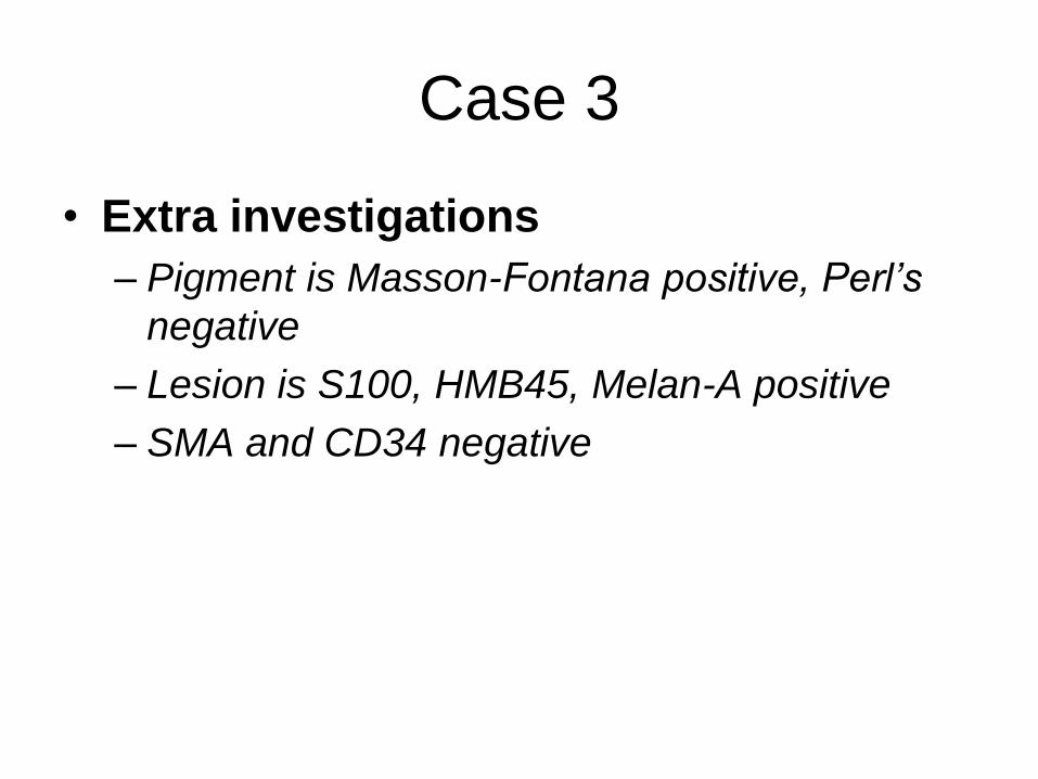
Case 3
• Extra investigations
– Pigment is Masson-Fontana positive, Perl’s
negative
– Lesion is S100, HMB45, Melan-A positive
– SMA and CD34 negative
Case 3
• Clinico-pathological correlation
– Benign lesions
– Small and blue macroscopically
– Excision curative
– BRAF / RAS mutations absent

Case 3
• Skin, arm - Blue naevus
Case 4
52F, lesions on lower legs
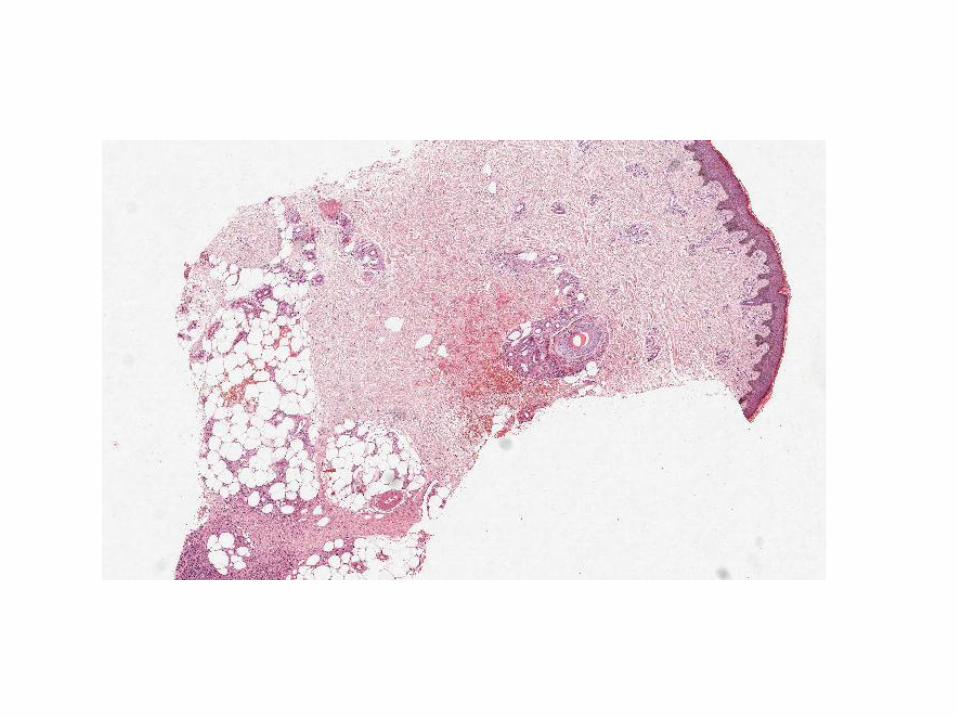
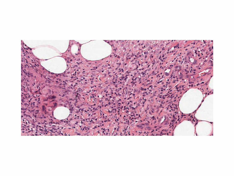
Case 4
• Description
– This is a punch biopsy of skin and
subcutaneous fat. The fat shows septal
inflammation composed of histiocytes,
lymphocytes and plasma cells. There is no
evidence of vasculitis, necrosis or neoplasia.
The overlying skin shows mild venous stasis-
related features only
Case 4
• Interpretation
– This is erythema nodosum
– This is a septal panniculitis
– This is panniculitis
– This is an inflammatory process
Case 4
• Differential
– Only if you’re not sure
– Lobular panniculitis
• Secondary to pancreatitis
• Could suggest serum amylase
Case 4
• Extra investigations
– Fungal / ZN stains reasonable as
granulomatous inflammation
Case 4
• Clinico-pathological correlation
– F>M
– Lots of causes
• Drugs (COCP) – check history
• Infections (TB, Strep)
• Malignancies (usu. haematological)
• Crohn’s
• Sarcoid – recent chest X-Ray?
– EN is often reactive and self-limiting
Case 4
• Skin, lower leg – Erythema Nodosum
Case 5
M16, multiple skin lesions – excision
of one from arm
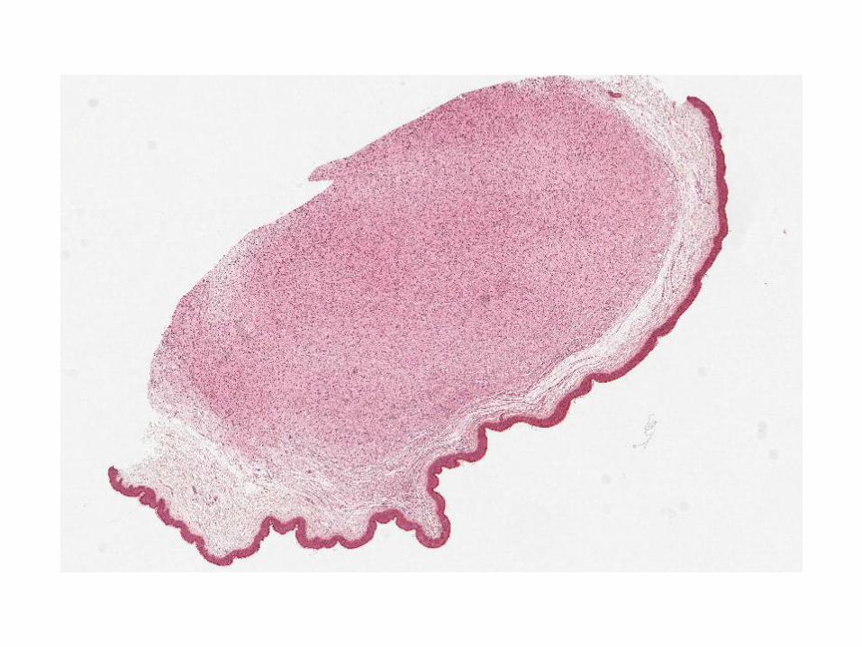
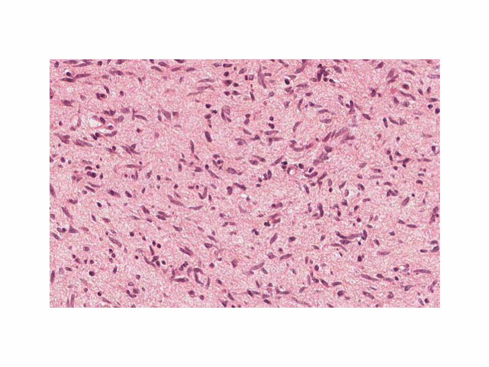
Case 5
• Description
– This is skin with a normal epidermis. The
dermis contains a well-circumscribed
proliferation of spindle cells with bland
buckled nuclei and admixed mast cells.
There are no features of atypia, mitotic activity
or necrosis
– This lesion is incompletely excised at the
deep margin

Case 5
• Interpretation
– This is a neurofibroma
– This is a benign neural lesion
– This is a benign mesenchymal / spindle cell
tumour
– This is a benign neoplasm
Case 5
• Differential diagnosis
– If not sure:
• Dermatofibroma not an unreasonable suggestion,
but unlikely to do you much good in a simple case
like this
Case 5
• Extra investigations
– Not necessary in reality, but could state that
neurofibromas are S100 positive (patchy)
Case 5
• Clinico-pathological correlation
– History of multiple lesions!
– NF-1 must be considered
• Café au lait spots, Lisch nodules
• Increased risk of MPNST
• Be firm that this case is benign (no mitoses etc)
– Otherwise excision is curative
Case 5
• Skin, arm - Neurofibroma
Case 6
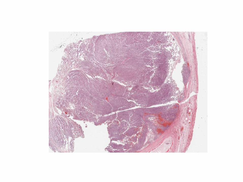
M66, testicular mass
Case 6
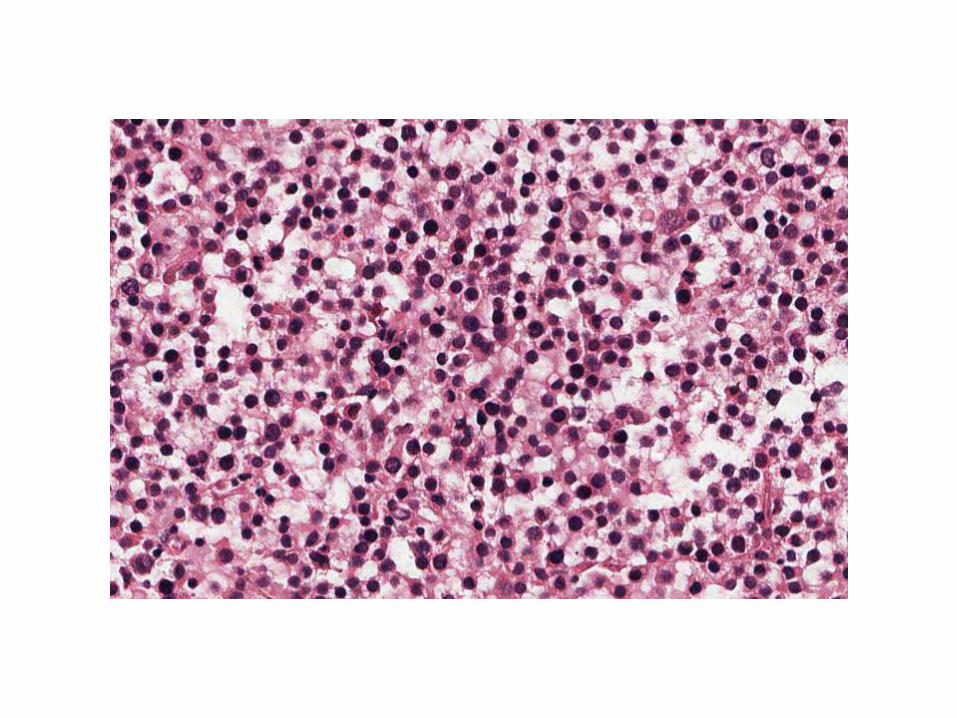
• Description
– This is a tumour composed of nests and
sheets of cells with round nuclei, prominent
nucleoli and delicate cytoplasm. There is no
evidence of a prominent lymphocytic
component. The tumour appears confined to
the testis and vascular invasion is not seen.
There is no evidence of IGCN.

Case 6
• Interpretation
– This is a spermatocytic seminoma
– This is a malignant germ cell tumour
– This is a malignant epithelioid neoplasm
Case 6
• Differential diagnosis
– Main one is classical seminoma
• Spermatocytic seminomas lack lymphocyte-rich
fibrous bands and usually affect older men
• They are also not associated with IGCN
• Can sort out on immuno if unsure
Case 6
• Extra investigations
– Immuno
• Spermatocytic seminoma
– C-KIT & OCT 3/4 +ve
– PLAP –ve
• Classical seminoma
– C-KIT, OCT 3/4 , PLAP +ve
Case 6
• Clinico-pathological correlation
– Older men
– Excellent prognosis
– Excision curative
– Stage and discuss at Urological MDT
Case 6
• Testicular mass – Spermatocytic
seminoma
Case 7
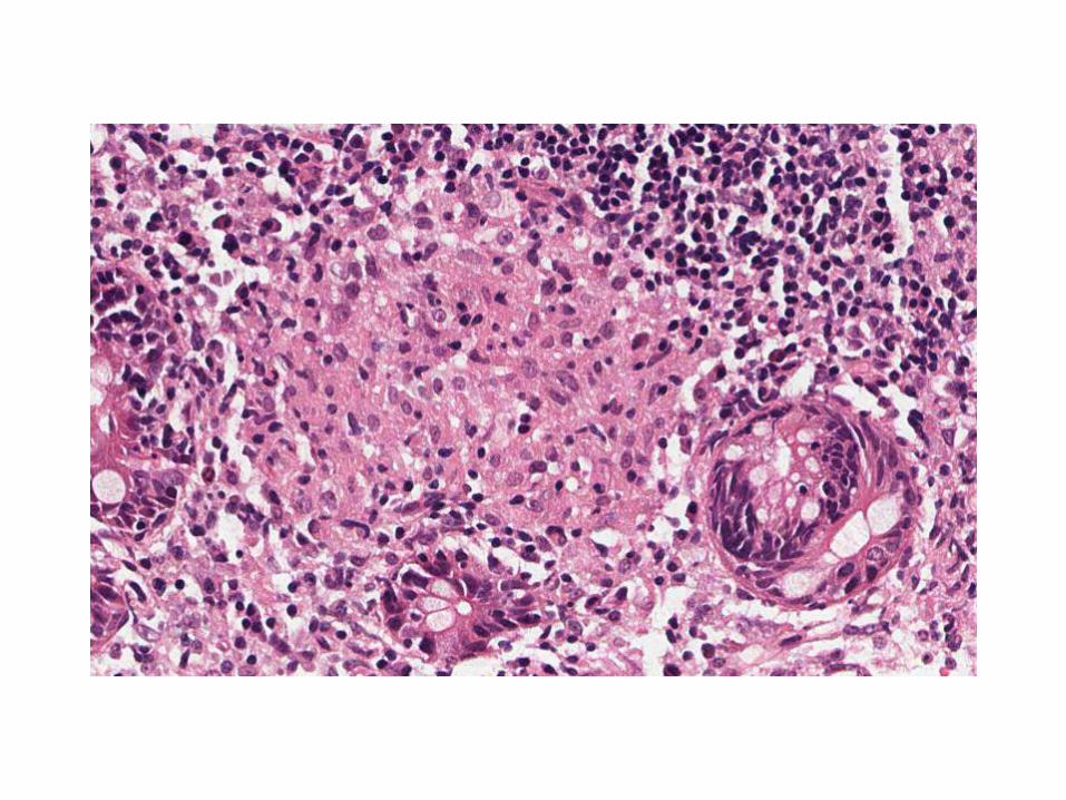
M18, diarrhoea, colorectal biopsies

Case 7
• Description
– Biopsies of large intestinal mucosa showing
mild crypt distortion, non-caeseating
granulomas within the lamina propria and a
mild increase in lamina propria cellularity
– No evidence of active inflammation,
ulceration, dysplasia or malignancy

Case 7
• Interpretation
– This is a granulomatous colitis
Case 7
• Differential diagnosis
– Necessary in this case
• Crohn’s disease
• Mycobacterial infection
• Fungal infection
• Sarcoidosis
• Reaction to tumour
Case 7
• Extra investigations
– ZN / fungal stains
– Mycobacterial PCR
Case 7
• Clinico-pathological correlation
– Needs examination of targeted colorectal and
ileal biopsies to investigate IBD
• Crohn’s disease associated with skip lesions,
fissuring
Case 7
• Colorectal biopsies – Granulomatous
colitis
Case 8
F42, lesion on arm
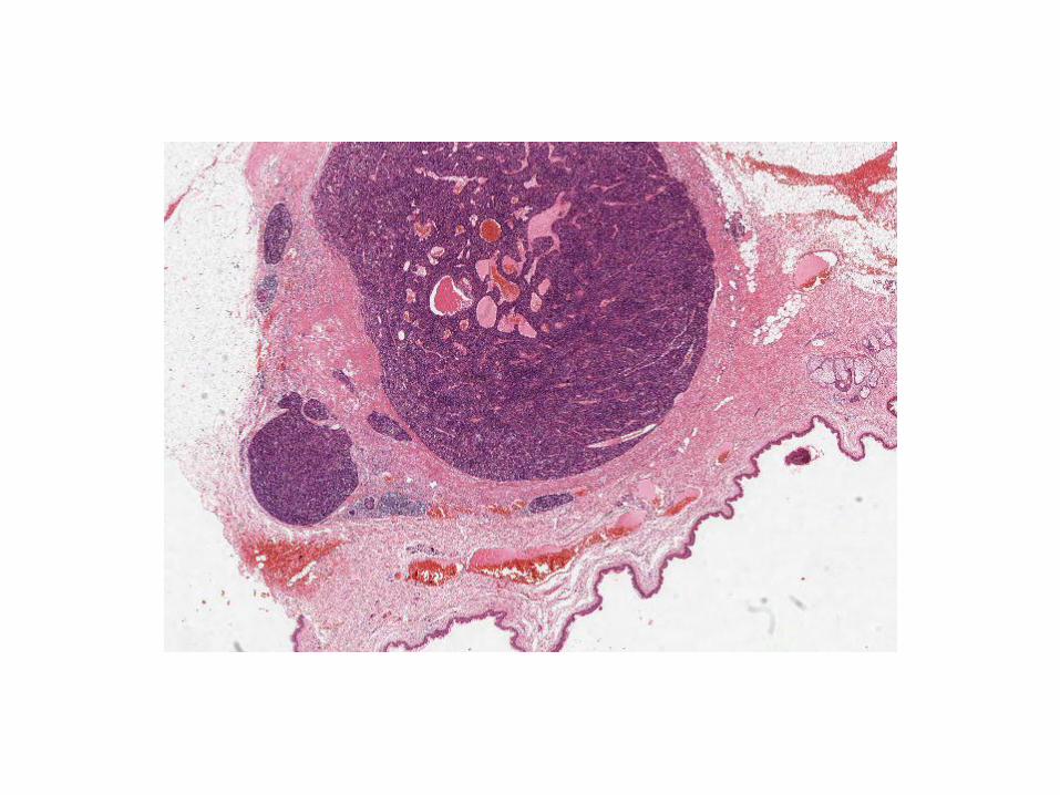
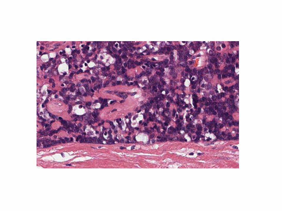
Case 8
• Description
– This is a well-circumscribed, vascular, dermal
lesion composed of small basaloid cells
admixed with larger pale epithelial cells and
lymphocytes. There is no epidermal
connection. There are no features of
malignancy.
– This lesion appears excised in the plane of
section examined
Case 8
• Interpretation
– This is an eccrine spiradenoma
– This is a benign eccrine tumour
– This is a benign adnexal tumour
– This is a benign tumour
Case 8
• Differential diagnosis
– Reasonable to include some other adnexal
tumours if you’re not sure
• Cylindroma
• Acrospiroma (Dermal duct tumour)
Case 8
• Extra investigations
– No relevant that I can think of
Case 8
• Clinico-pathological correlation
– Usually solitary lesions
– Classically painful (ANGEL)
– Malignant transformation rare
– Excision curative
– Multiple tumours associated with Brooke-
Spiegler syndrome
Case 8
• Lesion on arm – Eccrine spiradenoma
Case 9
M38, nausea and vomiting, gastric
biopsies
Case 9
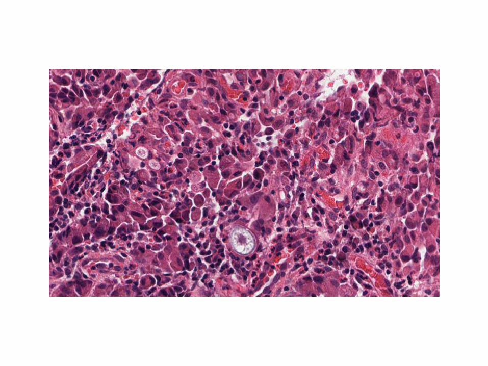
• Description
– These are biopsies of fundic-type gastric
mucosa showing diffuse infiltration of the
lamina propria by malignant tumour cells
– The cells show displaced nuclei and
occasional cytoplasmic vacuolation (“signet-
ring” morphology)
– No vascular invasion identified
– No evidence of background dysplasia
Case 9
• Interpretation
– This is poorly-differentiated (signet-ring)
adenocarcinoma
– This is a malignant epithelioid neoplasm
Case 9
• Differential diagnosis
– Poorly differentiated adenocarcinoma
– Lymphoma
– Melanoma
Case 9
• Extra investigations
– PAS stain - +ve in vacuoles
– Immuno
• Cytokeratin (CK 7) and CEA positive
• CK20, CD45, S100 and Melan-A negative
Case 9
• Clinico-pathological correlation
– Requires MDT correlation
– Classical “linitis plastica” picture at endoscopy
– May be resectable depending on extent of
spread
– Overall poor prognosis
– New evidence of role of Trastuzumab in Her-2
positive gastric cancer
Case 9
• Gastric biopsies – Poorly differentiated
(signet ring) adenocarcinoma
Case 10
F28, haematuria and pelvic pain,
bladder biopsies
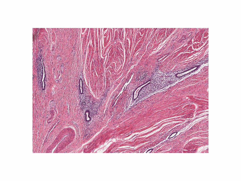
Case 10
• Description
– Bundles of smooth muscle infiltrated by
endometrial glands and stroma
– Foci of extravasated red blood cells and
pigment-laden macrophages
– No evidence of atypia or necrosis
– No in-situ disease
Case 10
• Interpretation
– This is endometriosis
Case 10
• Differential diagnosis
– No others of note
Case 10
• Extra investigations
– Not necessary, but can state that endometrial
foci would be CD10 and ER positive
Case 10
• Clinico-pathological correlation
– Endometriosis often multifocal (cervix, Pouch
of Douglas, ovaries)
– Increased risk of infertility
– Treatment involves hormones (COCP, coil) or
surgical
– Usually disappears post-menopause
Case 10
• Bladder biopsies - Endometriosis
Case 11
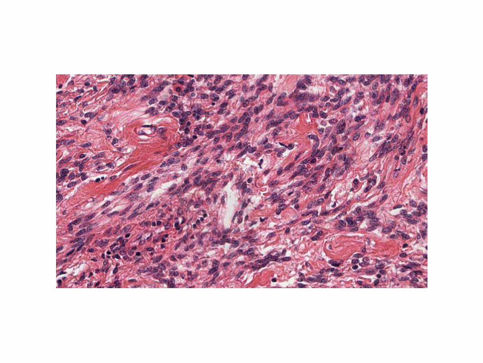
F61, hysterectomy. Lesion in
anterior myometrium
Case 11
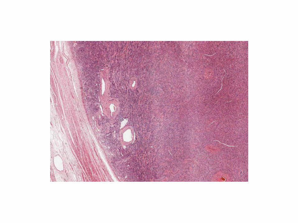
• Description
– Well-circumscribed cellular lesion composed
of spindle cells with cigar-shaped nuclei
– Minimal nuclear atypia, no increase in mitotic
activity and no necrosis
– Smooth, regular interface with surrounding
tissue
Case 11
• Interpretation
– This is a cellular leiomyoma
– This is a leiomyoma
– This is a benign smooth muscle tumour
– This is a smooth muscle tumour
Case 11
• Differential diagnosis
– Cellular leiomyoma
– Leiomyosarcoma
– STUMP
Case 11
• Extra investigations
– Correlate with macroscopic appearance
– Needs extensive sampling, particularly around
the edges of the lesion
• Search for areas of increased mitotic activity,
necrosis, infiltrative border
– Can confirm smooth muscle origin with SMA,
desmin, H-caldesmon
– Ki67 may be useful
Case 11
• Clinico-pathological correlation
– Correlate with imaging – any suspicious
ultrasound features?
– Cellular leiomyomas are benign
– Excision curative
Case 11
• Uterus – Cellular leiomyoma
Case 12
F30, rash on arms. IgA positive on
immunofluorescence studies
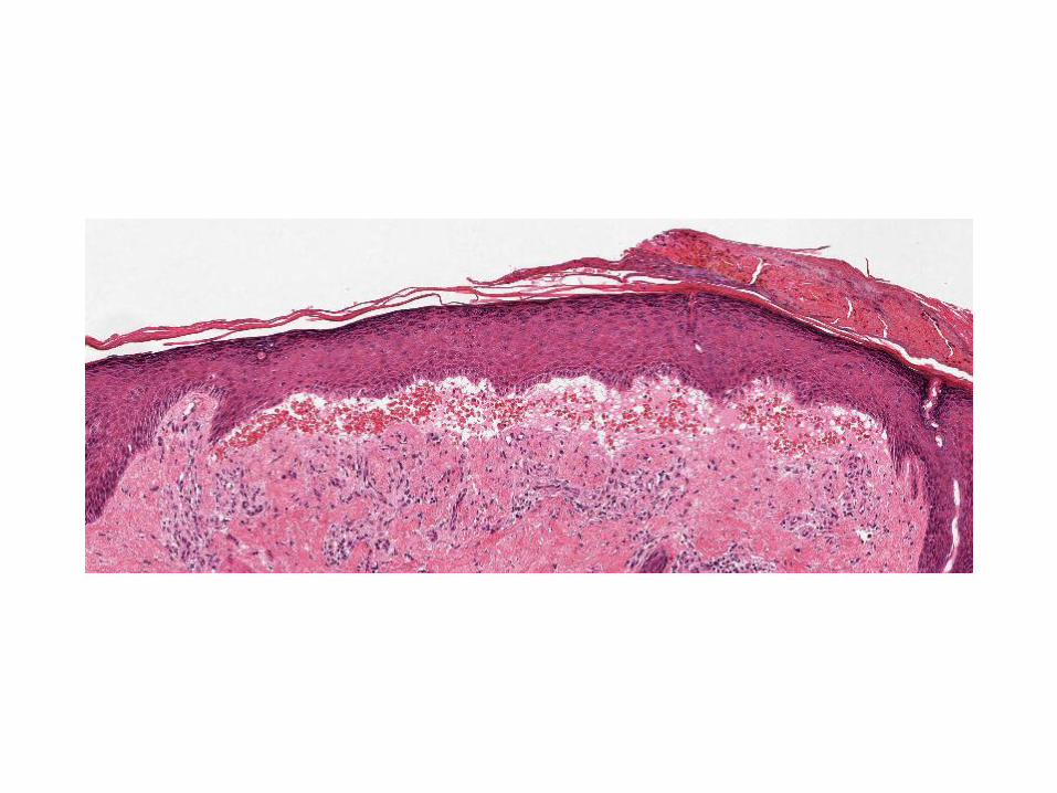
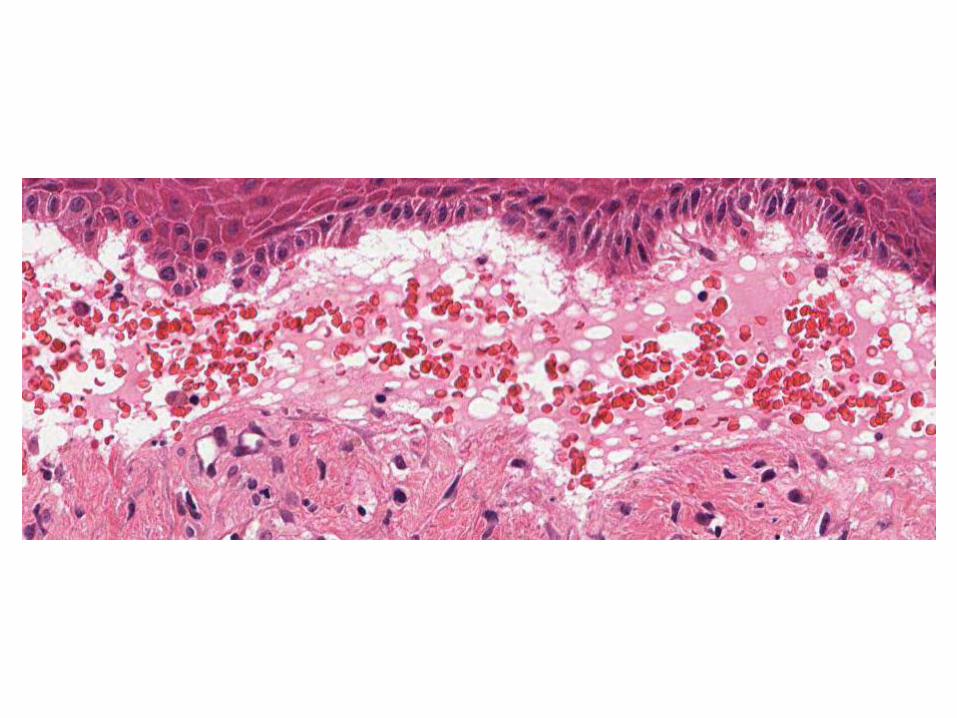
Case 12
• Description
– Skin with subepidermal clefting and
effacement of the dermal papillae
– Blister filled with blood, proteinaceous fluid,
inflammatory cells
– Fibrin on base of blister
– No evidence of neoplasia

Case 12
• Interpretation
– This is an inflammatory blistering process

Case 12
• Differential diagnosis
– Dermatitis herpetiformis
– Linear IgA disease
– Bullous SLE
– Bullous pemphigoid / EBA
• These are NOT supported by the IMF
• Only possible DDs if IMF not present
Case 12
• Extra investigations
– IMF main one, already provided
• Usually patchy and granular along BM in DH
• Deposits may also be seen in the dermis (in DH)
Case 12
• Clinico-pathological correlation
– Essential in this case
– Discuss history with clinicians
• ? Coeliac disease (Dermatitis Herpetiformis)
• ? Medication history (Linear IgA disease)
• ? Previous history of SLE or autoimmune disease
Case 12
• Skin, arm – Favour dermatitis
herpetiformis, see text
Summary – Short Surgicals
• Stick to a standard format
• Practise it, over and over again, on any case
• Work on degree of certainty
– If you’re prepared, you’ll be certain on around 75% of
the questions
– If uncertain take a step back, and keep to the
structured answer
• All this of course depends on good background
microscopy skills

Frozen Sections

Key tips
• Don’t panic
– The frozen sections are often very straightforward
– You have plenty of time
– Your first impression is usually the right one
– The Viva is usually quick and straightforward
– You can probably only get away with deferring to paraffin once
– Show initiative – if unsure, ask BMS to cut extra levels, show colleague
Common Cases
• Peritoneal nodules
– Metastatic tumour
• Ovarian
• GI
– Endometriosis
• Ureteric resection margins
– CIS vs normal
Common Cases
• Granulomatous inflammation – Differentials
– Clean the cryostat
• Resection margins – Skin
– Bone
– Pancreas
• Liver nodules – Met Ca
– Von Meyenburg complex
Common Cases
• Lymph nodes
– Benign / reactive
– Don’t diagnose lymphoma
• Beware the non-diagnosable cases
– Spindle cell lesions
Finally….
• Prepare well – Reading and slide exposure
• Practise over and over again
• Have a few days off before the exam
• Get a comfortable hotel
• Take chocolate and water into the exam – Not crisps
• Don’t dwell on things, at least until both days are over
• Go on holiday immediately afterwards