The fungal consortium of Andromeda polifolia in bog habitats
N.V. Filippova1 and M.N. Thormann2
1Yugra State University, Khanty-Mansiysk, Russia 2Aquilon Services Ltd., Edmonton, Canada
_______________________________________________________________________________________ SUMMARY (1) Andromeda polifolia (bog rosemary) is a common plant species in northern circumboreal peatlands. While not a major peat-forming species in most peatlands, it is characterised by a substantial woody below-ground biomass component that contributes directly to the accumulation of organic matter below the moss surface, as well as sclerophyllous leaf litter that contributes to the accumulation of organic matter above the moss surface. Rather little is known about the fungal communities associated with this plant species. Hence, we investigated the fungal consortium of A. polifolia in three distinct vegetation communities of ombrotrophic bogs near Khanty-Mansiysk, West Siberia, Russia, in 2012 and 2013. These vegetation communities were forested bog (Tr = treed), Sphagnum-dominated lawn (Ln), and Eriophorum-Sphagnum-dominated hummock (Er).
(2) In total, 37 fungal taxa, belonging to five classes and 16 families, were identified and described morphologically. Seven fungal species were previously known from Andromeda as host. Others are reported for the first time, thus considerably expanding the fungal consortium of this dwarf shrub. Most taxa were saprobic on fallen leaves of A. polifolia found amongst Sphagnum in the bog. Two taxa were parasitic on living plant tissues and one taxon was saprobic on dead twigs. Three taxa, recorded only on A. polifolia leaves and on no other plant species or materials, may be host-specific to this dwarf shrub.
(3) A quantitative analysis of the frequency of occurrence of all taxa showed that one taxon (Coccomyces duplicarioides) was very abundant, 64 % of the taxa occurred frequently, and 32 % of the taxa occurred infrequently. The mean Shannon diversity index of the community was 2.4.
(4) There were no statistical differences in the fungal community composition of A. polifolia in the three vegetation communities investigated in this study. Redundancy analysis suggested that some fungal taxa were positively, and others negatively, correlated with the water level relative to the moss surface in the bog.
(5) The information about the composition and structure of the fungal consortium of A. polifolia reported here could be supplemented using other techniques such as cultural and molecular methods. Nevertheless, the data presented improve our understanding of the different microbial communities functioning in peatlands and, thus, of carbon dynamics in these ecosystems. KEY WORDS: bog rosemary, fungi, diversity, Russia, West Siberia _______________________________________________________________________________________ INTRODUCTION Fungi play essential roles in ecosystem functioning as saprobes (saprotrophs), pathogens and mycorrhizal associates of most plant species. The fungal consortium associated with any particular plant species can consist of a few to several hundred species (Cannon & Sutton 2004). Recently, the microfungi associated with two peatland plants (Sphagnum fuscum (Schimp.) Klinggr. and Salix planifolia Pursh) in western Canada were investigated by the cultivation technique (Thormann et al. 2004); however, studies of this nature are uncommon for peatland plant species. Moreover, cultivation studies cannot provide a complete picture
of the true species richness in any one plant species or substrate; they should be supplemented by other means, including molecular techniques and/or direct observations.
Andromeda polifolia L. (bog rosemary) is a perennial evergreen dwarf shrub with a circumboreal range, growing mainly in Sphagnum-dominated bogs, but also in fens (Dierssen & Dierssen 1980). It is characterised by extensive rhizomatous growth, with roots penetrating the peat down to a depth of 45 cm, which results in > 75 % of its total biomass being below the moss surface (Wallén 1986, Malmer & Wallén 1986). Sclerophyllous leaf litter is deposited annually onto the moss surface and this, along with fine root turnover, contributes to the
N.V. Filippova & M.N. Thormann THE FUNGAL CONSORTIUM OF ANDROMEDA POLIFOLIA IN BOGS
accumulation of organic matter in peatlands. The geographical range of the plant is decreasing as a result of habitat loss or alteration. For example, it is considered rare in France, The Netherlands, England and Ireland, and has disappeared altogether from several European countries (Jacquemart 1998). In Siberia (Russia) and Canada, A. polifolia still occurs in the boreal to boreal-tundra zones as well as in the alpine zone in mountain ranges, where it inhabits bogs and fens as well as wet forests (Glooschenko et al. 1993, Malyschev 1997).
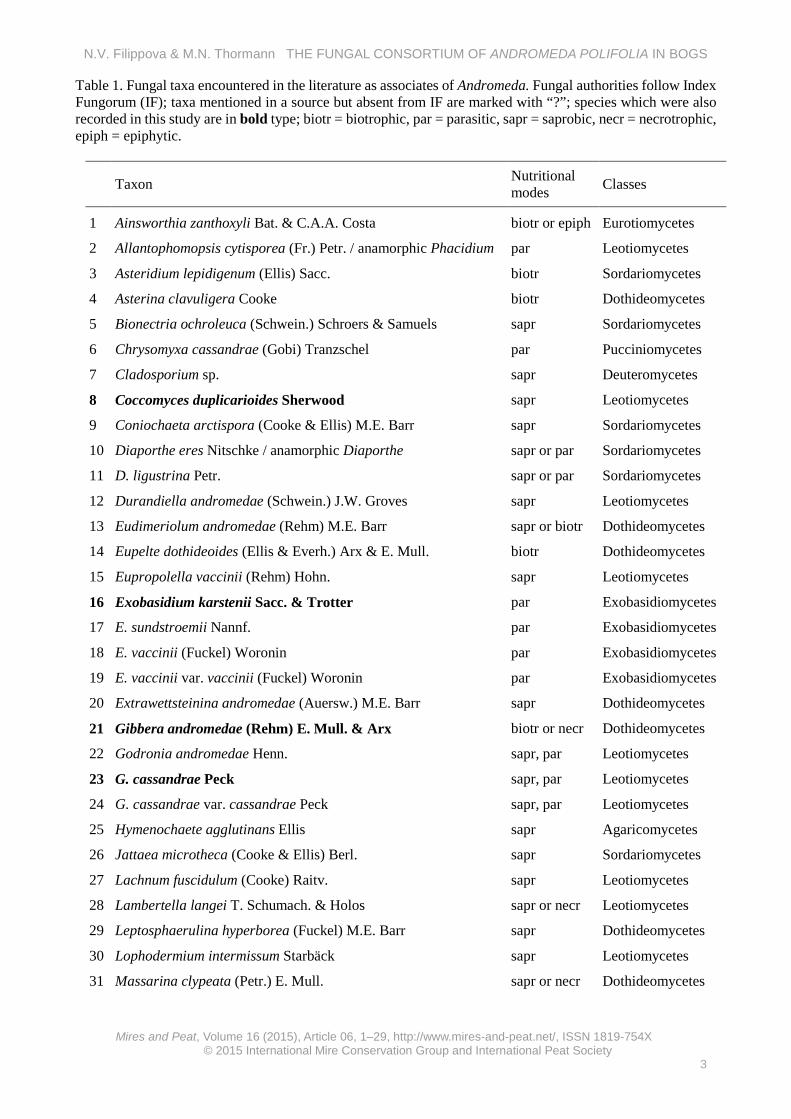
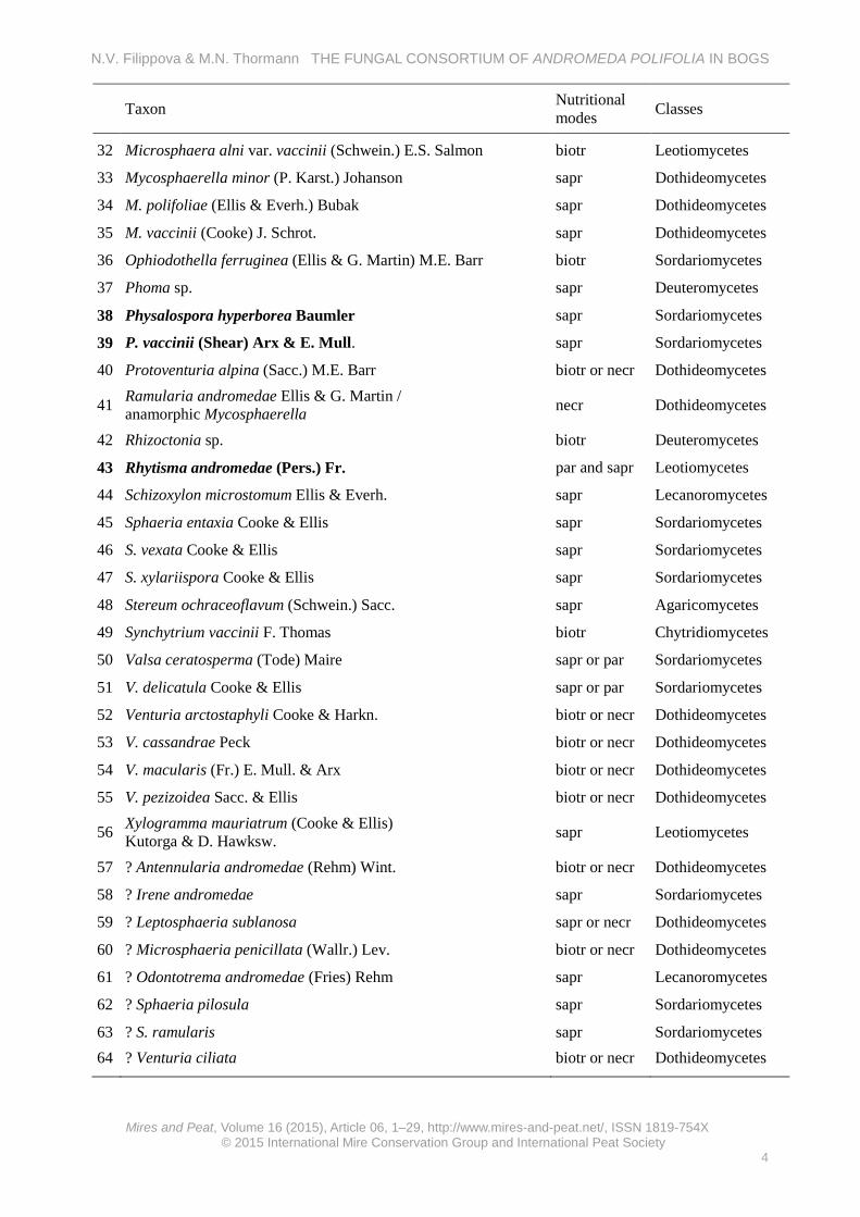
An analysis of the literature and electronic databases (Farr & Rossman 2015) showed 64 fungal taxa associated with the genus Andromeda, with 22 species specifically related to A. polifolia (mainly from the USA and Canada, but also from 14 European and Asian countries). Generally, there is a scarcity of publications specifically addressing the fungal consortium of Andromeda spp., with most reports originating from regional checklists and/or annotations in taxonomic treatments. From works particularly concerning the fungal consortium of A. polifolia, Eriksson (1970, 1974) provided an analysis of Discomycetes, Pyrenomycetes and Deuteromycetes on Ericales in Fennoscandia, and Remler (1979) described fungi on ericaceous hosts in the eastern Alps (including seven fungal species associated with A. polifolia). More recently, Jacquemart (1998) listed 14 fungal species that are parasitic on A. polifolia, his list being based on earlier publications (Dennis 1968, Remler 1979, Farr et al. 1989, Ellis & Ellis 1997). In addition, Jacquemart (1998) reported that A. polifolia normally formed ericoid mycorrhiza with Hymenoscyphus ericae. The already-reported 64 fungal species associated with Andromeda belong to three phyla (Ascomycota: 56 species; Basidiomycota: seven species; and Chytridiomycota: one species). Their taxonomic dispositions represent ten classes, 20 orders, 32 families and 46 genera (Table 1). The nutritional strategies of these taxa include parasitic (biotrophs and necrotrophs) and saprobic relationships with their host.
Despite the rarity or complete absence of A. polifolia in some European countries and its probably substantial contribution to the accumulation of organic matter in some peatlands, there has not yet been any concerted effort to specifically examine the fungal consortium associated with this important peatland plant species. The disappearance of the host could result in the extinction of its narrowly associated fungal species, along with their potential biochemical properties. Therefore, such host species should be of particular concern in conservation programmes. To identify them we require additional
information about their ecology. The carbon dynamics in peatland ecosystems are accomplished by suites of microbial communities functioning under different environmental conditions and on different substrata. Knowledge of specific properties of these communities and their responses to fluctuating environmental conditions will ultimately contribute to a better understanding of carbon dynamics and the sustainable use of peatland ecosystems.
The purpose of this study was to investigate the community structure of microfungi associated with A. polifolia leaves in bog habitats. Our hypotheses were: (1) the total fungal species richness of A. polifolia is
greater than 100 taxa; based on the fungal species richness of some other, more widely studied, plant species;
(2) some fungal species are host-specific, inhabiting only A. polifolia, because fungal host specificity differs among fungal groups and is considered to be a valuable character in fungal biodiversity estimates, which requires more attention in estimating the true biodiversity of ecosystems and specific substrata (Hawksworth 2001);
(3) the fungal community of A. polifolia litter differs according to vegetation community types, as was shown by Wiedermann et al. (2007); and
(4) the fungal community of A. polifolia litter differs under different environmental conditions, as was shown by Wiedermann et al. (2007).
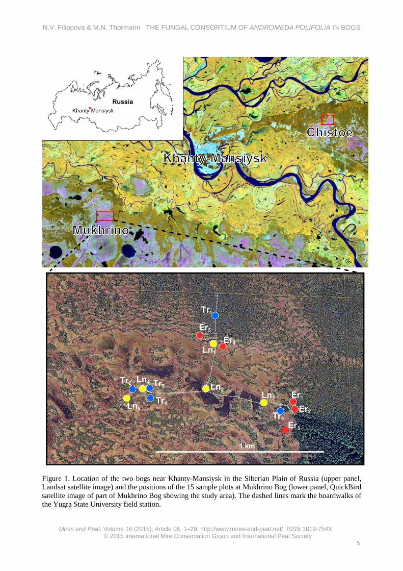
METHODS Study region and study sites The study region is near the town of Khanty-Mansiysk in the central Taiga of West Siberia (Figure 1). The climate is subarctic with short, cool summers and long, cold winters. Snow cover persists for about 180 days. The mean annual temperature is -1.3 °C, the mean temperature of the coldest month (January) being -19.8 °C and the mean temperature of the warmest month (July) being +18 °C. Total annual precipitation is 553 mm, with 70–80 % of this total falling in the summer months (Tryaszin 2007).
The terrain in this region is almost flat and this, in conjunction with the excess precipitation, has resulted in the development of vast peatland complexes consisting of mosaics of bogs and fens, which cover nearly 50 % of the landscape (Peregon et al. 2009). The study sites were two bogs near Khanty-Mansiysk: Chistoe Bog (61.06542 °N, 69.45911 °E) and Mukhrino Bog (60.89227 °N, 68.68259 °E) (Figure 1). The bogs are characterised
N.V. Filippova & M.N. Thormann THE FUNGAL CONSORTIUM OF ANDROMEDA POLIFOLIA IN BOGS
Table 1. Fungal taxa encountered in the literature as associates of Andromeda. Fungal authorities follow Index Fungorum (IF); taxa mentioned in a source but absent from IF are marked with “?”; species which were also recorded in this study are in bold type; biotr = biotrophic, par = parasitic, sapr = saprobic, necr = necrotrophic, epiph = epiphytic.
Taxon Nutritional modes Classes
1 Ainsworthia zanthoxyli Bat. & C.A.A. Costa biotr or epiph Eurotiomycetes
2 Allantophomopsis cytisporea (Fr.) Petr. / anamorphic Phacidium par Leotiomycetes
Figure 1. Location of the two bogs near Khanty-Mansiysk in the Siberian Plain of Russia (upper panel, Landsat satellite image) and the positions of the 15 sample plots at Mukhrino Bog (lower panel, QuickBird satellite image of part of Mukhrino Bog showing the study area). The dashed lines mark the boardwalks of the Yugra State University field station.
N.V. Filippova & M.N. Thormann THE FUNGAL CONSORTIUM OF ANDROMEDA POLIFOLIA IN BOGS
by three distinct vegetation communities, namely: forested (Tr = treed) pine - dwarf shrubs - Sphagnum, graminoid - Sphagnum lawn (Ln), and Eriophorum-Sphagnum communities (Er) (see Table A1 in Appendix 1). Treed pine - dwarf shrubs - Sphagnum communities are characterised by plants that are adapted to fairly dry conditions, with the mean summer water level being about 50 cm below the moss surface. They have a well-developed tree canopy consisting of Pinus sylvestris and Pinus sibirica. A dense understorey of dwarf shrubs is dominated by members of the Ericaceae including Ledum palustre and Chamaedaphne calyculata in equal proportions and, to a lesser degree, A. polifolia, Vaccinium uliginosum, Vaccinium vitis-idaea and Betula nana. The herbaceous vegetation stratum is dominated by Rubus chamaemorus, Oxycoccus microcarpus and Drosera rotundifolia, while the bryophyte stratum is dominated by Sphagnum fuscum on hummocks and Sphagnum magellanicum and Sphagnum angustifolium in hollows. This plant community was previously classified, in the floristic classification of bogs in the region (Lapshina 2010), as a Ledo - Sphagnetum fusci association (class Oxycocco - Sphagnetea).
In contrast to the treed bog community, the water level in the graminoid - Sphagnum lawns is near the moss surface (summer mean 8 cm below the moss surface) (Table A1). Trees are absent, and A. polifolia is the only dwarf shrub present in this community. The herbaceous vegetation stratum is dominated by Carex limosa, Scheuchzeria palustris, Baeothryon caespitosum, Rhynchospora alba, Drosera anglica, D. rotundifolia and Oxycoccus palustris. Sphagna are represented by several hydrophilic species including Sphagnum balticum, Sphagnum papillosum, Sphagnum jensenii and Sphagnum lindbergii. This community was previously classified, in the floristic classification of bogs in the region (Lapshina 2010), as a Scheuchzerio palustris - Sphagnetum cuspidati association (class Scheuchzerio-Caricetea nigrae).
The characteristics of Eriophorum - Sphagnum hummock communities are intermediate between those of the two communities above, and usually develop at their boundaries as ribs or sometimes more extensive areas (Table A1). Here, the growth of trees is prevented by higher water levels (summer mean 17 cm below the moss surface). The dwarf shrub stratum is well developed and dominated by A. polifolia, C. calyculata and B. nana. The herbaceous vegetation stratum is dominated by E. vaginatum forming dense tussocks, as well as Carex magellanicum and Carex pauciflora in the vicinity of bog - forest transitional zones. The
bryophyte stratum is dominated by S. balticum, S. magellanicum and S. papillosum with admixture of S. fuscum and S. capillifolium. This community was previously classified, in the floristic classification of bogs in the region (Lapshina 2010), as an Eriophoro vaginati - Sphagnetum baltici association (class Scheuchzerio - Caricetea nigrae). Sampling design A quantitative analysis of the fungal community of A. polifolia leaves at Mukhrino Bog was carried out in July and August 2013. Dead leaves deposited among Sphagnum capitula (0–3 cm deep) or lying on the moss surface were collected once during the expected maximum development phase of the fungal community (July–August), placed into plastic bags, and examined within 1–2 days of their collection. The leaves differed in age by several years and were at various stages of decomposition.
The minimum number of leaves (sample size) required for an adequate description of the fungal diversity per location was determined by randomly collecting and processing about 500 A. polifolia leaves, subsequently examining them micro-scopically, and then developing a species abundance curve. From this analysis it was determined that 300 leaves per plot would be an adequate basis for confident description of the species richness of A. polifolia leaves (Figure 2A), given the fact that we took five replicates for each plant community type (see below).
Fungal species observed during random observations of A. polifolia plant tissues in 2012 and 2013 (Chistoe and Mukhrino Bogs) were included in this study as well. In a related study (Filippova 2015), the litters of eleven other bog plant species were repeatedly examined in the same habitat from May to October 2013. From those results it was possible to determine whether or not the fungi observed on A. polifolia leaves were unique to this substratum, i.e. host-specific, or occurred on other plant litters too.
The alpha diversity (diversity within the same habitat) of the fungal community was examined in five 1-m2 plots located randomly in an area of radius about 500 m within Mukhrino Bog (300 randomly selected A. polifolia leaves per plot). To examine the beta diversity (diversity among habitats), the three types of bog vegetation were investigated: graminoid - Sphagnum lawns (Ln), treed pine - dwarf shrubs - Sphagnum fuscum bogs (Tr), and Eriophorum - Sphagnum hummocks (Er) (each plant community type was represented by five plots, making a total of 15 plots; Figure 1). These three plant community types represent the most common
N.V. Filippova & M.N. Thormann THE FUNGAL CONSORTIUM OF ANDROMEDA POLIFOLIA IN BOGS
Figure 2. Species accumulation curves: A) for fungi from Andromeda polifolia leaves in the pilot plot (Ln1) at Mukhrino Bog (the red circle indicates the adopted sample size of 300 leaves); and B) for fungi from A. polifolia for all samples (4,670 leaves) collected at Mukhrino Bog.
vegetation types in bogs in the region, and have characteristic hydrological regimes (water level position relative to the moss surface), pH, surface water electrical conductivity (EC) and plant communities (Table A1).
The leaf litter was examined for the presence of fungi under a Zeiss binocular microscope (magnification 8–50 times), and live fungal taxa were subsequently identified using morphological characteristics when examined in water and/or stained using several reactants and dyes (e.g. Lugol's solution (iodine plus potassium iodide dissolved in water) for testing amyloidity, Congo red for staining, and KOH for re-examination of dried collections) under a Zeiss AxioStar microscope (magnification 50–1000 times). Characteristic microscopic
morphological features of all fungal taxa were digitally photographed and sketched with Inkscape graphics software.
Leaves bearing fungal fruiting structures and all accessory files were accessioned to the Yugra State University fungarium. The fungal authorities followed Index Fungorum (2015), and the classification of the fungal taxa at various taxonomic ranks followed Eriksson (2005). Statistical analyses The number of fungal species per leaf (leaf occurrence) was noted; however, the abundance of fruiting structures of individual fungal taxa was not estimated in this study owing to the challenges associated with quantitative assessments of
N.V. Filippova & M.N. Thormann THE FUNGAL CONSORTIUM OF ANDROMEDA POLIFOLIA IN BOGS
microfungal reproductive structures on organic substrata. Species accumulation curves were plotted using sample-based calculations, where the sample is a single leaf with its fungal associates. In an effort to facilitate descriptive comparisons among the species abundance data, a six-part scale was used (Arnolds 1992), whereby Class I represented species with 1–3 occurrences (rare), Class II: 4–10 (uncommon), Class III: 11–30 (fairly abundant), Class IV: 31–100 (abundant), Class V: 101–300 (very abundant), and Class VI: species with more than 300 occurrences.
Statistical analyses were performed using open-source R software. Exploratory analyses of abundance classes, species occurrences, species richness per plot and alpha diversity indices were made using the ‘vegan’ package. Species accumulation curves were plotted using the “Specaccum” function, where an “exact” method was used with unconditional confidence intervals (Colwell et al. 2004); estimations of expected species richness were made using the “Poolaccum” function, with the Chao 2 estimator (Gotelli & Colwell 2001). Beta diversity was computed using distance matrices, clustering and constrained ordination methods in different packages available in the R software (e.g. vegan, ade4, gclus, and cluster), following Borcard et al. (2011). The best method for the cluster analysis was chosen on the basis of a cophenetic correlation computed between original dissimilarity matrices and the cophenetic distances. The cophenetic correlation coefficient is a measure of how faithfully a dendrogram preserves the pairwise distances between the original un-modelled data points and is a common method in the field of biostatistics. The method with the highest cophenetic correlation was employed in the analysis (Unweighted Pair-Group Method using arithmetic Averages (UPGMA)). The optimal number of clusters in the dendrogram was estimated by fusion level values, the Silhouette optimal number of clusters and the Mantel optimal number of clusters (Borcard et al. 2011), which provided a range from 2 to 5 clusters per dendrogram. We decided to select the dendrogram with three clusters, taking into account the initial hypothesis about the difference between the three community types. Redundancy analysis was performed using function rda (vegan) with the Hellinger pre-transformed species occurrence matrix and the environmental variables matrix. Rare taxa with < 10 occurrences were removed prior to the analysis (thereby increasing the power of the analysis), so that only the remaining 21 taxa were included. To assess the significance of the explained variation, the anova.cca (vegan) function (using 999 permutations) was employed to carry out a permutation test.
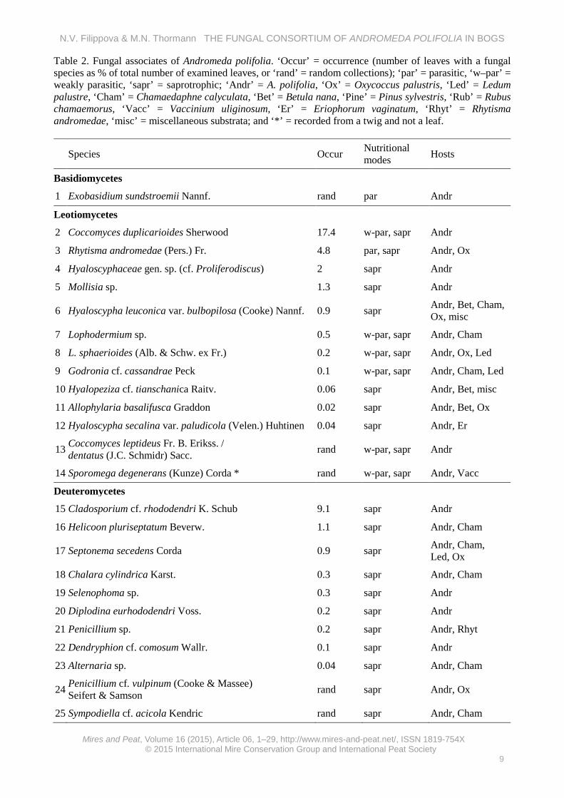
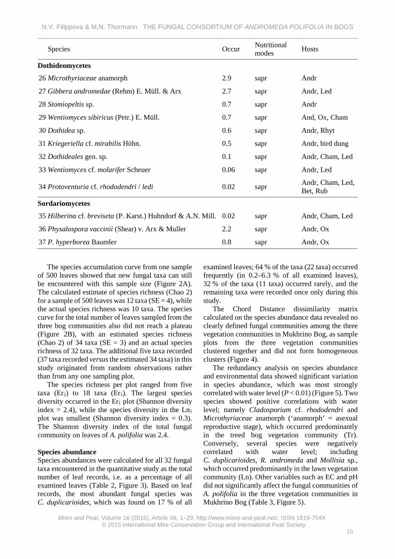
RESULTS Fungal community composition A total of 37 fungal taxa belonging to five classes, 14 families and 30 genera (excluding the unidentified taxa) was recorded from A. polifolia (Table 2, Appendix 2). All taxa except one (the basidiomycete Exobasidium sundstroemii) had ascomycete affinities, with members of the Leotiomycetes being the most prevalent (13 taxa), followed by members of the Deuteromycetes (11 taxa), Dothideomycetes (9 taxa), and Sordariomycetes (3 taxa). Fifteen taxa could be reliably identified to species, 12 taxa had close affinities to known species (marked as ‘cf.’ hereafter), eight taxa could be identified only to genus, and two taxa could be identified only to family.
Most taxa were encountered on the fallen leaves of A. polifolia, although one taxon (Sporomega degenerans) was observed on dead twigs and parasitic taxa were recorded from live leaves. Two taxa are parasitic (E. sundstroemii and Rhytisma andromeda), six taxa may be weakly parasitic (Coccomyces duplicarioides, Coccomyces cf. leptideus/dentatus, S. degenerans, Godronia cf. cassandrae, Lophodermium sphaerioides and Lophodermium sp.), two taxa inhabited old stroma of R. andromeda (Dothidea sp. and Penicillium sp.), and the remaining taxa were saprobic on the leaf litter. An analysis of other substrata revealed that most of the fungal taxa recorded from A. polifolia also occurred on other bog plant litters, i.e. they were not host-specific; however, of the ten taxa recorded only from A. polifolia, three were commonly collected on this host and did not occur on other plants; therefore, they could be considered truly host-specific. These were Coccomyces duplicarioides, Hyaloscyphaceae gen. sp. (cf. Proliferodiscus) and Rhytisma andromeda. The remaining taxa were collected less commonly, and it was impossible to determine whether they truly occur only on A polifolia or also on other plant species. Species richness Determination of the presence/absence of fungal species by examination of their reproductive structures on the leaf litter of A. polifolia showed that only about half (55 %) of all the examined leaves were surficially colonised by fungi. The remaining leaves were devoid of surficial fungal reproductive structures but they were colonised by fungi, i.e. mycelium was apparent. About 10 % of the leaves were colonised by two taxa and 1.6 % of the leaves were colonised by three taxa, but most leaves were colonised by a single fungal taxon only.
N.V. Filippova & M.N. Thormann THE FUNGAL CONSORTIUM OF ANDROMEDA POLIFOLIA IN BOGS
Table 2. Fungal associates of Andromeda polifolia. ‘Occur’ = occurrence (number of leaves with a fungal species as % of total number of examined leaves, or ‘rand’ = random collections); ‘par’ = parasitic, ‘w–par’ = weakly parasitic, ‘sapr’ = saprotrophic; ‘Andr’ = A. polifolia, ‘Ox’ = Oxycoccus palustris, ‘Led’ = Ledum palustre, ‘Cham’ = Chamaedaphne calyculata, ‘Bet’ = Betula nana, ‘Pine’ = Pinus sylvestris, ‘Rub’ = Rubus chamaemorus, ‘Vacc’ = Vaccinium uliginosum, ‘Er’ = Eriophorum vaginatum, ‘Rhyt’ = Rhytisma andromedae, ‘misc’ = miscellaneous substrata; and ‘*’ = recorded from a twig and not a leaf.
The species accumulation curve from one sample of 500 leaves showed that new fungal taxa can still be encountered with this sample size (Figure 2A). The calculated estimate of species richness (Chao 2) for a sample of 500 leaves was 12 taxa (SE = 4), while the actual species richness was 10 taxa. The species curve for the total number of leaves sampled from the three bog communities also did not reach a plateau (Figure 2B), with an estimated species richness (Chao 2) of 34 taxa (SE = 3) and an actual species richness of 32 taxa. The additional five taxa recorded (37 taxa recorded versus the estimated 34 taxa) in this study originated from random observations rather than from any one sampling plot.
The species richness per plot ranged from five taxa (Er2) to 18 taxa (Er1). The largest species diversity occurred in the Er1 plot (Shannon diversity index = 2.4), while the species diversity in the Ln5
plot was smallest (Shannon diversity index = 0.3). The Shannon diversity index of the total fungal community on leaves of A. polifolia was 2.4. Species abundance Species abundances were calculated for all 32 fungal taxa encountered in the quantitative study as the total number of leaf records, i.e. as a percentage of all examined leaves (Table 2, Figure 3). Based on leaf records, the most abundant fungal species was C. duplicarioides, which was found on 17 % of all
examined leaves; 64 % of the taxa (22 taxa) occurred frequently (in 0.2–6.3 % of all examined leaves), 32 % of the taxa (11 taxa) occurred rarely, and the remaining taxa were recorded once only during this study.
The Chord Distance dissimilarity matrix calculated on the species abundance data revealed no clearly defined fungal communities among the three vegetation communities in Mukhrino Bog, as sample plots from the three vegetation communities clustered together and did not form homogeneous clusters (Figure 4).
The redundancy analysis on species abundance and environmental data showed significant variation in species abundance, which was most strongly correlated with water level (P < 0.01) (Figure 5). Two species showed positive correlations with water level; namely Cladosporium cf. rhododendri and Microthyriaceae anamorph (‘anamorph’ = asexual reproductive stage), which occurred predominantly in the treed bog vegetation community (Tr). Conversely, several species were negatively correlated with water level; including C. duplicarioides, R. andromeda and Mollisia sp., which occurred predominantly in the lawn vegetation community (Ln). Other variables such as EC and pH did not significantly affect the fungal communities of A. polifolia in the three vegetation communities in Mukhrino Bog (Table 3, Figure 5).
N.V. Filippova & M.N. Thormann THE FUNGAL CONSORTIUM OF ANDROMEDA POLIFOLIA IN BOGS
Figure 3. Species occurrences (% of total) for fungi from Andromeda polifolia at Mukhrino Bog.
DISCUSSION Taxonomic and ecological structure The taxonomic diversity of fungi associated with living and decomposing leaves of A. polifolia in bogs appears to be lower than that of other phanerogams and herbaceous plant species (Cannon & Sutton 2004). We identified 37 taxa from the leaves of A. polifolia in Mukhrino Bog, which was more than in previous accounts from Eriksson (1970, 1974) and Remler (1979) and exceeds the total number in earlier reports on A. polifolia (22 taxa). This makes the present study of fungi associated with A. polifolia the
most comprehensive one yet. Seven of our taxa have previously been reported from A. polifolia (Table 1) and the remaining 30 taxa from our study represent new records for A. polifolia, increasing the list of fungi reported from this ericaceous plant to about 50 taxa, thereby rejecting Hypothesis 1: that there would be more than 100 species. Due to the taxonomic similarity of A. polifolia to other Andromeda species, it is likely that the total fungal consortium associated with the genus exceeds 100 taxa, particularly since we examined only leaves and not other plant tissues such as roots, stems, and/or branches. The number of fungi from this important and widespread member of
N.V. Filippova & M.N. Thormann THE FUNGAL CONSORTIUM OF ANDROMEDA POLIFOLIA IN BOGS
Figure 4. Cluster dendrogram, derived by average agglomerative clustering method, of a matrix of chord distance among plots with boxes around the three selected groups, for fungi from Andromeda polifolia at Mukhrino Bog.
Figure 5. Redundancy analysis triplot of the Hellinger-transformed species abundance data constrained by three environmental variables (pH, EC and water level), scaling 1, for fungi from Andromeda polifolia at Mukhrino Bog.
N.V. Filippova & M.N. Thormann THE FUNGAL CONSORTIUM OF ANDROMEDA POLIFOLIA IN BOGS
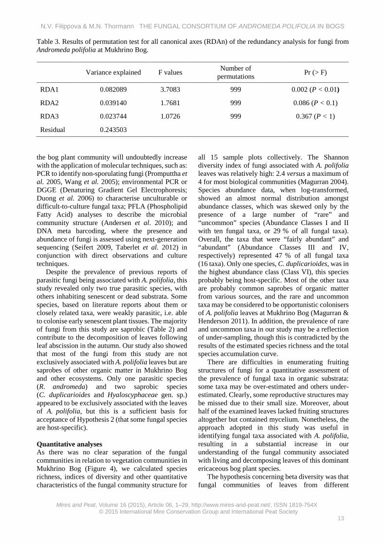
Table 3. Results of permutation test for all canonical axes (RDAn) of the redundancy analysis for fungi from Andromeda polifolia at Mukhrino Bog.
Variance explained F values Number of permutations Pr (> F)
RDA1 0.082089 3.7083 999 0.002 (P < 0.01)
RDA2 0.039140 1.7681 999 0.086 (P < 0.1)
RDA3 0.023744 1.0726 999 0.367 (P < 1)
Residual 0.243503 the bog plant community will undoubtedly increase with the application of molecular techniques, such as: PCR to identify non-sporulating fungi (Promputtha et al. 2005, Wang et al. 2005); environmental PCR or DGGE (Denaturing Gradient Gel Electrophoresis; Duong et al. 2006) to characterise unculturable or difficult-to-culture fungal taxa; PFLA (Phospholipid Fatty Acid) analyses to describe the microbial community structure (Andersen et al. 2010); and DNA meta barcoding, where the presence and abundance of fungi is assessed using next-generation sequencing (Seifert 2009, Taberlet et al. 2012) in conjunction with direct observations and culture techniques.
Despite the prevalence of previous reports of parasitic fungi being associated with A. polifolia, this study revealed only two true parasitic species, with others inhabiting senescent or dead substrata. Some species, based on literature reports about them or closely related taxa, were weakly parasitic, i.e. able to colonise early senescent plant tissues. The majority of fungi from this study are saprobic (Table 2) and contribute to the decomposition of leaves following leaf abscission in the autumn. Our study also showed that most of the fungi from this study are not exclusively associated with A. polifolia leaves but are saprobes of other organic matter in Mukhrino Bog and other ecosystems. Only one parasitic species (R. andromeda) and two saprobic species (C. duplicarioides and Hyaloscyphaceae gen. sp.) appeared to be exclusively associated with the leaves of A. polifolia, but this is a sufficient basis for acceptance of Hypothesis 2 (that some fungal species are host-specific). Quantitative analyses As there was no clear separation of the fungal communities in relation to vegetation communities in Mukhrino Bog (Figure 4), we calculated species richness, indices of diversity and other quantitative characteristics of the fungal community structure for
all 15 sample plots collectively. The Shannon diversity index of fungi associated with A. polifolia leaves was relatively high: 2.4 versus a maximum of 4 for most biological communities (Magurran 2004). Species abundance data, when log-transformed, showed an almost normal distribution amongst abundance classes, which was skewed only by the presence of a large number of “rare” and “uncommon” species (Abundance Classes I and II with ten fungal taxa, or 29 % of all fungal taxa). Overall, the taxa that were “fairly abundant” and “abundant” (Abundance Classes III and IV, respectively) represented 47 % of all fungal taxa (16 taxa). Only one species, C. duplicarioides, was in the highest abundance class (Class VI), this species probably being host-specific. Most of the other taxa are probably common saprobes of organic matter from various sources, and the rare and uncommon taxa may be considered to be opportunistic colonisers of A. polifolia leaves at Mukhrino Bog (Magurran & Henderson 2011). In addition, the prevalence of rare and uncommon taxa in our study may be a reflection of under-sampling, though this is contradicted by the results of the estimated species richness and the total species accumulation curve.
There are difficulties in enumerating fruiting structures of fungi for a quantitative assessment of the prevalence of fungal taxa in organic substrata: some taxa may be over-estimated and others under-estimated. Clearly, some reproductive structures may be missed due to their small size. Moreover, about half of the examined leaves lacked fruiting structures altogether but contained mycelium. Nonetheless, the approach adopted in this study was useful in identifying fungal taxa associated with A. polifolia, resulting in a substantial increase in our understanding of the fungal community associated with living and decomposing leaves of this dominant ericaceous bog plant species.
The hypothesis concerning beta diversity was that fungal communities of leaves from different
N.V. Filippova & M.N. Thormann THE FUNGAL CONSORTIUM OF ANDROMEDA POLIFOLIA IN BOGS
vegetation types (graminoid - Sphagnum lawns (Ln), treed pine - dwarf shrubs - Sphagnum fuscum bogs (Tr), and Eriophorum - Sphagnum hummocks (Er)) in Mukhrino Bog would differ, and that some of the environmental variables measured as part of this study would explain the differences. This was explored first via a dissimilarity matrix analysis and second via a redundancy analysis. We hypothesised that sample plots from the same vegetation type would be united by a high similarity index, while sample plots from different vegetation types would not be so united. This was not the case, as sample plots from different vegetation types formed heterogeneous clusters (Figure 4), so we reject Hypothesis 3 (that the fungal community differs according to vegetation community type).
The cluster dendrogram showed three distinct fungal community clusters, with the first cluster being represented by two sample plots from the treed bog vegetation type (Tr1 and Tr4), the second cluster by three sample plots from Eriophorum hummocks and lawn habitats (Er1, Er4, and Ln1), and the third cluster by sample plots from all three vegetation types (Figure 4).
The permutation test for the redundancy analysis showed that water level relative to the moss surface correlated significantly with some fungal taxa, thereby at least partly supporting Hypothesis 4 (that the fungal community of A. polifolia litter differs in response to differing environmental conditions).
The abundances of two species were highly negatively correlated with low treed-bog water levels, and those two taxa (C. cf. rhododendri and Microthyriaceae anamorph) could be considered as being adapted to drier litter conditions (Figure 5). Conversely, the abundances of three taxa (C. duplicarioides, R. andromeda and Mollisia sp.) were correlated positively with high water levels and, thus, they could be regarded as more hydrophilic taxa. Neither EC nor pH correlated significantly with any of the fungal taxa associated with A. polifolia leaves (Figure 5). However, these preliminary analyses need to be viewed with caution, as there are other environmental variables that may influence the fungal community associated with A. polifolia leaves, including leaf moisture content, chemical composition of the leaf, temperature and stage of decomposition, among others. These variables should form the focus of future studies to improve our understanding of fungal community dynamics and carbon dynamics in peatlands.
A long-term monitoring program was initiated at Mukhrino Bog in 2014, in an effort to elucidate the relationships between fungal taxa and environmental variables.
ACKNOWLEDGEMENTS For their helpful discussions on species identification, we thank: Eugene Popov, Hans-Otto Baral, Mateusz Wilk and Andrew Miller; and for help with revisions to this manuscript we thank Aimee Matthiessen. This work was funded by a grant (# 2014/505) to Yugra State University from the Ministry of Education and Science of the Russian Federation. REFERENCES Andersen, R., Grasset, L., Thormann, M.N.,
Rochefort, L. & Francez, A.-J. (2010) Changes in microbial community structure and function following Sphagnum peatland restoration. Soil Biology & Biochemistry, 42, 291–301.
Arnolds, E. (1992) The analysis and classification of fungal communities with special reference to macrofungi. In: Winterhoff, W. (ed.) Fungi in Vegetation Science, Handbook of Vegetation Science. Springer, The Netherlands, 7–47.
Barr, M.E. (1968) The Venturiaceae in North America. Canadian Journal of Botany, 46, 799–864.
Barr, M.E. (1970) Some amerosporous ascomycetes on Ericaceae and Empetraceae. Mycologia, 62, 377–394.
Barr, M.E. (1972) Preliminary Studies on the Dothideales in Temperate North America. University of Michigan, Ann Arbor, MI, USA. 663 pp.
Borcard, D., Gillet, F. & Legendre, P. (2011) Numerical Ecology with R. Springer, New York, NY, USA, 306 pp.
Cannon, P.F. & Sutton, B.C. (2004) Microfungi on wood and plant debris. In: Mueller, G.M., Bills, G.F. & Foster, M.S. (eds.) Biodiversity of Fungi. Inventory and Monitoring Methods. Elsevier Academic Press, Amsterdam, The Netherlands, 217–302.
Colwell, R.K., Mao, C.X. & Chang, J. (2004) Interpolating, extrapolating, and comparing incidence-based species accumulation curves. Ecology, 85, 2717–2727.
Dennis, R.W.G. (1968) British Ascomycetes. J. Cramer, Vaduz, 585 pp.
Dierssen, K. & Dierssen, B. (1980) The distribution of communities and community complexes of oligotrophic mire sites in Western Scandinavia. In: Gehu, J.M. Colloques Phytosociologiques VII: La Végétation Des Sols Tourbeux, Lille, 1978, J. Cramer, Vaduz, 95–119.
N.V. Filippova & M.N. Thormann THE FUNGAL CONSORTIUM OF ANDROMEDA POLIFOLIA IN BOGS
Duong, L.M., Jeewon, R., Lumyong, S. & Hyde, K.D. (2006) DGGE coupled with ribosomal DNA phylogenies reveal uncharacterized fungal phylotypes on living leaves of Magnolia liliifera. Fungal Diversity, 23, 121–138.
Ellis, M.B. (1971) Dematiaceous Hyphomycetes. Commonwealth Mycological Institute, Kew, 608 pp.
Ellis, M.B. & Ellis, J.P. (1997) Microfungi on Land Plants: an Identification Handbook. RP Richmond Publishers, Slough, UK, 869 pp.
Eriksson, B. (1970) On Ascomycetes on Diapensiales and Ericales in Fennoscandia 1. Discomycetes. A.-B. Lundequistskabokhandeln, Uppsala, Sweden, 71 pp.
Eriksson, B. (1974) On Ascomycetes on Diapensiales and Ericales in Fennoscandia. 2. Pyrenomycetes. Svensk Botanisk Tidskrift, 68, 192–234.
Eriksson, B. (ed.) (2005) Outline of Ascomycota - 2005. Myconet, 11, 1–113.
Farr, D.F., Bills, G.F., Chanuris, G.P. & Rossman, A.Y. (1989) Fungi on Plants and Plant Products in the United States. American Phytopathological Society, Saint Paul, MN, USA, 1252 pp.
Filippova, N.V. (2015) On the communities of fungi of raised bogs in taiga belt of West Siberia: 2. Microfungi on plant litter. Mycology and Phytopathology, 4(3), 164–172.
Glooschenko, W.A., Roulet, N.T., Barrie, L.A., Schiff, H.J. & McAdie, H.G. (1993) Wetlands of Canada and Greenland. In: Whighan, D., Dykyjova, D. & Hejny, S. (eds.) Wetlands of the World: Inventory, Ecology and Management, Kluwer Academic Publishers, Boston, 415–514.
Goos, R.D., Abdullah, S.K., Fisher, P.J. & Webster, J. (1986) The anamorph genus Helicoon. Transactions of the British Mycological Society, 87, 115–122.
Gotelli, N.J. & Colwell, R.K. (2001) Quantifying biodiversity: procedures and pitfalls in the measurement and comparison of species richness. Ecology Letters, 4, 379–391.
Graddon, W.D. (1977) Some new Discomycete species: 4. Transactions of the British Mycological Society, 69, 255–273.
Grove, W.B. (1935) The British Coelomycetes. V. 1. Cambridge University Press, Cambridge, UK, 488 pp.
Groves, J.W. (1965) The genus Godronia. Canadian Journal of Botany, 43, 1195–1276.
Groves, J.W. (1969) Notes on the genus Encoeliopsis. Canadian Journal of Botany, 47, 1319–1331.
Hawksworth, D.L. (2001) The magnitude of fungal diversity: the 1.5 million species estimate revisited. Mycological Research, 105, 1422–1432.
Hawksworth, D.L. & Sivanesan, A. (1976) New and interesting microfungi from Slapton, South Devonshire: Ascomycotina III. Transactions of the British Mycological Society, 67, 477–483.
Huhtinen, S. (1990) A monograph of Hyaloscypha and allied genera. Karstenia, 29, 45–252.
Index Fungorum (2015) Online at: http://www.indexfungorum.org, accessed 20 Feb 2015.
Jacquemart, A.-L. (1998) Andromeda polifolia L. Journal of Ecology, 86, 527–541.
Johnston, P.R. (1994) Ascospore sheaths of some Coccomyces, Hyphoderma, and Lophodermium species (Rhytismataceae). Mycotaxon, LII, 221–239.
Lapshina, E.D. (2010) Rastitel'nost' Bolot Yugovostoka Zapadnoy Sibiri (Wetland Vegetation of South-East Part of West Siberia). Redaktsionno-izdatel'skiytsentr NGU, Novosibirsk, Russia, 186 pp. (in Russian).
Luttrell, E.S. (1946) The genus Stomiopeltis (Hemisphaeriaceae). Mycologia, 38, 565–586.
Magurran, A.E. (2004) Measuring Biological Diversity. Blackwell Publishers, Malden, MA, USA, 256 pp.
Magurran, A.E. & Henderson P.A. (2011) Commonness and Rarity. In: Magurran, A. (ed.) Biological Diversity: Frontiers in Measurement and Assessment, Oxford University Press, New York, USA, 97–103.
Malmer, N. & Wallén, B. (1986) Inorganic elements above and below ground in dwarf shrubs on a subarctic peat bog. Oikos, 46, 200–206.
Malyschev, L.I. (1997) Flora Sibiri. T. 11 (Flora Sibiriae. V. 11). Nauka, Novosibirsk, Russia, 295 pp. (in Russian).
Miller, A.N., Huhndorf, S.M. & Fournier, J. (2014) Phylogenetic relationships of five uncommon species of Lasiosphaeria and three new species in the Helminthosphaeriaceae (Sordariomycetes). Mycologia, 106, 3, 505–524.
Minter, D.W. (1996a) IMI descriptions of fungi and bacteria. Rhytisma andromedae. Mycopathologia, 136, 163–165.
Minter, D.W. (1996b) IMI descriptions of fungi and bacteria. Coccomyces leptideus. Mycopathologia, 136, 147–149.
Minter, D.W. (2007) IMI descriptions of fungi and
N.V. Filippova & M.N. Thormann THE FUNGAL CONSORTIUM OF ANDROMEDA POLIFOLIA IN BOGS
Peregon, A., Maksyutov, S. & Yamagata, Y. (2009) An image-based inventory of the spatial structure of West Siberian wetlands. Environmental Research Letters, 4, 045014.
Promputtha, I., Jeewon, R., Lumyong, S., McKenzie, E.H.C. & Hyde, K.D. (2005) Ribosomal DNA fingerprinting in the identification of non-sporulating endophytes from Magnolia liliifera (Magnoliaceae). Fungal Diversity, 20, 167–186.
Raitviir, A. (2004) Revised Synopsis of the Hyaloscyphaceae. Estonian Agricultural University, Tartu, Estonia. 133 pp.
Raj, T.R.N., Kendrick, B. (1976) A Monograph of Chalara and Allied Genera. Wilfrid Laurier University Press, Waterloo, ON, Canada, 201 pp.
Raschle, P. (1977) Taxonomische Untersuchungen an Ascomyceten aus der Familie der Hyaloscyphaceae Nannfeldt (Taxonomic Studies of Ascomycetes of the Family Hyaloscyphaceae Nannfeldt). Dissertation der Naturwissen-schaften, ETH Zürich, Switzerland, 236 pp. (in German).
Remler, P. (1979) Ascomyceten auf Ericaceen in den Ostalpen. J. Cramer, Vaduz, 321 pp.
Scheuer, C. (1988) Ascomyceten auf Cyperaceen und Juncaceen im Ostalpenraum (Ascomycetes on Cyperaceae and Juncaceae in the Eastern Alps). J. Cramer, Berlin, Germany, 274 pp. (in German).
Schubert, K. (2005) Morphotaxonomic Revision of Foliicolous Cladosporium Species (Hypho-mycetes). Dissertation, Mathematisch-Naturwissenschaftlich-Technischen Fakultät (mathematisch-naturwissenschaftlicher Bereich) der Martin-Luther-Universität, Halle-Wittenberg, Germany, 116 pp.
Seifert, K.A. (2009) Progress towards DNA barcoding of fungi. Molecular Ecology Resources, 9, 83–89.
Seifert, K.A., Morgan-Jones, G. & Gams, W. (2011) The Genera of Hyphomycetes. CBS-KNAW Fungal Biodiversity Centre, Utrecht, The Netherlands, 1003 pp.
Sherwood, M.A. (1980) Taxonomic studies in the Phacidiales: the genus Coccomyces (Rhytismataceae). Occasional Papers of the Farlow Herbarium of Cryptogamic Botany, 15, 1–120.
Taberlet, P., Coissac, E., Pompanon, F., Brochmann, C. & Willerslev, E. (2012) Towards next-generation biodiversity assessment using DNA metabarcoding. Molecular Ecology, 21, 2045–2050.
Thormann, M.N., Currah, R.S. & Bayley, S.E. (2004) Patterns of distribution of microfungi in decomposing bog and fen plants. Canadian Journal of Botany, 82, 710–720.
Tryaszin, V.G. (2007) Klimat Khanty-Mansiyska i okrestnostey (Climate of Khanty-Mansiysk and its surroundings). In: Bulatov, V.T. (ed.) Geografiya i Ekologiya goroda Khanty-Mansiyska i ego Prirodnogo Okruzheniya (Geography and Ecology of Khanty-Mansiysk and its Surroundings). Izdatelstvo OAO "Infirmazionno-izdatelskiyzentr", Khanty-Mansiysk, Russia, 34–49 (in Russian).
Wallén, B. (1986) Above and below ground dry mass of the three main vascular plants on hummocks on a subarctic peat bog. Oikos, 46, 51–56.
Wang, Y., Guo, L.D. & Hyde, K.D. (2005) Taxonomic placement of sterile morphotypes of endophytic fungi from Pinus tabulaeformis (Pinaceae) in northeast China based on rDNA sequences. Fungal Diversity, 20, 235–260.
Wiedermann, M.M., Nordin, A., Gunnarsson, U., Nilsson, M. & Ericson, L. (2007) Global change shifts vegetation and plant-parasite interactions in a boreal mire. Ecology, 88, 454–464.
_______________________________________________________________________________________ Author for correspondence: Nina V. Filippova, Yugra State University, Stroiteley Street 1, Shapsha Village, Khanty-Mansiyskiy Autonomous Okrug, 628508, Russia. E-mail: [email protected]
N.V. Filippova & M.N. Thormann THE FUNGAL CONSORTIUM OF ANDROMEDA POLIFOLIA IN BOGS
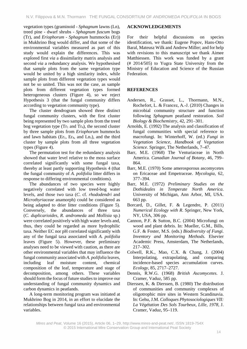
Appendix 2 Checklist with annotations The following checklist presents species annotations in alphabetical order. Each annotation provides the following information: herbarium accession data (substrate of collection; herbarium number in collection; date), macro- and micro-morphological descriptions based on our collections, notes on the ecology and abundance of each taxon in our peatlands, and a brief discussion on taxonomic issues if warranted. All descriptions are supported by schematic illustrations, where scale bars = 10 µm unless noted otherwise. All morphological dimensions marked with asterisks (*) originated from live specimens; morphological dimensions without asterisks originated from preserved specimens. Allophylaria basalifusca Graddon (Figure A1-A) On fallen leaves of A. polifolia, O. palustris and Betula nana among Sphagnum in a raised bog community. Specimens examined: Kh-4262, Kh-4319, Kh-4355, Kh-4370 (5.7.13, 20.7.13, 9.8.13, 13.8.13, 8.8.14).
Apothecia scattered on undersides of leaves, cupulate, stipitate, up to 130 μm long, the same width, outer surface greyish, base of stipe darker to almost black, hymenium surface greyish. Excipulum of thick-walled (some gelatinous) hyphae of textura porrecta/prismatica, cells 2.5 μm broad, end cells at the edge fusoid, obtuse, about 15 × 4.5 µm, with greenish vacuoles, cells of the base of stipe are shorter and more irregular; asci clavate, with crozier, with euamyloid ring, *60–71.5 × 8–9.8 µm; paraphyses cylindrical, with septa in lower part, rarely branched, in upper part with small ellipsoid or round vacuoles and slightly enlarged, *62 × 2.8 μm; spores fusoid, heteropolar, with many small oil guttules, *16.7 (14.6–19.6) × 2.8 (2.4–3.1) μm (n = 13).
Saprobic on leaves of Quercus (Graddon 1977, Ellis & Ellis 1997) and on leaves of Alnus, Betula, and Vaccinium myrtillus (H.-O. Baral, pers. comm.). Occurred rarely in this study (one record; on 0.02 % of examined leaves).
Specimen description coincided well with the species description except for some smaller spores in our specimens (Graddon 1977). Allophylaria basalifusca is probably synonymous with Calycellina leucella (P. Karst.) Dennis ex E. Müll., which also has an excipulum made of some gel but differs in having white (not black) stipe base (H.-O. Baral, pers. comm.). Alternaria sp. (Figure A1-B) On fallen leaves of A. polifolia and Chamaedaphne calyculata among Sphagnum in a raised bog community. Specimen examined: Kh-4218 (19.6.13).
Conidiophores arising in groups, ascending, simple, cylindrical, septate, brown, about 50 × 5 μm; conidia not in chains, straight, obclavate, with short beak, broadly rounded at base, brown, smooth, with 5–7 transverse septa, young conidia 40–62 × 13–14 μm, overmature conidia elongate up to 100–120 μm long and form a kind of cluster of conidiogenous cells on beak, producing micro-conidia.
Conidia fusoid, brown, septate, with short to long stalks, 64–133 × 11–15 (broadest part) μm (n = 10).
Alternaria spp. are saprobic on various substrata or parasitic causing plant diseases (Ellis 1971). Teleomorphic stage: Pleosporaceae (Lewia and others) (Seifert et al. 2011). Occurred rarely in this study (two records; on 0.04 % of examined leaves). Chalara cylindrica Karst. (Figure A1-C) On fallen leaves of A. polifolia and C. calyculata among Sphagnum in a raised bog community. Specimen examined: Kh-4159 (29.5.13).
Colonies effuse, brown, hairy. Phialophores simple, cylindrical, septate, brown, roughened at the base, up to 40 μm long and 3.5 μm wide; phialides lageniform, 25 (20–30) μm long; ventrum 18–21 × 3–3.6 μm, collarette cylindrical, 5–6.7 × 1–1.6 μm, transition between ventrum and collarette not abrupt; phialoconidia hyaline, cylindrical, 5.8 (5.1–6.7) × 1.4 (1.1–1.6) μm (n = 10).
Saprobic on various substrata, previously reported from bark, scales, and needles of Picea, Abies and Eucalyptus (Ellis 1971, Raj & Kendrick 1976). Teleomorphs of the genus s.l. have been reported from five orders within the Ascomycota, including two genera which were also found on A. polifolia in our study: Allophylaria and Calycellina. Abundant in this study, albeit easily overlooked due to very small size (on 0.34 % of examined leaves).
Our specimen’s characteristics differ from the species description in having a shorter collarette and a less-pronounced transition between the ventrum and the collarette (Raj & Kendrick 1976). Cladosporium cf. rhododendri K. Schub. (Figure A1-D) On fallen leaves of A. polifolia among Sphagnum in a raised bog community. Specimens examined: Kh-3690, Kh-4333, (6.8.12, 26.7.13).
Colonies densely covering upper side of leaf, in dense tufts; stromata present, from textura globosa/angularis, cells brown, smooth, 5–6 μm; conidiophores scattered to grouped in dense clusters, straight to some flexuous, unbranched, septate, smooth; conidiogenous cells integrated, terminal and intercalary, 70–170 μm long, 4–5 μm wide in middle of length, narrower in upper part; conidia obovoid, fusiform, subcylindrical, 0–2 septate, rough-walled, 18.6 (14–22.3) × 5.3 (4.7–6.1) μm (n = 20).
Previously described from living leaves of Rhododendron sp., may be parasitic (Schubert 2005), other Cladosporium spp. may be saprobic or parasitic (Ellis 1971). Teleomorphic stage: Mycosphaerellaceae (Davidiella) (Schubert 2005). Abundant in this study (on 9.1 % of examined leaves, second-most-abundant taxon in this study) and most prevalent in the treed bog, showing a correlation with low bog water levels.
The identity of this taxon was based on only one fungal taxon with similar morphology having previously been reported from this plant (Schubert 2005).
N.V. Filippova & M.N. Thormann THE FUNGAL CONSORTIUM OF ANDROMEDA POLIFOLIA IN BOGS
Coccomyces duplicarioides Sherwood (Figure A1-E) On fallen leaves of A. polifolia among Sphagnum in a raised bog community. Specimens examined: Kh-0995, Kh-4153 (29.9.9, 28.5.13, 23.9.14).
Hysterothecia subcuticular on upper side of bleached leaves, orbicular, first appear as black pustules, then grow to become cylindrical and expanded in upper part to turbinate, up to 1.5 mm width, 0.5 mm height, outer surface blackish, hymenium surface grey. Asci clavate, with long stalk, tip with protruding wart, IKI negative, 180–238 × 15–18.5 μm; paraphyses strongly circinate, not enlarged in upper part (1.2 μm); ascospores filiform, slightly tapering below, with two gelatinous caps at both ends (upper cap globose and lower cap elongated), and with sheath surrounding entire spore, with some tiny oil guttules inside, *60 (54–65) × 3 (2.7–3.3) μm (with sheath about 13 μm width) (n = 24, length measured without gelatinous appendages).
Saprobic or weakly parasitic. Sherwood (1980) specified “recently dead plant material” as the primary substrate for this genus, with most species being host- specific (supported by this study). The most abundant taxon in this study (on 17.4 % of examined leaves) and appeared to be positively correlated with low water level in the bog lawns. Under-developed hysterothecia could be observed throughout the season, with mature apothecia starting to appear in late autumn after the first frosts (end of September to early October); overmature apothecia were observed after the snow melt in spring.
The specimen description coincides well with a species description by Sherwood (1980), except for the gelatinous spore caps, which were not described by that author. These structures could be lost in dry specimens and, therefore, be missed in the description; however, this feature is considered to be an important characteristic in the taxonomy of the genus (Johnston 1994). Coccomyces cf. leptideus (Fr.) B. Erikss. / dentatus (J.C. Schmidt) Sacc. (Figure A1-F) On fallen leaves of A. polifolia among Sphagnum fuscum on a hummock in a raised bog community. Specimen examined: Kh-4347 (8.8.13).
Apothecia on upper leaf surface, about 1 mm in diameter, nearly orbicular, surrounded by black bent edge with 4–6 teeth, hymenium surface yellowish. Asci clavate, with long stalks, with crozier, 8-spored, *121–134 × 12–15 μm; paraphyses cylindrical, bent, irregularly bulged in upper part (up to 5 μm broad); ascospores filiform, with small gelatinous cap covering upper end, with weak sheath surrounding entire spore, *70.2 (81.2–101.2) × 1.9 (1.4–2.3) μm (n = 20).
Saprobic or weakly parasitic. Previously reported from a wide host range, including members of the Ericaceae (Sherwood 1980). A single random collection made in this study from one leaf in a treed bog.
The specimen description in this study mostly matches the species description of C. leptideus (Sherwood 1980), including the host (different members of the Ericaceae); however, C. leptideus has somewhat broader spores. This taxon could also be C. dentatus, which has also previously
been collected from leaves of members of the Ericaceae (Sherwood 1980, Minter 1996b). Dendryphion cf. comosum Wallr. (Figure A1-G) On fallen leaves of A. polifolia among Sphagnum. Specimen examined: Kh-4338 (28.7.13).
Conidiophores arise singly or 2–3 in cluster, reddish-brown, up to 400 μm long, 20 μm wide in middle, with swollen base, straight, septate, smooth, thick-walled (2.5 μm), with two primary branches in upper part and up to eight secondary branches; conidia rarely observed, overmature, some spherical rough cells (conidial parts?) were located near the conidiophores, about 7 μm in diameter.
Saprobic on dead stems of different plants (Ellis 1971). Teleomorphic stage: Pleosporaceae (Index Fungorum 2015). Uncommon in this study (on 0.15 % of examined leaves).
Scarce collections with overmature conidiophores (conidia mostly lost) prohibited a reliable identification of this taxon; however, it has similar morphological characteristics to Dendryphion comosum (Ellis 1971). Diplodina eurhododendri Voss. (Figure A1-H) On fallen leaves of A. polifolia among Sphagnum. Specimen examined: Kh-4335 (26.7.13, 9.6.14, 21.7.14).
Pycnidia scattered on leaf underside (often on ribs), immersed, soon erumpent, subglobose, brownish, 150–200 μm in diameter; conidiophores hyaline, smooth, branched, cylindrical with narrowed upper part; conidiogenous cells phyalidic; conidia fusoid, hyaline, smooth, 1-septate, not constricted, some slightly curved, 11.2 (9.5–14.1) × 2.8 (2.5–3.2) μm (n = 13).
Reported from living leaves of Eurhododendron (Grove 1935) and from Rhododendron spp. (Farr & Rossman 2015). Teleomorphic stage: Encoeliopsis rhododendri (Groves 1969, Farr & Rossman 2015). Fairly abundant in this study (on 0.27 % of examined leaves) in different bog habitats.
Our specimen differs from the species description in that it is saprobic (found on fallen dead leaves) and originated from an alternative host (Andromeda as opposed to Ledum, although both are members of the Ericaceae); morphological characteristics were similar. Dothidea sp. (Figure A2-A) On attached dead leaves of A. polifolia affected by Rhytisma andromeda. Specimen examined: Kh-4340 (29.7.13).
Ascocarps immersed in Rhytisma’s stroma, appearing at the surface as irregular warts with orange punctation, globose, up to 100 μm in diameter, pore more or less pronounced; walls made by layers of brown irregular cells; asci about 40 × 10 μm; paraphysoid tissue not developed; spores fusoid, hyaline, two-celled, slightly heteropolar, with deep constriction, some broken into two parts, about 15–17 × 2.5–4 μm (n = 5).
Saprobic or parasitic on Rhytisma. Fairly abundant on leaves colonised by Rhytisma in Eriophorum communities in this study (on 0.6 % of examined leaves).
N.V. Filippova & M.N. Thormann THE FUNGAL CONSORTIUM OF ANDROMEDA POLIFOLIA IN BOGS
Dothideales gen. sp. (Figure A2-B) On fallen leaves of A. polifolia, C. calyculata and L. palustre among Sphagnum. Specimen examined: Kh-4236 (24.6.13, 23.6.14, 6.8.14).
Ascomata scattered on both sides of the leaf, spherical, superficial, setose around the pore, and with descending hyphae in lower part, up to 150 μm in diameter. Setae brown, septate, thick-walled, straight and short near the pore, bent and longer outward, 40–80 μm long, 8 μm broad at base, narrowing to obtuse tip; descending hyphae brown, septate, about 1 μm broad; asci clavate, attached to stalks which connected together in bunches, 20–23 × 8–9.6 μm, dehiscence mode unclear; hamathecium from elongated thin elements surrounded by gelatinous substance; spores hyaline, two-celled, with deep constriction, with many small to medium oil guttules *10 (9–10.8) × 3.5 (3.3–3.9) μm (n = 22).
Saprobic, uncommon in this study (on 0.13 % of examined leaves). Gibbera andromedae (Rehm) E. Müll. & Arx (Figure A2-C) On fallen leaves of A. polifolia and L. palustre among Sphagnum shoots in a raised bog. Specimens examined: Kh-3659, Kh-4241 (4.8.12, 26.6.13).
Ascomata scattered or grouped, rarely formed around the perimeter of circular spots, on upper or lower sides of fallen leaves, globose to conical, setose over upper part, up to 180 μm in diameter, brown. Setae straight or curved in basal part, sharp, up to 100 μm long, 6 μm wide in lower part, with enlarged bulbous base up to 16 μm in width; excipulum from light brown angular cells about 11 μm in diameter; asci fissitunicate, oblong, 51–73 × 13–17 μm; ascospores two-celled, with deep constriction, slightly heteropolar, yellow-green, olivaceous, with several medium and small oil guttules, ascospores *24 (23–24.8) × 8.8 (8–9.3) μm (n = 20), smaller when preserved 19 (17.4–22.5) × 5.9 (4.9–7.6) μm (n = 16).
Although Barr (1968) reported the species as being parasitic on living or weakened leaves (A. glaucophylla), it was collected only from dead and well-decayed leaf litter in this study. Abundant in this study (on 2.7 % of examined leaves).
The ascospore size of our live specimens was larger than stated in the species description, but once the ascospores from this study were preserved, their dimensions were similar to those given in the literature (Barr 1968). Godronia cf. cassandrae Peck (Figure A2-D) On fallen leaves of A. polifolia, dead twigs of C. calyculata and L. palustre among Sphagnum in a raised bog community. Specimens examined: Kh-4209: A. polifolia; Kh-3808, Kh-4211, Kh-4294: C. calyculata; Kh-4229: L. palustre (16.6.13, 21.8.12, 18.6.13, 15.7.13, 24.6.13).
Apothecia erumpent, single on upper side and leaf ribs, sessile, subglobose to urceolate, surface furfuraceous, without clear ribs, brown to yellowish brown near the edge, edge dirty yellow (when young), 0.4 mm in diameter. The apothecia from A. polifolia that we studied were
under-developed (contained immature asci). Saprobic or weakly parasitic, G. cassandrae cf.
vaccinii is causing disease of cultivated plants (Groves 1965). Rare in this study (on 0.1 % of examined leaves).
Underdeveloped apothecia prohibited definitive identification of the specimen. The morphology of the apothecia was similar to that in G. cassandrae, which was frequently collected from both C. calyculata and L. palustre in the same place; therefore, this taxon was tentatively assigned to this species. Alternatively, this taxon could be G. andromeda Henn. (the species described from A. polifolia); however, the identity of G. andromeda itself is questionable, because good reference collections are lacking (Groves 1965). Helicoon pluriseptatum Beverw. (Figure A2-E) On fallen leaves of A. polifolia and C. calyculata among Sphagnum. Specimen examined: Kh-4158 (29.5.13).
Mycelium rarely branched, light brown, 3–4.5 μm broad; conidia light brown, 50–53 μm broad (n = 10, length was not measured, since all conidia lie on the front side being quite flat), filament 6–7.3 μm broad (n = 10, the outer widest coil was measured), loosely coiled 6–8 times in three planes, with 3–4 septa per inner coil, and 23–26 septa per outer coil, forming blunt oval conidium.
Previously reported from decaying coniferous wood in moist places (Ellis 1971) and on decaying leaves of Betula, Quercus, Pinus and Rhododendron (Goos et al. 1986). Belonging to the group of aero-aquatic Hyphomycetes, with spores adapted for dispersal by water. Teleomorphic stage: Orbiliales or Sordariales (Seifert et al. 2011). Abundant in this study (on 1.1 % of examined leaves).
In comparison with the description in Goos et al. (1986), the conidia of the specimen in this study were larger, more coiled and more septate. Hilberina cf. breviseta (P. Karst.) Huhndorf & A.N. Mill. (Figure A2-F) On fallen leaves of A. polifolia, C. calyculata, and L. palustre among Sphagnum in a raised bog community. Specimens examined: Kh-4346 (2.8.13, 24.7.14, 6.8.14).
Ascomata scattered on underside of the leaf, ovoid, dark brown, base submerged in leaf tissue with protruding upper part, setose in upper part, up to 350 μm wide, 430 μm long. Setae up to 200 μm long, brown, thick-walled, septate, up to 15 μm broad at base, narrowing to acute tip; excipulum from angular brown cells about 10 μm; asci cylindrical, with long stipe, spore-bearing part about 80–100 × 14–17 μm; paraphyses cylindrical, septate with constrictions, 5 μm wide in middle; ascospores narrow-cylindrical, with attenuated basal end, lower part curved in obtuse angle, 43.2 (40.7–45.6) × 2.5 (2.2–2.8) μm (n = 12).
Previously reported from dead wood (Miller et al. 2014). A single collection in this study, of one perithecium on a fallen leaf in treed bog.
The specimen is morphologically similar to H. breviseta, but needs further examination due to the complex systematics of this taxon (A. Miller, pers. comm.).
N.V. Filippova & M.N. Thormann THE FUNGAL CONSORTIUM OF ANDROMEDA POLIFOLIA IN BOGS
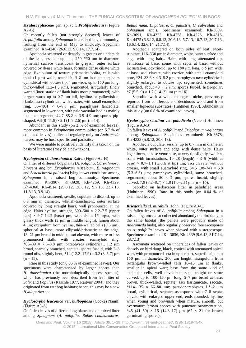
Hyaloscyphaceae gen. sp. (c.f. Proliferodiscus) (Figure A2-G) On recently fallen (not strongly decayed) leaves of A. polifolia among Sphagnum in a raised bog community, fruiting from the end of May to mid-July. Specimen examined: Kh-4240 (26.6.13, 9.6.14, 17.7.14).
Apothecia scattered or densely in groups on underside of the leaf, sessile, cupulate, 250–550 μm in diameter, hymenial surface translucent to greyish, outer surface covered by dense white hairs, forming dense ciliate white edge. Excipulum of textura prismatica/oblita, cells with thick (1 μm) walls, roundish, 5–8 μm in diameter; hairs cylindrical with obtuse tip, 4 μm wide, up to 150 μm long, thick-walled (1.2–1.5 μm), segmented, irregularly finely warted (incrustation of flank hairs more pronounced), with largest warts up to 0.7 μm tall, hyaline or yellowish at flanks; asci cylindrical, with crozier, with small euamyloid ring, 35–49.4 × 6–8.3 μm; paraphyses lanceolate, segmented in lower part, with pale vacuolar bodies mainly in upper segment, 44.7–85.2 × 3.9–4.9 μm; spores pip-shaped, 9.3 (8–11.8) × 2.1 (1.5–2.6) μm (n=14).
Abundant in this study (on 2 % of examined leaves), more common in Eriophorum communities (on 5.7 % of collected leaves), collected regularly only on Andromeda leaves, may be host-specific and parasitic.
We were unable to positively identify this taxon on the basis of literature (may be a new taxon). Hyalopeziza cf. tianschanica Raitv. (Figure A2-H) On litter of different bog plants (A. polifolia, Carex limosa, Drosera anglica, Eriophorum russeloum, E. vaginatum and Scheuchzeria palustris) lying in wet conditions among Sphagnum in a raised bog community. Specimens examined: Kh-3907, Kh-3920, Kh-4269, Kh-4329, Kh-4360, Kh-4514 (29.8.12, 30.8.12, 9.7.13, 23.7.13, 11.8.13, 3.9.14).
Apothecia scattered, sessile, cupulate to discoid, up to 0.8 mm in diameter, whitish-translucent, outer surface covered by long straight hairs, well pronounced at the edge. Hairs hyaline, straight, 300–500 × 2.2–7.5 (upper part) × 9.7–14.5 (base) μm, with about 15 septa, with glassy thick walls (2 μm in middle length), lumen about 4 μm; excipulum from hyaline, thin-walled cells (0.5 μm), spherical at base, more ellipsoid/prismatic at the edge, 13–21 μm broad in middle; asci clavate, with more or less pronounced stalk, with crozier, euamyloid ring, *66–89 × 7.6–8.8 μm; paraphyses cylindrical, 1.2 μm broad, scarcely branched, septate; spores fusoid, with 2–3 round oils, slightly bent, *14 (12.2–17.9) × 3.2 (3–3.7) μm (n = 15).
Rare in this study (on 0.06 % of examined leaves). Our specimens were characterised by larger spores than H. tianschanica (the morphologically closest species), which has previously been described from leaf litter of Salix and Populus (Raschle 1977, Raitviir 2004), and they originated from wet bog habitats; hence, this may be a new Hyalopeziza sp. Hyaloscypha leuconica var. bulbopilosa (Cooke) Nannf. (Figure A3-A) On fallen leaves of different bog plants and on mixed litter among Sphagnum (A. polifolia, Rubus chamaemorus,
Betula nana, L. palustre, O. palustris, C. calyculata and Sphagnum spp.). Specimens examined: Kh-3689, Kh-3691, Kh-4222, Kh-4258, Kh-4276, Kh-4316, Kh-4475 (6.8.12, 6.8.12, 20.6.13, 5.7.13, 10.7.13, 20.7.13, 16.6.14, 32.6.14, 21.7.14).
Apothecia scattered on both sides of leaf, short-stipitate, 116–330 μm in diameter, white, outer surface and edge with long hairs. Hairs with long attenuated tip, ventricose at base, some with septa at base, without incrustation, dextrinoid, up to 180 μm long, 6.5 μm wide at base; asci clavate, with crozier, with small euamyloid pore, *24–33.6 × 4.3–5.2 μm; paraphyses near cylindrical, slightly enlarged to obtuse tip, segmented, sometimes branched, about 40 × 2 μm; spores fusoid, heteropolar, *7 (5.5–9) × 1.7 (1.4–2) μm (n = 18).
Saprobic with a wide ecological niche, previously reported from coniferous and deciduous wood and from smaller ligneous substrates (Huhtinen 1990). Abundant in this study (on 0.8 % of examined leaves). Hyaloscypha secalina var. paludicola (Velen.) Huhtinen (Figure A3-B) On fallen leaves of A. polifolia and Eriophorum vaginatum among Sphagnum. Specimens examined: Kh-3678, Kh-4223 (5.8.12, 20.6.13).
Apothecia cupulate, sessile, up to 0.7 mm in diameter, white, outer surface and edge with dense hairs. Hairs lageniform, at base ventricose, at very tip slightly swollen, some with incrustations, 19–28 (length) × 3–5 (width at base) × 0.7–1.1 (width at tip) μm; asci clavate, without crozier, with small euamyloid ring, 54.5 (38–74) × 5.8 (5.3–6.6) μm; paraphyses cylindrical, some branched, segmented, about 50 × 2 μm; spores fusoid, slightly curved, 7.9 (7.2–8.7) × 1.8 (1.6–2.1) μm (n = 16).
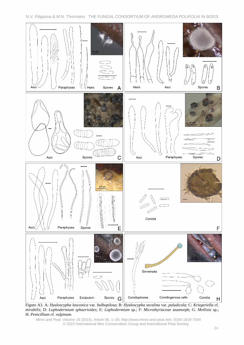
Saprobic on herbaceous litter in paludified areas (Huhtinen 1990). Rare in this study (on 0.04 % of examined leaves). Kriegeriella cf. mirabilis Höhn. (Figure A3-C) On fallen leaves of A. polifolia among Sphagnum in a raised bog, once also collected abundantly on bird dung in the same habitat (the pellets were probably made of Andromeda buds), also regularly observed free ascospores on A. polifolia leaves when viewed with a stereoscope. Specimens examined: Kh-3856, Kh-4339 (9.6.13, 31.7.14, 28.7.13).
Ascomata scattered on undersides of fallen leaves or densely on bird dung, black, conical with attenuated apical wart, with pronounced seta in upper part, superficial, up to 150 μm in diameter, 200 μm height. Excipulum from rectangular brown-walled cells 10–15 μm at flanks, smaller in apical wart; base from the same kind of excipular cells, well developed; seta straight or some curved, up to 100–150 μm long, 5–7 μm broad at base, brown, thick-walled, septate; asci fissitunicate, saccate, *114–135 × 66–80 μm; pseudoparaphyses 1.5–2 μm broad, cylindrical, septate; ascospores with 7–8 septa, clavate with enlarged upper end, ends rounded, hyaline when young and brownish when mature, smooth, but overmature brown spores with punctate ornamentation, *45 (41–50) × 16 (14.3–17) μm (62 × 21 for brown germinating spores).
N.V. Filippova & M.N. Thormann THE FUNGAL CONSORTIUM OF ANDROMEDA POLIFOLIA IN BOGS
Minter (2007) describes K. mirabilis as being saprobic on conifer needles. One of our collections originated from bird dung (probably digested Andromeda buds), where ascomata were abundant; ascomata on leaves were less abundant, suggesting a fimicolous and saprobic habit for this taxon. Fairly abundant in this study (on 0.5 % of examined leaves in bog communities).
Our specimen differed morphologically from K. mirabilis (Barr 1972, Minter 2007, Zhang et al. 2012) in spore size and in the presence of setae, and we were unable to find another taxon with similar morphological characters. Hence, our specimen may be a new Kriegeriella species. Lophodermium sphaerioides (Alb. & Schw. ex Fr.) (Figure A3-D) On fallen leaves of A. polifolia, L. palustre, and O. palustris among Sphagnum. Specimens examined: Kh-3658, Kh-4314 (4.8.12, 19.7.13, 6.8.14).
Hysterothecia circular to ellipsoidal, scattered or in groups, up to 10 on upper side of leaf, 0.3–0.6 mm in diameter, up to 0.3 mm high, subcuticular, with yellowish labium, hymenium grey, outer surface black. Flanks from outer layer of host epidermis and inner layer of fungus textura epidermoidea; palisade layer of labia well developed, light yellowish-brown, from prismatic cells arranged in columns, up to 5–6 cells in each column (30 × 5 μm); asci clavate, clamped, inamyloid, 79–88 × 6.4–10.2 μm; paraphyses filiform, 1.5–2.5 μm broad, with many small guttules, more or less circinate in upper part; ascospores filiform, 24 (21.5–26) × 1.3 (1.1–1.5) μm (n = 12).
Saprobic or weakly parasitic, previously reported from L. groenlandicum (Shoemaker & Egger 1981). Uncommon in this study (on 0.14 % of examined leaves). Lophodermium sp. (Figure A3-E) On fallen leaves of A. polifolia and C. calyculata among Sphagnum in a raised bog community. Specimens examined: Kh-3710, Kh- 4219, Kh-4264 (6.8.12, 19.6.13, 6.7.13).
Hysterothecia circular to ellipsoid, scattered on both sides of leaf, several on a single leaf, first appear as black pustules, later opening by a longitudinal more or less regular slit, without labile (or with pale greyish labile when wet?), 0.8–1.3 mm by long axis, up to 0.5 mm height. Asci clavate, clamped, 150 × 16.5 μm; paraphyses cylindrical, segmented, bent at tip, not enlarged, 3 μm wide; spores filiform, heteropolar, guttulate, with small gelatinous cap covering upper end, 73.2 (63–90.4) × 2.7 (2.3–3.4) μm (n = 11).
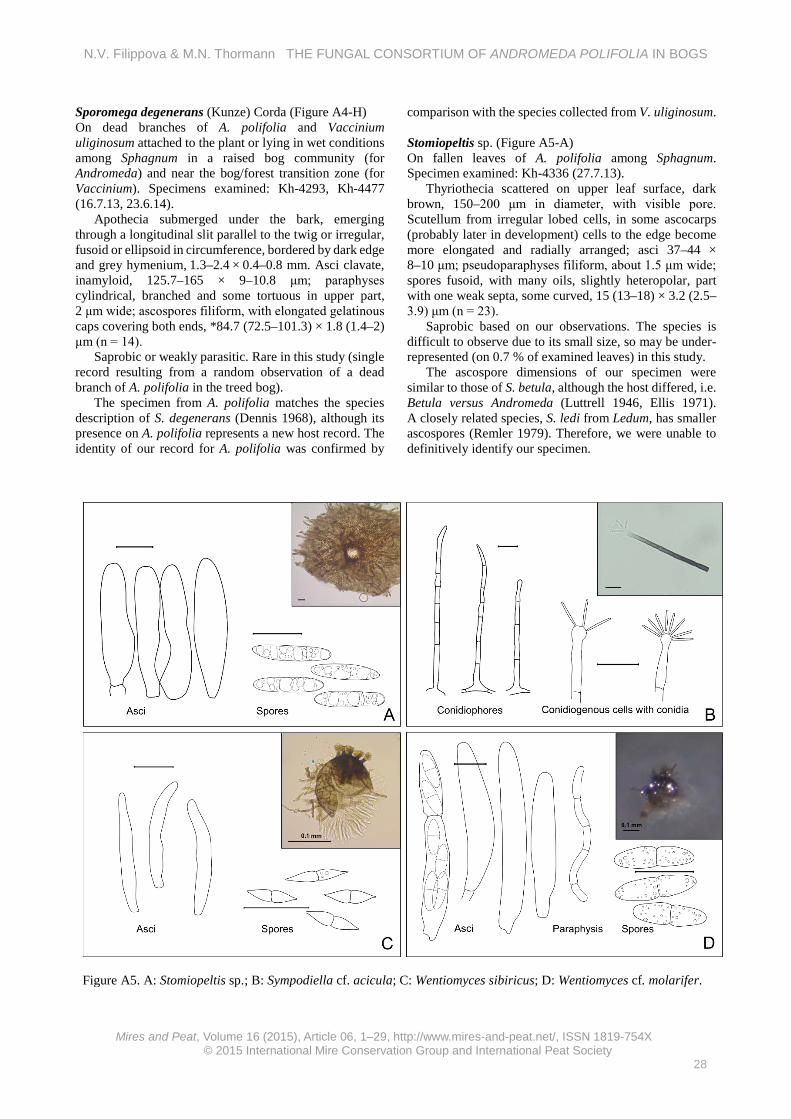
Saprobic or weakly parasitic. Fairly abundant in this study (on 0.5 % of examined leaves). Microthyriaceae anamorph (= Stomiopeltis?) (Figure A3-F) On fallen leaves of A. polifolia, on the same leaves as Stomiopeltis sp. or found without any teleomorphic stage. Specimen examined: Kh-4341 (29.7.13).
Pycnothyria orbicular, up to 100 μm in diameter, cells radially arranged, prismatic near centre, some elongated at edge, 3–5 μm; conidiogenous cells holoblastic, simple,
hyaline; conidia hyaline, cylindrical, one celled, about 6.5 × 1.2 μm (n = 5).
Very abundant in this study (on 2.9 % of examined leaves), its occurrence correlating positively with low water levels in treed bogs, and its abundance probably under-estimated due to the small size of the conidioma.
This anamorphic Microthyriaceae was fairly abundant on different leaves in the bog litter and could be related to different teleomorphs (e.g. Microthyrium and/or Trichothyrina spp.), which were recorded on different litter in the same habitat. We were unable to establish a definitive anamorph-teleomorph connection for this taxon. Mollisia sp. (Figure A3-G) On fallen leaves of A. polifolia among Sphagnum in a raised bog. Specimen examined: Kh-4224 (20.6.13).
Apothecia shallow-cupulate, sessile, hymenium grey with lighter edge, outer surface brownish, 430–800 μm in diameter. Excipulum from globose brown cells 10–15 μm at base, smaller to the edge, end cells clavate, 10 × 5 μm, with conspicuous refractive vacuoles disappearing in KOH; asci clavate, with obscure crozier, with euamyloid ring, 25–34 × 2.8–3.6 μm; paraphyses cylindrical, segmented, with enlarged upper segment, with abundant refractive vacuole disappearing in KOH without yellow reaction, 43.4 × 2.7 μm (width at enlarged upper part); spores fusoid, heteropolar, slightly curved, 6.2 (5–7.1) × 1.6 (1.5–2) μm (n = 22).
Saprobic. Abundant in this study (on 1.3 % of examined
leaves). Penicillium cf. vulpinum (Cooke & Massee) Seifert & Samson (Figure A3-H) On fallen leaves of A. polifolia and (if identical species) berries of O. palustris among Sphagnum. Specimens examined: Kh-4155, Kh- 4173 (28.5.13, 4.6.13).
Conidiophores in synnemata with long stalk up to 3 mm long, 150 μm wide, bluish head, 300 μm broad; conidia citriform, about 3.4 × 2.2 μm.
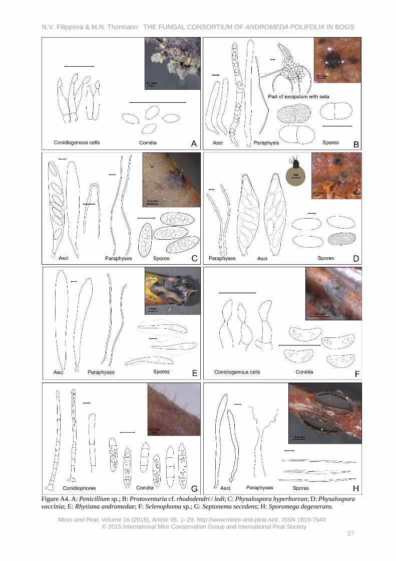
Saprobic. Rare in this study (single collection from Andromeda leaves, but commonly seen on O. palustris berries). Penicillium sp. (Figure A4-A) On fallen leaves of A. polifolia previously infected by Rhytisma andromeda. Specimen examined: Kh-4334 (26.7.13).
Conidiophores in synnemata, densely covering old stromas of R. andromeda; conidia citriform, about 3.3 × 1.8 μm (n = 7).
Saprobic. Fairly abundant in this study (on 0.2 % of examined leaves). Protoventuria cf. rhododendri (Höhn.) M.E. Barr / ledi (M.E. Barr) M.E. Barr (Figure A4-B) On fallen leaves of A. polifolia, C. calyculata, L. palustre, Betula nana and Rubus chamaemorus among Sphagnum. Specimens examined: Kh-4214, Kh-4225, Kh-4260 (5.7.13, 16.6.14, 18.8.13, 20.6.13).
Ascomata scattered to 5–15 in groups on both leaf surfaces, superficial, with abundant hyphae radiating from the ascomata and spreading at the surface, depressed-
N.V. Filippova & M.N. Thormann THE FUNGAL CONSORTIUM OF ANDROMEDA POLIFOLIA IN BOGS
globose, about 200 μm broad, with visible pore, setose in upper part. Seta brown, sharp, bent, up to 60 μm long, 6 μm wide at base; asci fissitunicate, cylindrical, without clamps, 74–85 × 8–10 μm; pseudo-paraphyses abundant, cylindrical; spores two-celled, greenish-olivaceous, with warty ornamentation, slightly heteropolar, *14.6 (13–16) [17.7] × 6.1 (5.4–6.6) μm (n = 14), smaller in dead state: 11.9 (10.6–13.4) × 5.4 (4.6–6.5) μm (n = 22).
Saprobic on leaves and branches of Rhododendron spp. (Barr 1968). Rare in this study (single collection in the Eriophorum community, but commonly collected from dead leaves of other bog plants).
Given the large variation in ascospore sizes in our specimens, a definitive identification to either P. rhododendri or P. ledi was not possible (Barr 1968). Physalospora hyperborea Baumler (Figure A4-C) On fallen leaves of A. polifolia and O. palustris among Sphagnum in a raised bog community. Specimen examined: Kh-4337 (28.7.13, 17.7.14).
Perithecia globose, submerged under epidermis with apical wart protruding at the surface, scattered or in small groups on both leaf surfaces. Excipulum of lower part from brownish rectangular cells 20–30 μm in diameter, apical wart from darker small cells; asci cylindrical, short-stipitate, becoming broader with age, with refractive ring, 130–150 × 18.3–20.8 μm; paraphyses abundant, cylindrical, segmented with constrictions, about 5 μm wide; ascospores ellipsoid, with many medium oil guttules, surrounded by thin gelatinous coat, *20.3 (16.8–22.2) × 7 (6.2–8.1) μm (n = 30).
Saprobic on dead leaves of Cassiope tetragona, Ledum decumbens and Phyllodoce caerulea (Barr 1970). Abundant in this study (on 0.8 % of examined leaves). Physalospora vaccinii (Shear) v. Arx & Muller (Figure A4-D) On fallen leaves of A. polifolia and O. palustris among Sphagnum in a raised bog community. Specimens examined: Kh-3669, Kh-4165, Kh-4252, Kh-4263, Kh-4352 (04.08.2012, 01.06.2013, 03.07.2013, 06.07.2013, 31.07.2014, 09.08.2013, 17.07.2014).
Perithecia spherical, submerged under epidermis, with erumpent at surface dark tip (wart) covered with seta, up to 250 μm in diameter, scattered or several in groups on both leaf surfaces. Excipulum of lower part from hyaline rectangular cells about 20 μm in diameter, smaller and dark pigmented in upper wart; setae up to 130 μm long; asci broadly elliptic, about 150 × 45 μm, with refractive apical annulus; paraphyses abundant, cylindrical, some narrowed to the tip, septate with constrictions, about 7.5 μm wide at base; ascospores ellipsoid, filled with tiny oil guttules, wall with punctate ornamentation visible well in over-mature spores, 32.7 (31–35) × 17.4 (16.2–18.3) μm (n = 14).
Saprobic on dead leaves of different Oxycoccus spp., also found on leaves of A. polifolia and C. calyculata (Barr 1970). Very abundant in this study (on 2.2 % of examined leaves).
Rhytisma andromedae (Pers.) Fr. (Figure A4-E) On living plants of A. polifolia, conidial and teleomorphic stages causing sometimes widespread infections, apothecia also collected from fallen leaves in litter. Specimens examined: Kh-0193, Kh-4261, Kh-3642 (15.6.08, 5.7.13, 4.8.12).
Stroma and conidioma formed on upper leaf surface, pustulate or covering the entire leaf surface with a common shield. Conidioma black flat pustules, conidial mass emits through small pore surrounded by shallow depression, conidial mass sticky, whitish. Ascomata in stroma, pustulate, black, later opening by irregular slits, hymenium grey. Asci clavate, about 200 × 25 μm; paraphyses cylindrical, segmented, sometimes branched, 2–3 μm wide, only slightly enlarged in upper part; ascospores clavate, with gelatinous sheath, with amorphous internal oil content, 63.7–81.3 × 7.6–9.2 μm (width at broadest part) (n = 9). Conidia cylindrical, obtuse-ended, 6.8 (6.2–7.6) × 1.6 (1.5–1.7) μm (n = 11).
Parasitic (perhaps necrotrophic) and saprobic at later stages of teleomorphic state development (Minter 1996a). Very abundant in this study (on 4.8 % of examined leaves); although considered to be host-specific, it was also collected from O. palustris in the same habitat.
The species is easily recognised, and our description coincides well with previous reports (Minter 1996a); however, our observations showed that the conidial mass is released through small pores surrounded by shallow depressions, as opposed to cracks (Minter 1996a). Selenophoma sp. (Figure A4-F) On fallen leaves of A. polifolia among Sphagnum. Specimen examined: Kh-4257 (5.7.13).
Pycnidia single or several on leaf underside, often on ribs, immersed with seen at the surface small dark pore, spherical and flattened, black, up to 0.5 mm in diameter. Conidiophores absent; conidiogenous cells phialidic, hyaline, cylindrical with narrowed upper part; conidia aseptate, lunate, 7.4 × 3 μm (n = 5).
Saprobic based on our observations. Fairly abundant in this study (on 0.3 % of examined leaves). Septonema secedens Corda (Figure A4-G) On fallen leaves of A. polifolia and different ericoid dwarf shrubs (C. calyculata, L. palustre, and Oxycoccus palustris). Specimen examined: Kh-4156 (29.5.13).
Conidiophores scattered forming hairy surface at leaf upper side, straight, cylindrical with enlarged basal cell, brown, 200 μm long, 6.5 μm wide in middle part, smooth; conidiogenous cells in upper part of conidiophore; conidia formed in chains, cylindrical with quite acute ends, septate, brown, smooth, 25 (18.6–30.5) × 5.9 (5.3–6.7) μm (n = 22).
Saprobic on wood, bark of Betula, Pinus, Vitis and others (Seifert et al. 2011). Teleomorphic stage: Mytilinidiaceae (Seifert et al. 2011). The species is difficult to observe due to its small size, and its apparent rarity in this study (on 0.9 % of examined leaves) may be conservative.
N.V. Filippova & M.N. Thormann THE FUNGAL CONSORTIUM OF ANDROMEDA POLIFOLIA IN BOGS
Sporomega degenerans (Kunze) Corda (Figure A4-H) On dead branches of A. polifolia and Vaccinium uliginosum attached to the plant or lying in wet conditions among Sphagnum in a raised bog community (for Andromeda) and near the bog/forest transition zone (for Vaccinium). Specimens examined: Kh-4293, Kh-4477 (16.7.13, 23.6.14).
Apothecia submerged under the bark, emerging through a longitudinal slit parallel to the twig or irregular, fusoid or ellipsoid in circumference, bordered by dark edge and grey hymenium, 1.3–2.4 × 0.4–0.8 mm. Asci clavate, inamyloid, 125.7–165 × 9–10.8 μm; paraphyses cylindrical, branched and some tortuous in upper part, 2 μm wide; ascospores filiform, with elongated gelatinous caps covering both ends, *84.7 (72.5–101.3) × 1.8 (1.4–2) μm (n = 14).
Saprobic or weakly parasitic. Rare in this study (single record resulting from a random observation of a dead branch of A. polifolia in the treed bog).
The specimen from A. polifolia matches the species description of S. degenerans (Dennis 1968), although its presence on A. polifolia represents a new host record. The identity of our record for A. polifolia was confirmed by
comparison with the species collected from V. uliginosum. Stomiopeltis sp. (Figure A5-A) On fallen leaves of A. polifolia among Sphagnum. Specimen examined: Kh-4336 (27.7.13).
Thyriothecia scattered on upper leaf surface, dark brown, 150–200 μm in diameter, with visible pore. Scutellum from irregular lobed cells, in some ascocarps (probably later in development) cells to the edge become more elongated and radially arranged; asci 37–44 × 8–10 μm; pseudoparaphyses filiform, about 1.5 μm wide; spores fusoid, with many oils, slightly heteropolar, part with one weak septa, some curved, 15 (13–18) × 3.2 (2.5–3.9) μm (n = 23).
Saprobic based on our observations. The species is difficult to observe due to its small size, so may be under-represented (on 0.7 % of examined leaves) in this study.
The ascospore dimensions of our specimen were similar to those of S. betula, although the host differed, i.e. Betula versus Andromeda (Luttrell 1946, Ellis 1971). A closely related species, S. ledi from Ledum, has smaller ascospores (Remler 1979). Therefore, we were unable to definitively identify our specimen.
Sympodiella cf. acicola Kendric (Figure A5-B) On fallen leaves of A. polifolia and C. calyculata among Sphagnum. Specimen examined: Kh-3700 (6.8.12).
Colonies effuse, loose hairy. Conidiophores single, straight, not branched, near 100 μm long, 3 μm wide, basal cell enlarged; conidiogenous cells form in upper slightly enlarged part, blastic; arthroconidia cylindrical, with truncate ends, in chains, about 10 × 2 μm (n = 5).
Saprobic on rotten needles of Pinus (Ellis 1971). Teleomorphic stage: Pleosporales (Venturiaceae) (Seifert et al. 2011). Not uncommon in our study, with several collections originating from random observations of fallen A. polifolia leaves. Wentiomyces sibiricus (Petr.) E. Müll. (Figure A5-C) On fallen leaves of A. polifolia, C. calyculata and O. palustris among Sphagnum in a raised bog community. Specimen examined: Kh-3687 (6.8.12).
Ascocarps scattered or in groups on both leaf surfaces, globose, superficial, brown, with thick rosette-shaped processes around apical pore, with descending hyphae in lower part, up to 50 μm in diameter. Peridium from rectangular cells near 8 μm broad; processes 15 μm long, with rosette-shaped apex 8 μm broad; asci fissitunicate, cylindrical, about 30 × 5 μm; hamathecium well developed from cylindrical elements; ascospores 2-celled, fusoid with middle constriction, with acute ends, 8 (7–9) × 1.7 (1.5–2) μm (n = 13).
Saprobic on Vaccinium myrtillus, but also on Filipendula and probably on Larix decidua (Hawksworth & Sivanesan 1976). Abundant in this study (on 0.7 % of examined leaves). Wentiomyces cf. molarifer Scheuer (Figure A5-D) On fallen leaves of A. polifolia and L. palustre among Sphagnum. Specimens examined: #4238 (24.6.13, 16.6.14, 6.8.14).
Ascomata scattered on both leaf surfaces, near globose, up to 80 μm in diameter, superficial, in upper part with seta, lower part covered by descending hyphae. Setae flattened and with sharp upper side (knife-shaped), bent, brown, septate, some branched, up to 120 μm long and 6 μm wide at base; peridium from rectangular brown cells 5–7 μm; descending hyphae 2.5–3 μm broad, brown; asci cylindrical, 43–61 × 6–9.3 μm; hamathecium well developed, from cylindrical elements, 2.4 μm broad; ascospores hyaline, two-celled, slightly heteropolar, with deep constriction, with many small oil guttules, *14 (11.4–15.5) × 4.7 (3.4–5.9) μm (n = 26).
Saprobic. The species was originally described from fallen leaves of Carex spp. Rare in our study (on 0.06 % of examined leaves).
Our specimen was morphologically similar to W. molarifer (Scheuer 1988); however, setae morphological differences prohibited us from achieving a definitive identification.