THE JOURNAL OF BIOLOGICAL CHEMISTRY Vol. 260, No. 11, Issue of June 10, pp. 7072-7077,1985 Printed in U. S. A. The Gene for the ,8 Subunit of Bovine Luteinizing Hormone Encodes a Gonadotropin mRNA with anUnusually Short 5’4Jntranslated Region* (Received for publication, August 9, 1984) Jeffrey B. Virgin$, Bernard J. SilverF, Arlen R. Thomason11 , and John H. Nilson4 From the Departments of $Pharmacology and §Medicine, Schoolof Medicine, Case Western Reserve University, and llCleveland Veterans Administration Medical Center, Cleveland, Ohio 44106 and IIAmgen, Inc., Thousand Oaks, California 91320 Both cDNA and genomic clones encoding the j3 sub- unit of bovine luteinizing hormone (LH) have been isolated and characterized. The nucleotide sequence was determined for the entire geneand 776 base pairs of 5’-flanking sequence. The mRNA cap site and po- lyadenylation site were mapped by primer extension and S1 nuclease protection, respectively. The bovine LHj3 gene spans less than 1.1 kilobase pairs and has three exons encoding a 550 nucleotide mRNA (exclud- ing the poly(A) tail). Bovine LHj3 is a single-copy gene, in contrast to human LHj3, which is a member of the LH/chorionic gonadotropin j3 subunit multigene family. Comparison of the bovine LHj3 gene with the human LHj3lchorionicgonadotropingene family reveals a high degree of nucleotide sequence homology, both within the genes and in the 5”flanking sequences. Despite this extensive sequence conservation, there is a major difference between the two species in the selection of a promoter site. As a result, the bovine LHj3 gene produces an mRNA with an usually short 5“untrans- lated region of only 6-1 1 nucleotides. LH’ is a pituitary gonadotropin which regulates gonadal steroidogenesis, gametogenesis, andovulation (1). Follicle- stimulating hormone and CG are two other gonadotropins closely relatedtoLHinstructureand biological activity. While LH and follicle-stimulating hormone are synthesized and secreted by the anterior pituitary, CG is synthesized in the placenta and, thus far, has been found only in primates and horses (2, 3). The gonadotropins are members of the glycoprotein hormone family, which also includes another pituitary hormone with a different biological activity, thyroid- stimulating hormone. Each of the glycoprotein hormonesis composed of two noncovalentlyassociatedsubunits, a and p (4). Within a *This work was supported by Research Grants AM 28559 and AM01316 from the Public Health Service and by Grant PCM-8309164 from the National Science Foundation. J. B. V. is a predoctoral student supported by the Pharmacological Science Training Program Grant GM 07382. The costs of publication of this article were defrayed he hereby marked “advertisement” in accordance with 18 U.S.C. in part by the payment of page charges. This article must therefore Section 1734 solely to indicate this fact. ’ The abbreviations used are: LH, luteinizing hormone; FSH, fol- licle-stimulating hormone; CG,chorionic gonadotropin; TSH, thy- roid-stimulating hormone; bLH@, @ subunit of bovine-luteinizing hormone; hCG& @ subunit of human chorionic gonadotropin; SSC, standard saline citrate (1 X = 0.15 M NaC1, 0.015 M sodium citrate); TBE, Tris/borate/EDTA buffer (1 X = 0.089 M Tris borate, 0.089 M boric acid, 0.002 M EDTA); bp, base pairs; kb, kilobase pairs; Pipes, 1,4-piperazinediethanesulfonic acid. species, these hormones share an identical (Y subunit, while their P subunits are unique and confer hormonalspecificity. The gene encoding the a subunit is present as a single copy in both humans and cattle (5, 6); thus, human pituitary and placental a subunits are derived from the same gene. In contrast, the P subunits of human LH and CG are members of a multigene family which includes 7 CGP genes and 1 LHP gene (7,8). It is not known whether the LHPICGP gene family is uniqueto humans or common to other species. The synthesis and assembly of the gonadotropins are among the most complex of all polypeptide hormones (4). This is due in part to their subunit structure and in part to the independ- ent, yet coordinate regulation of synthesis of their two sub- units (9, 10) from geneslocated on different chromosomes (11, 12). To begin studying the regulation of gonadotropin biosynthesis in more detail, we have characterized the genes encoding the a and /3 subunits of bovine LH. We have previ- ously reported the characterization of the bovine a subunit cDNA and gene (6, 13). Here, we report the characterization of cDNA and genomic clones for bovine LHP. In comparing the bovine LHP gene with the human LHPICGP gene family, we have found that theoverall gene structure and nucleotide sequence have been strongly conserved. However, there is a surprising difference in the transcription start sites, which suggests that promoter recognition for these gonadotropin /3 subunit genes may be species- or tissue-specific. METHODS~ RESULTS AND DISCUSSION Isolation and Characterization of an LHP cDNA Clone- Because the amino acid sequence of the bovine LHP subunit is known (14), we prepared a probe for screening the cDNA library by synthesizing oligodeoxynucleotides complementary to the predicted coding sequence for a pentapeptide region. Successful use of this approach required the synthesis of 32 individual oligodeoxynucleotides, representing the entire complement of all the possible coding sequences for amino acids 19-23 of bovine LHP. To select the authentic comple- mentary sequence, each individual oligodeoxynucleotide was hybridized to pituitary RNA (13). The oligodeoxynucleotide Portions of this paper (including “Methods” and Figs. 1, 4, and 5) are presented in miniprint at the end of this paper. Miniprint is easilyread with the aid of a standard magnifyingglass.Full size photocopies are available from the Journal of Biological Chemistry, 9650 Rockville Pike, Bethesda, MD 20814. Request Document No. 84M-2491, cite the authors, and include a check or money order for $2.80 per set of photocopies. Full size photocopies are also included in themicrofilm edition of the Journal that is available from Waverly Press. 7072
Transcript
THE JOURNAL OF BIOLOGICAL CHEMISTRY Vol. 260, No. 11, Issue of June 10, pp. 7072-7077,1985 Printed in U. S. A.
The Gene for the ,8 Subunit of Bovine Luteinizing Hormone Encodes a Gonadotropin mRNA with an Unusually Short 5’4Jntranslated Region*
(Received for publication, August 9, 1984)
Jeffrey B. Virgin$, Bernard J. SilverF, Arlen R. Thomason11 , and John H. Nilson4 From the Departments of $Pharmacology and §Medicine, School of Medicine, Case Western Reserve University, and llCleveland Veterans Administration Medical Center, Cleveland, Ohio 44106 and IIAmgen, Inc., Thousand Oaks, California 91320
Both cDNA and genomic clones encoding the j3 sub- unit of bovine luteinizing hormone (LH) have been isolated and characterized. The nucleotide sequence was determined for the entire gene and 776 base pairs of 5’-flanking sequence. The mRNA cap site and po- lyadenylation site were mapped by primer extension and S1 nuclease protection, respectively. The bovine LHj3 gene spans less than 1.1 kilobase pairs and has three exons encoding a 550 nucleotide mRNA (exclud- ing the poly(A) tail). Bovine LHj3 is a single-copy gene, in contrast to human LHj3, which is a member of the LH/chorionic gonadotropin j3 subunit multigene family. Comparison of the bovine LHj3 gene with the human LHj3lchorionic gonadotropin gene family reveals a high degree of nucleotide sequence homology, both within the genes and in the 5”flanking sequences. Despite this extensive sequence conservation, there is a major difference between the two species in the selection of a promoter site. As a result, the bovine LHj3 gene produces an mRNA with an usually short 5“untrans- lated region of only 6-1 1 nucleotides.
LH’ is a pituitary gonadotropin which regulates gonadal steroidogenesis, gametogenesis, and ovulation (1). Follicle- stimulating hormone and CG are two other gonadotropins closely related to LH in structure and biological activity. While LH and follicle-stimulating hormone are synthesized and secreted by the anterior pituitary, CG is synthesized in the placenta and, thus far, has been found only in primates and horses (2, 3). The gonadotropins are members of the glycoprotein hormone family, which also includes another pituitary hormone with a different biological activity, thyroid- stimulating hormone.
Each of the glycoprotein hormones is composed of two noncovalently associated subunits, a and p (4). Within a
*This work was supported by Research Grants AM 28559 and AM01316 from the Public Health Service and by Grant PCM-8309164 from the National Science Foundation. J. B. V. is a predoctoral student supported by the Pharmacological Science Training Program Grant GM 07382. The costs of publication of this article were defrayed
he hereby marked “advertisement” in accordance with 18 U.S.C. in part by the payment of page charges. This article must therefore
Section 1734 solely to indicate this fact. ’ The abbreviations used are: LH, luteinizing hormone; FSH, fol-
licle-stimulating hormone; CG, chorionic gonadotropin; TSH, thy- roid-stimulating hormone; bLH@, @ subunit of bovine-luteinizing hormone; hCG& @ subunit of human chorionic gonadotropin; SSC, standard saline citrate (1 X = 0.15 M NaC1, 0.015 M sodium citrate); TBE, Tris/borate/EDTA buffer (1 X = 0.089 M Tris borate, 0.089 M boric acid, 0.002 M EDTA); bp, base pairs; kb, kilobase pairs; Pipes, 1,4-piperazinediethanesulfonic acid.
species, these hormones share an identical (Y subunit, while their P subunits are unique and confer hormonal specificity. The gene encoding the a subunit is present as a single copy in both humans and cattle (5, 6); thus, human pituitary and placental a subunits are derived from the same gene. In contrast, the P subunits of human LH and CG are members of a multigene family which includes 7 CGP genes and 1 LHP gene (7,8). It is not known whether the LHPICGP gene family is unique to humans or common to other species.
The synthesis and assembly of the gonadotropins are among the most complex of all polypeptide hormones (4). This is due in part to their subunit structure and in part to the independ- ent, yet coordinate regulation of synthesis of their two sub- units (9, 10) from genes located on different chromosomes (11, 12). To begin studying the regulation of gonadotropin biosynthesis in more detail, we have characterized the genes encoding the a and /3 subunits of bovine LH. We have previ- ously reported the characterization of the bovine a subunit cDNA and gene (6, 13). Here, we report the characterization of cDNA and genomic clones for bovine LHP. In comparing the bovine LHP gene with the human LHPICGP gene family, we have found that the overall gene structure and nucleotide sequence have been strongly conserved. However, there is a surprising difference in the transcription start sites, which suggests that promoter recognition for these gonadotropin /3 subunit genes may be species- or tissue-specific.
METHODS~
RESULTS AND DISCUSSION
Isolation and Characterization of a n LHP cDNA Clone- Because the amino acid sequence of the bovine LHP subunit is known (14), we prepared a probe for screening the cDNA library by synthesizing oligodeoxynucleotides complementary to the predicted coding sequence for a pentapeptide region. Successful use of this approach required the synthesis of 32 individual oligodeoxynucleotides, representing the entire complement of all the possible coding sequences for amino acids 19-23 of bovine LHP. To select the authentic comple- mentary sequence, each individual oligodeoxynucleotide was hybridized to pituitary RNA (13). The oligodeoxynucleotide
Portions of this paper (including “Methods” and Figs. 1, 4, and 5) are presented in miniprint at the end of this paper. Miniprint is easily read with the aid of a standard magnifying glass. Full size photocopies are available from the Journal of Biological Chemistry, 9650 Rockville Pike, Bethesda, MD 20814. Request Document No. 84M-2491, cite the authors, and include a check or money order for $2.80 per set of photocopies. Full size photocopies are also included in the microfilm edition of the Journal that is available from Waverly Press.
7072
Characterization of the Gene for the p Subuni t of Bovine LH 7073
which hybridized to an mRNA of the estimated size for LHP (data not shown) was used to screen the cDNA library. Positive colonies were characterized by restriction mapping and Southern blot hybridization (15). The largest cDNA insert was subcloned into the PstI site of M13mp8 for se- quence analysis by the dideoxy-chain termination method (16). This cloned DNA fragment contained a sequence coding for 14 amino acids of the signal peptide and for amino acids 1-98 of the mature LHP protein (see below). It lacked coding sequence for the carboxyl-terminal region and for the amino terminus of the signal peptide and the 5'- and 3"untranslated regions. Rescreening the library with the partial cDNA clone did not produce any longer cDNA sequences.
Bovine LHP Is a Unique Gene-To estimate the size and number of bovine LHP genes, the bLH@-cDNA fragment was used to probe a genomic blot (Fig. 1). The restriction pattern appears simple since the cDNA hybridizes to a single genomic fragment with three different enzymes (Fig. 1A). These results suggest that the length of the LHP gene does not exceed the size of the BamHI fragment, which is approximately 4 kh. Although the cDNA fragment used as a probe did not repre- sent the entire mRNA sequence, further analysis of the LHP gene structure confirmed that the fragments detected in the genomic blot contain the entire gene (see below).
The simple restriction pattern of the genomic DNA suggests there is a single bovine LHP gene. To more precisely deter- mine the number of LHP genes, increasing amounts of the bLHp-cDNA plasmid were mixed with a constant amount of bovine genomic DNA and analyzed by Southern blot hybrid- ization (Fig. 1B). The band indicated by the arrow represents the linearized plasmid DNA, whereas the higher molecular weight band represents the EcoRI genomic fragment which contains the LHP gene (compare to the EcoRI lane in Fig. 1A). The intensity of the hybridization signal of the EcoRI genomic fragment is equivalent to the intensity of the plasmid band corresponding to one gene copy/haploid genome. A similar result has been obtained by dot hybridization analysis (data not shown). Thus, in the bovine genome there does not appear to be a family of closely related LHPICGP genes, as there is in the human genome.
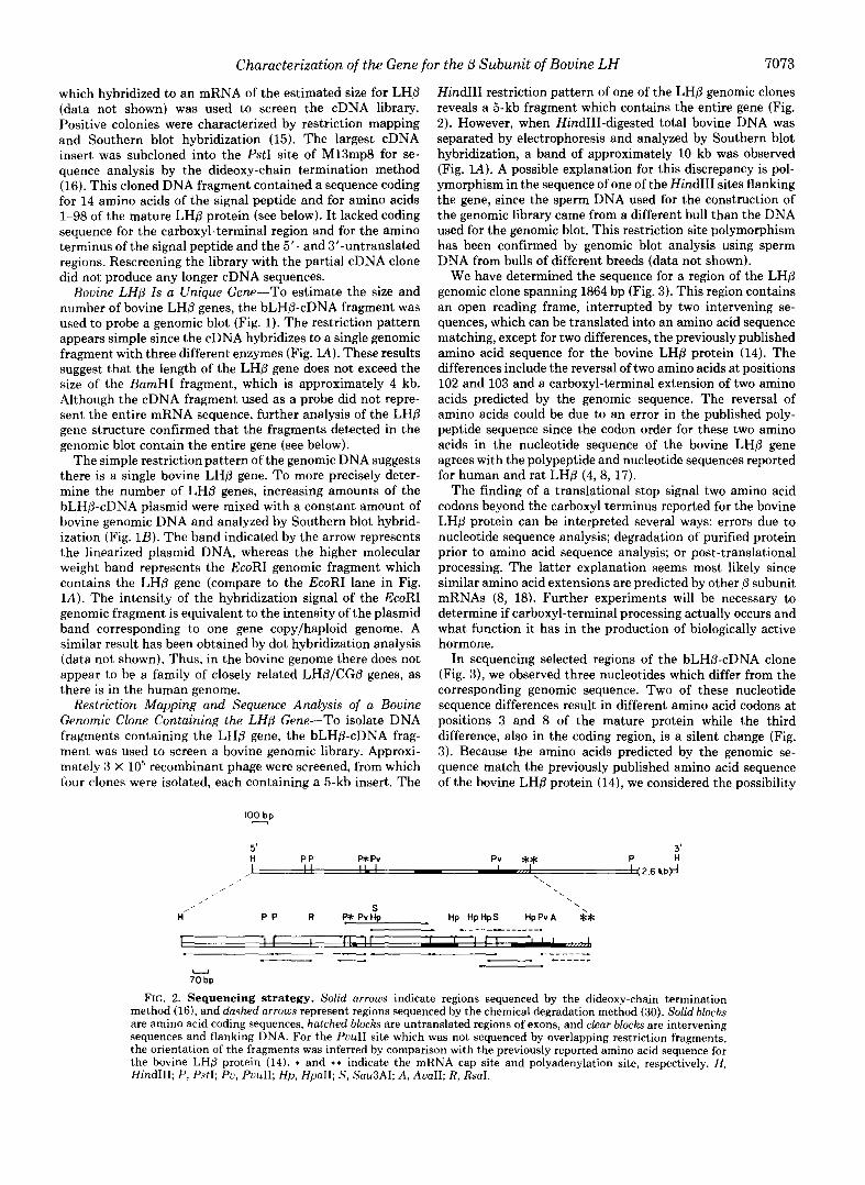
Restriction Mapping and Sequence Analysis of a Bovine Genomic Clone Containing the LHP Gene-To isolate DNA fragments containing the LHP gene, the bLHP-cDNA frag- ment was used to screen a bovine genomic library. Approxi- mately 3 X lo5 recombinant phage were screened, from which four clones were isolated, each containing a 5-kb insert. The
I O E p
5' H PP p* Pv
HindIII restriction pattern of one of the LHP genomic clones reveals a 5-kb fragment which contains the entire gene (Fig. 2). However, when HindIII-digested total bovine DNA was separated by electrophoresis and analyzed by Southern blot hybridization, a band of approximately 10 kb was observed (Fig. lA). A possible explanation for this discrepancy is pol- ymorphism in the sequence of one of the HindIII sites flanking the gene, since the sperm DNA used for the construction of the genomic library came from a different bull than the DNA used for the genomic blot. This restriction site polymorphism has been confirmed by genomic blot analysis using sperm DNA from bulls of different breeds (data not shown).
We have determined the sequence for a region of the LHP genomic clone spanning 1864 bp (Fig. 3). This region contains an open reading frame, interrupted by two intervening se- quences, which can be translated into an amino acid sequence matching, except for two differences, the previously published amino acid sequence for the bovine LHP protein (14). The differences include the reversal of two amino acids a t positions 102 and 103 and a carboxyl-terminal extension of two amino acids predicted by the genomic sequence. The reversal of amino acids could be due to an error in the published poly- peptide sequence since the codon order for these two amino acids in the nucleotide sequence of the bovine LHP gene agrees with the polypeptide and nucleotide sequences reported for human and rat LHP (4,8, 17).
The finding of a translational stop signal two amino acid codons beyond the carboxyl terminus reported for the bovine LHP protein can be interpreted several ways: errors due to nucleotide sequence analysis; degradation of purified protein prior to amino acid sequence analysis; or post-translational processing. The latter explanation seems most likely since similar amino acid extensions are predicted by other p subunit mRNAs (8, 18). Further experiments will be necessary to determine if carboxyl-terminal processing actually occurs and what function it has in the production of biologically active hormone.
In sequencing selected regions of the bLHp-cDNA clone (Fig. 3), we observed three nucleotides which differ from the corresponding genomic sequence. Two of these nucleotide sequence differences result in different amino acid codons at positions 3 and 8 of the mature protein while the third difference, also in the coding region, is a silent change (Fig. 3). Because the amino acids predicted by the genomic se- quence match the previously published amino acid sequence of the bovine LHP protein (14), we considered the possibility
Pv ** P H 3'
. . I 1 1 1 1 I I - , , ! (2.6 hb# , , \ ,
\ \ ,
H S
P P R FWPvHp Hp HpHpS HpPvA $* I I I r~ I I r I I ~ I 1 1 1
- "" - """. 1 - I . , 4 - - - - - " - """ "
70 bp U
FIG. 2. Sequencing strategy. Solid arrows indicate regions sequenced by the dideoxy-chain termination method (161, and dashed arrows represent regions sequenced by the chemical degradation method (30). Solid blocks are amino acid coding sequences, hatched blocks are untranslated regions of exons, and clear block are intervening sequences and flanking DNA. For the PuuII site which was not sequenced by overlapping restriction fragments, the orientation of the fragments was inferred by comparison with the previously reported amino acid sequence for the bovine LHP protein (14). * and * * indicate the mRNA cap site and polyadenylation site, respectively. H, HindIII; P, PstI; Pu, PuuII; Hp, HpaII; S, Sau3AI; A , AuaII; R, Rsal.
+€a 40 400 Gly Val Asp Pro M Val Ser Fhe Pro Val Ala Leu Ser Cqs Hs Cqs Gly Pro Cqs Arg Leu Ser Ser Thr kp Cys Gly Gly Pro Arg GGA GTG GAc ATG GTC TCC l l C CCC GTG GCC CTC AGC TGT UX TGT GGA CCC TCC Cac CTC A(€ Aa: ACT c4c TIX GCG G T CCC A(T\ "- "_ "- "- " "_ "_ "_ "- "- "t
410 420 Thr Gln R.0 Leu Ala Cqs Asp His Pro Pro Leu Pro kp ne Leu F%e Leu sc Acc CAA CCC l l G Gcc TGT c4c CPC CCC GCG CTC CIA W ATC CTC l l C CTC TAA GGAmCCCCAClTWTCCCATGCCCATCCTAPCTCTGG
n IUUWTGCPI;T COgggg-3'
FIG. 3. Nucleotide sequence of the bovine LHB gene. Flanking regions and intervening sequences are shown in lower case letters. The mRNA sequence is in capital letters. The predicted amino acids are numbered beginning with -20 a t methionine, the amino terminus of the signal peptide. The first amino acid of the mature protein is +l. The margins of the bLHP-cDNA clone are indicated by arrows below the genomic sequence. The regions of the cDNA which have been sequenced are also indicated the nucleotides and predicted amino acids which differ in the cDNA sequence are shown below the genomic sequence; dashes represent nucleotides which are the same in the cDNA and genomic sequences. The boxed arrows above the genomic sequence at the 5' and 3' ends indicate the mRNA cap site and polyadenylation site, respectively. The eukaryotic consensus TATAA (25) and AATAAA (20) sequences are shown in boxes.
Characterization of the Gene for the p Subuni t of Bovine LH 7075
I -400 I -360 -320 CCGCTG-------GAGCAGGGTTGAGGCTGCAGCCAATCACCATC~TTGT~GAGGG~GGTGCTGCAGCCTCTGCCffiGTCCCCTCATGATAGGTAGAGCGGCGTTCACAAGGC . . . . . . . . . . . . . . . . . . . . . . . . . . . . . . . . . . . . . . . . . . . . . . . . . . . . . . . . . . . . . . . . . . . . . . . . . . . . . . . . . . . . . . . . . . . . . . . . . . . . . A A A ( ; C C T C A A G T A G A G G A G G G r r G A G G C T T C A A T C C A G C - - - - - -
FIG. 6. Comparison of the 5’-flanking regions and cap sites of the bovine LHB gene and human CGB genes. The sequence comparison program of Wilbur and Lipman (24) was used for alignment to achieve maximum nucleotide sequence homology between the 5”flanking and 5’-untranslated regions of the bovine LHP gene and one of the human CG@ genes (CG5) previously reported by Talmadge et al. (8). Regions of homology near the cap sites (+I) are shown in bores. The consensus eukaryotic TATA sequence (25) in underlined.
that the differences in the cDNA sequence are the result of sequencing artifacts. However, since the sequencing ladder for the cDNA is unambiguous in these regions, it is more likely that these nucleotide differences are the result of allelic polymorphism in the bovine LHP gene.
Exon and Intron Mapping-The location of the first exon was determined initially by blot hybridization of pituitary RNA. The probes were synthesized from M13 subclones con- taining each strand of the 380-bp HindIII-PstI, and 320-bp PstI-PstI fragments at the 5’ end of the genomic clone, and one strand of the 100-bp PstI-PuuII fragment. RNA tran- scripts were detected with only one probe, from the 100-bp PstI-PuuII fragment (data not shown). The small size of the first exon, and the lack of recognition sequences for restriction endonucleases, precluded determination of the 5’ end of the LHP gene by mapping with S1 nuclease (19). Thus, we deter- mined the precise location of the mRNA cap site by extension of a bLHP-cDNA probe hybridized to pituitary RNA (Fig. 4). The longest primer-extended product (arrou, lane 1 of Fig. 4B) is 418-419 nucleotides, giving a length of 109-114 nucleo- tides beyond the 3‘ end of the primer (Fig. 4A). This places the mRNA cap site only 6-11 nucleotides 5’ to the AUG initiation codon (Fig. 3).
The mRNA polyadenylation site was identified by an S1 nuclease-protection assay (19), using as a probe a 600-bp
restriction fragment spanning the 3‘ end of the gene (Fig. 5). The length of the protected DNA (lane S I , Fig. 5) is 154-157 nucleotides, which places the mRNA polyadenylation site 15- 18 nucleotides 3‘ to the consensus AAUAAA polyadenylation signal (20).
The beginning of the coding region, the 3‘ end of the first exon, and the 5’ end of the second exon are inferred by homology with the human LHP gene (8), since the available bLHP-cDNA sequence does not extend to the 5’ end of the second exon (Fig. 3). The other exon-intron junctions are inferred by alignment with the published amino acid sequence for the bovine LHP protein (14). Also, the sequences at these exon-intron junctions closely match the consensus eukaryotic splice junction sequences (21).
Promoter Selection for the Gonadotropin P Subunit Genes May Be Tissue-specific-While short 3-20 nucleotide 5’-un- translated regions have been reported for other eukaryotic genes ( 2 2 ) , this is an unexpected finding for the bovine LHP gene since the closely related human CGP genes have 5’- untranslated regions in excess of 350 nucleotides (8, 23). Further comparison of the bovine LHP gene with the human LHPICGP gene family, by alignment to achieve maximum nucleotide sequence homology (24), reveals greater than 70% homology throughout most of their lengths (data not shown). Also, the positioning of the exon-intron junctions in their
7076 Characterization of the Gene for the 0 Subuni t of Bovine LH
respective mRNA sequences have been exactly conserved. Thus, the only major difference between the bLHP and hCGP genes is in the lengths of their transcribed regions.
The reason for the different transcription start sites in the two species is not readily apparent upon comparison of the nucleotide sequences near the start sites (Fig. 6). There is a 172-bp region of 76% homology (large box in Fig. 6) immedi- ately 5’ to the ATG initiation codon, which contains a con- sensus “TATA” sequence (underlined). The position of the TATA sequence in the bovine LHP gene 5’ to the mRNA cap site is consistent with its position in other eukaryotic genes transcribed by RNA polymerase I1 and with its function in the initiation of transcription (25). In contrast, the homolo- gous TATA sequence in the hCGP gene is located in the 5’- untranslated region. Since the region around the TATA se- quence is so highly conserved between the two species, it is surprising that the bLHp and hCGp genes have different transcription start sites.
Further comparison of the 5”flanking regions around the hCGP cap site reveals a 27-bp region of 81% homology (small box in Fig. 6). This region, which contains 23 nucleotides 5’ to the hCGp cap site, does not contain a consensus TATA sequence. The closest match to the consensus TATA sequence in the 5’-flanking region of the hCG@ genes is the sequence TCAAGTA (26), which occurs immediately 5’ to the 27-bp homologous region. Interestingly, this sequence does not ap- pear at the corresponding position in the bovine LHP gene. Several other viral and cellular genes lack TATA sequences near the mRNA cap site (27,28), indicating that this sequence is not absolutely required for transcription initiation by RNA polymerase 11. Thus, other eukaryotic promoter elements which direct initiation of transcription of the hCGP genes in the human placenta are either not present in the bovine LHP gene or inactive in the bovine pituitary.
The above results suggest that promoter recognition for the bLHP and hCGP genes may be species-specific or tissue- specific. Although tissue-specific selection of alternate pro- moters has been reported for the mouse a-amylase gene in the parotid gland and liver (29), both cap sites are fireceded by a TATA sequence. Thus, it will be of interest to more fully define the non-TATA promoter of the gonadotropin subunit genes through mutagenesis of LHP and hCGP genes, followed by transfection into placental cell lines which synthesize gonadotropins.
Acknowledgments-We would like to thank Marian Nejedlik, Rob- ert Umek, Cornelius Boerkoel, Wessel Dirksen, and Dr. Ray Goodwin for their assistance in various phases of this project. In addition, we wish to thank Dr. Dennis Luck for providing bovine sperm DNA and for his assistance with the genomic blot, and Drs. Frank Martin and Miguel Castro for synthesis of the oligodeoxynucleotides.
Addendum-After this manuscript was submitted for publication, a report appeared by Jameson et al. (J . Biol. Chem. 259,15474-15480) describing the characterization of the rat LHP gene. Like the bovine LHB gene, it is present as a single copy and has a 5”untranslated region of only 7 nucleotides. In addition, the bovine and rat LHP genes share 83% nucleotide sequence homology in their coding regions and 73% homology in the proximal 150-bp of 5”flanking sequence. Beyond 150 bp into the 5”flanking region, the homology drops to less than 30%. Thus, the proximal 5”flanking region of the gonado- tropin 0 subunit genes is highly conserved across three species, suggesting a possible function in the regulation of gene expression.
REFERENCES
1. Catt, K. J., and Pierce, J . G. (1978) in Reproduction Endocrinology (Yen, S. S. C., and Jaffe, R. B., ed) pp. 34-62, Saunders, Philadelphia
2.
3.
4.
5.
6.
7.
8.
9. 10.
11.
12.
13.
14.
15. 16.
17.
18.
19. 20.
21. 22. 23.
24.
25.
26.
27.
28.
29.
30.
31.
32.
33.
34.
35. 36.
37.
38.
39.
40.
Canfield, R. E., Birken, S., Morse, J . H., and Morgan, F. J . (1976) in Peptide Hormones (Parsons, J. A., ed) pp. 299-315, Univer- sity Park Press, Baltimore
Ward, D. N., Moore, W. T., Jr., and Burleigh, B. D. (1982) J , Protein Chem. 1, 263-280
Pierce, J . G., and Parsons, T. F. (1981) Annu. Reu. Biochem. 5 0 ,
Fiddes, J. C., and Goodman, H. M. (1981) J. Mol. Appl. Genet. 1 ,
Goodwin, R. G., Moncman, C. L., Rottman, F. M., and Nilson, J .
Boorstein, W. R., Vamvakopoulos, N. C., and Fiddes, J . C. (1982)
Talmadge, K., Vamvakopoulos, N. C., and Fiddes, J . C. (1984)
Hussa, R. 0. (1981) Ligand Review 3 , Suppl. 2,5-43 Nilson, J. H., Nejedlik, M. T., Virgin, J. B., Crowder, M. E., and
Nett, T. M. (1983) J. Biol. Chem. 258,12087-12090 Bordelon-Riser, M. E., Siciliano, M. J., and Kohler, P. 0. (1979)
Somatic Cell Genet. 5 , 597-613 Hardin, J. W., Riser, M. E., Trent, J. M., and Kohler, P. 0.
(1983) Proc. Natl. Acad. Sci. U. S. A. 80, 6282-6285 Nilson, J. H., Thomason, A. R., Cserbak, M. T., Moncman, C.
L., and Woychik, R. P. (1983) J. Biol. Chem. 258,4679-4682
465-495
3-18
H. (1983) Nucleic Acids Res. 11,6873-6882
Nature 300,419-422
Nature 3 0 7 , 37-40
Maghuin-Rogister, G., and Hennen, G. (1973) Eur. J. Biochem. 39,235-253
Southern, E. M. (1975) J. Mol. Biol. 9 8 , 503-517 Sanger, F., Nicklen, S., and Coulson, A. R. (1977) Proc. Natl.
Acad. Sci. U. S. A. 7 4 , 5463-5467 Chin, W. W., Godine, J. E., Klein, D. R., Chang, A. S., Tan, L.
K., and Habener, J. F. (1983) Proc. Natl. Acad. Sci. U. S. A. 80,4649-4653
Maurer, R. A., Croyle, M. L., and Donelson, J. E. (1984) J. Biol. Chem. 259,5024-5027
Berk, A. J., and Sharp, P. A. (1977) Cell 1 2 , 721-732 Proudfoot, N. J., and Brownlee, G. G. (1976) Nature 2 6 3 , 211-
Mount, S. M. (1982) Nucleic Acids Res. 10,459-472 Kozak, M. (1984) Nucleic Acids Res. 12,857-872 Policastro, P., Ovitt, C. E., Hoshina, M., Fukuoka, H., Boothby,
M. R., and Boime, I. (1983) J. Biol. Chem. 2 5 8 , 11492-11499 Wilbur, W. S., and Lipman, D. J . (1983) Proc. Natl. Acad. Sci. U.
Breathnach, R., and Chambon, P. (1981) Annu. Reu. Biochem.
Fiddes, J . C., and Talmadge, K. T. (1984) Recent Prog. Horm.
Baker, C. C., Herisse, J., Courtois, G., Galibert, F., and Ziff, E.
Lee, M.G-S., Lewis, S. A., Wilde, C. D., and Cowan, N. J . (1983)
Schibler, U., Hagenbiichle, O., Wellauer, P. K., and Pittet, A. C.
Maxam, A. M., and Gilbert, W. (1980) Methods Enzymol. 65,
Nilson, J. H., Thomason, A. R., Horowitz, S., Sasavage, N. L., Blenis, J., Albers, R., Saker, W., and Rottman, F. M. (1980) Nucleic Acids Res. 8, 1561-1573
Sasavage, N. L., Nilson, J . H., Horowitz, S., and Rottman, F. M. (1982) J. Biol. Chem. 2 5 7 , 678-681
Caruthers, M. H. (1981) in Recombinant DNA, Proceedings of the
ed) pp. 261-272, Elsevier/North-Holland, Amsterdam Third Cleveland Symposium on Macromolecules (Walton, A. G.,
Murray, N. E., Brammar, W. J., and Murray, K. (1977) Mol. Gen.
Benton, W. D., and Davis, R. W. (1977) Science 1 9 6 , 180-182 Genetics 1 5 0 , 53-61
Maniatis, T., Fritsch, E. F., and Sambrook, J. (1982) Molecular Cloning, A Laboratory Manual, Cold Spring Harbor Laboratory, Cold Spring Harbor, New York
Rigby, P. W. J., Dieckmann, M., Rhodes, C., and Berg, P. (1977)
Camper, S. A,, Luck, D. N., Yao, Y., Woychik, R. P., Goodwin, R. G., Lyons, R. H., Jr., and Rottman, F. M. (1984) DNA 3,
Grainger, R. M., Hazard-Leonards, R. M., Samaha, F., Hougan, L. M., Lesk, M. R., and Thomsen, G. H. (1983) Nature 3 0 6 , 88-91
Characterization of the Gene for the p Subunit of Bovine LH 7077
The Gene f o r t he 8 Submit of Bovine Luteinizing Hormone Encodes a Gonadotropin mRNA w t h an Unusually Short 5'-Untranslated Region
Je f f rey B. Virgin. Bernard J. Silver, Arlen R . Thanason. and John H. Nl lson
METHODS
b o v i n e p i t u i t a r y cDNA l i b r a r y h a s b e e n d e s c r i b e d e lsewhere (31, 32) . ~_ Cons t ruc t i on and sc reen inq o f t h e cDNA l i b r a r y . The p r e p a r a t i o n o f a
Ol igodeoxynucleot ide probes, 14 n u c l e o t i d e s i n l e n g t h . were s y n t h e s i z e d b y a s o l i d phase. phosphi te chemical method (33) . and end- label led a t the i r 5 ' - t e r m i n i w i t h T4 p o l y n u c l e o t i d e k i n a s e and [ -3zPlATP fo r zc reen lng t he cDNA l i b r a r y (13). A p a r t i a l bLHn-cDNA c lone was i s o l a t e d ( s e e " R e s u l t s and 01scussion") and used to screen a bovine genomic l i b ra ry .
supp l i ed by Or. Paul L. Jackson, Los Alamos Nat ional Laboratory . It was S c r e e n i n q a g e n o m i c l i b r a r y . A b o v i n e g e n o m i c l i b r a r y was generously
p r e p a r e d b y l i g a t i n g b o v i n e sperm DNA, d i g e s t e d t o c o m p l e t i o n w i t h H i n d I l l , i n t o t h e H i n d I l l s i t e o f A-590 f34l. The l i b r a r y was screened f35. 361 w i t h a probe prepared by n ick - t rans la t ing (37) t h e p u r i f i k Pst I i nse r t 'fr& the bLHn- cDNA plasmid with Cm-32PldCTP (3000 CiInmal. hersham) to obtain an approximate spec i f i c ac t i v i t y o f 108 cpmfug.
Genomic B l o t . The prepara t ion o f bov ine sperm DNA has been descr ibed e l s e w h e i e B F A Southern b l o t (15) of bovine genomic DNA was prepared. and probed w i t h n i c k - t r a n s l a t e d i n s e r t from the bLHn-cDNA plasmid. Hybr id izat ion and wash condi t ions have been previously described (6). To reduce non-specific b i n d i n g o f r a d i o a c t i v e p r o b e t o t h e f i l t e r . t h e p r o b e i n t h e h y b r i d i z a t i o n so lu t i on was pre-incubated w i th a b lank p iece o f Gene Screen (39).
and tEche:ical de$adation method o f Maxam and G i lbe r t (30) were ~3% DNi sequence analysis. To p repare s ing le -s t randed temp la tes f o r t he cha in t e r m i n a t i o n r e a c t i o n s , r e s t r i c t i o n f r a g m e n t s were subcloned i n t o M13mp8 and H13mp9 (40) . For sequence analysis by the chemical degradat ion method, r e s t r i c t i o n f r a g m e n t s w e r e 3 ' e n d - l a b e l l e d b y d i g e s t i n g a n d t h e n r e p a i r i n g r e s t r i c t i o n s i t e s w i t h 5 ' - p r o t r u d i n g ends u s l n g t h e l a r g e f r a g m e n t o f DNA po l ymerase (Boehr inge r Mannhe im) and o n e o r more o f t h e a p p r o p r i a t e deoxynucleoside Co-32Pltriphosphates.
DNA Se uenc in . Both t he cha in t e rm ina t ion me thod o f Sanger e t a l ( 1 6 )
the 5 - i n m LHn nRNL TO i n i t i a te syn thes i s of the cDNA primer. 5 ug Primer extension. A single-stranded cDNA pr imer was prepared f o r napping
o f message-sense M13:bLHn-template DNA were hybr id ized wi th 2.4 ng MI3 s ingle- strand primer (17 nucleotides, PL Biochemicals) i n 22 e l o f 0.09 M N a C I , 0.013 M Tris-HCI (pH 7.5). 0.013 M " J C I p The hybr id i za t i on m ix tu re was heated a t 65OC f o r 5 m i n . f o l l o w e d b y i n c u b a t i o n a t 22OC for 20 min. For the s t h e s i s r e a c t i o n . 2 u n i t s o f t h e l a r g e f r a g m e n t o f DNA polymerase. 40 uCi [ D - ~ ~ P I ~ C T P (800 Cifmmol, hersham). and deoxynucleoside t r iphosphates were added t o t h e hybr id iza t ion mix tu re , vh ich was d i l u t e d t o 40 ul t o ob ta in t he f o l l ow ing f i na l concen t ra t i ons : 0.125 mM each dATP. dGTP, and dTTP. 0.05 M NaCl. 0.007 M T r i s - HCI (pH 7.5). 0.007 M MgC12. A f t e r i n c u b a t i o n a t 2 2 O C fo r 30 min . dCTP was
ano the r 30 min. The double-stranded product was d iges ted w i th Bg l I , then added t o a f i n a l c o n c e n t r a t i o n o f 0.125 m M , and t h e i n c u b a t i o n c o n t i n u e d f o r
p r e c i p i t a t e d w i t h e t h a n o l . The DNA was denatured by resuspending i n 0.05 M NaOH. 0.01 M EDTA. 10% glycerol , and 0.1 % brmocresol green. and incuba t ing a t 950C fo r 5 min. The strands were separated by e lec tmphores is in a 1.5% agamse g e l i n 1X TEE. and the s ing le -s t randed p robe was e l u t e d f r o m t h e g e l b y electrophoresis (36). The probe was fu r the r pu r i f i ed by e lec tmphores i s i n a 6% denaturing wlyacrylamide gel (30).
TO map the 5' end of the LHn mRN4. 5 X IO5 cpn o f t h e Ml3:bLHn-cDliA pr imer w e r e h y b r i d i z e d t o 2.2 ug s t e e r p i t u i t a r y p o l y ( A ) - c o n t a i n i n g RNA i n 10 u1 50%
s u l f a t e f o r 2 h a t 45% The hybr id iza t ion reac t ion was PreciDi tated i n ethanol formamide, 0.01 M Pipes (pH 6.4). 0.4 M NaCl, 0.001 M EDTA, 0.11 sodium dodecyl
and resuspended i n b u f f e r as described (31). except t h a t . i t contained 1 mM each dATP, dGTP. dCTP, dTTP. and 480 U f m l AMY reve rse t ransc r ip tase (L i f e Sc iences Inc., S t . Petersburg. FL). The 25 u l r e a c t i o n was i n c u b a t e d f o r 1 h a t 37%
gel (30). The products were analyzed by electrophoresis i n a 6% denaturing polyacrylamide
denatui-ed b y b o i l i n g f o r 3 m i n i n t h e sane hyb r id i za t i on bu f fe r desc r ibed f o r SI Mapping. A double-s t randed. 3 ' end- label led probe was prepared. and
pr imer ex tens ion , except tha t i t contained 80% formamide Ten ug o f s t e e r pi tu i tary poly (A)-containing RNA were hybr id ized wi th 5 X I & cpn o f probe i n
h y b r i d i z a t i o n , 100 u l o f b u f f e r c o n t a i n i n g 0.05 M potass ium ace ta te (pH 4.5), 10 u l o f t h e h y b r i d i z a t i o n b u f f e r f o r 3 h a t 600C. A t t h e end o f t h e
0.3 M NaCl, 0.001 M ZnSO4, 25 ugfml denatured salmon sperm DNA. and 1000 u n i t s O f 51 nuclease ( A s p e r q i l l u s m, Boehringer Mannheim) were added t o h y b r i d i z a t i o n b u f f e r c o n t a i n i n g e i t h e r t h e p r o b e a l o n e or the p robe p lus RNA. Incubation was fo r 1 h a t 37OC. The protected probe was analyzed as described for the Drimer-extended Droducts.
A. B E H
23- - 9.4 - 6.6-
4.3-
*r
a
2.3- 2.0 -
B. I 2 5 1020
c
A.
3'
B. M lCTAG2
3- 1353 0
1078 - 872 0
803 0 -
1 Bg'l 3' 5' " """"""_
Bgl I Bgl I + -
M13mp8 clone containing the m e s s a g e - s e n s m t r a n d o f t h e bLHn cDNA (boxed Fig. 4. 5'-end mapping by primer extension. A. Pr imer syn thes is : an
reg ion ) was used a s a t e m p l a t e f o r s y n t h e s i s o f t h e bLH6-cDNA p r i m e r ( s o l i d arrow). The condi t ions o f the react ion permi t ted spthes is o f the complete MI3
con ta ins 251 n u c l e o t i d e s o f t h e bLHn-cDNA coding region sequence, p lus an (-) s t r a n d (dashed arrow). The pr imer . p roduced by d iges t ion w i th Bg l I,
a d d i t i o n a l 54-5 n u c l e o t i d e s o f homopolymer G and M13 sequence. B. Pr imer
b y r e v e r s e t r a n s c r i p t a s e f r o m b o v i n e p i t u i t a r y p o l y ( A ) - c o n t a i n i n g RNA, extension: The 3gP-labelled Ml3:bLHe slngle-stranded cDNA pr imer was elongated
followed by electrophoresis and autoradiography. Lane 1: the products of the r e v e r s e t r a n s c r i p t a s e r e a c t i o n . Lane 2: t h e p u r i f i e d Ml3:bLHn-cDNA primer. Lane M: X-be I l l markers. Lanes C,T.A.G sequencing ladder from dideoxy-chain te rm ina t ion reac t i ons used as a s i z ing s tandard . S i zes a re i n bp. The a v o w indicates the longest primer-extended product.
I fragment was i s o l a t e d and 3' md- labe l l ed us ing t he l a rge f ragmen t o f DNA Fig. 5. )'-end mappinq by 3 nuclease. k Probe synthesis: an Ava 11-Pst
polymerase to repair the 5 ' -protruding end generated by d igest ion wi th Ava I I .
l a b e l l e d n u c l e o t i d e s (*) a t t h e 3' end. One-hal f o f the sample o f l a b e l l e d Both [m-3ZP]dATP and [m-32P]TTP were included i n t h e r e a c t i o n , l e a v i n g t w o
fragment was used f o r S mapping (19). w h i l e t h e o t h e r h a l f w a s sequenced b y chemica l degradat ion (40). B. 51 mapping: the 3 ' end- label led probe was h y b r i d i z e d t o s t e e r p i t u i t a r y p o l y ( A ) - c o n t a i n i n g RNA, f o l l o w e d b y d i g e s t i o n w i t h SI nuclease. The SI -p ro tec ted p roduc ts were e lec t rophoresed bes ide a sequencing ladder. produced by chemical degradation o f t he probe. to p rec ise ly de te rm ine t he l eng th o f t he p ro tec ted f ragmen ts . Lanes A>C, T t C , and C: p roduc ts o f the chemica l degradat ion sequenc ing reac t ions . Lane SI: SI-
nuclease i n t h e absence o f RNA. The sequence AAUAAA i s the mRNA canplement of p ro tec ted p roduc ts ( ind ica ted by the a r row) . Lane P: p robe d iges ted w i th 51
the cDNA sequence.
digested t o canplet ion w i th the designated enzyme, e lec t rophoresed through a Fig. 1. Bovine LHB genomic b lo t . A. Ten ug o f b o v i n e sperm DNA were
0.7% agarose gel, and t ransferred to Gene Screen (NEN) according to the protocol supp l i ed by t he manu fac tu re r . The b l o t was h y b r l d i z e d w i t h n i c k - t r a n s l a t e d i n s e r t frm the bLHn-cDNA plasmid. B=BamH I , E=EcoR I. H=Hind 111. Sires are i n d i c a t e d i n kbp. B. To est imate the number of LHn genes, bLHn-cONA plasmid DNA was added i n amounts equivalent to I . 2, 5 , IO. and 20 copies per haploid genome t o IO ug bovine sperm DNA p r i o r t o d i g e s t i o n w i t h EcaR I and b lo t hybr id iza t ion . The arrow indicates the band corresponding to the l inearized plasmid DNA.