119
C h a p t e r 9 The General and Special Senses PowerPoint® Lecture Slides prepared by Jason LaPres Lone Star College - North Harris Copyright © 2010 Pearson Education, Inc.
| Date post: | 20-Mar-2018 |
| Category: |
Documents |
| Upload: | vuongtuyen |
| View: | 221 times |
| Download: | 3 times |

Copyright © 2010 Pearson Education, Inc.
C h a p t e r
9
The General and Special Senses
PowerPoint® Lecture Slides
prepared by Jason LaPres
Lone Star College - North Harris
Copyright © 2010 Pearson Education, Inc.
Copyright © 2010 Pearson Education, Inc.
9-1 Sensory receptors connect
our internal and external
environments with the nervous
system
Copyright © 2010 Pearson Education, Inc.
Sensory Receptors
• Specialized cells that monitor specific conditions
in the body or external environment
• When stimulated, a receptor passes information
to the CNS in the form of action potentials along
the axon of a sensory neuron
Copyright © 2010 Pearson Education, Inc.
Sensory Receptors
• Sensation
– The arriving information from these senses
• Perception
– Conscious awareness of a sensation

Copyright © 2010 Pearson Education, Inc.
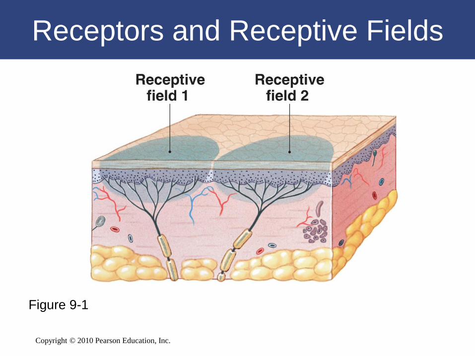
Sensory Receptors
• The Detection of Stimuli
– Receptor sensitivity:
• Each receptor has a characteristic sensitivity
– Receptive field:
• Area is monitored by a single receptor cell
• The larger the receptive field, the more difficult it is
to localize a stimulus
Copyright © 2010 Pearson Education, Inc.
Receptors and Receptive Fields
Figure 9-1
Copyright © 2010 Pearson Education, Inc.
Sensory Receptors
• The Interpretation of Sensory Information
– Arriving stimulus:
• Takes many forms:
– physical force (such as pressure)
– dissolved chemical
– sound
– light

Copyright © 2010 Pearson Education, Inc.
Sensory Receptors
• The Interpretation of Sensory Information
– Sensations:
• Taste, hearing, equilibrium, and vision provided by
specialized receptor cells
• Communicate with sensory neurons across
chemical synapses
Copyright © 2010 Pearson Education, Inc.
Sensory Receptors
• Adaptation
– Reduction in sensitivity of a constant stimulus
– Your nervous system quickly adapts to stimuli
that are painless and constant

Copyright © 2010 Pearson Education, Inc.
Sensory Receptors
• General Senses
– Describe our sensitivity to:
• Temperature
• Pain
• Touch
• Pressure
• Vibration
• Proprioception
Copyright © 2010 Pearson Education, Inc.
Sensory Receptors
• Special Senses
– Olfaction (smell)
– Vision (sight)
– Gustation (taste)
– Equilibrium (balance)
– Hearing
Copyright © 2010 Pearson Education, Inc.
Sensory Receptors
• Stimulation of a receptor produces action potentials
along the axon of a sensory neuron
• The frequency and pattern of action potentials
contain information about the strength, duration, and
variation of the stimulus
• Your perception of the nature of that stimulus
depends on the path it takes inside the CNS
Copyright © 2010 Pearson Education, Inc.
9-2 General sensory receptors
can be classified by the type
of stimulus that excites them
Copyright © 2010 Pearson Education, Inc.
Classifying Sensory Receptors
• General sensory receptors are divided into
four types by the nature of the stimulus that
excites them
– Nociceptors (pain)
– Thermoreceptors (temperature)
– Mechanoreceptors (physical distortion)
– Chemoreceptors (chemical concentration)
Copyright © 2010 Pearson Education, Inc.
Pain
• Nociceptors (also called pain receptors)
– Are common in the superficial portions of the
skin, joint capsules, within the periostea of
bones, and around the walls of blood vessels
– May be sensitive to temperature extremes,
mechanical damage, and dissolved chemicals,
such as chemicals released by injured cells
Figure 15–2
Copyright © 2010 Pearson Education, Inc.
Pain
• Nociceptors
– Are free nerve endings with large receptive
fields:
• Branching tips of dendrites
• Not protected by accessory structures
• Can be stimulated by many different stimuli
• Two types of axons: Type A and Type C fibers

Copyright © 2010 Pearson Education, Inc.
Pain
• Nociceptors
– Myelinated Type A fibers:
• Carry sensations of fast pain, or prickling pain,
such as that caused by an injection or a deep cut
• Sensations reach the CNS quickly and often
trigger somatic reflexes
• Relayed to the primary sensory cortex and receive
conscious attention
Copyright © 2010 Pearson Education, Inc.
Pain
• Nociceptors
– Type C fibers:
• Carry sensations of slow pain, or burning and
aching pain
• Cause a generalized activation of the reticular
formation and thalamus
• You become aware of the pain but only have a
general idea of the area affected
Copyright © 2010 Pearson Education, Inc.
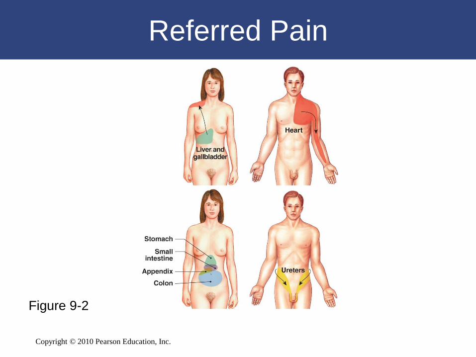
Referred Pain
Figure 9-2

Copyright © 2010 Pearson Education, Inc.
Temperature
• Thermoreceptors
– Also called temperature receptors
– Are free nerve endings located in:
• The dermis
• Skeletal muscles
• The liver
• The hypothalamus
Copyright © 2010 Pearson Education, Inc.
Temperature
• Thermoreceptors
– Temperature sensations:
• Conducted along the same pathways that carry
pain sensations
• Sent to:
– the reticular formation
– the thalamus
– the primary sensory cortex (to a lesser extent)

Copyright © 2010 Pearson Education, Inc.
Touch, Pressure, and Position
• Mechanoreceptors
– Sensitive to stimuli that distort their plasma
membranes
– Contain mechanically gated ion channels whose
gates open or close in response to
• Stretching
• Compression
• Twisting
• Other distortions of the membrane
Copyright © 2010 Pearson Education, Inc.
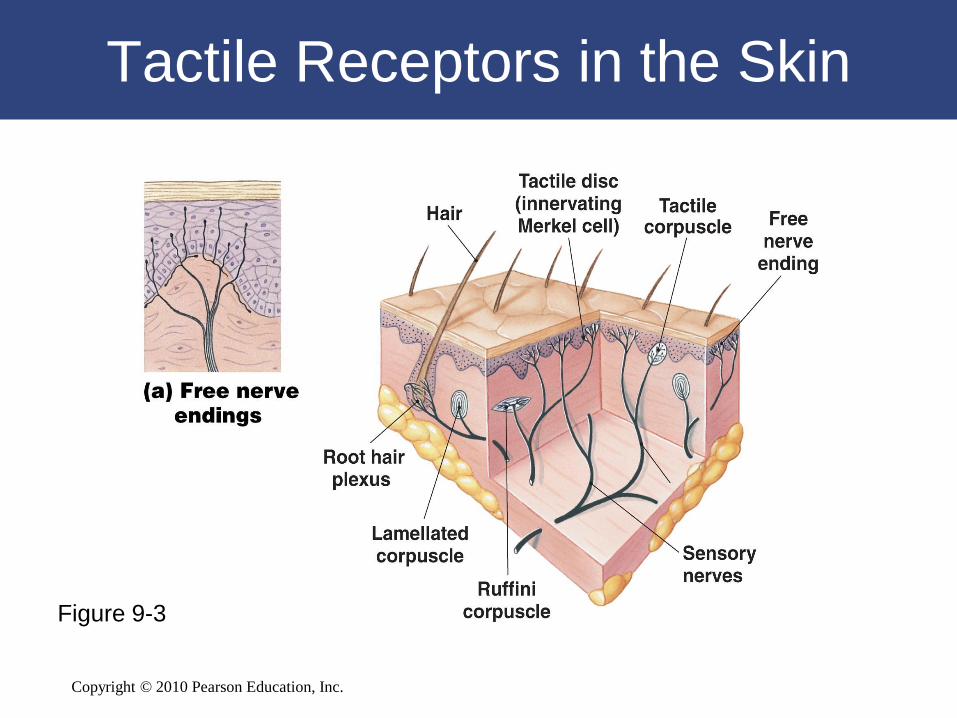
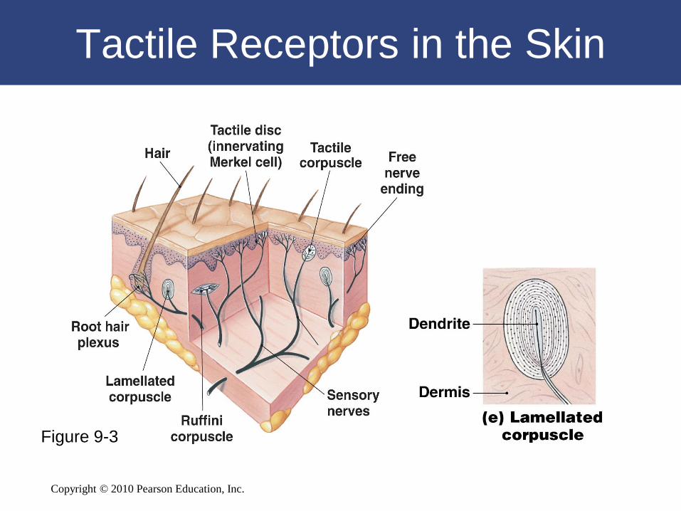
Touch, Pressure, and Position
• Tactile receptors
– Provide the sensations of touch,
pressure, and vibration:
• Touch sensations provide information
about shape or texture
• Pressure sensations indicate degree of
mechanical distortion
• Vibration sensations indicate pulsing or
oscillating pressure
Copyright © 2010 Pearson Education, Inc.
Tactile Receptors in the Skin
Figure 9-3

Copyright © 2010 Pearson Education, Inc.
Tactile Receptors in the Skin
Figure 9-3

Copyright © 2010 Pearson Education, Inc.
Tactile Receptors in the Skin
Figure 9-3

Copyright © 2010 Pearson Education, Inc.
Tactile Receptors in the Skin
Figure 9-3
Copyright © 2010 Pearson Education, Inc.
Tactile Receptors in the Skin
Figure 9-3

Copyright © 2010 Pearson Education, Inc.
Tactile Receptors in the Skin
Figure 9-3

Copyright © 2010 Pearson Education, Inc.
Touch, Pressure, and Position
• Baroreceptors
– Monitor change in pressure
– Consist of free nerve endings that branch
within elastic tissues in wall of distensible
organ (such as a blood vessel)
– Respond immediately to a change in
pressure, but adapt rapidly

Copyright © 2010 Pearson Education, Inc.
Baroreceptors
Figure 9-4

Copyright © 2010 Pearson Education, Inc.
Touch, Pressure, and Position
• Proprioceptors
– Monitor:
• Position of joints
• Tension in tendons and ligaments
• State of muscular contraction
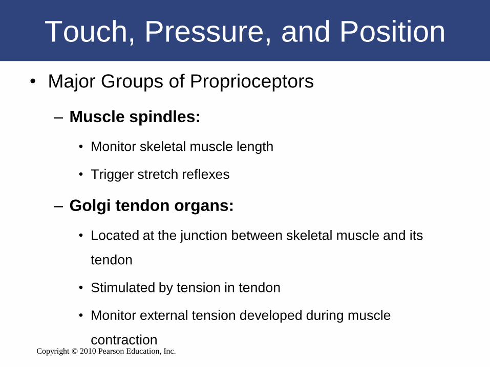
Copyright © 2010 Pearson Education, Inc.
Touch, Pressure, and Position
• Major Groups of Proprioceptors
– Muscle spindles:
• Monitor skeletal muscle length
• Trigger stretch reflexes
– Golgi tendon organs:
• Located at the junction between skeletal muscle and its
tendon
• Stimulated by tension in tendon
• Monitor external tension developed during muscle
contraction
Copyright © 2010 Pearson Education, Inc.
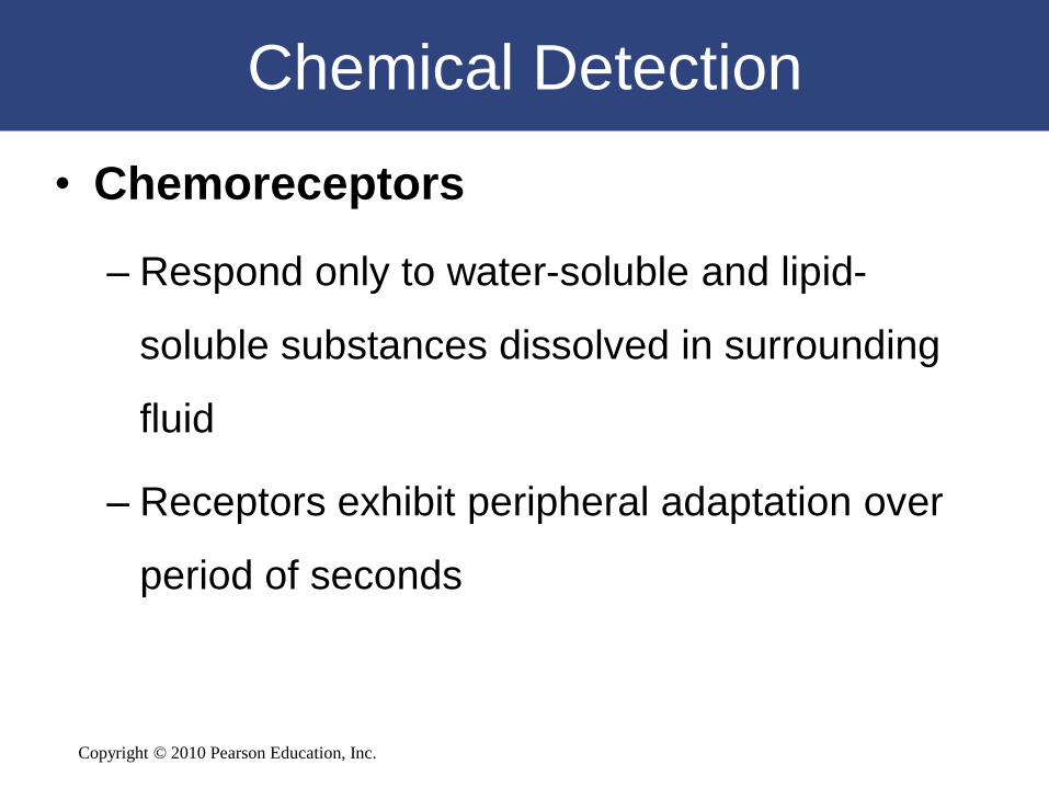
Chemical Detection
• Chemoreceptors
– Respond only to water-soluble and lipid-
soluble substances dissolved in surrounding
fluid
– Receptors exhibit peripheral adaptation over
period of seconds

Copyright © 2010 Pearson Education, Inc.
Classifying Sensory Receptors
• Chemoreceptors
– Located in the:
• Carotid bodies:
– near the origin of the internal carotid arteries on each side of
the neck
• Aortic bodies:
– between the major branches of the aortic arch
– Receptors monitor pH, carbon dioxide, and oxygen
levels in arterial blood

Copyright © 2010 Pearson Education, Inc.
Chemoreceptors
Figure 9-5
Copyright © 2010 Pearson Education, Inc.
9-3 Olfaction, the sense of smell,
involves olfactory receptors
responding to chemical stimuli
Copyright © 2010 Pearson Education, Inc. Figure 17–1a
Smell (Olfaction)
• Olfactory Organs
– Provide sense of smell
– Located in nasal cavity on either side of
nasal septum
– Made up of two layers:
• Olfactory epithelium
• Lamina propria

Copyright © 2010 Pearson Education, Inc.
The Olfactory Organs
Figure 9-6

Copyright © 2010 Pearson Education, Inc.
Smell (Olfaction)
• Olfactory Glands
– Secretions coat surfaces of olfactory organs
• Olfactory Receptors
– Highly modified neurons
– Olfactory reception:
• Involves detecting dissolved chemicals as they interact with
odorant-binding proteins
Copyright © 2010 Pearson Education, Inc.
Smell (Olfaction)
• Olfactory Pathways
– Axons leaving olfactory epithelium:
• Collect into 20 or more bundles
• Penetrate cribriform plate of ethmoid
• Reach olfactory bulbs of cerebrum where first synapse
occurs
• Axons leaving olfactory bulb:
– travel along olfactory tract to reach olfactory cortex,
hypothalamus, and portions of limbic system

Copyright © 2010 Pearson Education, Inc.
Smell (Olfaction)
• Olfactory Discrimination
– Can distinguish thousands of chemical stimuli
– CNS interprets smells by the pattern of receptor
activity
• Olfactory Receptor Population
– Considerable turnover
– Number of olfactory receptors declines with age
Copyright © 2010 Pearson Education, Inc.
9-4 Gustation, the sense of taste,
involves taste receptors
responding to chemical stimuli

Copyright © 2010 Pearson Education, Inc.
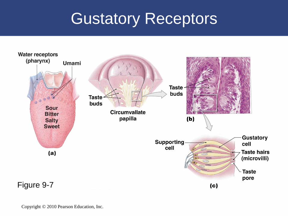
Taste (Gustation)
• Gustation provides information about the
foods and liquids consumed
• Taste receptors (or gustatory receptors)
are distributed on tongue and portions of
pharynx and larynx
– Clustered into taste buds
Copyright © 2010 Pearson Education, Inc.
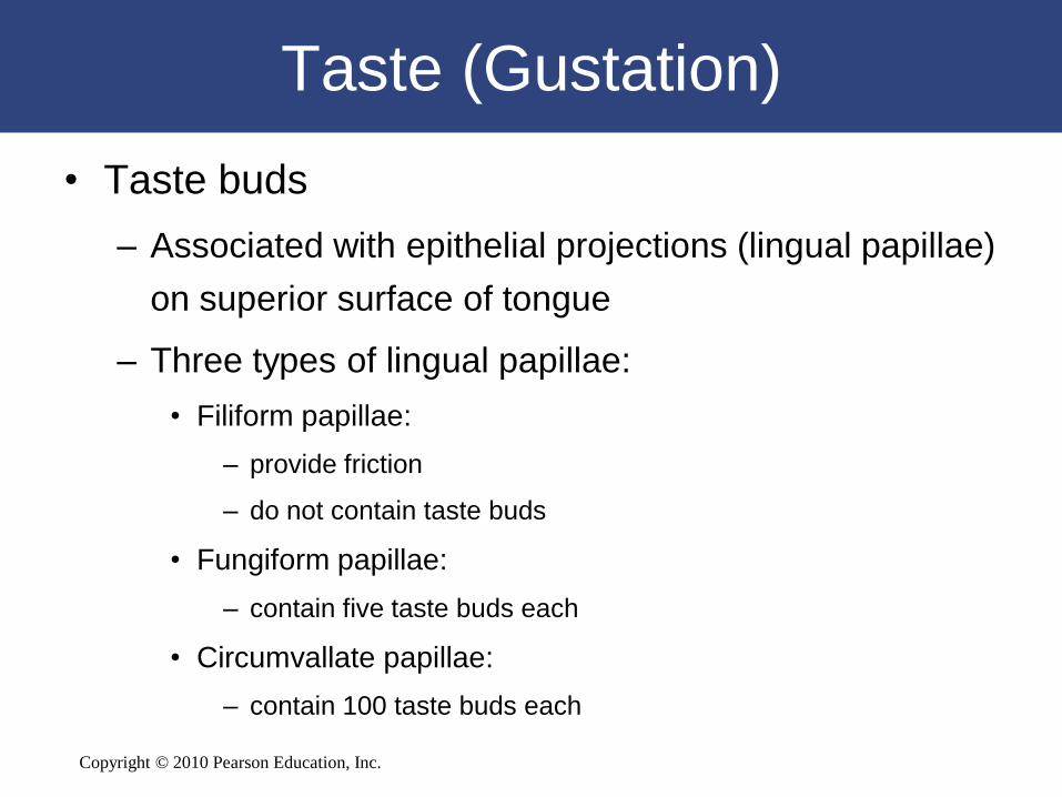
Taste (Gustation)
• Taste buds
– Associated with epithelial projections (lingual papillae)
on superior surface of tongue
– Three types of lingual papillae:
• Filiform papillae:
– provide friction
– do not contain taste buds
• Fungiform papillae:
– contain five taste buds each
• Circumvallate papillae:
– contain 100 taste buds each
Copyright © 2010 Pearson Education, Inc.
Gustatory Receptors
Figure 9-7
Copyright © 2010 Pearson Education, Inc.
Taste (Gustation)
• Gustatory Discrimination
– Primary taste sensations:
• Sweet
• Salty
• Sour
• Bitter

Copyright © 2010 Pearson Education, Inc.
Taste (Gustation)
• Additional human taste sensations
– Umami:
• Characteristic of beef/chicken broths and Parmesan cheese
• Receptors sensitive to amino acids, small peptides, and
nucleotides
– Water:
• Detected by water receptors in the pharynx
Copyright © 2010 Pearson Education, Inc.
Taste (Gustation)
• Gustatory Discrimination
– Dissolved chemicals contact taste hairs
– Bind to receptor proteins of gustatory cell
– Salt and sour receptors:
• Chemically gated ion channels
• Stimulation produces depolarization of cell
– Sweet, bitter, and umami stimuli:
• G proteins:
– gustducins

Copyright © 2010 Pearson Education, Inc.
9-5 Internal eye structures
contribute to vision, while
accessory eye structures provide
protection
Copyright © 2010 Pearson Education, Inc.
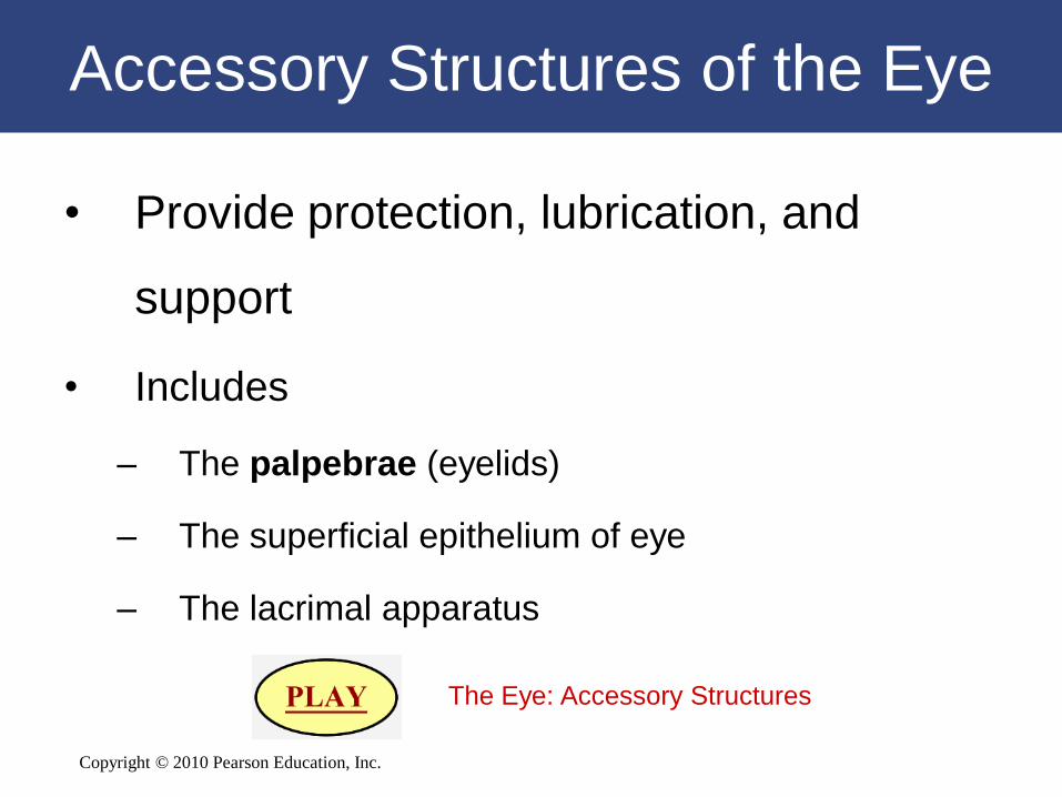
Accessory Structures of the Eye
• Provide protection, lubrication, and
support
• Includes
– The palpebrae (eyelids)
– The superficial epithelium of eye
– The lacrimal apparatus
The Eye: Accessory Structures

Copyright © 2010 Pearson Education, Inc.
Accessory Structures of the Eye
Figure 9-8a
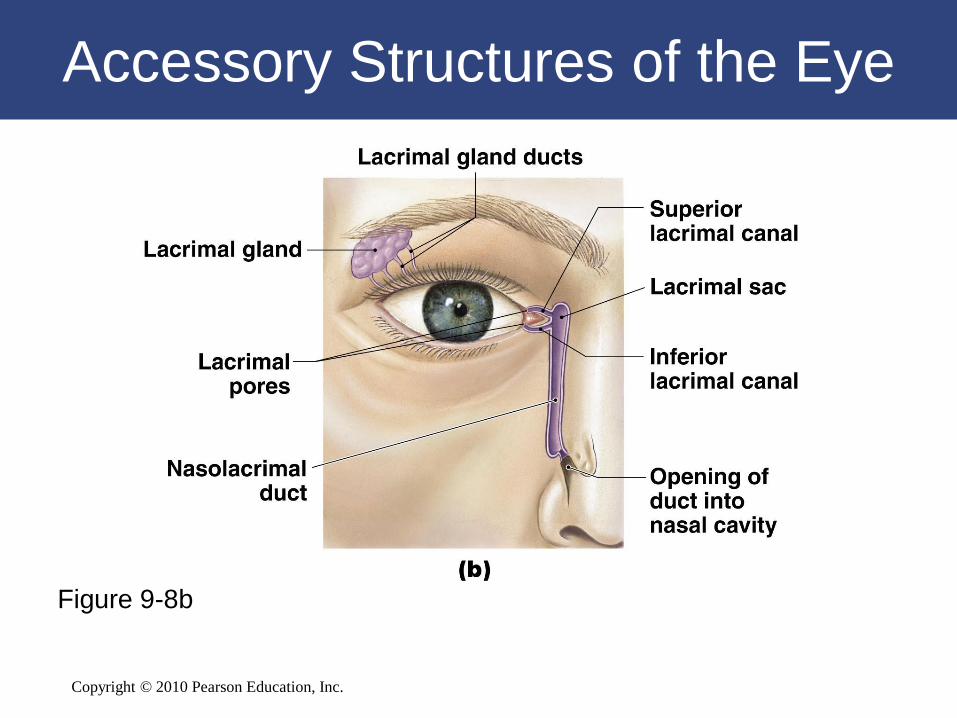
Copyright © 2010 Pearson Education, Inc.
Accessory Structures of the Eye
Figure 9-8b
Copyright © 2010 Pearson Education, Inc.
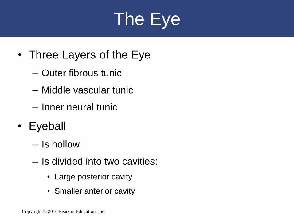
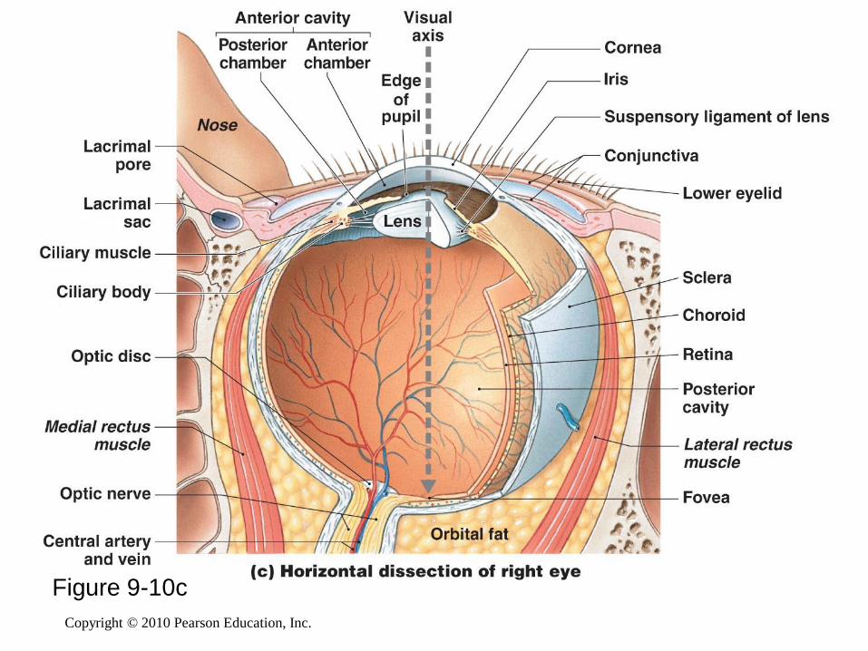
The Eye
• Three Layers of the Eye
– Outer fibrous tunic
– Middle vascular tunic
– Inner neural tunic
• Eyeball
– Is hollow
– Is divided into two cavities:
• Large posterior cavity
• Smaller anterior cavity

Copyright © 2010 Pearson Education, Inc.
The Extrinsic Eye Muscles
Figure 9-9

Copyright © 2010 Pearson Education, Inc.
Copyright © 2010 Pearson Education, Inc.
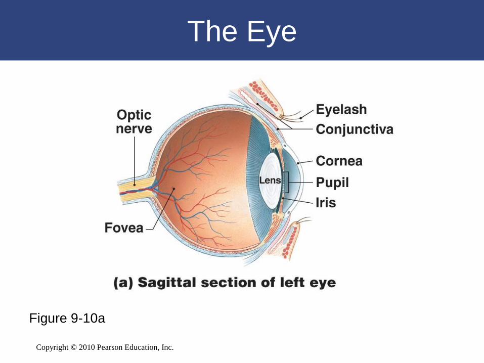
The Eye
Figure 9-10a
Copyright © 2010 Pearson Education, Inc.
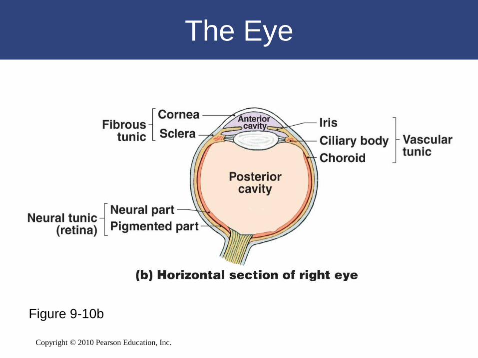
The Eye
Figure 9-10b
Copyright © 2010 Pearson Education, Inc.
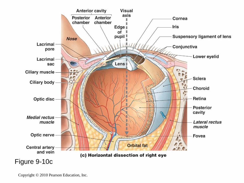
Figure 9-10c
Copyright © 2010 Pearson Education, Inc.
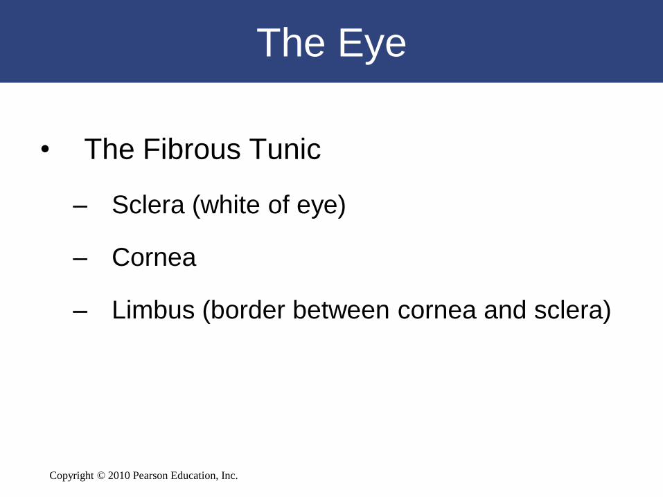
The Eye
• The Fibrous Tunic
– Sclera (white of eye)
– Cornea
– Limbus (border between cornea and sclera)
Copyright © 2010 Pearson Education, Inc.
The Eye
• Vascular Tunic (Uvea) Functions
– Provides route for blood vessels and lymphatics that
supply tissues of eye
– Regulates amount of light entering eye
– Secretes loose and reabsorbs aqueous humor that
circulates within chambers of eye
– Controls shape of lens, which is essential to focusing
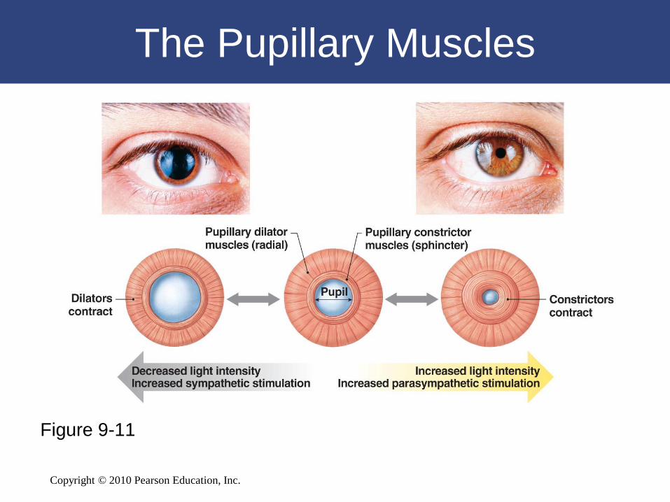
Copyright © 2010 Pearson Education, Inc.
The Pupillary Muscles
Figure 9-11

Copyright © 2010 Pearson Education, Inc.
The Eye
• The Neural Tunic (Retina)
– Outer layer called pigmented part
– Inner neural part:
• Contains visual receptors and associated neurons
• Rods and cones are types of photoreceptors:
– rods:
» do not discriminate light colors
» highly sensitive to light
– cones:
» provide color vision
» densely clustered in fovea, at center of macula
lutea
Copyright © 2010 Pearson Education, Inc.
Figure 9-10c

Copyright © 2010 Pearson Education, Inc.
Retinal Organization
Figure 9-12

Copyright © 2010 Pearson Education, Inc.
Retinal Organization
Figure 9-12
Copyright © 2010 Pearson Education, Inc.
Retinal Organization
Figure 9-12

Copyright © 2010 Pearson Education, Inc.
The Eye
• The Neural Tunic (Retina)
– Inner neural part:
• Bipolar cells:
– neurons of rods and cones synapse with ganglion cells
• Horizontal cells:
– extend across outer portion of retina
• Amacrine cells:
– comparable to horizontal cell layer
– where bipolar cells synapse with ganglion cells
Figure 17–6a

Copyright © 2010 Pearson Education, Inc.
The Eye
• The Chambers of the Eye
– Ciliary body and lens divide eye into:
• Large posterior cavity (vitreous chamber)
• Smaller anterior cavity:
– anterior chamber:
» extends from cornea to iris
– posterior chamber:
» between iris, ciliary body, and lens

Copyright © 2010 Pearson Education, Inc.
The Eye
• Smaller anterior cavity
– Aqueous humor:
• Fluid circulates within eye
• Diffuses through walls of anterior chamber into canal of
Schlemm
• Re-enters circulation
– Intraocular pressure:
• Fluid pressure in aqueous humor
• Helps retain eye shape

Copyright © 2010 Pearson Education, Inc.
The Eye
• Large Posterior Cavity (Vitreous Chamber)
– Vitreous body:
• Gelatinous mass
• Helps stabilize eye shape and supports
retina

Copyright © 2010 Pearson Education, Inc.
The Eye Chambers
Figure 9-14

Copyright © 2010 Pearson Education, Inc.
LASIK

Copyright © 2010 Pearson Education, Inc.
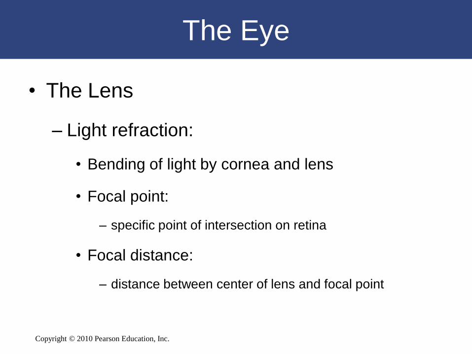
The Eye
• The Lens
– Lens fibers:
• Cells in interior of lens
• No nuclei or organelles
Copyright © 2010 Pearson Education, Inc.
The Eye
• The Lens
– Light refraction:
• Bending of light by cornea and lens
• Focal point:
– specific point of intersection on retina
• Focal distance:
– distance between center of lens and focal point

Copyright © 2010 Pearson Education, Inc.
The Eye
Figure 9-15

Copyright © 2010 Pearson Education, Inc.
The Eye
• Light Refraction of Lens
– Accommodation:
• Shape of lens changes to focus image on retina
– Astigmatism:
• Condition where light passing through cornea and lens is not
refracted properly
• Visual image is distorted
– Visual acuity:
• Clarity of vision
• ―Normal‖ rating is 20/20

Copyright © 2010 Pearson Education, Inc.
The Eye
Figure 9-15

Copyright © 2010 Pearson Education, Inc.
Image Formation
Figure 9-16

Copyright © 2010 Pearson Education, Inc.
9-6 Photoreceptors respond
to light and change it into
electrical signals essential
to visual physiology
Copyright © 2010 Pearson Education, Inc.
Visual Physiology
• Rods
– Respond to almost any photon, regardless of
energy content
• Cones
– Have characteristic ranges of sensitivity
Copyright © 2010 Pearson Education, Inc.
Visual Physiology
• Anatomy of Rods and Cones
– Outer segment with membranous discs
– Inner segment:
• Narrow stalk connects outer segment to inner segment
– Visual pigments:
• Is where light absorption occurs
• Derivatives of rhodopsin (opsin plus retinal)
• Retinal: synthesized from vitamin A
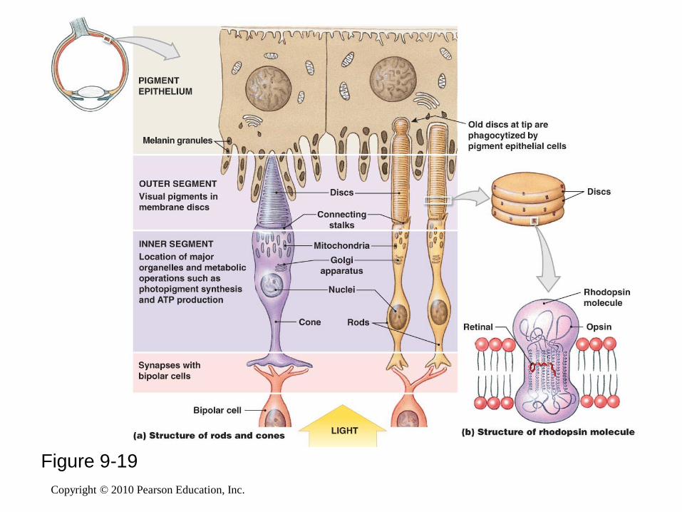
Copyright © 2010 Pearson Education, Inc.
Figure 9-19

Copyright © 2010 Pearson Education, Inc.
Visual Physiology
• Photoreception
– Photon strikes retinal portion of rhodopsin molecule
embedded in membrane of disc
• Opsin is activated
• Bound retinal molecule has two possible configurations:
– 11-cis form
– 11-trans form

Copyright © 2010 Pearson Education, Inc.
Visual Physiology
Figure 9-20
Copyright © 2010 Pearson Education, Inc. Figure 17–16
Visual Physiology
• Color Vision
– Integration of information from red,
green, and blue cones
– Color blindness:
• Inability to detect certain colors
Copyright © 2010 Pearson Education, Inc.
Visual Physiology
• Light and Dark Adaptation
– Dark:
• Most visual pigments are fully receptive to
stimulation
– Light:
• Pupil constricts
• Bleaching of visual pigments occurs

Copyright © 2010 Pearson Education, Inc.
Visual Physiology
• The Visual Pathways
– Begin at photoreceptors
– End at visual cortex of cerebral hemispheres
– Message crosses two synapses before it
heads toward brain:
• Photoreceptor to bipolar cell
• Bipolar cell to ganglion cell

Copyright © 2010 Pearson Education, Inc.
Figure 9-21

Copyright © 2010 Pearson Education, Inc.
9-7 Equilibrium sensations
originate within the inner ear,
while hearing involves the
detection and interpretation of
sound waves
Copyright © 2010 Pearson Education, Inc.
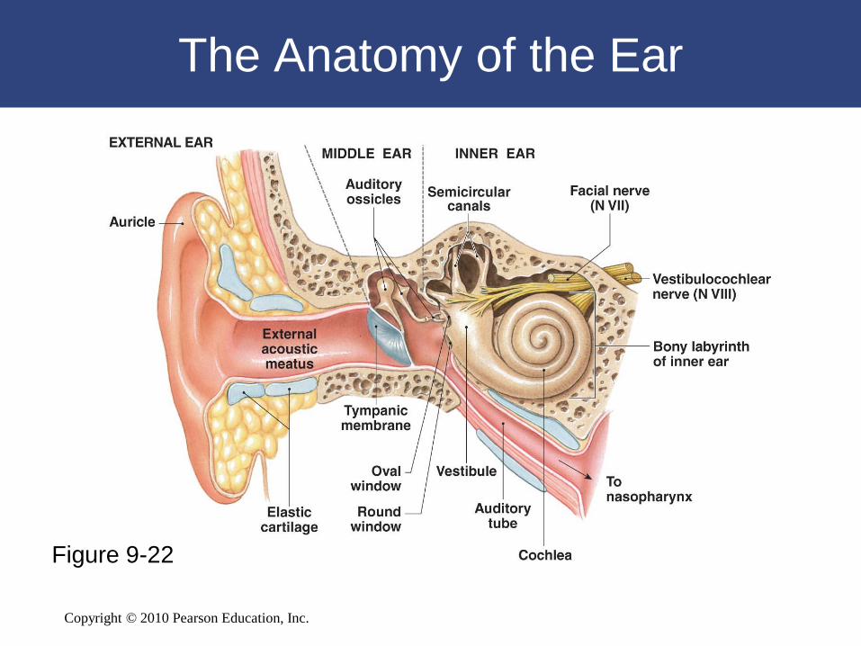
Anatomy of the Ear
• The External Ear
– Auricle:
• Surrounds entrance to external acoustic meatus
• Protects opening of canal
• Provides directional sensitivity
– External acoustic meatus:
• Ends at tympanic membrane (eardrum)
– Tympanic membrane:
• Is a thin, semitransparent sheet
• Separates external ear from middle ear
Copyright © 2010 Pearson Education, Inc.
The Anatomy of the Ear
Figure 9-22
Copyright © 2010 Pearson Education, Inc.
The Ear
• The Middle Ear
– Also called tympanic cavity
– Communicates with nasopharynx via auditory tube:
• Permits equalization of pressures on either side of tympanic
membrane
– Encloses and protects three auditory ossicles:
• Malleus (hammer)
• Incus (anvil)
• Stapes (stirrup)

Copyright © 2010 Pearson Education, Inc.
The Structure of the Middle Ear
Figure 9-23

Copyright © 2010 Pearson Education, Inc.
The Ear
• The Inner Ear
– Contains fluid called endolymph
– Bony labyrinth surrounds and protects membranous
labyrinth
– Subdivided into:
• Vestibule
• Semicircular canals
• Cochlea
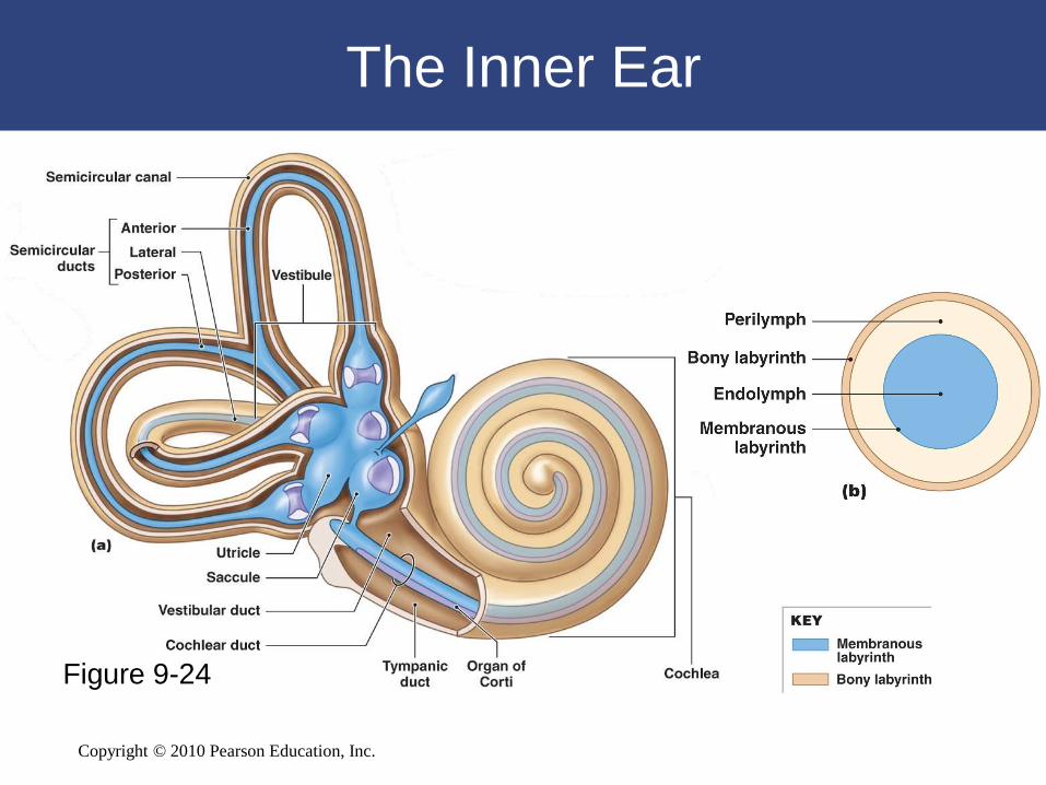
Copyright © 2010 Pearson Education, Inc.
Figure 9-24
The Inner Ear
Copyright © 2010 Pearson Education, Inc.
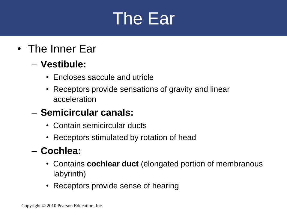
The Ear
• The Inner Ear
– Vestibule:
• Encloses saccule and utricle
• Receptors provide sensations of gravity and linear
acceleration
– Semicircular canals:
• Contain semicircular ducts
• Receptors stimulated by rotation of head
– Cochlea:
• Contains cochlear duct (elongated portion of membranous
labyrinth)
• Receptors provide sense of hearing
Copyright © 2010 Pearson Education, Inc.
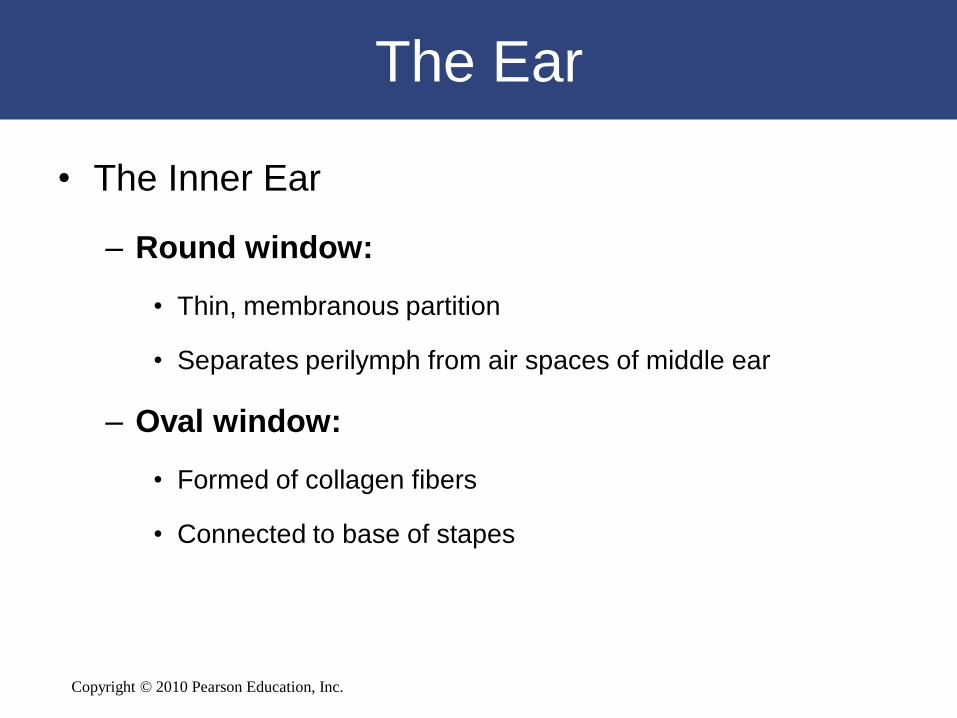
The Ear
• The Inner Ear
– Round window:
• Thin, membranous partition
• Separates perilymph from air spaces of middle ear
– Oval window:
• Formed of collagen fibers
• Connected to base of stapes

Copyright © 2010 Pearson Education, Inc.
Equilibrium
• Sensations provided by receptors of vestibular
complex
• Hair cells
– Basic receptors of inner ear
– Provide information about direction and strength of
mechanical stimuli
Copyright © 2010 Pearson Education, Inc.
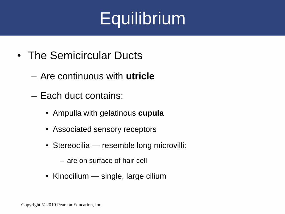
Equilibrium
• The Semicircular Ducts
– Are continuous with utricle
– Each duct contains:
• Ampulla with gelatinous cupula
• Associated sensory receptors
• Stereocilia — resemble long microvilli:
– are on surface of hair cell
• Kinocilium — single, large cilium

Copyright © 2010 Pearson Education, Inc.
The Semicircular Ducts
Figure 9-25 a,b,c

Copyright © 2010 Pearson Education, Inc.
Equilibrium
• The Utricle and Saccule
– Provide equilibrium sensations
– Are connected with the endolymphatic duct, which
ends in endolymphatic sac
– Maculae:
• Oval structures where hair cells cluster
– Statoconia:
• Densely packed calcium carbonate crystals on surface of
gelatinous mass
• Otolith (ear stone) = gel and statoconia

Copyright © 2010 Pearson Education, Inc.
Equilibrium
Figure 9-25 a,d

Copyright © 2010 Pearson Education, Inc.
Equilibrium
Figure 9-25 e

Copyright © 2010 Pearson Education, Inc.
Pathways for Equilibrium Sensations
• Vestibular receptors
– Activate sensory neurons of vestibular ganglia
– Axons form vestibular branch of
vestibulocochlear nerve (VIII)
– Synapse within vestibular nuclei

Copyright © 2010 Pearson Education, Inc.
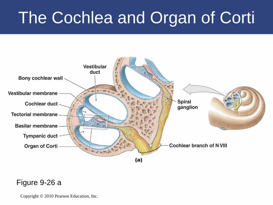
Hearing
• Cochlear duct receptors
– Provide sense of hearing
Copyright © 2010 Pearson Education, Inc.
Figure 9-26 a
The Cochlea and Organ of Corti

Copyright © 2010 Pearson Education, Inc.
Figure 9-26 b
The Cochlea and Organ of Corti
Copyright © 2010 Pearson Education, Inc.
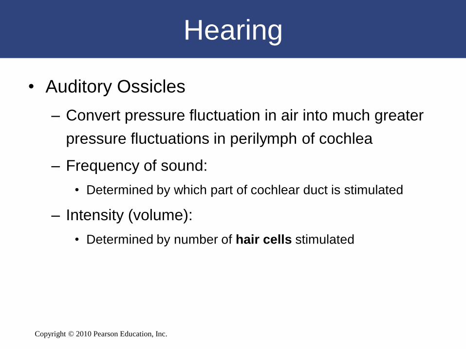
Hearing
• Auditory Ossicles
– Convert pressure fluctuation in air into much greater
pressure fluctuations in perilymph of cochlea
– Frequency of sound:
• Determined by which part of cochlear duct is stimulated
– Intensity (volume):
• Determined by number of hair cells stimulated

Copyright © 2010 Pearson Education, Inc.
Sound and Hearing
Figure 9-27
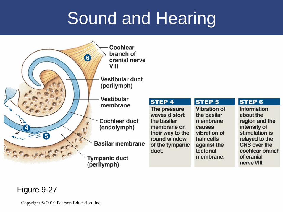
Copyright © 2010 Pearson Education, Inc.
Sound and Hearing
Figure 9-27
Copyright © 2010 Pearson Education, Inc.
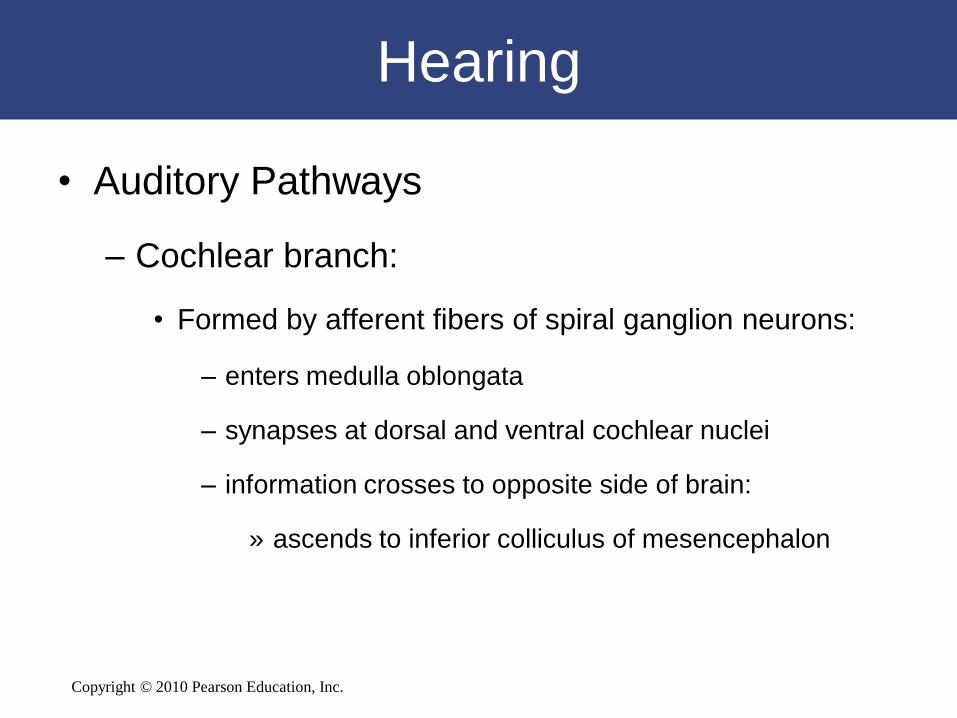
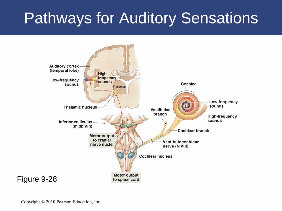
Hearing
• Auditory Pathways
– Cochlear branch:
• Formed by afferent fibers of spiral ganglion neurons:
– enters medulla oblongata
– synapses at dorsal and ventral cochlear nuclei
– information crosses to opposite side of brain:
» ascends to inferior colliculus of mesencephalon
Figure 17–31

Copyright © 2010 Pearson Education, Inc.
Hearing
• Auditory Pathways
– Ascending auditory sensations:
• Synapse in medial geniculate nucleus of thalamus
• Projection fibers deliver information to auditory
cortex of temporal lobe
Copyright © 2010 Pearson Education, Inc.
Pathways for Auditory Sensations
Figure 9-28

Copyright © 2010 Pearson Education, Inc.
9-8 Aging is accompanied
by a noticeable decline in
the special senses
Copyright © 2010 Pearson Education, Inc.
Smell and Aging
• Olfactory neuron recycling slows, leading
to decreased sensitivity

Copyright © 2010 Pearson Education, Inc.
Taste and Aging
• Number of taste buds is reduced, and
sensitivity is lost
Copyright © 2010 Pearson Education, Inc.
Vision and Aging
• Lens stiffens
• Lens clouds
• Blood vessels grow in retina
Copyright © 2010 Pearson Education, Inc.
Hearing and Aging
• Loss of elasticity in tympanic membrane


