The HDE Summary of Safety and Probable Benefit What It Is and Does Under section 520(m) of the Federal Food, Drug, and Cosmetic Act (FD&C), the Secretary may approve a Humanitarian Device Exemption (HDE) application, which exempts the device from the effectiveness requirements of sections 514 and 515. Although the device is exempt from demonstrating effectiveness, there must be sufficient information to show a probable benefit for its intended use and indication for use. The Summary of Safety and Probable Benefit (SSPB) is an FDA document (originally submitted by the applicant and modified by FDA) which is intended to present a reasoned, objective, and balanced critique of the scientific evidence which served as the basis for the approval of the HDE. The SSPB documents that there is reasonable assurance of safety and probable benefit for the device as labeled based on the nonclinical and clinical information described in the HDE. The SSPB is a summation of both the positive and negative aspects of the scientific evidence. For a HDE to be approved, the potential risks versus possible benefits assessment must favor this action in conjunction with a comparison to all the possible alternatives. The SSPB summarizes these judgements. The SSPB is a publicly releasable document, which the applicant is required to submit under 21 CFR 814.104(b)(4). The applicant's SSPB often differs from FDA's objective, i.e., it contains only the positive aspects of the device; it contains marketing language, etc. Consequently, revision is often needed. Since minor variations in SSPB formats are possible, it is best to consult previously released SSPBs (preferably recent ones for similar devices) for guidance and to consult with the HDE staff. Since the SSPB deals with scientific/technical data and will be read by many non-professionals, it is best to keep the format clear by grouping study objective(s), study methods, results, interpretation(s), and conclusion(s) for each study or substudy. An overall statement/conclusion integrating the individual study outcomes in light of safety and probable benefit should be written. The SSPB should be as concise as possible. Note: In creating this template, parts of the General Program Memorandum #G91-1: Device Labeling Guidance was incorporated. This guidance can be referenced at the following web page: http://www.fda.gov/cdrh/g91-1.html HDE H110002 Summary of Safety and Probable Benefit Page 1
Transcript
The HDE Summary of Safety and Probable BenefitWhat It Is and Does
Under section 520(m) of the Federal Food, Drug, and Cosmetic Act (FD&C), the Secretarymay approve a Humanitarian Device Exemption (HDE) application, which exempts thedevice from the effectiveness requirements of sections 514 and 515. Although the device isexempt from demonstrating effectiveness, there must be sufficient information to show aprobable benefit for its intended use and indication for use. The Summary of Safety andProbable Benefit (SSPB) is an FDA document (originally submitted by the applicant andmodified by FDA) which is intended to present a reasoned, objective, and balanced critiqueof the scientific evidence which served as the basis for the approval of the HDE. The SSPBdocuments that there is reasonable assurance of safety and probable benefit for the device aslabeled based on the nonclinical and clinical information described in the HDE. The SSPB isa summation of both the positive and negative aspects of the scientific evidence. For a HDEto be approved, the potential risks versus possible benefits assessment must favor this actionin conjunction with a comparison to all the possible alternatives. The SSPB summarizesthese judgements.
The SSPB is a publicly releasable document, which the applicant is required to submit under21 CFR 814.104(b)(4). The applicant's SSPB often differs from FDA's objective, i.e., itcontains only the positive aspects of the device; it contains marketing language, etc.Consequently, revision is often needed. Since minor variations in SSPB formats arepossible, it is best to consult previously released SSPBs (preferably recent ones for similardevices) for guidance and to consult with the HDE staff. Since the SSPB deals withscientific/technical data and will be read by many non-professionals, it is best to keep theformat clear by grouping study objective(s), study methods, results, interpretation(s), andconclusion(s) for each study or substudy. An overall statement/conclusion integrating theindividual study outcomes in light of safety and probable benefit should be written. TheSSPB should be as concise as possible.
Note: In creating this template, parts of the General Program Memorandum #G91-1:Device Labeling Guidance was incorporated. This guidance can be referenced at thefollowing web page: http://www.fda.gov/cdrh/g91-1.html
HDE H110002 Summary of Safety and Probable Benefit Page 1
Device Trade Name: Argus' II Retinal Prosthesis SystemApplicant's Name.& Address: Second Sight Medical Products, Inc. 12744
San Fernando Road, Building 3 Sylmar,CA 91342
Humanitarian Device Exemption (HDE) Number: HI 10002
Humanitarian Use Device (HUD) Designation #09-0216Number:
Date of Humanitarian Use Device (HUD) May 28, 2009Designation:
Date(s) of Panel Recommendation: September 29, 2012
Date of Good Manufacturing Practice Inspection: October 18, 2011
Date of Notice of Approval to Applicant: February 13, 2013
I. INDICATIONS FOR USE
The Argus II Retinal Prosthesis System is intended to provide electrical stimulation of theretina to induce visual perception in blind patients. It is indicated for use in patients withsevere to profound retinitis pigmentosa who meet the following criteria:
* Adults, age 25 years or older.* Bare light or no light perception in both eyes. (If the patient has no residual light
perception, then evidence of intact inner layer retina function must be confirmed.)* Previous history of useful form vision.* Aphakic or pseudophakic. (If the patient is phakic prior to implant, the natural
lens will be removed during the implant procedure.)* Patients who are willing and able to receive the recommended post-implant
clinical follow-up, device fitting, and visual rehabilitation'.
The Argus II implant is intended to be implanted in a single eye, typically the worse-seeing eye.
'The indication for use statement has been modified from that granted for the HUD designationrequest #09-0216. The HUD designation states that the device is indicated "to provide electricalstimulation of the retina to elicit visual perception in blind subjects with severe to profound retinitispigmentosa."The current IFU further clarifies the intended population.
HDE H1 10002 Summary of Safety and Probable Benefit Page 2
III. CONTRAINDICATIONS
* Ocular diseases or conditions that could prevent the Argus II System fromworking (e.g. optic nerve disease, central retinal artery or vein occlusion, historyof retinal detachment, trauma, severe strabismus, etc.).Ocular structures orconditions that could prevent the successful implantation of the Argus II Implantor adequate healing from surgery (e.g. extremely thin conjunctiva, axial length<20.5 mm or >26 mm; corneal ulcers, etc.).
* Ocular diseases or conditions (other than cataracts) that prevent adequatevisualization of the inner structures of the eye (e.g. comeal opacity, etc.).
* Inability to tolerate general anesthesia or the recommended antibiotic and steroidregimen associated with the implantation surgery.
* Metallic or active implantable device(s) (e.g. cochlear implant) in the head.* Any disease or condition (e.g. significant cognitive decline, etc.) that prevents
understanding or communication of informed consent, fitting of the Argus IISystem, or post-operative follow-up. A pre-operative psychological evaluationmay be recommended to confirm the patient is not contraindicated based on thiscnterion.
* Predisposition to eye rubbing.0
IV. WARNINGS AND PRECAUTIONS
WARNINGS:
* Failure to follow the recommended surgical procedure for implanting theArgus II Implant may increase the risk of adverse events and damage to theimplant.
* Individuals implanted with an Argus II Implant should not undergo shortwave or microwave diathermy. High currents induced in the implant electrodescan cause tissue damage or serious injury. Diathermy may also cause permanentdamage to the implant.
* Individuals implanted with an Argus II Implant should not undergoelectroconvulsive therapy (ECT) as ECT may cause tissue damage or permanentdamage to the implant.
* If lithotripsy or high output ultrasound must be used, do not focus thetreatment beam near the Argus II Implant. Exposure of the Argus II Implant tothese therapies may harm the patient or damage the implant.
* The Argus 11 Implant has been classified as an MR Conditional device.Individuals with an Argus II Implant may undergo a magnetic resonanceimaging (MRI) procedure ONLY if it is performed using a 1.5 or 3.0 Tesla MRI
HDE H110002 Summary of Safety and Probable Benefit Page 3
System and ONLY following the MRI Instructions provided later in this insert.Individuals with an Argus 11 Implant should not enter a room housing an MRISystem that has a rating other than 1.5 or 3.0 Tesla, even if the Argus II System isnot being used. The external equipment (i.e. VPU and glasses) should remainoutside the MR system room, as severe harm to people in the MR system roomcould occur. If any pain is experienced during the MRI procedure the patientshould be instructed to notify the technician immediately.The Argus II System may interfere with the operation or accuracy of medicalmonitoring, diagnostic or life support equipment. Do not use the Argus IISystem within 3 feet (0.9 meters) of this type of equipment. If interference occurs,turn off the Argus II VPU or extend the distance between yourself and theaffected equipment.
* Do not use monopolar electrosurgical equipment in individuals with an ArgusII Implant. Monopolar electrosurgical equipment may cause damage to theimplant or to tissue surrounding the implant.
PRECAUTIONS
* In the event of any undesirable sensation when using the Argus II System (forexample, pain), immediately halt operation of the system by removing the ArgusII Glasses or turning off the Argus II VPU.
* At any time after implantation, Argus II patients have a risk of conjunctivalcomplications which, if left untreated, may result in conjunctival erosion whichcould lead to endophthalmitis. Argus II recipients should be vigilant in reportingany new symptoms of foreign body sensations, tearing and/or pain promptly totheir eye care professional. Long-term professional monitoring for lateconjunctival issues is necessary.
* The long-term effects of chronic electrical stimulation are unknown. Sucheffects may include deterioration of the retina or optic nerve. These effects maylead to deterioration of residual native vision and/or visual response to the ArgusII System and could preclude subsequent replacement of the Argus II Implantwith another retinal implant.
* Individuals with an Argus II Implant should only use a VPU that has beenspecifically programmed for them by their clinician or Second Sight personnel.Use of a different VPU may be ineffective in providing visual information andmay cause physical discomfort from overstimulation.
* To avoid unsafe stimulation, do not use a VPU configured for Operating Roomuse for anything other than pre-implantation testing, testing during implantation,or initial fitting testing.
* Individuals with an Argus II Implant should avoid physical impact or extremedirect pressure to the eye as this may result in eye trauma, movement or damageto the Argus II Implant. If this occurs, consult your physician.
* Individuals with an Argus II Implant should avoid eye rubbing as this maydislodge the implant or cause eye irritation.
HDE H110002 Summary of Safety and Probable Benefit Page 4
* Individuals with an Argus II Implant should continue to use their other mobilityaids (e.g. canes, dogs, etc.) at all times.
* Use of the Argus II System during pregnancy and nursing has not beenevaluated.
PRECAUTIONS REGARDING OTHER MEDICAL PROCEDURES
A. GENERAL INFORMATION (APPLICABLE TO ALL PROCEDURES):
* Individuals needing to undergo any of the procedures listed below, should informhis or her doctor about the existence of a retinal prosthesis in the eye. The doctorshould contact Second Sight at 1-818-833-5060 for more information.
* Before having any medical or test procedure that involves the use of othermedical equipment, individuals with an Argus II Implant should remove theArgus II Glasses and VPU.
* Once the procedure is complete, that individual should have the Argus II Implanttested as soon as possible to make sure it is still functioning properly. Damage tothe implant may not be immediately detectable.
B. INFORMATION ABOUT SPECIFIC PROCEDURES:
* Magnetic Resonance Imaging (MRI) - Refer to the Warnings section above
* The use of laser, phacoemulsification, or fragmatome may damage the Argus IIImplant. If these procedures must be used in an implanted eye, do not direct thelaser beam at the implant. Extra caution should be used when performing theseprocedures intraocularly as visualization:of the implant may be obscured.
* The use of bipolar electrosurgical equipment may damage the Argus II Implant.Use caution when using this equipment near the implant.
* CT Scans or Diagnostic Ultrasound may be performed in individuals with anArgus II Implant. However, if a scan or ultrasound is performed in the regionwhere the Argus II Implant is located, the implant may create an image artifactmaking the scan unreadable in this region.
* Use of defibrillators or therapeutic ionizing radiation to the head maypermanently damage the Argus II Implant. However, this should not preclude orchange the way in which these treatments are delivered. The Argus II Implantshould be tested by a qualified clinician or Second Sight personnel as soon aspossible following the procedure or defibrillator activation to confirm that it isstill functioning properly. Damage to the implant may not be immediatelydetectable.
HDE H1 10002 Summary of Safety and Probable Benefit Page 5-'3
* The effects of cobalt treatment and linear acceleration techniques on the implantare unknown.
C. ELECTROMAGNETIC INTERFERENCE (EMI):
Electromagnetic interference is a field of energy (electrical, magnetic, or both) created byequipment found in public environments that may be strong enough to interfere with thenormal operation of the Argus II System. The Argus 11 System meets internationalstandards for electromagnetic compatibility and is designed to continue to operate in a"safe mode" in the presence of any electromagnetic interference which would normallybe encountered during every day use of the Argus II System. It is important to note,however, that in certain circumstances, electromagnetic interference could cause thefollowing:
* Serious injury. Exposure of the implant to EMI may result in the implant heatingand damaging nearby retinal tissue.
* Damage to the Argus II implant. Damage to the implant may requirereplacement, or result in loss of, or irreversible change in the performance of theArgus II System.
* Unexpected Turning off of the Argus II VPU. EMI may cause the VPU to turnoff unexpectedly.
* Interruption of Stimulation. EMI may cause a momentary interruption ofstimulation.
Argus II System users should be advised that upon entering an environment which maybecausing interference with the Argus II System, they should move away from theequipment or object thought to be causing the interference, if possible, turn off theequipment or object causing the interference, tell the equipment operator or the doctorwhat happened and, if they continue to experience interference or think that the Argus IISystem is not working as well as it did before they encountered the interference, tocontact their doctor.
D. POSSIBLE INTERFERENCE WITH OTHER ELECTRONIC DEVICES
* Theft or metal detectors (such as those located in entrances to public buildingsand department stores) and airport or security screening devices may temporarilyinterrupt Argus 11 stimulation if the Argus II System is used within 1 yard (0.9meters) of them. Normal operation will resume when you move away from theseitems. When possible, it is best to avoid these devices or turn the VPU off when
HDE H1 10002 Summary of Safety and Probable Benefit Page 6Iq
passing through these systems. Individuals with an Argus II Implant should showtheir ID card to any attendant in the area who may be able to assist them inbypassing the devices. If unavoidable, walk through the scanner and promptlymove away from the area. Do not lean on these scanners or linger in their path.
* Electronic Article Surveillance (EAS) systems, EAS Tag Deactivators, andRadiofrequency identification (RFID) systems may temporarily interrupt ArgusII stimulation if the Argus II System is used within 3.5 yards (3.2 meters) of them.Normal operation will resume when you move away from these items. RFIDsystems and EAS systems and tag deactivators send out energy fields thatwirelessly communicate with tags that are attached to objects such asmerchandise, materials and people. These systems are used for security, theftprevention, tracking and inventory control and they are usually found in retailstores, libraries, government buildings, warehouses and offices. For example,security tags attached to clothing contain RFID tags.
* Electrostatic Discharge (ESD) may interfere with normal operation or causedamage to the electrical components of the Argus JJ System. Common situationsthat create static electricity include putting on or removing clothes, or draggingfeet across a carpet or rug when there is less than 30% relative humidity. Careshould be taken to avoid handling the VPU and glasses when static electricity ispresent.
* The Argus I System may interfere with the normal operation of some models ofhearing aids. Hearing aids should be tested prior to implantation, to ensureproper functioning of both the Argus II System and the hearing device.
* Some home appliances (for example, microwaves ) and some devices withantennae (for example, cell phones, and cordless phones) may temporarilyinterrupt Argus II stimulation if the Argus II System is being used near them. Thetable below lists the distance at which interruption of stimulation may occur withthese systems.
Table 1: Separation Distances
Interruption of stimulation may occur if device isoperated within this distance of the Argus II
Type of device System
Another Argus IISsthem 7 inches (17.5 cm)System
Cell phone I inch (2.5 cm)
Cordless phone 1 inch (2.5 cm)
Bluetooth device 1 inch (2.5 cm)
Microwave oven I inch (2.5 cm)
HDE H 110002 Summary of Safety and Probable Benefit Page 7
WiFi Access Point 8 inches (20 cm)
Wireless Router 8 inches (20 cm)
Normal operation will resume when you move away from these items.
* The Argus II System operates using wireless technology which could interferewith the safe operation of an airplane. Patients should not turn on the Argus IISystem on an airplane.
* Commercial electrical equipment (such as arc welders, induction furnaces orresistance welders), communication equipment (such as microwave transmitters,linear power amplifiers and high-power amateur transmitters), high voltage lines,power lines or generators, electric steel furnaces, or large magnetizedspeakers may temporarily interrupt Argus II System function. Normal operationwill resume when you move away from these items.
Additional information on specific environments and recommended separationdistances is provided in the Electromagnetic Environments section of the ProductInsert.
V. DEVICE DESCRIPTION.
The Argus II System is composed of three subsystems: The retinal prosthesis (implant),the Externals System and the Fitting System. The implant works in unison with theExternals System to deliver electrical stimulation to the retina. The third subsystem(Fitting System) is used in conjunction with the implant and Externals System in theclinic when fitting the device to the patient.
PRINCIPLE OF OPERATION
The principle of operation of the Argus II System is shown schematically in Figure 1.
HDE H1 10002 Summary of Safety and Probable Benefit Page 8
FIGURE 1: SCHEMATIC OVERVIEW OF THE ARGUs 1I SYSTEM
A video camera that is attached to glasses worn by the patient captures a video image.The camera signal is sent to a Video Processing Unit (VPU), worn by the patient on a beltor strap, which processes the camera image and transforms it into electrical stimulation
patterns. The electrical stimulation data are then sent to a receiving/transmitter coilmounted on the glasses which sends both data and power wirelessly via radio-frequency
(RF) telemetry to the implanted retinal prosthesis. The implant receives the radio-
frequency commands and delivers stimulation to the retina via a thin film electrode arraythat is secured to the retina with a retinal tack. The pulses are intended to mimic thefunction of photoreceptor cells (which have lost most, if not all, of their fuinctionality inthis patient population) by inducing cellular responses in the remaining neurons thattravel through the optic nere to the visual cortex, where they are perceived as
phosphenes of light.
A fitting process is periodically performed with the patient in the clinic to develop thestimulation parameters used to process the video images from the camera. This isaccomplished through use of the Clinician Fitting System (CFS) which consists of fittingsoftware with a graphical user interface running on a laptop computer. The patient
specific configuration files developed from the fitting process are downloaded to theVPU where they are available for use by the patient.
A description of each of the subsystems follows.
IMPLANT SUBSYSTEM
This subsystem is composed of the Argus 11 Retinal Prosthesis and the Argus 11 RetinalTack. Each is described below.
HDE H1 10002 Summary of Safety and Probable Benefit Page 9
cm-7
RETINAL PROSTHESIS
The retinal prosthesis, which is implanted in and on the eye and electrically stimulates theretina, consists of the following four major components:
* A small hermetic package that contains the electronics to receive power and to drivethe electrical stimulation of the electrodes. A suture tab assembly encircles thepackage.
* A coil that receives power and receives and transmits data to/from the externalprimary coil (on the glasses).
* A thin film electrode cable and array that is electrically connected to the package andtransmits the stimulation signals to the retina via 60 exposed platinum electrodeswhich are arranged in a 6 x 10 pattern. The electrode array is secured to the retinawith a retinal tack. The implant has 55 of these electrodes enabled for use.
* A scleral band that allows the implant to be secured to the outside of the eye.
An illustration of the Argus 11 Retinal Prosthesis showing each of its components isprovided in Figure 2. Scleral Band
ElectronicsCase
RetinalTack
InternalCoil Electrode Array
FIGURE 2: ARGUS II RETINAL PROSTHESIS
The Argus II Retinal Prosthesis is intended to be implanted in just one eye, typically theworse-seeing eye. The retinal prosthesis is provided sterile.
RETINAL TACKS
Two retinal tacks are included with the retinal prosthesis. Modeled after a standard retinaltack, which was once commonly used to repair retinal detachments, the Argus II RetinalTack is used to affix the electrode array to the retina. A single tack is used to secure thearray. The second tack acts as a back-up. The tack is constructed from a biocompatible
HDE H1 10002 Summary of Safety and Probable Benefit Page 10
titanium alloy used in other commercially available retinal tacks. The retinal tackincludes a sharply pointed end for piercing through the retina, choroid, and sclera, and anelongated, cylindrical shaft. The tack also includes an enlarged head and flange as well asa spring that applies a gentle compressive force to hold the array down. When securingthe electrode array to the retina, the retinal tack is placed through a tack hole on theelectrode array.
EXTERNALS SUBSYSTEM
The Externals System is composed of the Argus II Glasses and the Argus II VideoProcessing Unit. Each is described below.
GLASSES
The Argus II Glasses provide a convenient and discreet way to house the video camerafor capturing images and the radio-frequency system needed to power and communicatewith the implant.
A small, light-weight video camera is mounted in the center of the frame above the nose-piece. The telemetry coils and radio-frequency system are mounted on the ear piece. Theposition of the coil housing is adjustable to provide comfort to the patient and to allowthe external coil assembly to be optimally positioned relative to the implant coil toachieve a good communication link for patients with different facial structures.
The Argus II OR coil, although used only during the implantation surgery, is categorizedas part of the Externals System. It is comprised of the same telemetry coil and RE systemused in the glasses. The OR coil is used to test functionality of the implant duringsurgical implantation. It is placed in a sterile sleeve in the operating room.
VIDEO PROCESSING UNIT (VPU)
The Argus II VPU, which powers and controls the implant, is comprised of a case,buttons, connectors, rechargeable battery and digital circuit boards. The buttons are largeand shaped so that they can be easily identified by touch. There is one connector thatconnects the VPU to the glasses that can be easily connected by a sightless person. TheVPU can also be connected to the Argus II Clinical Fitting System to allow testing of theimplant and programming of the VPU ("patient fitting").
The VPU acquires video input from the camera and converts it into a digital format.Filters, such as edge detection and contrast adjustment, may be then applied. The imageis then reduced to a 6 x 10 resolution using a downscaling filter. This representation ofthe image is then mapped to stimulation intensity using customized look-up tables thathave been derived during the patient fitting process. A check is performed to assure thatthe overall current and the maximum charge per phase are within safety limits. Thestimulation parameters are then sent via telemetry to the implant.
HDE H110002 Summary of Safety and Probable Benefit Page 1119
Prior to use, the VPU is configured to the patient's implant utilizing the Argus II VPU-Implant Matching CD.
FITTING SUBSYSTEM
The primary components comprising the fitting subsystem are the Argus II ClinicianFitting System (CFS), the Argus II Psychophysical Test System (PTS), and the Argus IICommunication Adapter (CA). Supporting accessories include various cables, USBdrives, user input device and touch screen monitor. The components are described below.
CLINICIAN FITTING SYSTEM (CFS)
The CFS consists of dedicated fitting software with a graphical user interface running ona laptop computer. The CFS is used to perform diagnostic tests of the system (e.g.,electrode impedance), measure patient response to stimulation (e.g., thresholds), andconfigure the system stimulation parameters and video processing strategies for eachpatient.
The CFS includes software modules that allow the clinician to quickly administer tests tomeasure important perceptual parameters, such as electrical stimulation threshold. Basedon these perceptual parameters, the fitting software allows the clinician to customconfigure the transformation between the video image and electrode stimulationparameters thereby optimizing the retinal prosthesis for each patient.
The patient-specific configuration files developed from the fitting process can bedownloaded to the VPU where they are available for use by the patient. The stimulationparameters are checked by CFS (in addition to the VPU) to ensure that only safestimulation is delivered.
The CFS laptop is provided with a power supply unit for recharging the PC battery and acable for connecting the CFS laptop to the Communication Adapter. The CFS softwareand VPU firmware ensure that the stimulation delivered to the patient are withinspecified safety limits.
PSYCHOPHYSICAL TEST SYSTEM (PTS)
The PTS is an optional system component that consists of a laptop with an applicationprogramming interface (API) that the clinician can utilize to run scripts (programs) forevaluating patient performance with the Argus II System. The PTS is connected to theCFS. The PTS laptop is provided with a power supply unit for recharging the PC batteryand a cable for connecting the PTS laptop to the CFS laptop. The CFS software and VPUfirmware ensure that the stimulation parameters of the PTS developed scripts are withinspecified safety limits.
HDE H110002 Summary of Safety and Probable Benefit Page 12
COMMUNICATION ADAPTER (CA)
The CA provides an optically isolated communication channel between the CFS and theVPU, thus preventing the flow of electric current between the CFS and VPU.
TOUCH SCREEN MONITOR
The touch screen monitor is an off-the-shelf component that is used as a tool for fittingthe patient and for performing psychophysical tests in the clinic. The monitor is suppliedwith a medical grade power supply.
OTHER ACCESSORIES
Other accessories used during the fitting process include cables for connecting the FittingSystem to Externals System (Argus II CA-VPU Cable, Argus II CFS-CA Cable, and theArgus II CFS-PTS Cable) and USB drives for obtaining access to the Fitting System andfor collecting transferring, and archiving data (Argus II USB Security Drive, Argus IIUSB Video Settings Drive, Argus 1I USB Transfer Drive, Argus II Archive Drive). Avideo gamepad controller and touch screen monitor for collecting patient responses mayalso be employed in the clinic.
VI. ALTERNATIVE PRACTICES AND PROCEDURES
There are no commercialized treatment options currently available for RP patients.Traditionally, the approach to vision rehabilitation in patients with RP has been to use theremaining vision with the assistance of optical aides. In patients with no remaining usefulvision, tools to maximize auditory or tactile information (e.g. Braille, cane travel, etc.)are used.
VII. MARKETING HISTORY
The Argus 1I System received approval for commercial distribution in the EuropeanEconomic Area (EEA) on February 10, 2011. Second Sight began commerciallydistributing the device in Europe in October 2011. The device has not been marketed inthe US.
The Argus II Retinal Prosthesis System has not been withdrawn from marketing for anyreason relating to the safety and effectiveness of the device.
HDE H110002 Summary of Safety and Probable Benefit Page 13zil
VIII. POTENTIAL ADVERSE EFFECTS OF THE DEVICE ON HEALTH
The following potential device- or implant surgery- related adverse events were notobserved during the clinical trial:
* Facial nerve stimulation, transient electrical shock, skin burn due toexcessive heating of the external equipment, or retinal tissue damage dueto mechanical trauma, excessive stimulation or excessive heating of theimplant.
* Failure or damage to the Argus II Implant requiring it to be explanted.* Fall or bump resulting from use of the Argus II System.* Risks known to be associated with standard vitreo-retinal surgery, peeling
of an epiretinal membrane and use of a scleral band: suprachoroidalhemorrhage, intrusion/extrusion of the scleral band, and macular hole.
* Risks known to be associated with the removal of the lens using clearcornea phacoemulsification: cortical drop in vitreous or vitreous prolapse.
* Risks known to be associated with canthotomy: improper apposition of theeyelids and chronic irritation at the lid margin.
* Risks known to be associated with the use of general anesthesia, steroidsand antibiotics: chest pain, urinary retention, myocardial infarction,pulmonary embolism, deep vein thrombosis, respiratory failure, blood lossrequiring transfusion, systemic infection, prolonged hospitalization, andallergic reaction to anesthesia.
IX. SUMMARY OF PRECLINICAL STUDIES
LABORATORY STUDIES
Objectives:. The objectives of the laboratory studies were to test performance, safety andreliability of the Argus II System. Testing was performed on a component level, sub-assembly level, device level and system level. Provided below is a summary of testing.Testing has been categorized by subsystems comprising the Argus I System.
SUMMARY OF LABORATORY STUDIES: IMPLANT
A summary of the bench testing performed on the implant components, subassemblies,and the final device is provided in Table 2.
TABLE 2: SUMMARY OF LABORATORY STUDIES FOR IMPLANT & IMPLANTCOMPONENTS/SUB-ASSEMBLIES
Implant Component: Array
HDE H110002 Summary of Safety and Probable Benefit Page 14
Implant Component: Array
Flexure Fatigue Test: Testing was performed to verify that the electrode array canwithstand the flexure forces which might occur during or after implantation. Thespecification was developed with a margin of safety from normal stresses anticipateddiring actual use. Flexure fatigue testing was performed on samples soaked in saline. Adetermination was made of the number of flexure cycles until trace failure, as defined asloss of electrical continuity, was observed. The array was determined to meetspecification.
Active Soak Test:.Testing was performed to verify that the electrode array meets itslong-term electrical stimulation specification, i.e., that it permits stimulation at themaximum charge density limit at 30 Hz (baseline frequency) for the equivalent of 5years in saline. Test samples were exposed to accelerated aging conditions. Arraysstimulating at accelerated active stimulation reached the equivalent of 32 years of real-time use with no significant changes in voltage waveform.
Stimulation Test: Testing was performed to verify that the array satisfies its electricalspecifications for insulation resistance, capacitance between traces, and electrode seriesresistance and capacitance. Specification was based on the requirement that currentlevels be isolated so as not to induce percepts on adjacent channels. Testing wasperformed in the presence of saline. The array was determined to meet specification.
Mechanical Test: Testing was performed to verify that the electrode array continues tomeet its specification for shape and curvature after exposure to expected thermalexcursions, sterilization, and the forces of surgical implantation. s.
Implant Component: Coil
Flexure / Soak Test: Testing was performed to verify that the coil meets its electricalspecifications (inductance, quality factor, and self resonance frequency) after beingexposed to the forces which might occur during or after implantation. Implant coils weretested under accelerated aging conditions under a maximum electrical load. Coils weresubjected to a flexure test to introduce mechanical stress on the coils prior to lifetimesoak test. The coil was determined to meet its specified tolerances.
Mechanical Test: Tear strength testing was performed to verify that the coil suture tabcould withstand the tear forces incurred during implantation of the device. The suturetabs met acceptance criteria.
Implant Component: Application Specific Integrated Circuit (ASIC)
Performance Test: Testing was performed on the retina chip (i.e., ASIC) to verify that itmeets defined specifications for (1) Power supply and reset characteristics (2) Receivercharacteristics (3) Electrode driver output characteristics (4) System controlcharacteristics (5) Back telemetry characteristics and (6) Test interface. The ASIC metspecifications.
HDE H1 10002 Summary of Safety and Probable Benefit Page 1513
Implant Sub-assembly: Package
Corrosion Test: Samples of the package were subjected to active and passive soaking tostudy corrosion effects on the package's lifetime. Testing included EDX/SEM analysisand electrochemical impedance spectroscopy (EIS) measurements of the hardcase. Noevidence of any corrosion including galvanic corrosion occurred on native metal of thecases. Moreover, no change in impedance of the hardcase, which serves as a returnelectrode, was detected.
Environmental Testing: Hermetically sealed packages were subject to temperatureextremes, random vibration, and variations in atmospheric pressure to evaluatereliability of the implant to withstand the forces which may occur under normalconditions of storage, shipment and use. The hermetically sealed package of the implantmet established acceptance criteria for temperature cycling, random vibration, internalwater vapor content, and atmospheric pressure variation.
Mechanical Test / Design Analysis: A design analysis with tear strength testing wasperformed to verify that the suture tabs surrounding the hardcase could withstand thetear forces incurred during implantation of the device. The suture tabs met acceptancecriteria.
Implant (Assembled)
Soak Test: Test samples equivalent to the actual device interconnect were evaluated toverify that the flex interconnection of the array, coil and package meets the designlifetime specification of at least 5 years in vitro under active soak conditions with amaximum electrical load. Testing was performed under accelerated aging conditions.The interconnect met its design lifetime specification.
Design Analysis: In addition to the above interconnect subassembly soak test, a designanalysis was performed to assess the integrity of the coil attachment and the electrodearray interconnect to withstand flexural stresses. The flexure forces were determined tobe controlled by the design.
Environmental Testing: Packaged implants were subjected to random vibration,different atmospheric pressures, and temperature cycling (550 C and -10' C) to verifythat the device can withstand the forces which may occur under normal conditions ofshipping and use. Devices exposed to environmental testing continued to meetspecifications for electrical functionality and visual integrity.
Diagnostic Ultrasound: Implants were evaluated for their compatibility to ultrasoundexposure per EN 45502-1:1997. Devices exposed to ultrasound energy were able topower up with no change in the power characteristics. The safety checks that theimplant performs on start-up were also unaffected.
HDE H110002 Summary of Safety and Probable Benefit Page 16
Implant (Assembled)
Diagnostic MRI: Implants were evaluated for their compatibility to magnetic resonanceimaging (MRI) per EN 45502-1:1997. Testing demonstrated that the implant and retinaltack are "MR Conditional" in 1.5 Tesla/64MHz and 3.0 Tesla/128 MHz MRenvironments.
Particulate Testing: Implants were tested for particulates to verify that there is nounacceptable release of particulate matter when in contact with body fluid. Devicestested had net particle concentrations lower than the acceptance criteria for bothparticles greater than 2 pm and 5 pm.
Interconnect Flexural Stress Test: Samples mechanically identical in form to the devicewere attached to silicone eye models to simulate the actual implanted condition. Theseassemblies were affixed to a fixture that imparted an oscillating deflection thatsimulated the maximum accelerations that would be experienced by the deviceimplanted in the eye. The test was run for 5 years equivalent lifetime to verify therobustness of the interconnection between the coil, array and package. Test assemblieswere measured for resistance and leakage current to solution. The test samples metestablished acceptance criteria.
Device Lifetime - Active Soak Test: Implants were stimulated while soaking underaccelerated aging conditions and evaluated for functional performance. The testingfound that the implant operated as intended over the specified life of the device. Nosigns of corrosion on the package, braze or coil were observed.
Implant (Packaged & Sterilized)
Biocompatibility: Biological testing was performed on the implantable components inaccordance with ISO 10993-1. Testing included cytotoxicity, irritation, sensitization,acute systemic toxicity, pyrogenicity, subchronic toxicity, genotoxicity andimplantation. The implantable components passed these biological tests. Theimplantable components were also evaluated for biocompatibility in terms ofcarcinogenicity and chronic toxicity without the need for further testing per ISO 10993-1 and FDA G95-1 Guidelines. The testing and evaluation established that theimplantable components were suitable for long-term implantation.
Sterility: The implantable components are sterilized with ethylene oxide. Thesterilization process has been validated according to ANSI/AAMI/ISO 11135:1994 andEN 556 requirements for terminally-sterilized medical devices. The Second Sight ArgusII product family has been accepted into the validated processing group through anadoption study. Ethylene oxide (EtO) residual testing has demonstrated that thesterilization process will not leave residual toxin levels inappropriate for use in the eye.
HDE H1 10002 Summary of Safety and Probable Benefit Page 17
Implant (Packaged & Sterilized)
Dynamic Lifetime Test: Testing was performed on implants using a test model systemsimulating the stresses of saccadic micro-motion. During testing, the implants werepowered under the maximum anticipated use conditions. The on-going study is beingperformed under real time conditions in the presence of saline. At the time of the mostrecently collected data (424 days and 53 days for right and left eye implants,respectively), the implant is meeting specification. A minimum of 5 years real time datais to be collected.
Shipping, Packaging and Shelf-Life Evaluation: A series of tests were conducted tovalidate the sterile packaging in compliance with ISO 11607-1:2006. The testing(stability testing, microbial challenge and bubble test) demonstrated the long-termintegrity of the packaging and verified that the packaged components (i.e., the implantand tack) can safely withstand the stresses associated with shipping.
BENCH TESTING FOR THE EXTERNALS SUBSYSTEM AND THE FITTING
SUBSYSTEM
Bench testing has demonstrated the electrical safety and performance of the externals andfitting subsystems.
ELECTROMAGNETIC COMPATIBILITY
The Externals System and fitting subsystem accessories met electromagnetic interferenceand electromagnetic compatibility requirements based on IEC 60601-1-2 and EN 300330-1 for intentional and unintentional radiators. The tests demonstrated that the systemsmeet standards for radiated emissions from 30 to 1000 MHz, immunity from radiatedfields ranging from 80 to 2700 MHz, immunity from electrostatic discharges up to 8 kV,RF common mode immunity and power frequency magnetic field immunity. Allcommercially available fitting system accessories indicate compliance to their applicablestandards.
In addition to the electromagnetic compatibility testing described above, the Argus IISystem was evaluated for its electromagnetic compatibility with the following knownemitters:
* Security and Logistical Systems (includes radio frequency identification systems,metal detectors, electronic article surveillance systems, and tag deactivators)
* Cellular phones and other personal communication devices* Microwave ovens* US Coast Guard transmitters
The Argus II System was also evaluated for its ability to safely coexist with other ArgusII Systems.
HDE H110002 Summary of Safety and Probable Benefit Page 18
In the presence of these possible sources of electromagnetic interference, the Argus IISystem continues to meet its essential performance requirements (as defined in IEC60601-1-2) in that no unintended or unsafe stimulation is delivered.
BASIC SAFETY AND ESSENTIAL PERFORMANCE
The externals and fitting subsystems met all applicable requirements for medicalelectrical equipment and systems according to IEC 60601-1. Tests included requirementson protection from electrical shock, protection from mechanical hazards, protectionagainst excessive temperatures and other hazardous situations.
BIOCOMPATIBILITY
The applicable patient contacting components were tested for cytotoxicity, sensitization,and irritation in accordance with of ISO 10993-1:2003.. The components passed thetesting and were determined to meet the biocompatibility requirements for their intendeduse.
RELIABILITY/ENVIRONMENTAL TESTING
The Externals System passed all tests for operation at its minimum and maximumoperating temperature, humidity and pressure. The Externals System also passedvibration testing in accordance with EN 60068-2-64 Test Fh.
SPECIFIC ABSORPTION RATE AND CURRENT DENSITIES
The Externals System was analyzed and shown to be within applicable IEEE and ICNIRPsafety standards on specific absorption rate and current densities.
PACKAGING
Shipping and packaging integrity for all components of the Externals System and for thelaptops and communication adapter of the fitting system passed the ISTA Test Procedure2A. This testing included preconditioning for extremes in atmospheric conditions.
FUNCTIONAL
The externals and fitting subsystems passed a wide range of functional testing to internalrequirements covering all aspects of device and system usage.
ANIMAL STUDIES
Objectives. The objectives of the chronic animal testing were to aid the development ofthe Argus II implant and to verify the design intended for clinical use. The majority ofthis testing was performed with mechanical models (i.e. non-active devices) of the ArgusII implant to evaluate the mechanical design of the implant and the surgical implantation
HDE H110002 Summary of Safety and Probable Benefit Page 19
technique. The remainder of the testing was performed with partially or fully activeimplants, to test the functionality of the device.
In vivo animal testing was performed in healthy canines. The studies typically lasted for aperiod of 3 to 6 months, or until a serious adverse event occurred (e.g. retinal detachmentor unresolved infection). Summaries of the animal studies conducted during the designdevelopment and design verification process for the Argus II System are provided below.
MECHANICAL MODEL STUDIES
* Design Development Series: A total of 30 canines were implanted with variousmechanical models of the Argus II during the design development process. Duringthe design development process, the design and surgical implantation technique wereiterated many times to address feedback from surgeons and to incorporate changesintended to improve the clinical outcomes. Based on the results from these animals, afinal design configuration was determined and was implanted in 3 canines.
* Design Validation Series:
o A total of 3 canines were implanted with the design configuration of the Argus IIused in the beginning of the human clinical trial. This study demonstrated themechanical design was suitable and safe for use in humans.
o Midway through the clinical trial, the implant design was modified to improve itssafety and performance. This upgraded design was re-validated. An additional 6canines were implanted with mechanical models representative of an upgradeddesign. The upgraded design proved to be surgically feasible to implant. No fullthickness damage of the retina was observed and histological analysis showedgeneral preservation of the retina in the area to be stimulated.
ACTIVE IMPLANT STUDIES
* Design Development Series: Four canines were implanted with partial or fully activeversions of the Argus II during the design development process to assess devicefunctionality in animals.
* Validation Study for Temperature Rise: A temperature study. conducted in caninesverified that the outer surface temperature of the implant does not rise more than 20 Cabove the normal surrounding body temp of 37"C.
* Validation Study for Device Functionality:
o Three canines were implanted with fully active Argus II devices to verify thefunctionality of the device following implantation. All three implants met thecriterion of the primary endpoint: successful device functionality at 2 weeks postimplant. This demonstrated that the device successfully survived surgicalimplantation. In addition to meeting their primary end point, two of the devices
HDE H1 10002 Summary of Safety and Probable Benefit Page 20
maintained successful functionality for the duration of the 6 month study. Thethird device was explanted after the 3 month mark due to clinical issues with thecanine. However, at the 3 month mark, the device was also functioningsuccessfully.
o Following modification of the implant design midway through the clinical trial,validation was repeated in two additional canines utilizing devices thatincorporated the upgraded implant design. The primary endpoint was unchangedfrom the previous study. At the 2 week post implant time point, the devicesmaintained their functionality. The study was continued for an additional twoweeks with the devices continuing to function within specification.
EXPLANT STUDY
A total of 3 healthy canines that had been implanted with mechanical models of theArgus II were selected to have their device explanted to evaluate and refine theexplantation procedure. The study found that the device could be safely explanted.
X. SUMMARY OF CLINICAL INVESTIGATION
OVERVIEW OF CLINICAL INVESTIGATION
A prospective clinical trial was conducted to evaluate the safety and probable benefit ofthe Argus II Retinal Prosthesis System in providing visual function to blind subjects withsevere to profound retinitis pigmentosa. This was a non-randomized, single-armstudy.(due to the rarity of the patient population, a large, randomized clinical trial was notfeasible). Subjects served as their own controls on all tests that were performed with theSystem both ON and OFF.
A total of 30 subjects were enrolled at 10 centers (6 in the United States and 4 in Europe).This study was conducted under an approved Investigational Device Exemption in theU.S. and appropriate approvals were obtained from the European Competent Authoritiesto conduct this study. In addition, Institutional Review Boards and Ethics Committeesapproved this study at each center and provided ongoing review of the study.
SUBJECT SELECTION CRITERIA
The main criteria for enrollment in the trial were that a subject was required to:
* Have a confirmed history of retinitis pigmentosa [in the US] or outer retinaldegeneration [in Europe] with bare light perception or worse vision in both eyes;
* Have functional ganglion cells and intact optic nerve (as documented by full-fieldflash detection or electrically evoked response);
* Have a confirmed history of useful form vision; and* Be at least 25 years of age [in US and Switzerland] or at least 18 years of age [in
France and UK].
HDE H1I10002 Summary of Safety and Probable Benefit Page 21
The main reasons for exclusion from the study were if a subject had a disease orcondition that would impede the ability to implant the device, or would prevent theSystem from functioning for the duration of the trial, or that prevented adequatevisualization of the retina. Additional exclusion criteria were defined in the studyprotocol.
STUDY POPULATION
Thirty (30) subjects were implanted with the Argus II Retinal Prosthesis System.Twenty-nine (29) were diagnosed with retinitis pigmentosa (one of whom had Leber'sCongenital Amaurosis), and I with choroideremia. At baseline, all subjects werefunctionally blind and had bare light perception or worse in both eyes, defined as residualvision worse than 2.9 logMAR, and the ability to reliably detect at least a full-field flash(n = 29) or have an electrically evoked response (n=1). Most subjects' vision haddeclined to bare light perception in their mid-30's (median 38 years, range 20-69). Bythe time subjects were implanted with the Argus II device, they had been at bare lightperception for many years (median 17.5 years, range 1.5-25 years). As such, most werewell-adapted to being blind.
Of the thirty subjects recruited in the study, 21 were male and 9 female. The median ageat time of implant was around 58 years, and with the exception of one 27 year oldsubject, all subjects were between 45 and 77 years.
STUDY PERIOD
Enrollment in the study took place between June 6, 2007 and August 11, 2009. As ofMarch 15, 2012, all subjects had been implanted for a minimum of 2.5 years (with theexception of one subject explanted at 1.2 years). The average length of follow-up was3.5 years (range 2.6 - 4.8 years).
ADVERSE REACTIONS AND COMPLICATIONS
Throughout the trial, subjects were actively monitored for adverse events at the regularclinical visits and asked to visit the site at any time should they experience signs orsymptoms of adverse events between visits. Adverse events were reviewed andadjudicated by an Independent Medical Safety Monitor who adjudicated each event as toits relatedness (i.e. whether it was primarily device-, surgery-, or subject-related) andwhether or not it met the regulatory definition of a serious adverse event (SAE).
DEFINITION OF ADVERSE EVENTS
In the study, serious adverse events (SAEs) were medical occurrences that:
HDE H1 10002 Summary of Safety and Probable Benefit Page 22
* Necessitated medical or surgical intervention to preclude permanent impairmentof a body function or permanent damage to a body structure; or
* Caused permanent impairment of a body function or permanent damage to bodystructure; or
* Required hospitalization or prolonged hospitalization.
Events not meeting the above criteria were considered non-serious. All device- orsurgery-related events are summarized below.
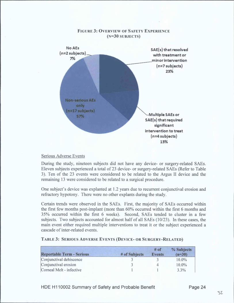
OVERVIEW OF SAFETY EXPERIENCE
Nineteen subjects (63%) experienced no, or only non-serious, adverse events. These non-serious events were treated routinely with medication or observation only. An additional7 subjects experienced SAEs that resolved with topical medication or minorinterventions.
The remaining 4 subjects were distinct from the other subjects in that they had a higherrate of adverse events due to a cascade of related events. In total, these 4 subjectsaccounted for 57% of all serious adverse events (SAEs) and 24% of all non-seriousadverse events. Refer to Figure 3.
HDE H1 10002 Summary of Safety and Probable Benefit Page 23
FIGURE 3: OVERVIEW OF SAFETY EXPERIENCE(N=30 SUBJECTS)
No AEs SAE(s) that resolved(n=2 subjects) with treatment or
7% .. minorintervention(n=7 subjects)
23%
Multiple SAEs orSAE(s) that required
significantintervention to treat
(n=4 subjects)13%
Serious Adverse Events
During the study, nineteen subjects did not have any device- or surgery-related SAEs.Eleven subjects experienced a total of 23 device- or surgery-related SAEs (Refer to Table3). Ten of the 23 events were considered to be related to the Argus II device and theremaining 13 were considered to be related to a surgical procedure.
One subject's device was explanted at 1.2 years due to recurrent conjunctival erosion andrefractory hypotony. There were no other explants during the study.
Certain trends were observed in the SAEs. First, the majority of SAEs occurred withinthe first few months post-implant (more than 60% occurred within the first 6 months and35% occurred within the first 6 weeks). Second, SAEs tended to cluster in a fewsubjects. Two subjects accounted for almost half of all SAEs (10/23). In these cases, themain event either required multiple interventions to treat it or the subject experienced acascade of inter-related events.
TABLE 3: SERIOUS ADVERSE EVENTS (DEVICE- OR SURGERY-RELATED)
Non-Serious Adverse EventsAny adverse event that did not meet the definition of an SAE was considered to be a non-serious adverse event. These events normally resolved without treatment or were treatedwith medical management (i.e. they did not require surgical intervention). There were140 non-serious device-or surgery-related adverse events (in 28 subjects), of which 78were device-related and the remaining 62 were surgery-related. The following non-serious events were reported (number of events is indicated in parentheses): ocular pain(17), conjunctival congestion (11), epiretinal membrane (11), elective revision surgery(7), non-serious hypotony (7), suture irritation (7), choroidal detachment (6), uveitis (6),inflammatory conjunctivitis (5), retinal thickening with cystoid macular edema (CME)(5), ocular inflammation (4), retinal thickening with no cystic changes (4), vitreoushemorrhage (4), headache (3), high intraocular pressure (3), hyphema (3), keraticprecipitates (3), comeal vascularization (2), epiphora (lacrimation) (2), and foreign bodysensation (2). There was one reported case of each of the following events: 3600circumferential vitreous band traction, choroidal effusion, conjunctival cyst, conjunctivaldehiscence, conjunctival erosion, comeal abrasion, corneal dryness, comeal epithelialdefect, corneal filaments, corneal fold, comeal suture broken, decrease in lightperception, fibrosis around the tack, filamentary keratitis, nausea, nystagmus increase,ocular fibrin, proliferative vitreo-retinopathy, ptosis, serous retinal detachment, tractionalretinal detachment, retinal folds, retinoschisis, rubeosis, scleral patch displacement,scleritis, sub-conjunctival eyelashes, and vertigo..
Surgical Re-Interventions
Nine subjects required a surgical re-intervention(s) to treat a device- or surgery-relatedadverse event(s). Seven subjects had elective revision surgery. Refer to Table 4.
In cases where it was necessary to remove all or part of the implant and/or tack (i.e. 1case of explant and 3 cases where the retinal tack was removed to reposition the implantduring an elective revision surgery), no adverse sequelae occurred.
HDE H1 10002 Summary of Safety and Probable Benefit Page 25
TABLE 4: SURGICAL RE-INTERVENTIONS
# of Subjects # of Events % Subjects(n=30)Re-intervention to treat an AE: 9 26.7%
One Argus 11 Implant experienced an intermittent communication link beginning at 10months post-implant which led to a significant decline in the functionality of the device.This device eventually failed approximately 4 years post-implant; however, the deviceremained implanted.
SOURCES OF POTENTIAL BIAS IN THE ARGUS II CLINICAL TRIAL
PROTOCOL DEVIATIONS
Protocol compliance was monitored throughout the study and protocol deviations wererecorded and evaluated for their potential effect on study outcomes. Approximately 25%of deviations were due to a single exam or a follow-up visit being performed out ofwindow (i.e. a few days/weeks early or late). Approximately 25% of deviations were dueto minor modification made to a test method or test schedule in advance of a formalprotocol amendment. Fifteen percent (15%) were due to an exam not being performed inthe fellow eye (e.g., eye exam or fluorescein angiography). Fifteen percent (15%) werefor a test not being performed due to equipment malfunction or a medical reason (e.g.,OCT not possible due to lack of view into the eye). The remaining 20% of deviationswere due to a test or exam not being completely (or correctly) performed. In summary,these protocol deviations which occurred during the study did not significantly affect theevaluation of the safety and probable benefit of the Argus II System.
INVESTIGATOR BIAS
HDE H110002 Summary of Safety and Probable Benefit Page 26
The potential for investigator bias in the study was limited in several ways. A total of 10sites participated in the study. Each site enrolled between I to 7 subjects, with theaverage being 3 subjects per site. Therefore, no investigator contributed more than 25%of the subjects in the study.
The practice of vitreoretinal surgery and ophthalmic follow-up is consistent throughoutthe US and Europe, where the study was conducted. All principle investigators, and, ifdifferent, all retinal surgeons, were chosen following the same selection criteria. Allwere of internationally recognized competence and underwent the same training andstudy preparations. None of the investigators responsible for the assessment of clinicaladverse events and outcomes had financial conflicts of interest.
Together, these measures limited the potential for investigator bias in the study.
REGIONAL BIAS
This study was conducted in the US and Europe. Fourteen subjects were enrolled in theUS and 16 were enrolled in Europe. The study was analyzed to ensure that the datacollected from centers outside the US were valid and to demonstrate that the ethicalaspects, methods used during the study, the practice of ophthalmology, and the quality ofthe investigators was comparable for the US and European sites. Also, the prevalence ofRetinitis Pigmentosa was shown to be similar in both regions. In addition, an analysis ofthe demographics of the subjects revealed that those enrolled in the Europe werecomparable to those enrolled in the US. This analysis demonstrated that there were nosignificant regional differences that could have affected the outcomes in the study.
GENDER BIAS
The subgroup analysis stratified by gender demonstrated that the outcomes with theArgus II System were comparable for men and women and that neither group performedsubstantially better or worse than the other on any of main outcomes in the study. Therewere only 9 women enrolled in the study; therefore, some observed minor differencesmay have been due to the small sample size of women. Based on this analysis, there is noevidence of a gender effect or difference in outcomes with the Argus II System betweenmen and women.
HDE H1 10002 Summary of Safety and Probable Benefit Page 27
XI. RISK PROBABLE BENEFIT ANALYSIS
The Argus II System provided all 30 subjects with benefit as measured by visual functiontests, although this level of benefit was variable. All 30 subjects were able to see visualpercepts when the Argus II was electrically activated.
On the Square Localization test (i.e., object localization), subjects were consistently ableto perform better with the System ON versus System OFF over the course of the study.Figure 4 displays the observed mean accuracy which indicates the subjects' meandistance from the center of the target square. Error bars represent the mean of thestandard error.
On the Direction of Motion test, subjects were consistently able to perform better withthe System ON versus System OFF over the course of the study. Figure 2 displays theobserved mean accuracy which indicates the mean response error between the angledisplayed and the subject's response. Error bars represent the mean of the standard error.
On the Grating Visual Acuity test, the most difficult of the 3 tests, 27% of subjects wereable to reliably score on the scale (between 1.6 and 2.9 logMAR with a confidenceinterval within the scale) at least once with the System ON, while none of the Argus IIsubjects were able to score on the scale with the System OFF in either eye (Refer toTable 5).
HDE H1 10002 Summary of Safety and Probable Benefit Page 28
FIGURE 4: SQUARE LOCALIZATION (OBSERVED MEAN ACCURACY)
NOTE: Since this test was introduced midway through the study, the Baseline to 12-month results wereonly from subjects enrolled in 2009. Subjects enrolled in the study in 2007 and 2008 first performed thistest at either their 18- or 24-month follow-up visit.
HDE H1 10002 Summary of Safety and Probable Benefit Page 2957
FIGURE 5: DIRECTION OF MOTION (OBSERVED MEAN RESPONSE ERROR)
NOTE: Since this test was introduced midway through the study, the Baseline to 12 month results wereonly from subjects enrolled in 2009. Subjects enrolled in the study in 2007 and 2008 first performed thistest at either their 18 or 24 month follow-up visit.
TABLE 5: GRATING VISUAL ACUITY
% (n) of Subjects WhoseVisual Acuity Improved to
>2.9 LogMAR*
System ON 27% (8)
System OFF Implanted Eye 0% (0)
System OFF Fellow Eye 0% (0)
* Best result at any follow-up visit.
The Argus II System was also able to provide subjects with benefit as measured byobjectively-scored, partially-controlled functional vision tests. Subjects consistentlyperformed better with the Argus II System ON vs. OFF on orientation and mobility tests(finding a door and following a line, Figure 6 and Figure 7, respectively).
FIGURE 6: DOOR TASK AVERAGE SUCCESS RATES (OBSERVED MEANS)
HDE H1 10002 Summary of Safety and Probable Benefit Page 30
Self-report questionnaires of activities of daily living and quality of life indicated mildimprovement (Massof Activity Inventory) or no change (VisQOL), respectively.
An assessment of Argus II subjects in and around their home by independent, certifiedlow vision rehabilitation specialists was also performed. This assessment, called theFunctional Low-vision Observer Rated Assessment (FLORA) was designed to evaluatehow the Argus II System affected subjects' well-being and functional vision. It wasadded to the study in 2010 at which time subjects' length of follow-up ranged from 1.4 to3.7 years post-implant.
In no cases did the assessors report that the Argus I System had a negative impact onsubjects. In 77% of cases, assessors using the FLORA determined that the subject wasreceiving (or had received at one time) functional vision and/or well-being benefit fromthe Argus II System. Refer to Table 5.
HDE H1 10002 Summary of Safety and Probable Benefit Page 32
TABLE 6:. SUMMARY OF FLORA RESULTS (N=26 SUBJECTS)
20 (77%) 6 (23%)Note: 4 subjects did not participate in the FLORA.
CONCLUSIONS DRAWN FROM THE STUDIES
Pre-clinical in vitro and in vivo testing demonstrated the Argus II Retinal ProsthesisSystem meets applicable international standards and Second Sight-defined designrequirements. A 6 patient feasibility study with a first generation device (Argus 16)demonstrated that a retinal prosthesis could be implanted chronically in human subjects.Long-term data collected from this study demonstrated the safety and proof of conceptthat an electrode array could be used to stimulate the retina to elicit visual percepts inblind subjects. Data from the Argus 16 study provided important design input for thenext generation implant (Argus II), external equipment, and stimulation fitting strategies.
A 30 subject prospective clinical trial was conducted which demonstrated that the ArgusII System is safe and will provide a probable benefit to the indicated patient population.All subjects in the clinical trial had been implanted a minimum of 2.5 years follow-updata, with several subjects having over 4 years of follow-up data. The long-term safetyresults are acceptable, with the majority of events resolving with no or minimalintervention. Serious adverse events were clustered in a few subjects and most occurredwithin the first 6 months post-implant. Furthermore, based on a trend toward reducedadverse events as the trial progressed and more experience was gained with the device, itis likely that the safety profile of the Argus II System will continue to improve withincreasing surgical experience with these devices. The risk, therefore, is acceptable,especially when considering that the adverse events are occurring in blind eyes, for whichdecreased vision is not a significant risk. The performance analysis showed that amajority of subjects using the Argus II System have improved visual function that rangedbetween subjects from light perception to at least hand motion, or counting fingers vision.Assessments of subjects in their normal environments by low vision therapists alsodemonstrated that the majority of subjects received positive effects from the Argus IISystem in terms of well being and/or functional vision. These results represent asignificant improvement and benefit for these subjects, especially when considering thatthey have no other approved treatment options for their irreversible degenerative disease.
HDE H1 10002 Summary of Safety and Probable Benefit Page 33(41
When considering all the data, it has been demonstrated that the Argus II System posesan acceptable risk to people with severe to profound retinitis pigmentosa in exchange fora probable benefit - that of improvements in visual function, functional vision, and/orwell-being.
XII. PANEL RECOMMENDATION
At an advisory meeting held on September 28, 2012, the Ophthalmic Devices Panel ofthe Medical Devices Advisory Committee recommended that Second Sight MedicalProduct's HDE for the Argus® II Retinal Prosthesis System be approved.
XIII. CDRH DECISION
CDRH has determined that, based on the data submitted in the HDE, that the Argus® IIRetinal Prosthesis System will not expose patients to an unreasonable or significant riskor illness or injury, and the probable benefit to health from using the device outweighsthe risks of illness or injury, and issued an approval order on February 13, 2013.
XIV. APPROVAL SPECIFICATIONS
Directions for use: See the Physician's Labeling.
Hazards to Health from Use of the Device: See Indications, Contraindications,Warnings, Precautions and Adverse Events in the labeling.
Postapproval Requirements and Restrictions: See Approval Order.
XV. REFERENCES
General Program Memorandum #G91 - 1: Device Labeling Guidance.http://www.fda.gov/cdrh/g91 - .html
HDE H1 10002 Summary of Safety and Probable Benefit Page 34
Issued: 12/11/01Revised: 3/10/05
HDE H1 10002 Summary of Safety and Probable Benefit Page 35


![Summary of Safety and Probable Benefit - Food and Drug ... · Summary of Safety and Probable Benefit I. GENERAL IN],ORMATION Device Generic name(s): Enzyme-linked Immunosorbent Assay](https://static.documents.pub/doc/80x56/5ea7a86fe47688183e350c08/summary-of-safety-and-probable-benefit-food-and-drug-summary-of-safety-and.jpg)


