No. 4567. MARCH 11, 1911. The Hunterian Lecture ON THE ASSOCIATION OF DISEASE OF THE MOUTH WITH RHEUMATOID ARTHRITIS AND CERTAIN OTHER FORMS OF RHEUMATISM. Delivered at the Royal College of Surgeons of England on March 6th, 1911, BY KENNETH W. GOADBY, D.P.H. CAMB., M.R.C.S.ENG., L.R.C.P.LOND., L.D.S. R.C.S.ENG., HUNTERIAN PROFESSOR, ROYAL COLLEGE OF SURGEONS OF ENGLAND ; LECTURER ON BACTERIOLOGY AT THE NATIONAL DENTAL HOSPITAL, GREAT PORTLAND STREET, LONDON, W. MR. PRESIDENT, VICE-PRESIDENT, AND FELLOWS,-I wish to express my deep sensibilitv of the honour you have con- ferred upon me in electing me as one of vour Hunterian professors this year. The subject which I have with your consent chosen for this lecture is one which I feel sure would have appealed to that indefahgable worker in whose memory these lectures are established, and my aim will be, as a humble follower of John Hunter, to endeavour to correlate certain pathological facts in a new light. When investigating any of the diseases affecting the joints of the human body the investigator is at the very outset hampered by the nomenclature of his subject : rheumatoid arthritis, osteo-arthritis, rheumatic gout, arthritism, poly- arthritis, to cite a few of the hundred names applied to disease of the joints. In the earliest writings upon medicine, gout and rheumatism were confused and regarded as mani- festations of one disease ; in fact, Sydenham was of the opinion that rheumatism was a modern disease and unknown to the ancients. It is not necessary for the present purpose to critically survey the various divisions and subdivisions of diseases of the joints. A summary of the terminology and an epitome of the history will be found in Llewellyn Jones’s 2 book on arthritis deformans. REMARKS ON ETIOLOGY. Recently the leaning of most writers has been to regard swellings affecting the joints as divisible into (a) those affections which have their primary lesions in the capsule and peri-articular tissues; and (b) those in which the seat of the lesion is in the cartilages and bony portions of individual joints. Broadly speaking, the first group, com- prising affections of the peri-articular tissues, are generally regarded as of infective origin due to bacteria, either within the joint itself or to a toxin produced by organisms in some other part of the body ; as Llewellyn Jones points out, many cases of arthritis in which a bacteriological examination is negative closely resemble the cases of ascer- tained microbic origin, not only as regards the character of the local arthritic change, but also the general aspect of the patient, the mode of onset of the disease, and its clinical course and behaviour under treatment. The second group, in which the disease apparently commences in the bony and cartilaginous structures of the joints, is not usually con- sidered of direct infective origin. Goldthwait attaches great value to X ray photographs in differentiating infectious arthritis from that of his two other varieties, atrophic and hypertrophic arthritis, but Kienboch, and more recently Lindsay, have shown that in gonorrhceal arthritis the bones may not preserve their normal density. It is not only in gonorrhoxal arthritis, however, but even in ordinary septic processes without any definite arthritic affection, that thinning and alteration in the density of the bone take place. Moreover, a septic process of low virulence, but of long-continued action, in the mouths of human beings invariably produces 1 Opera, Sec. VI., c. 5. Arthritis Deformans, 1909. 3 Infectious Arthritis, Boston Medical and Surgical Journal, 1904. 4 Volkmann’s Sammlung Klinischer Vorträge, N.F., No. 315. 5 Bulletin of the Committee for the Study of Special Diseases, October, 1908, vol. ii., p. 106 alteration in the density of the bone in the neighbourhood of the infecting process,’as the accompanying radiograph shows (Fig. 1 ) The infection is not by any means always associated with a copious discharge of pus, but, as I pointed out in my Erasmus Wilson lecture in 1907, loss of density (rarefaction) takes place without the discharge of pus into the mouth, and such rarefaction may be accompanied by sclerosis. For some years the belief in the infective origin of arthritis deformans has been gaining ground, more especi- ally with that group of cases usually described as rheumatoid arthritis. The initial joint disturbances in acute poly- arthritis caused by the gonococcus, the acute onset of rheumatoid arthritis in which no organisms have been found, and the onset of acute articular rheumatism, exhibit similarity ; and further, the changes in the joints found in acute polyarthritis of demonstrable infective origin are closely related to the joint changes in which after careful bacteriological search no infecting agent was found. It is fully admitted that clinically an unbroken progressive series may be established from osteo-arthritis of unknown origin through rheumatoid arthritis, infective polyarthritis, to acute rheumatism, although individually each may be differentiated by certain typical symptoms. But it seems also an equally logical supposition to assume that such a series of cases, although perhaps not owing their cause to a common specific infective agent, may yet be due to infec- tive agents possessing differing degrees of infectivity or virulence. To sum up this preliminary survey of the subject, a large number of cases of arthritis deformans, using the term in its inclusive sense, are now recognised to be of infective origin, others are of definite neural origin, but between the two are a considerable number which, according to some observers, may be infective, according to others atrophic, neural, or even idiopathic. CLINICAL EVIDENCE. For some time my attention has been drawn to the presence of various so-called rheumatic symptoms in persons subject to disease of the jaws, particularly the type of disease known as pyorrhoea alveolaris, and associated with a dis- charge of pus from the gum margins and rarefaction of the alveolar process, and frequently the body of the bone. Lindsay in 1908 gives a report of 172 cases of rheumatoid arthritis in which particular inquiry was made for infective foci. In 88 of these cases an infective focus was found, 15 being men and 73 women, and 19 of the 73 women had definite pyorrhoea alveolaris. Lindsay further states that in the majority of the 172 cases some inflammation of the mouth existed, but only those cases where definite pus was to be found were included as pyorrhoea alveolaris. Lambert 7 in the same year says that of 190 cases 141 (76 per cent.) had badly decayed teeth or the teeth had dropped out and 49 (23’8 8 per cent.) showed teeth in fairly good condition ; these latter were the younger patients. For many years pyorrhoea alveolaris was classed by the majority of surgeons as "gout in the mouth," and even amongst dental surgeons a special anatomical form of tooth is often referred to as a " gouty " tooth, and it is worthy of note that the particular gouty or rheumatic tooth so-called is invariably one in which the antero-posterior diameter of the crown is greater than the antero-posterior diameter measured at the gum margin ; such teeth are liable to permit the accumulation of food in the interstitial spaces, to injuries of the gum and periosteum, and therefore to local infection. Strange as it may sound, I have often heard the pus oozing from the gum margins ascribed to the patient’s rheumatic diathesis. Two main types of disease of the alveolar processes of the jaws may be distinguished in relation to rheumatic affec- tions:-(a) Easily bleeding hypertrophied gums, wasting of alveolus, discharge of pus from the alveolar margins. Loosening of all or individual teeth. (b) Thickening of the alveolar process (sclerosis) with area of rarefaction between individual teeth; alteration in position of individual teeth without loosening ; showing atrophic gums, little or no discharge of pus. Microscopically pus may be found. In the more common types of mouth infection, where copious discharge of pus is found, where 6 Erasmus Wilson Lecture on Pyorrhoea Alveolaris, THE LANCET, March 9th, 1907, p. 633. 7 Bulletin of the Committee for the Study of Special Diseases, vol. ii., pp. 11-95. K
Transcript
No. 4567.
MARCH 11, 1911.
The Hunterian LectureON
THE ASSOCIATION OF DISEASE OF THEMOUTH WITH RHEUMATOID ARTHRITIS
AND CERTAIN OTHER FORMS OFRHEUMATISM.
Delivered at the Royal College of Surgeons of England onMarch 6th, 1911,
BY KENNETH W. GOADBY, D.P.H. CAMB.,M.R.C.S.ENG., L.R.C.P.LOND., L.D.S. R.C.S.ENG.,
HUNTERIAN PROFESSOR, ROYAL COLLEGE OF SURGEONS OF ENGLAND ;LECTURER ON BACTERIOLOGY AT THE NATIONAL DENTAL HOSPITAL,
GREAT PORTLAND STREET, LONDON, W.
MR. PRESIDENT, VICE-PRESIDENT, AND FELLOWS,-I wishto express my deep sensibilitv of the honour you have con-ferred upon me in electing me as one of vour Hunterianprofessors this year. The subject which I have with yourconsent chosen for this lecture is one which I feel sure wouldhave appealed to that indefahgable worker in whose memorythese lectures are established, and my aim will be, as ahumble follower of John Hunter, to endeavour to correlatecertain pathological facts in a new light.When investigating any of the diseases affecting the joints
of the human body the investigator is at the very outset
hampered by the nomenclature of his subject : rheumatoidarthritis, osteo-arthritis, rheumatic gout, arthritism, poly-arthritis, to cite a few of the hundred names applied todisease of the joints. In the earliest writings upon medicine,gout and rheumatism were confused and regarded as mani-festations of one disease ; in fact, Sydenham was of theopinion that rheumatism was a modern disease and unknownto the ancients.
It is not necessary for the present purpose to criticallysurvey the various divisions and subdivisions of diseases ofthe joints. A summary of the terminology and an epitomeof the history will be found in Llewellyn Jones’s 2 book onarthritis deformans.
REMARKS ON ETIOLOGY.
Recently the leaning of most writers has been to regardswellings affecting the joints as divisible into (a) thoseaffections which have their primary lesions in the capsuleand peri-articular tissues; and (b) those in which the seatof the lesion is in the cartilages and bony portions ofindividual joints. Broadly speaking, the first group, com-
prising affections of the peri-articular tissues, are generallyregarded as of infective origin due to bacteria, eitherwithin the joint itself or to a toxin produced by organismsin some other part of the body ; as Llewellyn Jones pointsout, many cases of arthritis in which a bacteriologicalexamination is negative closely resemble the cases of ascer-tained microbic origin, not only as regards the character ofthe local arthritic change, but also the general aspect of thepatient, the mode of onset of the disease, and its clinicalcourse and behaviour under treatment. The second group, inwhich the disease apparently commences in the bony andcartilaginous structures of the joints, is not usually con-sidered of direct infective origin.Goldthwait attaches great value to X ray photographs
in differentiating infectious arthritis from that of histwo other varieties, atrophic and hypertrophic arthritis,but Kienboch, and more recently Lindsay, have shownthat in gonorrhceal arthritis the bones may not preservetheir normal density. It is not only in gonorrhoxalarthritis, however, but even in ordinary septic processeswithout any definite arthritic affection, that thinning andalteration in the density of the bone take place. Moreover,a septic process of low virulence, but of long-continuedaction, in the mouths of human beings invariably produces
1 Opera, Sec. VI., c. 5.Arthritis Deformans, 1909.
3 Infectious Arthritis, Boston Medical and Surgical Journal, 1904.4 Volkmann’s Sammlung Klinischer Vorträge, N.F., No. 315.
5 Bulletin of the Committee for the Study of Special Diseases,October, 1908, vol. ii., p. 106
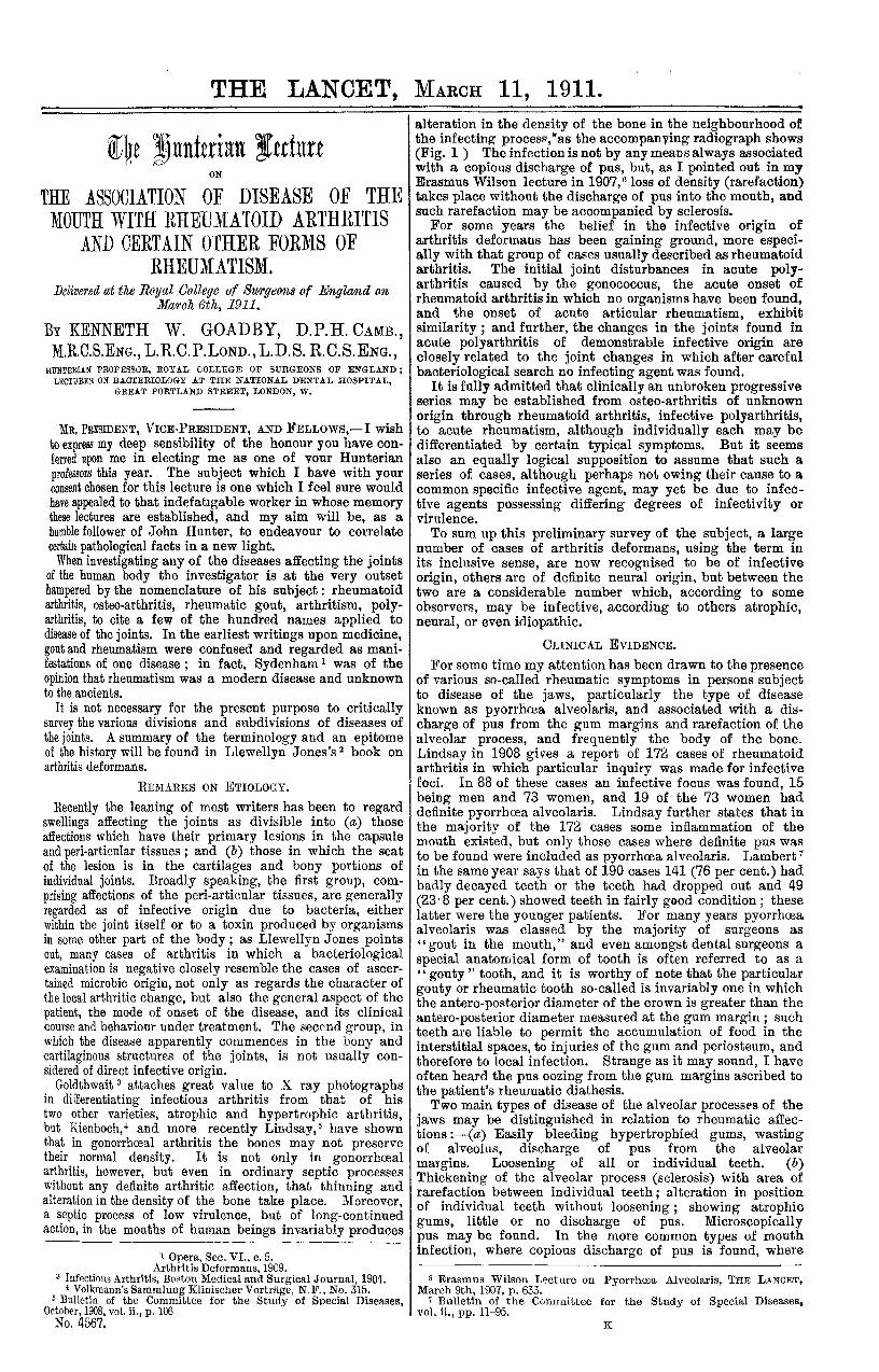
alteration in the density of the bone in the neighbourhood ofthe infecting process,’as the accompanying radiograph shows(Fig. 1 ) The infection is not by any means always associatedwith a copious discharge of pus, but, as I pointed out in myErasmus Wilson lecture in 1907, loss of density (rarefaction)takes place without the discharge of pus into the mouth, andsuch rarefaction may be accompanied by sclerosis.For some years the belief in the infective origin of
arthritis deformans has been gaining ground, more especi-ally with that group of cases usually described as rheumatoidarthritis. The initial joint disturbances in acute poly-arthritis caused by the gonococcus, the acute onset ofrheumatoid arthritis in which no organisms have been found,and the onset of acute articular rheumatism, exhibitsimilarity ; and further, the changes in the joints found inacute polyarthritis of demonstrable infective origin are
closely related to the joint changes in which after carefulbacteriological search no infecting agent was found.
It is fully admitted that clinically an unbroken progressiveseries may be established from osteo-arthritis of unknown
origin through rheumatoid arthritis, infective polyarthritis,to acute rheumatism, although individually each may bedifferentiated by certain typical symptoms. But it seemsalso an equally logical supposition to assume that such aseries of cases, although perhaps not owing their cause to acommon specific infective agent, may yet be due to infec-tive agents possessing differing degrees of infectivity or
virulence.To sum up this preliminary survey of the subject, a large
number of cases of arthritis deformans, using the term inits inclusive sense, are now recognised to be of infectiveorigin, others are of definite neural origin, but between thetwo are a considerable number which, according to some
observers, may be infective, according to others atrophic,neural, or even idiopathic.
CLINICAL EVIDENCE.
For some time my attention has been drawn to the presenceof various so-called rheumatic symptoms in persons subjectto disease of the jaws, particularly the type of diseaseknown as pyorrhoea alveolaris, and associated with a dis-
charge of pus from the gum margins and rarefaction of thealveolar process, and frequently the body of the bone.Lindsay in 1908 gives a report of 172 cases of rheumatoidarthritis in which particular inquiry was made for infectivefoci. In 88 of these cases an infective focus was found, 15being men and 73 women, and 19 of the 73 women haddefinite pyorrhoea alveolaris. Lindsay further states that inthe majority of the 172 cases some inflammation of themouth existed, but only those cases where definite pus wasto be found were included as pyorrhoea alveolaris. Lambert 7
in the same year says that of 190 cases 141 (76 per cent.) hadbadly decayed teeth or the teeth had dropped out and 49(23’8 8 per cent.) showed teeth in fairly good condition ; theselatter were the younger patients. For many years pyorrhoeaalveolaris was classed by the majority of surgeons as
"gout in the mouth," and even amongst dental surgeons aspecial anatomical form of tooth is often referred to as a" gouty " tooth, and it is worthy of note that the particulargouty or rheumatic tooth so-called is invariably one in whichthe antero-posterior diameter of the crown is greater than theantero-posterior diameter measured at the gum margin ; suchteeth are liable to permit the accumulation of food in theinterstitial spaces, to injuries of the gum and periosteum, andtherefore to local infection. Strange as it may sound, I haveoften heard the pus oozing from the gum margins ascribed tothe patient’s rheumatic diathesis.Two main types of disease of the alveolar processes of the
jaws may be distinguished in relation to rheumatic affec-tions:-(a) Easily bleeding hypertrophied gums, wastingof alveolus, discharge of pus from the alveolar
margins. Loosening of all or individual teeth. (b)Thickening of the alveolar process (sclerosis) with area ofrarefaction between individual teeth; alteration in positionof individual teeth without loosening ; showing atrophicgums, little or no discharge of pus. Microscopicallypus may be found. In the more common types of mouthinfection, where copious discharge of pus is found, where
6 Erasmus Wilson Lecture on Pyorrhoea Alveolaris, THE LANCET,March 9th, 1907, p. 633.
7 Bulletin of the Committee for the Study of Special Diseases,vol. ii., pp. 11-95.
K
640
the teeth themselves become altered in position and length,and which become entirely loosened owing to the destructionof their sockets, the appearance of the bone in the neighbour-hood of the tooth sockets is one of a general rarefyingosteitis. This is perhaps the more common type. The
accompanying skiagraphs show the extent to which rarefac-tion and perosis of the bone may occur, extending in somecases into the body of the bone itself. [A series of X rayphotographs were shown illustrating this point.] As thisrarefaction commonly gives no pain, the patient’s attentionis usually not called to the distressing condition untilthe teeth themselves are loose. Occasionally evidence ofreparative sclerosis is seen.
I have already drawn attention to the importance of thismouth disease as the origin of certain generalised infections,but the peculiar import of alveolar osteitis in arthritisdeformans lies in the fact that the alveolus of the jaw isthe position in the body where the bony tissues are exposedto septic infection, while owing to the absence of pain treat-ment is most often neglected, the insidiousness of the onsetmore often precludes its early diagnosis, and, perhaps moreimportant still, the infecting organisms become adapted tobony tissues.Lindsay states that in the Royal Mineral Water Hospital at
Bath particular attention is paid to the presence of infectiveconditions of the mouth, and cites two cases where theremoval of the septic teeth arrested but did not cure pro-gressive disease of the joints. Llewellyn Jones discussingdiagnosis says : " Strong presumptive evidence of infectiousarthritis may be drawn from the presence of any local sourceof infection, such as pyorrhoea alveolaris, otorrhcea, and soforth," and MacNamara states that he had traced manycases to the chronic poisoning of septic teeth. Luff, 9
Spender, Fayerweather," and many others consider thegastro-intestinal tract as the origin of infection, whileHunter,12 myself, and others have demonstrated the relation-ship of oral sepsis to gastro-intestinal affections. It is
truly said that septic diseases of the mouth are remarkablycommon and that relatively arthritis deformans is rare ; but,on the other hand, I can confirm Grünbaum’s 13 experiencethat few, if any, cases of arthritis deformans have soundmouths. Further, the bacteria I have isolated from themouth do not, as a rule, possess a high degree of virulenceeven when injected in large doses into laboratory animals,and it is not illogical to suppose micro-organisms of a lowtype of virulence habituated to the environment of bonytissue and peri-articular structures would be more likely toact as infecting agents in chronic processes than organismspossessing a high degree of virulence. This suppositionreceives considerable support from my own investigation,before detailing which it is necessary briefly to consider thepresent bacteriological knowledge of rheumatoid arthritis.
BACTERIOLOGY.The earliest bacteriological work on Arthritis Deformans
is that of Schüller, H who obtained fluid from the joints in atype of arthritis deformans clinically associated with earlyatrophy of the muscles, thickening of the synovial membranesof the joints and peri-articular structures, termed byhim as polyarthritis chronicc villosa bacilzaris. Schullerdescribed certain short bacillary forms which were easilydecolourisable by the staining methods he used ; in a laterpublication he says the organism stained by Gram’s method.The organisms were found but rarely in the joint fluid, theirseat of selection being the capsule and other structuresoutside the joint. Cultures were obtained by allowing fluidfrom the joint to drop through a cannula on to the culturemedia, or by triturating portions of the thickened synovialmembrane or capsule and inoculating from the mass on toculture tubes. SchüUer’s bacillus produced greyish-whitediscrete colonies on agar in from two to six days, and grewreadily on gelatin which was slowly liquefied. The organismremained alive on culture media for several months, retain-ing its virulence. Subcutaneous injections of 0’5 gramme
8 Proceedings of the Royal Medical and Chirurgical Society, ThirdSeries, vol. xi., p. 47.
9 THE LANCET, March 12th, 1910, p. 712. 10 Osteo-arthritis, 1889.11 American Journal of the Medical Sciences, 1905, July-December,
New Series, vol. cxxx.12 Severest Anæmias, &c.
13 The Clinical Journal, vol. xxxvi., 1910, p. 289.14 Verhandlungen der Deutschen Gesellschaft fiir Chirurgie, 1892 ;
Berliner Klinische Wochenschrift, Sept. 4th, 1893 ; American Journal ofthe Medical Sciences. December, 1906; Verhandlungen des Congressesfür Innere Medicin, Berlin, 1897.
to 1 gramme in rabbits were fatal in 24 hours ; injection of asimilar quantity into a joint produced swelling only, whichbecame permanent in character, and resulted in a persistentlimp. No pus was found in the joints, but the synovialmembrane in places showed villous and brown fringes,sections of which were shown to contain the injectedorganisms. They were polar-staining bacilli which formedspores. Cocci were often found together with the bacilli,but were not regarded as playing an important part.The next bacteriological work was that of Blaxalll;
in 1896, who examined fluid aspirated by Bannatyne from thejoints in rheumatoid arthritis and found that by prolongedstaining in concentrated solutions an organism could bedemonstrated in the synovial fringes. This organism showedmarked polar staining, the average length was 8;u, and theaverage breadth 0’ 6. Sometimes large numbers were foundcongregated round leucocytes. The organism almost com-pletely decolourised by Gram’s method, but not absolutely.Only two out of the 18 cases he examined showed organismsreferable to skin contamination. He also examined anumber of joint fluids in chronic synovitis, gonorrhcea, andother affections, without seeing this special form of organism.Attempts at cultivation were at first unsuccessful, but finallya growth was obtained on beef broth, in the form of a
slightly flocculent deposit compared to gold dust. Thebacilli were 3 to 4/ in length and showed the same polarstaining as in the direct smears. No chains were seen.Upon agar the colonies developed very slowly, and werenever larger than a pin’s point. The bacilli grew on milkwithout producing acid or precipitating the casein. Intra-venous inoculations into animals produced no definite re-
action, and the animals recovered; slight swelling of theinoculated joints was observed in two instances which,however, cleared up. Both Bannatyne 16 and Schuller’cases were ones in which no bony or cartilaginous changeswere present.
Kroh,1’ in his paper on Experimentelle Arthritis Deformans,gives an account of a number of cases of arthritis deformanswhich were produced experimentally in animals (rabbits).The method adopted was the resection of a portion of thejoint, leaving the joint itself unaffected by septic processes,but partially out of action, due to the removal either of
portions of the condyles or other anatomical parts of theknee-joints. No suppuration followed the small operation,but skiagrams taken at intervals after the operation showeda progressive change had taken place in the joints, causinghypertrophy of the remaining articular surfaces and the bonytissues supporting them, and producing a certain amountof swelling and deformity of the external portion of thejoint.
Poynton and Paine,18 Ainley Walker and Beaton,19 andBeattie made cultures from cases of acute rheumatism,
Poynton and Paine described a streptocneczcs ’I’ne16matimt8found in the lesions of rheumatic fever, and which, wheninoculated into animals, produced infective endocarditis, butthey say they have never personallv found the occurrence ofthis coccus in the joints of human beings. Beattie gives theresult of cultures made from a case of acute rheumaticfever, which died six days after admission to the Royat
15 THE LANCET, April 25th, 1896, p. 1120.is Ibid.
17 Deutsche Zeitschrift für Chirurgie, Leipzig, 1909, vol xcix., p. 425.18 THE LANCET, Sept. 22nd and 29th, 1900, pp. 861 and 932.
19 Brit. Med. Jour., 1903, vol. i., p. 237.
DESCRIPTION OF ILLUSTRATIONS.FIG. I.-X ray photograph showing loss of alveolus; the dotted line
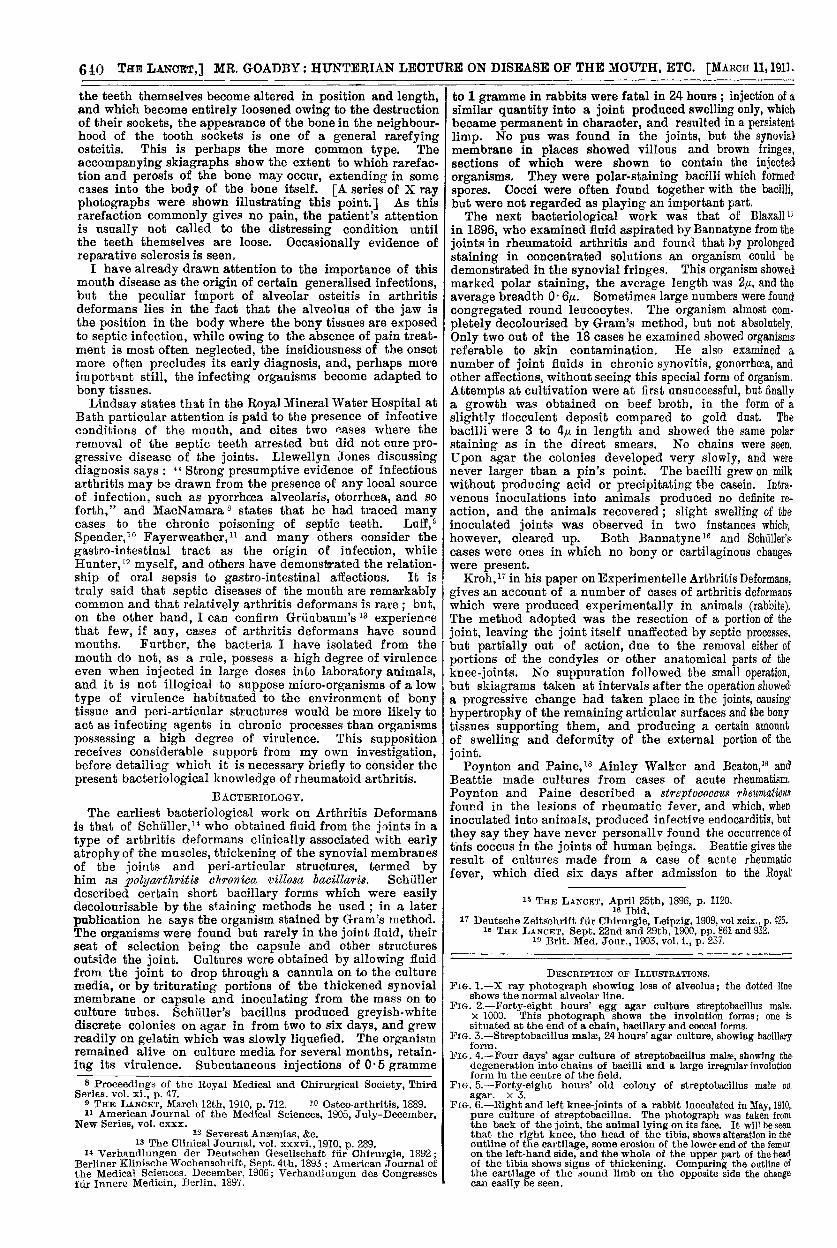
shows the normal alveolar line.FIG. 2.-Forty-eight hours’ egg agar culture streptobacillus mala.
x 1000. This photograph shows the involution forms; one issituated at the end of a chain, bacillary and coccal forms.
FIG. 3.-Streptobacillus malse, 24 hours’ agar culture, showing bacillaryform.
FIG. 4.-Four days’ agar culture of streptobacillus malse, showing thedegeneration into chains of bacilli and a large irregular involutionform in the centre of the field.
FIG. 5.-Forty-eight hours’ old colony of streptobacillus malae on, agar. x 3.five. 6.-Right and left knee-joints of a rabbit inoculated in May, 1910,
pure culture of streptobacillus. The photograph was taken fromthe back of the joint, the animal lying on its face. It will be seenthat the right knee, the head of the tibia, shows alteration in theoutline of the cartilage, some erosion of the lower end of the femuron the left-hand side, and the whole of the upper part of the headof the tibia shows signs of thickening. Comparing the outline ofthe cartilage of the sound limb on the opposite side the changecan easily be seen.
641MR. GOAI BY : HUNTERIAN LECTURE 014-disease OF THE MOUTH, ETO.
Fict. 1.
Fic.. 3.
Fir. 5.
FIG. 2.
F.G. 4.
FiG. 6
642
Infirmary, Edinburgh. Subcultures of the organism obtainedfrom the joints of the case were inoculated into animalsintravenously, the amount used being two whole agar tubes.The organism was recovered in pure culture from thetissues of the animals afterwards, the injections having pro-duced arthritic changes in four days. Two animals were in-oculated in the knee-joints, producing definite arthritis of anacute type which gradually subsided but never disappeared,the animals ultimately regaining their normal health. Novisceral lesions were found at the necropsy, but somethickening and infiltration remained round the joints.Cultivations from the fluid and from the cartilaginous surfaceswere sterile. For the control, streptococci obtained from thevegetation in a case of malignant endocarditis were inoculatedinto the knee-joints of two rabbits. The rabbits died fromstreptosoccal septicsemia with no trace of arthritic changes.
In 1903 Beaton and Ainley Walker described the culturalcharacters of an organism, a streptococcus, found in acuterheumatism. This was also found in cases of acute
rheumatism, chorea, and endocarditis. They considered itsimilar to that found by Paine and Poynton, and Beattie.Large doses of the organism produced death within 40 hoursin rabbits, six or more agar cultures being used. Moderatedoses caused effusion of the joints and tendon sheaths, anddeath up to as late as three weeks, whilst small doses of theorganism obtained from rabbits dead from infection with thestreptococcus produced polyarthritis in animals (rabbits).
In 1907 Walker 20 again returns to the subject, and comesto the conclusion that the organism is a streptococcus differ-ing in some respects from the ordinary streptococcuspyogenes.
It will be noted that the last three or four observers when
dealing with acute infection of rheumatic fever type foundthat the organisms produced an acute polyarthritis whichcleared up practically without a permanent lesion in the
joints, similar in this respect to the lesions of rheumaticfever in the human subject.Fayerweather worked at the bacteriology of arthritis
deformans, his cases belonging to Schuller’s type of poly-arthritis. Fayerweather investigated four cases, in all ofwhich were found bacilli, pleomorphic in type, alternatingbetween long rods with slightly rounded ends, and diplo-coccal or lanceolate forms, which ultimately grew out intodefinite bacilli. Two of the organisms produced lique-faction of blood serum, but the other two did not. Ofthe two which did not produce liquefaction, one was non-motile and the other motile, and the ron-motile one was notfound to produce spores. The organisms differed slightlyin their morphological and cultural characters. Ten rabbitswere inoculated from the cultures, either intravenously, sub-cutaneously, or into the knee-joints. Subcutaneous andintraperitoneal inoculation produced no effect whatever.Three rabbits were inoculated with cultures of the first
organism, a bacillus which produced some slight changeswhen inoculated into the knee-joints, whereas the third
organism, a diplococcal form, produced a uniform thicken-ing of the synovial capsule. No changes were seen in thebones, and no muscle wasting, and the animal was appa-rently quite normal.A point of some importance in Fayerweather’s work is
that although the organisms produced no effect wheninoculated subcutaneously or intravenously, in severalinstances change did occur in the joint, the synovial fringesbeing the chief structure affected.The bacteriological findings in articular rheumatism may
be divided into two series, the one acute-in which Poyntonand Paine, Ainley Walker and Beaton, and Beattie all agreein finding streytococci and chronic in which Schüller,Bannatyne, and Fayerweather found organisms of bacillarytypes, Schuller and Fayerweather both having producedlesions of the joints of rabbits by inoculation with cultures ofthe isolated bacilli.One other observer remains to be cited-namely, Painter 2i
-who treated nine cases of rheumatoid arthritis withvaccines prepared from the organism described by Fayer-weather and claimed by Schuller to be identical with the onehe first described. Unfortunately, no reference is made towhich of Fayerweather’s organisms was used. The opsonicindex to the organism was low in every case tested, but astock, and not an autogenous, vaccine was used. No
20 Brit. Med. Jour., May 25th, 1907, p. 1233.21 Boston Surgical and Medical Journal, Nov. 7th, 1907.
improvement was observed in any of the cases treated,although a definite exacerbation of the local joint symptomsfollowed the injection of the vaccine in all the cases.With the foregoing survey of the subject, I now turn to my
own researches on the
ESPECIAL RELATION OF RAREFYING ALVEOLAR OSTEITIS.TO ARTHRITIS DEFORMANS.
The amount of destruction of the bony tissues in the jaw isoften out of all proportion to the severity, or apparent severity,of the infection, and even when pus is found welling up fromthe gum sockets one cannot fail to be surprised at the amountof deep infection the bone has sustained.The following X ray photographs taken for me by my
friend Mr. Reid exhibit the loss of tissue and rarefaction
particularly well. [A series of X ray photographs of the jawwere shown illustrating this point.] ]
Fig. 1 shows in the lower jaw the extreme destructionwhich may take place ; not only the alveolus has been lost,but a distinct cavity has been formed in the body of the bonereaching downwards into the inferior dental canal. Thebicuspid is also deeply infected. In the upper jaw someattempt has been made at repair. The photographs are
merely examples of a large number which might be adducedto show the ravages that take place in the hard tissuesof the jaw under the influence of chronic suppurativelesions. It must be noted also that in many cases therewas little pus to be found. In Case 3 particularly itwas only with the greatest difficulty and even on micro.
scopical examination that definite pus was to be demon-strated. The teeth themselves were not loose, but of coursegave definite evidence on physical examination of loss of
bony attachment.The peculiar interest of these photographs is the presenceof rarefaction, together with sclerosis. Such a condition inmany ways is comparable to arthritis deformans, and wasthe starting-point of my inquiry as to the possibility of theproduction of experimental arthritis deformans in animals bythe use of the organisms, which evidently possessed a
peculiar predilection for destruction of bony tissues.In the first stage of my experiments I attempted to repro-
duce disease of the joints by the intravenous inoculation oforganisms isolated from such cases as the ones shownin the X ray photographs. To begin with, the pus itselfwas used for inoculation into rabbits intraperitoneally,subcutaneously, and intravenously. Only a few of these
experiments are given in the table of animal experiments,as in all instances in which such inoculation was per-formed no inflammatory reaction was produced in the
joints. Localised suppuration indeed was formed in manyinstances, and in not a few cases the animals died some-times from toxsemia, sometimes from generalised infection.Attempts were therefore made to determine which of thehorde of bacteria in the pus were the infecting agents.Resource was had to agglutination, the opsonic and phago-cyte index, estimations both before and after massage of theaffected parts, and to direct animal experiments. A numberof organisms were isolated, certain of which undoubtedlyproduced local suppuration when inoculated subcutaneously,and it was thought that perhaps these were the most likelyto give positive results. The method described by Beattiewas made use of, animals were inoculated intravenously withcultures of the organisms, and the animals afterwards exposedto cold. Beattie, who made use of this method, found thatcertain animals which he had injected with streptococciobtained from rheumatic cases exhibited signs of arthriticwhen the temperature of the laboratory in which they werehoused went down to freezing-point during temporary damageto the heating apparatus. None of my animals exposed tocold after inoculation showed signs of arthritis.Most of the organisms obtained from the alveolar pro-
cesses are non-motile, and therefore the method of agglutina-tion did not give particularly satisfactory results, and wasabandoned.The estimation of the opsonic index gave rather better
results, but it was found that not only one organism, butoften four or five different species isolated from the gummargins underwent variations in their opsonic index,particularly when the blood was taken before and after theoperation of scaling and cleaning out the gum pockets.Amongst the organisms isolated from the gum margin
one in particular seemed to give more variation in its opsonicindex than any of the others. This organism was at first
643
regarded as a Gram-negative streptococcus, but as Gram-negative streptococci are exceedingly rare a good deal ofattention was given to its morphology. It was found oncareful examination that the organism in question differedvery much in its general morphology, and in a certainnumber of other particulars, from the ordinary streptococcusof the mouth.
It is true that streptococcal forms exist in many instancesand, as will be seen in Fig. 2, a fairly well-marked strepto-coccal form is seen, but with a tendency to elongation of theindividual elements. Not only this, but there is definiteirregular staining, giving the suggestion of polar-stainingbacilli. This polar staining is much better demonstratedwith Loffier’s blue. It is, of course, known that strepto-cocci when grown for any length of time in the laboratoryhave a tendency to form bacillary types ; and it is also wellknown, as pointed out by von Lingelsheim in Kolle and,Wassermann’s "Handbuch der Pathogenen Microorganismen,"that alternations of bacillary and coccal forms are notuncommon in streptococcal chains giving the well-knownappearance of a Morse alphabet. Further, when growingbadly or on unsuitable media, and particularly on potato,the streptococcus rapidly involutes and produces a
variety of irregular involution forms, so that thedifficulty of distinguishing the streptobacillus from the
streptococcus is very great. Nevertheless, after a con-
siderable amount of investigation and the stainingof a large number of films in various ways I havefinally come to the conclusion that the oxganism which I amdescribing is a streptobacillus. Its closest morphologicalrelation, as far as I can discover, is the streptobacillus ofulcus molle described by Ducrey, and the drawings given inKolle and Wassermann’s " Handbuch," p. 434, and particu-larly p. 429, in which the involution forms are given, are inalmost every particular identical with the morphology ofthis organism.
MORPHOLOGY.When growing freely the organism is like Ducrey’s
bacillus, a definite streptobacillus, but in colonies takendirect from the plates on first plating a diplobacillaryform is common. (Fig. 3 22). At first sight these diplo-bacilli closely resemble diplococci, but if care is exer-
cised, particularly in staining by Lomer’s methyleneblue, it will be seen that the organisms are in realitysmall diplobacilli with well-marked polar staining. Youngcultures (12 hours) stain by Gram’s method, but the
organism very rapidly loses the power of Gram-stain-
ing and except for the larger involution forms and forhere and there scattered granules in individual organisms,decolourisation takes place ; in fact, this peculiarity firstled me to regard the organism as Gram-negative, but thequestion of Gram-staining is entirely one of the age of theculture. In the older cultures, and even in some of the
younger cultures where the organism is not growingfreely, there are certain curious forms which may or maynot be regarded as involution forms, and they may be ifdivided into three types : 1. Large, oval, swollen forms, ,,2 to 3jM in width, and showing a marked transverse striationwhen stained with L6ffler’s blue, the ends frequentlypointed, but may be rounded. 2. Large coccal forms,2 and even 3 in diameter, irregular in shape and asso-
ciated in clusters. (Fig. 4.) 3. Oval, elongated, and shuttle-shaped forms occurring frequently in the length of the chain,taking the stain for the most part deeply. At times thesepointed swollen forms are seen at the termination of a thread,and they may exhibit transverse striation. (See Fig. 2.)The various photographs show the pleomorphism that may
be met with. Fig. 3 gives very frankly a diplobacillary formwhich is common in freshly isolated cultivations. Fig. 4gives the streptobacillary form, showing, amongst othercharacters, the tendency to swollen elements in the thread,large swollen and irregular involution forms. In Fig. 4particularly one large involution form is seen in the centre ofthe field. In the centre of the field of Fig. 2 a large irregu-larly stained form is seen at the end of one of the threads.Figs. 3, 4, 5, and 6 are from the same cultivations taken atdifferent days. The organism may, I think, with proprietybe termed pleomorphic, but there are other charactersthan the pleomorphism which tend to divide it from thestrp.ntnnnf.fi.
22 This name suggested for the organism as identifying it with thejaw lesions, from Lat. mala—the jaw bone.
CULTURAL CHARACTERS.
Fig. 5 is a 48-hours old colony of the organism on agar,and gives the typical form of this organism. It will beseen at once that it differs from the ordinary streptococcalcolonies, which are regular, hyaline, and faintly granular.It may, perhaps, bear some sort of resemblance to thecolonies of the pneumococcus, but the organism itself in noway resembles Fraenkel’s organism. The organism is afacultative anaerobe, and grows best of all in deep stabcultures on agar. Seemingly it requires a small per-centage of oxygen present as it grows much better inthe depths of a glucose formate stab than it does on a
glucose formate streak in an atmosphere of hydrogen. Thereaction of the medium is another important factor. Nogrowth takes place at all on media + 10 or neutral. + 3 isthe optimum reaction. Nutrose and ascetic agars both givefair development, but by no means as good a growth as agarmade with egg albumin. On broth little or no growth is
seen ; generally a small quantity of precipitate collects at thebottom of the tube, but no flakes are seen down the sides ofthe tube, and general turbidity of the medium rarely takesplace, neither is any pellicle formed.
I find that notwithstanding considerable care the organismdies out with provoking rapidity, and that unless constantsubcultures are made on alkaline agar the cultures dieout in about a week’s time. They die out more rapidly at thetemperature of the room than they do at blood heat ; infact, I find from experience that a week’s residence at 15° C.almost invariably results in the death of the organism. Thethermal death-point is 520 C. for half an hour. The
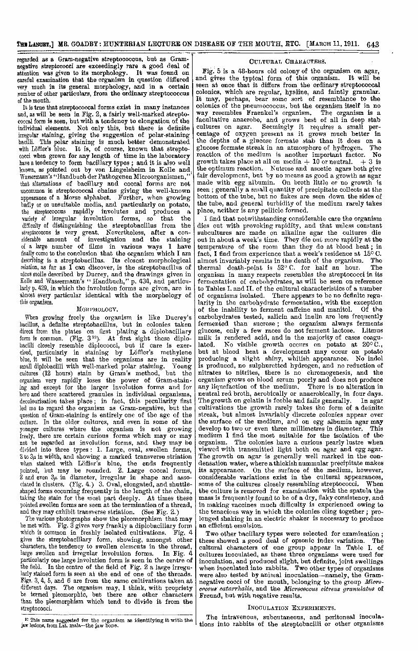
organism in many respects resembles the streptococci in itsfermentation of carbohydrates, as will be seen on referenceto Tables I. and II. of the cultural characteristics of a numberof organisms isolated. There appears to be no definite regu-larity in the carbohydrate fermentation, with the exceptionof the inabilitv to ferment caffeine and manitol. Of the
carbohydrates tested, salicin and inulin are less frequentlyfermented than sucrose ; the organism always fermentsglucose, only a few races do not ferment lactose. Litmusmilk is rendered acid, and in the majority of cases coagu-lated. No visible growth occurs on potato at 20° C.,but at blood heat a development may occur on potatoproducing a slight shiny, whitish appearance. No indolis produced, no sulphuretted hydrogen, and no reduction ofnitrates to nitrites, there is no chromogenesis, and the
organism grows on blood serum poorly and does not produceany liquefaction of the medium. There is no alteration inneutral red broth, aerobically or anaerobically, in four days.The growth on gelatin is feeble and fails generally. In agarcultivations the growth rarely takes the form of a definitestreak, but almost invariably discrete colonies appear overthe surface of the medium, and on egg albumin agar maydevelop to two or even three millimetres in diameter. Thismedium I find the most suitabie for the isolation of theorganism. The colonies have a curious pearly lustre whenviewed with transmitted light both on agar and egg agar.The growth on agar is generally well marked in the con-densation water, where a thickish nummular precipitate makesits appearance. On the surface of the medium, however,considerable variations exist in the cultural appearances,some of the cultures closely resembling streptococci. Whenthe culture is removed for examination with the spatula themass is frequently found to be of a dry, flaky consistency, andin making vaccines much difficulty is experienced owing tothe tenacious way in which the colonies cling together ; pro-longed shaking in an electric shaker is necessary to producean efficient emulsion.Two other bacillary types were selected for examination ;
these showed a good deal of opsonic index variation. Thecultural characters of one group appear in Table I. ofcultures inoculated, as these three organisms were used forinoculation, and produced slight, but definite, joint swellingswhen inoculated into rabbits. Two other types of organismswere also tested by animal inoculation-namely, the Gram-negative cocci of the mouth, belonging to the group Mioro-onceus’ catarrhalis, and the Miorooocoo8 citret68 9’l’anulatu8 ofFreund, but with negative results.
INOCULATION EXPERIMENTS.
The intravenous, subcutaneous, and peritoneal inocula-tions into rabbits of the streptobacilli or other organisms
644
produced no effect or symptoms of arthritis in rabbits (seeTable HI.). At the suggestion of Mr. Clement Lucas I pro-ceeded to the direct joint inoculation, with the result thatsymptoms indistinguishable from arthritis deformans havebeen established in 18 rabbits. The organisms which haveproduced this joint change have in 15 instances been the-streptobacillus of the type the cultural characters of whichare given in Table I., but differing from one another incertain particulars with regard to their carbohydrate fermen-tation and their growth on litmus milk (see Table 1.); butboth these media are ones in which organisms exhibitpeculiarities not sufficient to differentiate them into definitespecies. In three rabbits a slight amount of joint changehas resulted from the inoculation of the organisms given firstin the table of cultural characteristics (Table I.), but in nocase has the swelling or effect upon a joint been so markedin degree nor so definite as with the streptobacilli.
as the inoculation. Distinct wasting of the extensor muscleswas also seen, and the joint became slightly flexed when theanimal was at rest. In a number of other instances animalsinoculated later than this one, particularly in three animalswhich were inoculated in January of this year, semi-flexionof the inoculated joint has already occurred.
Mr. Reid kindly obtained X ray photographs of a numberof these animals for me, and I wish to tender him my sincerethanks for the trouble which amongst other things entailedtransporting much apparatus to the laboratory for the
purpose of taking the photographs of these experimentalanimals.
Fig. 6 shows the antero-posterior view of the right and leftknees of the arthritic rabbit. The head of the tibia and thelower end of the femur on the outer side both show altera.tions in the cartilage and commencing destruction of the
articular surfaces of the joint, but the photographs, of
TABLE L-Cu.ltzral Ckaraote’1’S of Organisms InlJoulattJd.
{A-Nos. 1-3 & 23. Bacilli irregular and diplo-, faintly staining with ordinary stains; red granules in three-day cultures with
rv-! methylene blue.MORPHOLOGY B-Nos. 4-22 & 24. Bacilli, oval-pointed and spindle-shaped, coccal forms common, diplo- or strepto-bacilli, large, irregular, and swollen involution forms, these taking Gram’s stain darkly, other forms oftendecolourised.
A Microscopically : Undulate or entire, marmorate, coarsely granular.) Macroscopically: Round medium, opalescent, opaque, raised, moist.
C J i r;J Microscopically Lacerate or lobate, sometimes lobulate, hyaline with grumous centre, or with irregular central darker
OLONIES area.
B Macroscopically: Transparent and translucent, with opalescent sheen, slightly irregular or undulate, occasionally with central point.C. M. B. = Carbolic methylene blue. A = Acid reaction. + = Complete reaction. ±= Slow and feeble reaction. No reaction.
In Table III. I have summarised as far as possible the.inoculation experiments of 33 animals, which have been
injected with cultures of the organisms I have been dis-cussing. Case 34, Rabbit 10, was inoculated in May,1910, with an emulsion of 0 5 c.c. of a 24 hours’ culture ofa streptobacillus (432C). This organism was a typicalvariety of the streptobacillus, showing the curious shuttle-shaped involution forms and the long streptobacillary forms.On May 26th there was well-marked thickening of the
capsule with some creaking in the joint, which was
swollen, but not tender nor greatly thickened. The knee-jointprogressively increased in size, and on Jan. 13th, 1911, thereappeared to be some slight thickening of the knee-joint onthe opposite side, and at the same time there was also to befelt a slight thickening of the ankle-joint on the same side
course, do not show the alterations in the synovial structures.The joint itself is typically that of arthritis deformans, thephotograph being taken nine months after inoculation, onlyone injection having been made. The alteration in the headof the tibia and the thinning of the tuberosity are definitelyshown. There appears also from the photograph to be somesclerosis in the head of the tibia and the lower end ofthe femur, recalling to some extent the rarefaction associatedwith sclerosis shown in the earlier photographs of the localdisease of the jaw. A small foreign body is to be seen onthe outer side of the joint and to-day there appears to bethe commencement of osteophytic nodules around the marginof the head of the tibia.
Another rabbit was inoculated on the same day of May,1910, but with a culture of a streptobacillus obtained from
645
another case. Well-marked swelling occurred on the outerside of the joint at the end of a month, and in four months’time the right knee was still slightly swollen. On Dec. 5ththe animal was inoculated again, this time with a wholeagar culture from another case exhibiting rheumatoidsymptoms, but in this instance in the ear vein and not
locally. No increase of swelling took place in the knee-joint; in fact, some improvement occurred for a short time.On Jan. 25th the animal was reinoculated on the outer sideof the same joint with 1 c. c. of an agar emulsion in 1 c. c.sterile saline, care being taken that no fluid went directInto the cavity of the joint. The inoculation was followedby considerably increased swelling of the joint, which byFeb. 20th was much thickened, so much so that theanimal was unable to extend the leg at the knee, andthe knee-joint was held flexed. On Feb. 13th a skia-
graph was taken by Mr. Reid, and very little change
other rabbit. These animals were inoculated from’casesin which arthritis deformans was a definite symptom,’but Ihave found that the streptobacillus is by no means confinedto such mouths as have rheumatoid arthritis.
Rabbits Nos. 15 and 30 are two instances of knee-jointinoculations, Rabbit No. 45 of intracapsular inoculations,Rabbit Nos. 47 and 49 of extracapsular inoculation with0’5 c.c. of saline emulsion of the streptobacillus obtainedfrom persons who were apparently in normal health, but whohad rarefying osteitis of the jaw.23 It will be seen that theseanimals exhibit well-marked swelling and deformity of theright knee-joints. These animals have received only oneinoculation, and Rabbit No. 47 was inoculated with a
culture, photographs of which I have shown on the screen.CASES.
I In going over my records of cases of diseases of the mouth
TABLE II.-Cnlt16/ral OhO/l’aoters of Streptobaoill1ls from Various Sources.
A = Acid reaction. + = Complete reaction. ± = Slow and feeble reaction. - = No reaction.
is to be seen in the skiagram notwithstanding thefact that well-marked thickening of the synovial mem-brane and the capsule were to be felt. The dissectionof this joint and the uninoculated joint on the other sideof the body are on the table, and show the thickenedcapsule and swollen synovial fringes as well as commencingdestruction of the joint with bony and cartilaginous changes.The appearance of the skiagraph suggests some alterationin the right knee-joint, but very much less than in the
I find that in the last 263 uncomplicated cases of varyingdegrees of infective processes of the alveolus, excluding allcases of growth and such cases as had complications, i.e.,as antral disease and all other similar infections, 49 gave adefinite history of rheumatic symptoms. These cases weredivisible into three main groups, but of course shaded
imperceptibly one into the other.
23 These animals were exhibited at the lecture.
646
TYPES OF CASES.
G’1’Oll.p A.-Definite arthritis of one or more joints, withpain, swelling, limitation of movement, and with or withoutwasting of one or other muscles in the neighbourhood of thejoint. Swelling of tendons or tendon sheaths in the
neighbourhood of joint or elsewhere. Muscular pains, diffuseand irregular in distribution and origin. Neuralgia of
trigeminal type. Anaemia.
Group C.-Occasional swelling and tenderness in joints ortendons, rapidly clearing up and leaving no objectivesymptoms. Irregular muscular pains, occasionally asso-
ciated with stiffness. Tingling, or "pins-and-needies"sensation in extremities. Local asphyxias or other vaso-motor phenomena. Intermittent lumbago. Occasionaltrigeminal neuralgia.
, These three types gave respectively 25, 16, and 8 cases.Of the total number of cases with a history of rheumatic
TABLE III.-Inocitlati6n Experiments.
The numbers in parenthesis under the heading " Organisin " refer to the numbers of the organism in Table I.
Group B.-No definite arthritis ; inflammation, or swellingof tendon sheath or tendon. Diffuse muscular pains,irregular in distribution and origin. Occasional swelling ofjoints or tendons, clearing up and leaving no objectivesymptoms. Palpitation, with irregularity of heart action.Tachycardia. Neuralgia, trigeminal type. Sciatic pain, notdefinitely referred to the sciatic nerve, but diffuse andreferred. Gastric and intestinal symptoms.
symptoms, 15 were males and 34 females. A brief synopsisof the 49 cases is appended.
NOTES OF CASES.
Group A.CASE I.-Aged 60. Failing health for several years, with recurrent
attacks of swelling in mouth. An acute attack diagnosed as foodpoisoning two years previously. Has had fleeting swellings in various
situations, mostly extensor surfaces of hands, and stiffness in shoulders.
647
Ulnar deflection both hands. Right index, metacarpo-phalangeal jointswollen. Mouth: condition very bad, much loss of tissue, alveolusabsorbed and rarefied. Vaccine therapy. Cured.C.isE 2.-Aged 42. Five and a half years previously had a little rheu-
matism which became subacute; treated at Buxton three months,much better. Following winter more rheumatism, with swelling injoints, knees, arms, and hands, especially the left, very tender at night,cramps, tingling in calf mostly preceding cramps. Treated again atBuxton and Aixe. Confinement next year in Malta ; sore tongue duringthe time at Malta; child also had sore mouth. Teeth extracted beforeleaving Malta; acute infection following; was stiff, could not move.Acute attack subsided, joints became less stiff. Present condition:right internal head of tibia thickened, knee full of fluid, much creak-ing, limitation of movement. Left knee freer movement, but creakingin joint. Thickening along patellar tendon, ulnar displacement of bothhands. Mouth: thickening of alveolar margins. Pus present in smallquantity; interdental rarefaction of bone. Vaccine therapy. Relieved.CASE 3.-Aged 30. In September, 1909, was going home from business
and got wet through. An acute attack of pain and swelling in most ofher joints. Was sent to bed ten weeks, and then to Bath, where she wasable to walk with two sticks. Was at Bath until Dec. 24th ; not pro-gressed at all. On May llth, 1910, right knee and ankle swollen; knee,thickening of upper and outer margin of head of tibia, painful onpressure; cedema over shin; pain and swelling of right hip ; lordosis,unable to walk upstairs without assistance; can just manage to walk onthe flat with a stick, which always brings on pain. Mouth : teeth andgums appear normal; slight ulceration at back of upper central incisors.History of occasional sore mouth, loss of bone between premolars inupper jaw. Pus microscopically only. Vaccine therapy. Cured.CASE4.-Pain in back and outer side of left thigh for six years.
Three years after commencement thickened synovial fringe diagnosed.Well-marked swelling of left knee, pain in certain positions, tenderspot outer and lower side of patella. Mouth: desquamative gingivitis,sclerosing osteitis upper jaw, mild in type; much thickening ofalveolus. Pus from three teeth. Vaccine therapy. No improvement.CASE 5.-Six years ago pain and diffuse swelling right knee and
right hand and wrist; pain disappeared, leaving stiffness; jointscreaked. Present time unable to use right wrist; thickening of tendonsheath of extensor of hand; slight ulnar displacement. Mouth :teeth have been projecting more and more for some three years.Little suppuration, but thickening of alveolus, slight thickening ofalveolar margin. Vaccine therapy. No improvement.CASE 6.-March, 1906. Right knee large swelling. Droitwich in
August. By October nearly well. At the end of October swellingrecommenced, and in March, 1907, left knee and left shoulderwere also considerably swollen with much pain. Both hands be-came swollen on dorsal surfaces. In June he went to Aix-les-Bains, did not improve, and was worse afterwards. Was treatedat home till beginning of 1908, when he was sent to Teneriffe. Duringthe whole of the time he had been taking iodides. At Teneriffeanother acute attack, in which all the joints that had been previouslyaffected became very much worse; had constant smell in nose andmouth, and taste of "burning hair." Unable to walk without theassistance of two people, one on either side. In February, 1909, swell-ing right and left knees, impossible to feel outline of joint; well-marked creaking in both; ulnar deflection both hands, right hand
fingers flexed, cannot straighten without acute pain. All tendonsheaths thickened; both shoulder-joints thickened, creaking, verypainful on passive movement. Has lost 2 stone in weight since March,1906, and a stone during the last year. Mouth : full of gold crownsand bridge work, with discharge of pus from almost every socket.Vaccine therapy. Cured.CASE 7.-Progressive polyarthritis, remissions and exacerbations, the
latter with fever. Pain, wasting of muscles. Mouth : all teeth lost,except two, through pyorrhoea. Not treated.CASE 8.-Age 45. Three years ago acute pain in left shoulder; swollen;
three days in bed; recurrence a year later, associated with cardiacattack; treated at Llandudno; in bed for three weeks. Pain recurredat intervals for three years, occasionally appearing in other joints.Latterly associated with pain, hypersesthesia and occipital neuralgia.Fusiform swelling of middle and ring fingers of both hands. Swellingexternal malleolus. Mouth: little or no suppuration, thickened alveoli ;teeth recently shifting in position without obvious cause. Vaccinetherapy. Cured.CASE 9.-Local asphyxia with coldness, tingling in both feet at
night, right foot "goes to sleep." Tingling in fingers ; occasionallyleft hand becomes quite cold for a short time. Pain in both kneesintermittent; swelling synovial membrane right knee; tender roundinternal tuberosity of left tibia. Mouth: some pus, only found micro-scopically, but composed of fresh polymorphonuclear cells. Has lostmost of teeth owing to progressive loosening. Last few years constantrecurrence of water blisters on lips. Vaccine therapy. Relieved.CASE 10.-Aged 54. Six years’ history of neuritis in arms and legs,
associated with swelling of tendons in hands, stiffness and creaking inleft shoulder. Constant lumbago, occipital neuralgia. Fusiformswelling ring finger of left hand. Mouth: has had receding gums for20 years. Little pus, alveolus thickened, some loss of alveolus in inter-dental spaces. Vaccine therapy. Cured.CASE 11.-Swelling of both knees, thickening of synovial membrane,
no apparent alteration in joint surfaces, ulnar deflection of one hand.First noticed rheumatic symptoms five years ago; from that date tothe present time has been progressively stiff in all the joints, and withmuscular pains, generally worse at night. Quite unable to walk nowmore than a few hundred yards, even then only with excessive fatigueand pain in joints. Appetite bad, has lost weight; considerabledepression. Mouth: considerable pus discharge from gums. X rayphotograph shows rarefaction and sclerosis. Vaccine therapy. Cured.CASE 12.-Rheumatoid arthritis diagnosed in both great toes eight
years previous to examination. Acute neuritis, swelling, pain, and lossof movement in left shoulder, made worse by electrical treatment.Recurrent trigeminal neuralgia, vasomotor disturbances in fingers.Mouth: bad taste in mouth, gastric fermentation. Pus from the upperJaw, mainly incisor region, four years. Thinning and porosis ofalveolus, no thickening. Vaccine therapy. Still under treatment.CASE 13.-Attacks of acute rheumatism affecting all the joints,
necessitating confinement to bed for six weeks. Treated at WoodhallSpa. All joints still creaking three years after attack. Right knee muchswollen and thickened synovial membrane, patches of fibrositis in theangle of the scapula, and persistent lumbago, and pain over both sacro-iliac joints, worse at night; hands swollen and a little ofdematous,
swelling not confined to extensor tendons, but diffuse. (Edema, ofshins. Mouth : gums cedematous, spongy, bleeding. Right uppercentral incisor quite loose. Pus. Alveolus upper and lower jawthickened, sclerosed. Vaccine therapy. Cured.CASE 14.-Twenty-nine years previously puerperal septicaemia. Nine-
teen years later gradual development of thickening, pain, swelling,loss of movement in right knee which persisted, becoming progressivelyworse for the last ten years; one knee 18 inches in circumference, theother 19 inches ; crepitations in both. Left wrist-joint swollen, dorsalsurfaces, extensor tendons also. Mouth : no pus. Few teeth loose.Osteoporosis. Vaccine therapy. Improved.CASE 15.-Vaso-motor disturbances with hands becoming white and
cold. Rheumatic pain both shoulders. Acute attack affecting bothknees, right more than left. Swelling of peri-articular tissue, crepita-tions in right knee, not in left; right knee remaining slightly swollen,but with little pain. Intestinal intcxication, mucous colitis. Mouth :localised area of purple and oedematous gums, principally upper jaw onleft side. Four teeth discharging pus. "Gouty" teeth. Two goldcrowns, which were removed, badly fitting; roots found thin andtransparent. Vaccine therapy. Relieved.CASE 16.-On March 7th, 1908, started with pain behind left ear,
without obvious cause. Progressive rheumatism affecting the rightshoulder and left thigh; several chills and shivering fits, followed bypain in joints, temperature in the evening going up to 1000 F. Attacksubsided, but the following spring another acute attack, with fever,pains in the head and neck which came on every afternoon and withany change of temperature; unable to move head. Pain in severaljoints, shoulder stiffness and creaking. Had a specific history, buthad been treated for four years. After an X ray examination of thechest it was decided that it was possibly a case of early tuberculardisease, and the patient was sent to a sanatorium, where, however, hederived no benefit. He had lost much weight, and was entirely unableto follow his profession. Subsequent history: on examination con-siderable amount of thickening in left shoulder-joint and in rightknee, with recurring pain described as " toothache " in the neck. Hyper-2esthesia over all cranial superficial nerves. Mouth: small degree ofrarefying osteitis in lower jaw, rather more in upper jaw; a good dealof alveolar thickening. Vaccine therapy. Cured.CASE 17.-First noticed stiffness in 1908, became gradually worse.
No pain except in right arm. In June, 1910, unable to walk. Generalwasting of muscles, right knee-joint particularly fixed. Hands andfeet shrunken, with acute pain in affected muscles subsequently.Worse at night. Mouth: 15 septic roots, swollen and hypertrophiedgum around them, no macroscopic pus, small quantity of pus foundmicroscopically. Considerable hypertrophy, thickening of alveolarprocess in upper jaw. Vaccine therapy. No improvement.CASE 18.-Pain in both knee-joints, occurring irregularly, and worse
at night. Commenced after bad attack of fever, followed by blackwater,in Nigpria. From this time gums bled ; constant bad taste in mouth.Latterly pain in left tendo Achillis. Slight oedema of knees, wrists,and shins. Mouth : gums swollen, partially detached from teeth,alveolus bare in places, considerable amount of pus. Vaccine therapy.Cured.CASE 19.-Has had rheumatic gout for the last six years ; has been
treated at Bath regularly twice a year. First noticed loosened teeth ayear previous to the rheumatoid symptoms. A good deal of swelling andthickening in both knees, crepitations left shoulder, slight ulnardeviation in left hand. Mouth : gums purple, nodular ; has acuteexacerbations of pain with swelling of alveolus. No pus formation.Considerable rarefaction of alveolus in upper jaw ; in lower jaw somethickening. Roots of molar teeth almost denuded of attachment.Vaccine therapy. Relieved.CASE 20.-Recurrent shivering fits, associated with acute aching in
arms and legs; both knees swollen ; left hip very painful; considerablelimitation of movement. Latterly attacks have been more frequent;has been treated at Bath, Marienbad, Aix, and many other watering-places. Mouth: no suppuration; strongly marked alveolar thickeningin upper and lower jaws ; gums pale ; wasting of os marginum. Vaccinetherapy. Not improved.CASE 21.—Hyperaesthesia and recurrent pain right arm and shoulder;
ulnar displacement left hand; thickening of second phalangeal joint,left ring finger. Mouth : much pain in gums ; two crowned teeth, bothtender. No obvious pus around gum margins. Both crowns removed.Right upper molar large cavity in bone at root; tooth much thinuedand periosteum hypersemic. Thickened and sclerosed alveolus. Vaccinetherapy. Cured.CASE 22.-Swelling and chronic pain sometimes in both knees;
creaking on movement; tender on pressure. Crepitations both elbowsand shoulder-joints. Frequent rashes on thighs and chest appear anddisappear quickly. Tingling and loss of sensation in arms. Mouth :chronic pyorrhoea, many years’ duration. Considerable alveolarthickening; much loss of bone in lower jaw.CASE 23.-Left lower maxilla, thickening and pain following suppura-
tion around lower molar ; lower molar removed. Subsequent necrosis,followed by operation. Recurrent swelling and pain; no fluctuation;no pus. Chronic swelling temporo-maxillary joint on same side;thickening of capsule; alteration of movement. Pain worse at night.Six months after suppuration and necrosis of the jaw had acute attackof pain in right knee, which subsided, but left creaking in the joint.Vaccine therapy. Cured.CASE 24.-Early chronic arthritis, both knees; irregular swellings
affecting ,joints of both hands; fusiform swelling, two fingers of lefthand; right index finger extensor tendons swollen, painful; unable towear rings. Severe left-sided trigeminal neuralgia, with ptosis on sameside. Mouth: considerable loosening of majority of teeth; uppercentrals very loose. Much pus. Vaccine therapy. Relieved.CASE 25.-History of gout and rheumatism for several years. Oeca-
. sional attacks of pain with swelling in the knee-joints; left knee, slightly enlarged. Recurrent lumbago and irregular muscular pains.
. with white flakes of desquamation. Enlargement of lymphatic glands’
over submaxilla. Frontal headache. Vaccine therapy. Cured.
Group B.CASE 26.-Diffuse muscular pains in shoulders and arms, irregular in
. onset, but leaving stiffness sufficient to prevent playing tennis.Recurrent stiffness and pain in lumbar region. Pain in right gluteal) region and referred down sciatic nerve. Sciatic nerve not tender at
foramen. Stiffness of calf muscles. Worse at night. Mouth: one-
, third of teeth lost through caries. Discharge of pus lower incisorla 2
648
region, and molar region upper and lower right. No hypertrophy of$;um. Sclerosis of alveolus, well-marked denudation between severalteeth. Vaccine therapy. Cured. ,CASE 27.-Diffuse muscular pains in shoulders and back, swelling of
extpnsor tendons of left hand. Stiffness over both knee joints. Com-plains of recurrent rheumatism. Trigeminal neuralgia. Mouth : allteeth removed two years previously for pyorrhoea. Thickening oflymphatic channels both sides of neck. Slight thickening ofivmnhatic glands of posterior cervical triangle left side. Vaccinetherapy. Cured.CASE 28.—Suffered from muscular rheumatism for six years. Recur-
rent neuralgias, mostly trigeminal. stiffness and pain at back of head.recurrent b’tt with long intervals. No focal spots of neuralgia. AtWoodhall Spa six years ago, suffered from rheumatism ever since.Mouth been treated for last ten years for discharge of prs. Consider-able bony deficiencies in too’h sockets with rarefaction as well asiselerosis. Vaccine therapy. No improvement.CASE 29 -Aged 18. History of recurring attacks of fainting from no
particular cause. Much acne. Irregular, diffuse, rheumatic pains,<)ften at night, occasionally leaving stiffness of shoulders and unable totake violent exercise after. Last attack had swelling of left hand andradius. Crepitation in right knee. Mouth: discharge of pus fromsockets for several years ; this despite all attempts at treatment. Fivetooth sofkets still suppurating. Vaccine therapy. Relieved.CASE 30.-Pain in right shoulder last three or four years; unable to
put on coat comfortably ; keeps him awake at night. Suffers fromrecurrent vaso-motor spasm of feet, with subnormal temperature.Mouth: little pus; thickened alveolus showing much loss, particularly’upper jaw. Most teeth extracted, remainder one only loose. Somerarefaction ; alveolus mostly thickened and sclerosed. Vaccine therapy.Cured.
CASE 31.-Pain in joints fleeting. not persistent or resulting inchanges. Fermentative gastritis. Neurasthenia. Mouth: thickenedand sclerosed alveolus, œdema of gums, slight pus formation. Osteo-fPOrosis in molar region of upper jaw. Vaccine therapy. Relieved.CASE 32.-Rheumatic pains in various parts of the body with
-cm,,t-ional swellings of joints associated with fever, disappearing aftershort time and not leaving any joint deformity. Swelling of hands.:aiso disappearing and leaving no thickening. Neuritis. Acuteneuralgia. trigeminal, mostly first and second divisions. Rarefyingosteitis with sclerosis Suppuration slight, but definite pus formation.Vaccine therapv. Relieved.
CASE 33.-History of occasional rheumatic pain. diffuse, not very-detinite. Diffuse, rarefying osteitis of alveolus, left upper jaw ; central,lateral, and canine entirely denuded of bone Much pus formation,several other teet,h in upper jaw also loose. Lower gums cedematous;alveolus thickened. During course of treatment for alveolar diseasesudden onset of an attack of arthritis in left knee, no fluid in joint,swelling of the internal svnovial membrane and capsule, creaking onpassive movement, gradually disappearing without leaving deformityof joint.
CASE 34.-Irregular rheumatism generally in shoulders, occasionallyin knee-joints, no swelling. and so long as he takes plenty of exercisehas no return. Constant fermentative indigestion. Mouth: for threeyears has had well-marked suppuration along the gum margins. Largequantity of pus now found, a good deal of alveolar infection. Vaccinetherapy. Cured.CASE 35 -Vaso-motor spasm of hands preventing writing, together
with tingling and occasional burning sensation in upper arm. Re-current stiff necks, some fibrositis in deltoids and left angle of scapula,no swelling, no definite articular lesions. Mouth : has lost all molarand bicuspid teeth from pyorrhoea. At the present time lower centrals.and upper centrals standing, much coated with deposit. Discharge of,pus from inflamed and hypertrophied gums. Vaccine therapy. Relieved.CASE 36.-Bilateral enlargement of first metatarsals of both feet.
Neuritis left arm ; ansemic. Good deal of neurasthenia. Mouth : copiousdischarge of pus, much rarefaction, but with sclerosis in severalsituations Vaccine therapy. Relieved.
CASE 37.-Neuritis right hand. preceded by attacks of coldness.Always suffers from rheumatism when exposed to wet. Swelling, pain,and some thickening of left temporo-maxillary articulation. Teethmearly all loose, for eight years progressing. Pus welling from all
- sockets. Very great hypertrophy of gums, blue and turgid. Rarefac-
tion of alveolus of lower teeth, so far advanced that hypodermic needletmight be passed between roots of lower teeth entirely throughthe jaw. Vaccine therapy. Cured.CASE 38.-Slight arthritis of shoulder on left side and left knee.
Vaso-motor spasms, particularly affecting face. Acute attack of rheu-
matic type, followed by anginal attacks and myocarditis. Intestinalindigestion. Mouth: acute paroxysmal pain of individual teeth.Sclerosis. All extracted teeth roots transparent. No suppuration, but.extracted teeth highly inflamed periosteum. Vaccine therapy. Cured.CASE 39.-Patient aged 18 years. Acute pain, temperature 104° F.
!Pain in all joints, so tender could not be touched. Quite well in arfortnight. For two years had alternations of general rheumatic pains- with attacks in feet, hands, shoulders, or knee. Frequently attack inbig toe. Harrogate, Marienbad, Buxton. Mouth: much hyperæmiaof gum margin, particularly anterior portion of mouth. No pus. Rare-rfaction of alveolus. Vaccine therapy. Relieved.
CASE 40. -Occasional rheumatic pains, no definite joint lesions.’Mouth: has lost nearly all teeth from loosening. Has had acuteexacerbations with pus formation in mouth after last three confine-ments. Two children both suffered from rheumatic fever. Vaccinertherapy. Relieved.
CASR 41.-Had acute endocarditis when two years and three monthsold. Five to six months after brother had had similar disease with’high temperature and-heart infection. First attack corresponded to,time his mother was suffering from acute pyorrhoea. Recurrrentattacks, fleeting pain in joints, praecordial pain, breathless. Mouth:present condition gingivitis with marked suppuration around severaltemporary teeth, mucosa of mouth generally inflamed. Vaccinertherapy. Cured.
Group C.CASE 42.-Patient aged 18 years. Irregular trigeminal neuralgia.
;Stiffness and pain left temporo-maxillary joint. Muscular cramps,irregular, at night on three occasions. Mouth : diffuse gingivitis withhypertrophy of gum. All teeth much discharge of pus. Trouble com-
mencing after acute attack of measles. Vaccine therapy. Cured.CASE 43.-Subsequent to an attack of influenza suffered from
palpitation and diffuse muscular pains, aching in joints which persistedfor six months. Temperature of 101° F. at intervals at night, lastingthree or four months. Occasionally swelling and stiffness in hands andfeet; right hand goes to sleep at times. Heart ringing first sound,slight roughness. Haemoglobin 75 per cent. Mouth: has had badtaste in mouth ever since influenza, and has noticed recession, bleeding,and discharge from the gums. Well-marked infection of the alveolusin many situltions. Few teeth loose. Vaccine therapy. Relieved.CASE 44.-Recurrent attacks of sacro-iliac neuralgia, followed by
stiffness in lumbar region. No history of’ generalised rheumatism.Mouth : lower central incisors loose. discharge of pus from upper andlower gums. Rarefaction of bone. Vaccine therapy. Cured.CASE 45.-Patient aged 50 years. Recurrent attacks of asthma since
35 years of age. Attacks of colitis. Various operations for nasal polypisuppuration. Last four years emphysema, with occasional attacks ofbronchitis. Recurrent lumbago. Intestinal indigestion. Occasionalswelling of tendons of back of hand. Recurrent stiff neck. Mouth:takes great care. Little or no pus found, but well-marked interstitialdeficiencies, thinning of bone, sclerosis of alveolus. Vaccine therapy.Relieved.CASE 46.-Attacks of unaccountable sleeplessness, with sudden loss of
sensation and feeling in one arm. Rheumatism which interferes withhis golf. Mouth : suppuration acute left upper canine, most teeth lostthrough pyorrhoea, much shrinkage of alveolus, except where teeth arestanding, where, particularly in the upper jaw, considerable thickeninghas taken place. Vaccine therapy. Cured.CASE 47.-Irregular attacks of stiffness and pain in knees and right
hip. Dyspepsia, three years’ history. Mouth: acute suppuration inmouth. All teeth affected, gums swollen, oedematous, much wastingof alveolus, little or no thickening. Vaccine therapy. Cured.CASE 48.-History of mild polyarthritis, many years’ standing. At
times knee-joint becomes swollen and tender; slight thickening rightknee, some loss of movement. Mouth: much alveolar thickening, leftside of upper jaw being at least twice the normal size, Suppurationaround roots of upper molar teeth, much elongated. Rarefaction ofbone around teeth. Vaccine therapy. Cured.CASE 49 -Last three or four vears has had irregular attacks of
lumbago, with rheumatic pain in shoulders and down right arm.Occasionally swelling of fingers of right hand ; no permanent enlarge-ment of joints, but sometimes definite swellmg which makes it difficultto wear her rings. Mouth: mouth and gums normal, with exception ofright upper bicuspid tooth, which is dead, and from which a chronicsinus has existed for the last six years. Alveolus around this bicuspidtooth is thinned and almost entirely remover. Some deeper infectionint) the body of the jaw. Antrum not affected.
For the sake of brevity the section dealing with the
bacteriology of the mouth is confined to the consideration ofthose cases in which animal experiments have been made.Animal experiments were made in respect of 17 of thecases: 8 in Group A, Nos 2. 4, 14, 16, 17, 21, 24,and 25 ; 7 in Group B, Nos. 31. 32, 31, 35, 36. 38,and 40 ; 2 in Group C, Nos. 44 and 49. In all the49 cases a bacteriological investigation was made and
organisms similar in general characters to those describedin Table II. were isolated. In addition to these cases
giving a definite history, 12 non-rheumatic cases of rarefyingalveolar osteitis were submitted to bacteriological examina-tion, the streptobacillus isolated from them and used foranimal experiments. The organisms obtained resembledthose from the rheumatoid cases. The two groupsof cases are indicated in Table III., cases from50 onwards being those in which no rheumatic symptomswere observable. The bacteria isolated from the cases
which were not suffering from arthritis or other rheumaticsymptoms in several instances caused swellings and pro-gressive changes when inoculated into the knee-joints ofrabbits. I have also isolated the streptobacillus from casesof posterior rhinitis, chronic pharyngitis, quinsy, anddisease of the antrum of Highmore. I have also isolated thestreptobacillus from the urine of four cases in which it waspresent in the mouth. In my last 100 cases of mouth diseasesexamined bacteriologically of all varieties excepting dentalcaries I have met with it 65 times.
It will be seen from the notes of many of my cases thata considerable amount of crown and bridge work had beeninserted in the unfortunate persons’ mouths, and I cannotexpress too great a horror of that potent predisposing causeof infection of the jaws-namely, the ill-fitting, ill-adjusted,
ill-conceived, and pernicious cap crown. To the objectorwho demurs to this condemnation I would only retort thatit is a device which separates the normal gum edge fromits attachment to the tooth, that rarely, if ever, fits closelyto the contour of its encircled victim, but invariably projectsin such a manner that debris of food is trapped under itsrim and inevitably promotes ulceration, periosteal infection,and, sooner or later, rarefying osteitis and suppuration.
TREATMENT.The first suggestion that treatment of the mouth infection
might also influence the arthritis was given by a case whosemouth was resplendent with gold crowns, Case No. 6. A
bacteriological examination was made, the infectingorganisms isolated, and an autogenous vaccine prepared.
649
Preliminary inoculatioas were givtn and all the worst
and badly infected teeth removed. The vaccine was
then given regularly, first at periods of ten days, thenat fortnightly intervals for six months, and after that atintervals of six weeks for a year. The first inoculations wereassociated with exacerbation of the joint symptoms. The
patient made a slow but steady recovery. The left knee-
joint, which had undergone some bony change, has remainedpermanently thickened, particularly the outer margin ofthe head of the tibia, but the thickening of the tendonsheaths of the hands has disappeared, together withthe joint swelling; the patient is able to walk without
pain, and has so far recovered the use of his limbs that hewas able so long ago as last September to mow his own
grass plot. He has gained about 2 stones in weight, andhas been able to return to his occupation. Four freshvaccines were made during the period of treatment; the doseto commence with was 10,000,000, but the later doses were150,000,000 to 200,000,000, on each occasion being thestreptobacillus described above, the organism in this case
being Nos. 125B and 237B on the list in Table II. The lineof treatment of this case is that adopted as general routine.In nearly all of my early cases distinct increase of the arthriticpain and swelling was caused by the first inoculation;latterly the preliminary dose has been reduced to avoid thisexacerbation.Case No. 16 was another of the earlier cases, but in this
instance the organisms were tested on mice, mainly for viru-lence, no intravenous or other inoculation having been madeinto rabbits; in fact, it was not until May of last yearthat I commenced direct inoculation into the knee-jointsof animals. It will be seen on reference to this case that theamount of inflammation in the mouth itself was practicallynil. The patient had, however, lost a number of his teeth,and those which remained showed a small but definiteamount of rarefying osteitis in the lower jaw. There wasalso a considerable amount of alveolar thickening. Therewere no other obvious sources of infection, although carefulsearch was made of all the orifices, and the bacteria wereisolated and tested against the patient’s blood. Resourcewas therefore had to immunisation by means of a vaccineprepared from the patient’s mouth. Almost from the firstinoculation a diminution took place in the slight degree offever from which the patient constantly suffered, the
temperature generally being raised to 100° at night. The in-oculations were carried out regularly and progressive improve-ment took place. The specific treatment was also continued,as at one time some parietal thickening occurred which wasthought to be of specific origin. The patient made a gradualbut complete recovery, having lost all stiffness, all neuraigia,and, having regained his normal weight, has been enabled toreturn to his business ; more important still, he is nowable to play golf again without difficulty. The two miceinoculated with the patient’s organisms recovered withoutany ill effects.Case No. 3 was another in which a very slight amount of
mouth infection was to be found. She had a history ofindigestion, her throat was hyperæmic, there was occasionalpain in the mouth and palate, and the latter showed slightulceration behind the central upper incisors. There was
slight but definite infection of the alveolar process, butwithout very careful examination this could not be detected,as to all appearances the patient’s teeth and gums werenormal; on careful examination with a thin platinum probeit was found that here and there the gums were detachedand distinct loss of alveolar process in the interdentalposition had occurred. The streptobacillus was isolatedfrom this patient, who received injections at weekly intervals,and latterly at more extended periods. At the first threeinjections increase of pain and swelling of the affected partsoccurred. She was under treatment from May, 1910, toDecember of the same year. By this time all the swelling inthe knee had entirely disappeared, the joint was quitenormal, the patient had discarded not only her crutches andher stick, but was able to dance.
Case No. 23 is of interest because it was the only one inthe whole series of 49 cases in which there was anysuggestion of infection either in or in the neighbourhood ofthe temporo-maxillary articulation, and in this case theorganisms found differed from those obtained from the
majority of the cases. The organism from this case
belonged to the type given at the head of Table II. in thatthe organisms produced no fermentation of carbohydrate.
The swelling ertirely disappeared after eight inoculationsand has not returned.
Forty-five out of the 49 cases were treated with auto-genous vaccines of the streptobacillus malas, 21 in Group A,16 in Group B, and 8 in Group C. Of these cases 23 havebeen cured, inasmuch as their arthritic symptoms haveresolved sufficiently for them to go about their normal avoca-tions ; of these, 11 belonged to Group A, 7 to Group B, and14 to Group C. Four cases in Group A and 1 in Group Crefused treatment, and of the remaining cases 8 in Group Aare still under treatment, 4 have shown marked improve-ment, and in 4 so far no improvement appears to have takenplace, whilst 2 cases ceased treatment after a couple ofmonths, having been in no way benefited, but in no case wasthe condition in any way aggravated. In Group B 7 casesare still under treatment and 2 were in no way benefited.In Group C 4 cases have been cured and the remainder arestill under treatment.
TABLE IV.
The routine method of treatment which I have adopted inall these cases has been inoculation with autogenous strepto-bacillary vaccines, followed by attention to the local mouthinfection. Where extraction was deemed necessarv, and the
question of extraction has generally been decided with theassistance of X ray photographs, preliminary immunisationof two or three doses of vaccine has always been undertakenbefore the removal of the infected tooth. I am of opinionthat harm may result from the free removal of even looseteeth in sufferers from arthritis deformans unless preliminarytreatment is adopted, and I have found, as a matter of
practice, that increased local swelling and pain does occurin many instances in arthritis deformans after the removalof teeth, and may even follow an ordinary process of scalingtartar-coated teeth.
In conclusion, it must be understood that my contributionto the study of arthritis deformans coincident with alveolarosteitis does not seek to establish a specific organism as theetiological factor in all cases of rheumatoid arthritis, infec-tive fibrositis, muscular rheumatism, &c., but that in diseasesof the jaws, especially rarefying alveolar osteitis, a strepto-bacillus is present, pure cultivations of which inoculatedinto or around the knee-joints of rabbits have producedsymptoms similar to and indistinguishable from arthritis de-formans ; that in such disease of the mouth and jaws thetreatment of the disease locally and by means of auto-
genous vaccine prepared from the streptobacillus has beenassociated with amelioration and disappearance of thearthritic symptoms in a greater number of instances thancan be ascribed to fortuitous coincidence.
A BRIGHTON WORKHOUSE SCHEME. - TheBrighton board of guardians at its last meeting agreed inprinciple to the complete separation of the infirmary inmatesfrom the workhouse inmates, a separation to be effected bythe elimination of all the children from the workhouse and
accommodating them at Warren Farm School, which is alsounder the jurisdiction of the guardians. This removal wouldprovide the accommodation needed at that institution forthe classification of the sick. The daily average number ofinmates of the workhouse is 1350, of which number about500 are maintained in the infirmary. The nursing staffnumbers 40, comprising a superintendent-nurse, a nightsister, 10 charge nurses, 11 staff nurses, 15 probationers, and2 male nurses. The guardians were almost unanimous onthe matter, 23 voting for the scheme and only 1 against.The vote was an endorsement of the view taken by the com-mittee which considered the question that "in all prob-ability the time is not far distant when Poor-law infirmariesof the size of that of Brighton will be under separate adminis-tration from the workhouse."