Considerable progress has been made both in under-standing the basic immune mechanisms of kidney disease and in translating these findings to clinical therapies. Sophisticated animal studies combined with the analysis of clinical samples have led to a pre-cise knowledge of the autoimmune targets and of the mechanisms responsible for kidney injury. Kidney diseases are highly prevalent and cost-intensive, but many discoveries in renal immunology are not widely known in the immunological community, although they are often relevant to diseases that affect other organs.
In this Review, we discuss recent advances in our understanding of immune-mediated kidney diseases, emphasizing those of particular relevance to the wider immunology community and those that have led to a better understanding of basic immunological mechan-isms. We have had to be selective in the topics consid-ered and so have excluded a discussion of acute kidney injury, kidney transplantation and alloimmunity, as well as of systemic diseases with associated kidney disease, such as type 2 diabetes and hypertension, that are not primarily caused by the immune system, despite the involvement of innate (and possibly adap-tive) immune responses in the renal injury they cause. Here, we discuss the innate immune mechanisms of kidney injury and introduce novel concepts about the
role of the cellular immune responses that drive renal disease. Moreover, we summarize recent discoveries about complement- and antibody-mediated nephritis, and we discuss kidney pathologies that are mediated by renal autoantigen-specific antibodies, especially those that are induced by crossreactive microorganism-specific antibodies. Finally, we describe how the disruption of kidney function and kidney pathologies can influence systemic immune responses.
Kidney-resident immune cellsIn the kidneys, toxic waste products of metabolism are removed from the blood by nephrons. Each nephron contains one glomerulus, which functions as a size-selective filter that retains molecules above ~50 kDa in the blood. Compounds of lower molecular mass pass through the glomerular filter, enter the tubular system and are excreted with the urine unless they are re absorbed by the tubular epithelium (BOX 1). The kidneys produce several hormones that directly or indirectly affect immune responses, including vita-min D, which regulates bone homeostasis and phago-cyte function, erythropoietin, which is induced in response to hypoxia to regulate erythropoiesis, and renin, which induces angiotensin and aldosterone to regulate electrolyte balance, extracellular osmolarity and blood pressure.
1Institutes of Molecular Medicine and Experimental Immunology (IMMEI), Rheinische Friedrich-Wilhelms-Universität, Sigmund-Freud-Str. 25, 53105 Bonn, Germany.2III. Medizinische Klinik, Universitätsklinikum Hamburg-Eppendorf, Martinistrasse 52, 20246 Hamburg, Germany.3Medizinische Klinik und Poliklinik IV, Ludwig-Maximilians Universität München, Ziemssenstr. 1, 80336 München, Germany.4Clinical Institute of Pathology, Medical University of Vienna, Währinger Gürtel 18–20, A-1090 Vienna, Austria.e-mails: [email protected]; [email protected]; [email protected]; [email protected] authors contributed equally to this work.doi:10.1038/nri3523Published online 16 September 2013
The immune system and kidney disease: basic concepts and clinical implicationsChristian Kurts1, Ulf Panzer2, Hans-Joachim Anders3 and Andrew J. Rees4
Abstract | The kidneys are frequently targeted by pathogenic immune responses against renal autoantigens or by local manifestations of systemic autoimmunity. Recent studies in rodent models and humans have uncovered several underlying mechanisms that can be used to explain the previously enigmatic immunopathology of many kidney diseases. These mechanisms include kidney-specific damage-associated molecular patterns that cause sterile inflammation, the crosstalk between renal dendritic cells and T cells, the development of kidney-targeting autoantibodies and molecular mimicry with microbial pathogens. Conversely, kidney failure affects general immunity, causing intestinal barrier dysfunction, systemic inflammation and immunodeficiency that contribute to the morbidity and mortality of patients with kidney disease. In this Review, we summarize the recent findings regarding the interactions between the kidneys and the immune system.
R E V I E W S
738 | OCTOBER 2013 | VOLUME 13 www.nature.com/reviews/immunol
NephronsAnatomically and functionally independent kidney units that each consist of one glomerulus and one tubule. The nephron delivers urine into collecting ducts that empty into the renal pelvis and, through the ureters, into the urinary bladder.
GlomerulusAn anatomical structure that is located in the kidney cortex and that filters blood into the tubular system.
TubulointerstitiumThe space between the tubuli and glomeruli, which contains capillaries, fibroblasts and dendritic cells, and thus is an important site for the progression of nephritis.
Bacterial pyelonephritisA bacterial infection of the kidney, mostly due to uropathogenic Escherichia coli that ascend through the urethra, bladder and ureter into the kidneys.
Under homeostatic conditions, the resident immune cells of the kidneys include dendritic cells (DCs) and macrophages, as well as a few lympho-cytes1–4. DCs are restricted to the tubulointerstitium and are absent from the glomeruli1,2. In mice, kidney DCs are CD11c+CD11b+F4/80+CX3CR1+CD8−CD205− and have a transcriptome that is typical of DCs resident in various non-lymphoid tissues5,6. Kidney DCs are derived from monocytes and from common DC pre-cursors (CDPs), but in contrast with other organs, some CDP-derived kidney DCs express CD64 (also known as FcγRI)7. Kidney DCs function as sentinels in homeostasis, local injury and infection3,8. They rap-idly produce neutrophil-recruiting chemokines dur-ing bacterial pyelonephritis, which is the most prevalent kidney infection8. Neutrophils can also be recruited by tubular epithelial cells, but not as quickly as by DCs. Mice lacking expression of CX3C-chemokine recep-tor 1 (CX3CR1) have a selective reduction in kidney DC numbers9. There is also a high renal expression of its ligand CX3C-chemokine ligand 1 (CX3CL1)10, which suggests that the CX3CR1–CX3CL1 chemokine pair are important for DC recruitment to the kidney and
that CX3CR1 might be a specific therapeutic target to modulate DC numbers in the kidneys. In renal ischae-mia (which is relevant in kidney transplantation) and in ureteral obstruction, renal DCs promote tissue injury by producing pro-inflammatory cytokines11,12. Basic leucine zipper transcriptional factor ATF-like 3 (BATF3)-dependent CD103+ tissue DCs, which can cross-present antigens to CD8+ T cells, are rare and their function in the kidney is unclear13. Macrophages are preferentially found in the renal medulla and cap-sule1 and have homeostatic and repair functions14. There are also mast cells in the kidney tubulointer-stitium but their function is poorly understood15–17. In addition, the role of innate-like lymphocytes is currently unclear. Finally, the renal lymph nodes rep-resent a priming site for nephritogenic T cells during renal inflammation18,19.
Low-molecular-mass proteins can pass through the glomerular filter but are reabsorbed and degraded by tubular epithelial cells. However, some of these proteins are captured by renal DCs or reach the renal lymph nodes by lymphatic drainage within seconds after filtra-tion20. Importantly, filtered proteins are concentrated in
Box 1 | Basic kidney anatomy and physiology
The kidneys purify toxic metabolic waste products from the blood in several hundred thousand functionally independent units called nephrons. A nephron consists of one glomerulus and one double hairpin-shaped tubule that drains the filtrate into the renal pelvis. The glomeruli located in the kidney cortex are bordered by the Bowman’s capsule. They are lined with parietal epithelial cells and contain the mesangium with many capillaries to filter the blood. The glomerular filtration barrier consists of endothelial cells, the glomerular basement membrane and visceral epithelial cells (also known as podocytes). All molecules below the molecular size of albumin (that is, 68 kDa) pass the filter and enter the tubule, which consists of the proximal convoluted tubule, the loop of Henle and the distal convoluted tubule. An intricate countercurrent system forms a high osmotic gradient in the renal medulla that concentrates the filtrate. The tubular epithelial cells reabsorb water, small proteins, amino acids, carbohydrates and electrolytes, thereby regulating plasma osmolality, extracellular volume, blood pressure and acid–base and electrolyte balance. Non-reabsorbed compounds pass from the tubular system into the collecting ducts to form
urine. The space between the tubules is called the interstitium and contains most of the intrarenal immune system, which mainly consists of dendritic cells,
TubulesHairpin-like structures that receive filtered blood. The tubular epithelium reabsorbs water, electrolytes, nutrients and proteins. Each nephron has a single tubule, which defines proximal and distal tubules as parts of the nephron.
Chronic kidney disease(CKD). Chronic (and often progressive) impairment of renal functions, such as blood purification, barrier function of the glomerular filter, water, electrolyte and acid–base homeostasis, endocrine functions such as vitamin D processing, erythropoietin production and blood pressure regulation.
UraemiaEnd-stage chronic kidney disease, the treatment of which requires dialysis or kidney transplantation.
GlomerulonephritisA heterogeneous group of immune-mediated kidney diseases that initiate in the glomeruli.
PodocyteA visceral epithelial cell that covers the glomerular capillaries in the Bowman’s capsule. Podocytes are a component of the glomerular filtration barrier.
FibrocytesMonocyte-derived collagen- producing cells that have been suggested to contribute to kidney fibrosis.
Kidney fibrosisThe end stage of chronic kidney disease, when functional renal tissue has been replaced by fibrotic scar tissue and is usually accompanied by uraemia.
the kidney proximal tubules, where >85% of the fluid is reabsorbed. Thus, renal DCs and the renal lymph nodes receive low-molecular-mass antigens from the circulation at concentrations that are over tenfold higher than in any other tissue. BATF3-dependent DCs in the renal lymph nodes capture and cross-present these proteins to CD8+ T cells, which results in the programmed cell death 1 ligand 1 (PDL1)-mediated deletion of these T cells21. Thus, the renal lymph nodes have a special role in the development of immune tolerance against circulating innocuous low-molecular-mass proteins, such as food antigens and hormones.
Immune-mediated kidney diseaseThe kidneys are a frequent target of systemic immune and autoimmune disorders, including systemic autoimmunity and vasculitis, immune complex-related serum sickness and complement disorders. This is partly related to the size-selective and charge-dependent filtration process in the glomeruli that promotes glomerular immune com-plex deposition. In addition, immune responses against kidney-derived autoantigens can cause autoimmune kidney diseases.
In chronic kidney disease (CKD), low-molecular-mass compounds accumulate in the body, which causes uraemia. CKD affects approximately 10% of the Western population and is a serious social and economic burden, especially for those who progress to kidney failure and that require dialysis or transplantation. The tissue injury associated with CKD is commonly directly or indirectly caused by the immune system (BOX 2).
Direct immune-mediated injury often affects the glo-meruli, at least initially, which causes different forms of glomerulonephritis. Irreversible kidney damage occurs when inflammation spreads to the tubulointerstitium22–24. Various mechanisms that cause this spreading have been proposed: podocyte damage might facilitate leakage of the glomerular filtrate and detachment of tubular cells from their basement membrane25; destruction of glomerular capillaries might restrict the perfusion of their downstream tubulointerstitial capillaries and cause ischaemia26; pro-inflammatory cytokines from inflamed glomeruli might perfuse the tubulointerstitial capillaries and cause inflam-mation27; reabsorption of abnormal amounts of protein from the glomerular filtrate might induce stress responses in tubular epithelial cells28; and glomerular antigens might reach DCs in the adjacent tubulointerstitium, which in turn might stimulate infiltrating T cells to produce pro-inflammatory cytokines19. Tubulointerstitial mono nuclear cell infiltrates can contribute to continuing immuno-pathology and to progressive tissue remodelling, which lead to tubular atrophy and interstitial scarring, both by main-taining local chronic inflammation and by recruiting fibro-cytes29. The end state of CKD is kidney fibrosis — a state in which functional nephrons are replaced by fibrotic tissue.
Immune-mediated CKD can be induced by immune complex deposition, by innate immunity and by T cells that interact with kidney-resident immune cells. Importantly, these immune mechanisms generally con-tribute to the progression of CKD, even in non-immune-initiated forms of the disease, and therefore there are obvious implications for therapy.
Box 2 | Kidney disorders grouped by their involvement in immunity
Kidney disorders that are initiated and mainly mediated by an immune response•Renal infections with renotrophic pathogens, including uropathogenic Escherichia coli (UPEC), Hantan virus, BK virus,
Leptospira spp., Mycobacterium tuberculosis and HIV
•Extrarenal infections with renal manifestations, including septic kidney injury, immune complex-mediated nephritis (for example, post-infectious glomerulonephritis and endocarditis, hepatitis and virus-related immune complex glomerulonephritis), interstitial nephritis and HIV nephropathy
•Systemic autoimmunity against ubiquitous antigens with renal inflammation, including IgA nephropathy or Henoch–Schönlein purpura, lupus nephritis, Sjögren’s syndrome, anti-neutrophil cytoplasmic antibody (ANCA)-associated vasculitis, interstitial nephritis, secondary membranous nephropathy and antibody-mediated forms of atypical haemolytic uraemic syndrome (aHUS)
•Immune responses against renal antigens, including anti-glomerular basement membrane (anti-GBM) autoimmune disease, the autoimmune disease primary membranous nephropathy and allograft rejection
•Other systemic disorders that affect the kidneys and that have genetic (including, complement C3 glomerulonephritis and aHUS) or unclear (including, minimal change disease and renal sarcoidosis) causes
Kidney disorders that involve renal inflammation as a secondary mechanism•Systemic autoimmunity against ubiquitous antigens with renal manifestations causing renal vascular obstruction
and ischaemia, including scleroderma renal crisis, panarteritis nodosa, giant cell vasculitis or phospholipid antibody syndrome
•Other systemic disorders that affect the kidney, including genetic disorders such as hereditary defects of GBM or podocyte genes leading to focal segmental glomerulosclerosis and hereditary tubulopathies or polycystic disorders; disorders driven by toxins, including Shiga toxin-producing Escherichia coli-induced HUS, drug- or contrast media-induced kidney injury; crystal and paraprotein-related nephropathies; and disorders caused by metals or food-borne toxins and toxic forms of focal segmental glomerulosclerosis
•Disorders that affect haemodynamics and the vascular system can also affect the kidney, including atherosclerosis, embolism, macro- or microvascular stenosis, shock, hepato-renal syndrome, thrombotic microangiopathy, eclampsia, hyperfiltration-associated focal segmental glomerulosclerosis, global glomerulosclerosis
•Obstructive nephropathy or renal amyloidosis
R E V I E W S
740 | OCTOBER 2013 | VOLUME 13 www.nature.com/reviews/immunol
InflammasomeAn intracellular complex containing pattern recognition receptors that activate caspase 1. Caspase 1 activation induces pyroptotic cell death and interleukin-1β (IL-1β) and IL-18 secretion.
Innate immune responses in CKD. Clinical entities of kid-ney disease, such as post-ischemic and toxic acute kidney injury, as well as nephropathies that are induced by dia-betes, hypertension and crystal deposition, involve sterile inflammation. As in other organs, sterile renal inflamma-tion is induced by intrinsic damage-associated molecular patterns (DAMPs) that are either released from dying parenchymal cells or that are generated during extracellu-lar matrix remodelling30–33. The kidney hosts a large range of different parenchymal cell types, including tubular epithelial cells, and endothelial cells that express a subset of Toll-like receptors (TLRs; that is, TLR1 to TLR6) and inflammasome components, which suggests that these cells can respond to DAMPs and that they can induce innate immune responses and subsequent renal inflammation34. However, NLRP3 (NOD-, LRR- and pyrin domain-con-taining 3) inflammasome activation is limited to renal mononuclear phagocytes. The resulting inflammation depends on the nature of the stimulus (whether it is tran-sient, repetitive or persistent) and the renal compartment that is affected (FIG. 1); for example, glomerular deposi-tion of antibodies or immune complexes and the activa-tion of complement and Fc receptor signalling drives the several forms of immune complex glomerulonephritis that have been described (BOX 2; see below).
By contrast, ischaemia, toxins, crystals and urinary outflow obstruction target the tubulointerstitial com-partment, in which they drive sterile inflammation. Renal tubular epithelial cells are highly susceptible to intrinsic oxidative stress because of their high reabsorp-tive and secretory activity and because their capillary network is downstream of the glomerular capillaries, which renders the medullary part of the tubulointer-stitium susceptible to hypoxia, as occurs during renal hypoperfusion and shock. During sepsis and ischae-mia–reperfusion injury, necrotic tubular cells and neutrophils release high-mobility group box 1 protein (HMGB1), histones, heat-shock proteins, hyaluronan, fibronectin, biglycan and other DAMPs that activate TLR2 and TLR4 on renal parenchymal cells and renal DCs. Renal parenchymal cells and DCs then secrete chemokines that promote an acute neutrophil-dependent inflammatory response that mainly contributes to acute kidney injury35–37. Another important DAMP is ATP that triggers sterile inflammation in the kidneys via the NLRP3 inflammasome38. By contrast, adenosine receptor A2a signalling inactivates DCs and abrogates kidney injury39. The DAMP T cell immunoglobulin and mucin domain-containing protein 1 (TIM1; also known as kidney injury molecule 1) is induced on the
Figure 1 | Innate immune mechanisms in kidney inflammation. Renal cell necrosis or programmed forms of inflammatory cell death release damage-associated molecular patterns (DAMPs) into the extracellular space, where they activate pattern recognition receptors (PRRs). Renal dendritic cells and macrophages express numerous PRRs, whereas PRR expression is limited on renal non-immune cells. PRR ligation activates the cell, which results in cell type-specific consequences, such as the secretion of pro-inflammatory mediators that promote renal immunopathology. In the glomerulus, PRR activation in mesangial cells also stimulates their proliferation, for example, in mesangioproliferative forms of glomerulonephritis such as lupus nephritis, IgA nephropathy and hepatitis C virus-associated glomerulonephritis. PRR activation of endothelial and epithelial cells (including podocytes and tubular epithelial cells) in the glomerulus increases their permeability, which results in proteinuria, a clinically useful biomarker of glomerular vascular permeability, inflammation and damage. Moreover, the activation of endothelial and epithelial cells manifests as interstitial oedema and secretory dysfunction, for example, in septic acute kidney injury. CXCL2, CXC-chemokine ligand 2; GN, glomerulonephritis; HUS, haemolytic uraemic syndrome; IC-GN, immune complex glomerulonephritis; IFN, interferon; IL, interleukin; ROS, reactive oxygen species; TNF, tumour necrosis factor.
Haemolytic uraemic syndrome(HUS). A group of diseases, which are induced by infection with Shiga toxin-producing bacteria, or by genetic or acquired defects in complement regulators, that are characterized by microvascular injury and thrombosis, which results in haemolytic anaemia, thrombocytopenia and organ dysfunction (kidney and often brain).
Thrombotic thrombocytopenic purpura(TTP). A rare life-threatening disease, characterized by the development of platelet thrombi and microvascular injury, which results from either genetic or acquired defects of the enzyme a disintegrin and metalloproteinase with thrombospondin motifs 13 (ADAMTS13), which has a unique role in the homeostasis of the coagulation system.
surface of tubular epithelial cells and binds to CD300b (also known as CLM7) on myeloid cells, which drives neutrophil recruitment to the post-ischemic kidney31. The initial inflammatory response is amplified by infil-trating neutrophils and later by LY6Chi macrophages, which results in acute kidney injury. The cellular pathophysiology of ischemic acute kidney injury has recently been reviewed by others40.
Tubular cells are especially sensitive to the freely filtered low-molecular-mass toxins that they reabsorb from the tubular fluid. These toxins can accumu-late and induce tubular cell necrosis and subsequent TLR4-mediated tubulointerstitial inflammation41. The high osmolarity and varying pH of urine promotes the crystallization of small filtered molecules, such as uric acid, calcium oxalate, calcium phosphate, myoglobin and free immunoglobulin light chains in the tubules. The crystals obstruct the tubules and directly injure the epi-thelial cells that line them, which indirectly causes sterile inflammation; examples of such crystalline nephropa-thies include kidney stone disease, oxalate nephropathy, acute urate nephropathy, adenine nephropathy, cysti-nosis, rhabdomyolysis-induced acute kidney injury and myeloma-associated cast nephropathy. A recently dis-covered pathological mechanism of sterile renal inflam-mation is that crystals that reach the tubulointerstitial compartment can directly induce inflammation by activating the NLRP3 inflammasome in renal DCs34. In addition, urinary outflow obstruction causes renal sterile inflammation through multiple mechanisms. It remains to be clarified which kidney diseases will ben-efit most from selective therapeutic blockade of these aforementioned innate immune pathways. Persistent renal inflammation is usually associated with epithelial atrophy and aberrant mesenchymal cell repair, which is known as glomerulosclerosis or interstitial fibrosis. The direct contribution of innate immune responses to pro-gressive fibrosis remains an area of debate33,42. In addi-tion, NLRP3 has inflammasome-independent effects in the tubular epithelium; for example, NLRP3 and the adaptor molecule ASC are needed for SMAD2 and SMAD3 phosphorylation in response to transforming growth factor-β receptor 1 (TGFβR1) signalling43–45. As TGFβR1 signalling is an essential pathway for epithe-lial–mesenchymal transition and renal fibrosis, this non-canonical effect of NLRP3 contributes to renal scarring. Whether this process also contributes to other forms of CKD remains to be studied.
Uromodulin (also known as Tamm–Horsfall pro-tein) is a kidney-specific molecule that is synthesized by epithelial cells in the distal tubules and that is selec-tively released into the tubular lumen. Uromodulin is an adherent polymer that binds to particles, pathogens, crystals and cytokines in the urine and facilitates their elimination. Uromodulin deficiency aggravates uri-nary tract infections, crystal aggregation and cytokine- mediated luminal inflammation in the kidneys46. Uromodulin leaks into the interstitium after tubular injury and activates intrarenal DCs and blood monocytes via TLR4 and the NLRP3 inflammasome in a DAMP-like manner47,48. This provides another example of
endogenous molecules that function as immunostimula-tory danger signals when they escape their normal physi-ological compartment; uromodulin may also contribute to the systemic inflammation associated with CKD.
Taken together, these findings show that non-infec-tious triggers induce innate immune responses in the kidney that can cause inappropriate immunopathol-ogy. Distinct immune pathways contribute to certain types of renal sterile inflammation such as the NLRP3 inflammasome in crystalline nephropathies. It remains necessary to identify the predominant pathways in each of the many different kidney diseases. Furthermore, the non-canonical function of NLRP3 during TGFβ1R sig-nalling that was first described in kidney disease not only awaits validation in systemic immune regula-tion but also deserves further study in different renal epithelial cell types.
Complement dysregulation and CKD. Recent advances in complement biology have led to the reclassifica-tion of glomerular diseases that are characterized by complement deposition in the absence of concomitant antibody deposition49,50. Complement C3 glomerulopa-thies are caused by spontaneous and uncontrolled acti-vation of the alternative complement pathway because of mutations in the components or the molecules that regulate it, such as factor B, factor H, factor I, mem-brane cofactor protein and factor H-related proteins51–54. An autoimmune variant of C3 glomerulopathy is medi-ated by an autoantibody (known as C3 nephritic factor) that is specific for C3 convertase. C3 nephritic factor stabilizes the C3 convertase, which leads to unrestrained complement activation and the subsequent deposition of C3 in the kidneys, which is accompanied by variable pathomorphological findings (most often membrano-proliferative changes). The importance of recognizing C3 glomerulopathies as a separate clinical entity is emphasized by initial reports that indicate the effec-tiveness of treatment with the C5 inhibitor eculizumab (Soliris; Alexion Pharmaceuticals)55–57.
Thrombotic microangiopathy (TMA) is character-ized by microvascular injury and thrombosis, which results in haemolytic anaemia with erythrocyte frag-mentation, thrombocytopenia and organ dysfunc-tion. The kidney and brain are primarily affected by this disease and the functional impairment in these organs mainly determines the outcome of the patients. The classification, pathogenesis and treatment strate-gies of TMA remain controversial. Three major types of TMA are commonly identified: two forms of haemolytic uraemic syndrome (HUS), including Shiga toxin- producing Escherichia coli-induced HUS (STEC-HUS) and atypical HUS (aHUS), as well as thrombotic thrombo-cytopenic purpura (TTP). Recent studies have improved our knowledge of all three groups of disease.
Infection with Shiga toxin-producing E. coli, which cause haemorrhagic enteritis, is the most common cause of HUS in children. After translocation across the intes-tinal epithelium, the Shiga toxin is transported in the cir-culation by poorly defined mechanisms to capillary beds in target organs. In the kidneys, Shiga toxin binds to the
R E V I E W S
742 | OCTOBER 2013 | VOLUME 13 www.nature.com/reviews/immunol
Anti-neutrophil cytoplasmic antibody(ANCA). An autoantibody that is commonly found in pauci-immune focal necrotizing glomerulonephritis.
Crescentic glomerulonephritisA rapidly progressive form of glomerulonephritis characterized by the hyperproliferation of parietal epithelial cells, which is driven by T cell and macrophage infiltrates and by plasma components leaking through the glomerular filter.
Delayed-type hypersensitivity(DTH). An inappropriate T cell-initiated response to self or foreign antigens that is carried out by macrophages, eosinophils or cytotoxic T cells.
glycolipid receptor globotriaosylceramide (Gb3), which is highly expressed on the glomerular endothelium, thereby initiating the events that are responsible for microvascular cell injury. Shiga toxin directly induces the expression of P-selectin on human endothelial cells, and P-selectin then binds to and activates complement C3 via the alternative complement pathway, which leads to thrombus forma-tion in the microvasculature58. This can be prevented by treatment with a C3a receptor antagonist in a mouse model of STEC-HUS58. Children with STEC-HUS have complement hyperactivation59, and early reports docu-ment marked improvement in small numbers of patients shortly after treatment with eculizumab60. This is sup-ported by a clinical study that used eculizumab during the major STEC-HUS outbreak in northern Germany in 2011 (R. A. K. Stahl, personal communication).
Complement is also central to the pathogenesis of aHUS, which is a rare group of disorders that includes sporadic and familial diseases and that is often caused by uncontrolled complement activation as a result of innate or acquired defects in the regulatory components of the complement system. In particular, mutations in the genes that encode factor H, membrane cofactor pro-tein, factor I and thrombomodulin have a crucial role in aHUS61. Interestingly, the same mutations underlie C3 glomerulopathy (see above). Eculizumab has become the first-line therapy in aHUS62. How similar and/or identical defects in regulatory proteins of the alternative complement pathway lead to a range of phenotypical manifestations of systemic and renal disease remains to be fully elucidated.
TTP has been linked to reduced activity of a disintegrin and metalloproteinase with thrombospondin motifs 13 (ADAMTS13), which results from either genetic or a cquired defects, including the generation of ADAMTS13- specific autoantibodies. Reduced ADAMTS13 activity leads to the disruption of von Willebrand factor-multimer processing, the development of platelet thrombi and microvascular injury63.
The major advances in the field of C3 glomerulopathy and thrombotic microangiopathies now provide the basis for a new pathogenesis-based disease classification, and complement dysregulation is likely to be a general feature in all of these disease entities. Most importantly, this gain in understanding has resulted in the use of terminal com-plement inhibition as a first-line therapy in aHUS, and might also result in its use in the other forms of HUS in certain circumstances in the future61. Moreover, hyperac-tivation of C5a and its receptor may also be involved in other renal autoimmune diseases such as anti-neutrophil cytoplasmic antibody (ANCA)-associated vasculitis64.
T cell responses targeting the kidneyDTH in crescentic glomerulonephritis. Glomerular cres-cents, formed by proliferation of the glomerular pari-etal epithelial cells and infiltrating leukocytes, are the morphological hallmarks of the most aggressive form of glomerulonephritis that progresses rapidly towards kid-ney failure. Despite being first described 100 years ago, nephrotoxic nephritis remains one of the most widely studied mouse models of crescentic glomerulonephritis.
It is induced by injecting mice with heterologous anti-bodies specific for the glomerular basement membrane (GBM) (Supplementary information S1 (table)). Injury in this model was initially thought to be exclusively mediated by antibodies65. Subsequent studies sug-gested that there might also be roles for antigen-specific T cells66–68, and Holdsworth and colleagues69 established that T cell-dependent delayed-type hypersensitivity (DTH) responses to the heterologous immunoglobulins deposited in the kidney were an underlying mechanism of injury (FIG. 2).
Recent studies showed the following sequence of events to take place. In the first days following anti-body injection, innate immune cells, including neutro-phils, mast cells15 and interleukin-17 (IL-17)-producing γδ T cells70, mediate renal damage. T cells specific for the heterologous antibodies are simultaneously primed in the lymphatic tissues and start entering the kidneys. A first wave of T cells, starting 4 days after nephritis induction, consists of pathogenic T helper 17 (TH17) cells expressing CC-chemokine receptor 6 (CCR6) and retinoic acid receptor-related orphan receptor-γt (RORγt)71-74. Their activity is controlled by CXC-chemokine receptor 6 (CXCR6)-expressing regula-tory invariant natural killer T (iNKT) cells, which are recruited by immature renal DCs secreting CXC-chemokine ligand 16 (CXCL16)75. If inflammation fails to resolve, renal DCs eventually mature and recruit CXCR3+ TH1 cells by producing CXCL9 (REFS 76,77). TH1 cells encounter antigens presented by DCs in the context of upregulated co-stimulatory molecules and IL-12. Next, activated TH1 cells recruit more pro-inflammatory cells, including monocytes and fibro-cytes29, and stimulate mannose receptor-dependent macrophages78 to produce injurious mediators such as tumour necrosis factor (TNF) and nitric oxide69,72. As renal DCs are located in the interstitium but not within the glomeruli, the stimulation of TH1 cells takes place in the periglomerular space, adjacent to parietal epithelial cells. The proliferative response of parietal epithelial cells and immune cells contributes to the characteristic glomerular crescents. CCR6+ and CCR7+ regulatory T (TReg) cells may still be able to control inflammation at this stage79–81. The severity of the initial injury deter-mines the balance between pro-inflammatory and anti-inflammatory T cells in the tissue, and whether kidney disease resolves or progresses to fibrosis. After 14 days, host antibodies that have been raised against the heterologous antibodies increasingly contribute to kidney injury.
Although immunity in nephrotoxic nephritis is directed against a different antigen than in human crescentic glomerulonephritis, this model has been instrumental in elucidating the mechanisms that drive immune responses to glomerular antigens and has made crucial contributions to the design of novel therapies. However, the extent to which DTH is also responsi-ble for human crescentic glomerulonephritis remains uncertain. Furthermore, it would be desirable to study whether these cellular immune mechanisms are also relevant in other forms of glomerulonephritis.
T cell-mediated glomerular injury. The role of T cells in renal injury has long been controversial65–67. A recent study using transgenic mice showed that adoptively trans-ferred CD4+ TH cells and cytotoxic CD8+ T cells that are specific for glomerular antigens can injure the kidneys19. The resulting release of glomerular antigens starts a vicious circle involving antigen capture and presentation by renal DCs to TH cells, the production of chemokines and cytokines, the recruitment of more CD8+ T cells and macrophages, and increased renal damage.
These findings, together with those in nephro-toxic nephritis, emphasize the importance of crosstalk between mature renal DCs and TH cells; in both cases the removal of kidney DCs in mice by depletion19,82, by CX3CR1 blockade or by genetic knockout9,83 rapidly reduced the mononuclear cell infiltration and halted disease progression. Although the route by which glo-merular antigens reach DCs in the tubulointerstitium is still unclear, their ability to do so and to stimulate TH cells may contribute to the spreading of glomerular
Figure 2 | Cellular immune response in experimental crescentic glomerulonephritis. The time-dependent changes in the pro-inflammatory and anti-inflammatory functions of leukocyte subsets during the course of experimental crescentic glomerulonephritis (a nephrotoxic nephritis model) are shown. a | The clinical outcome of the disease mainly depends on the balance between pro-inflammatory and anti-inflammatory immune cells. Whether this concept is relevant to human crescentic glomerulonephritis remains to be shown. Neutrophil recruitment to the kidney starts several hours after the induction of nephrotoxic nephritis and is partly mediated by interleukin-17A (IL-17A)-producing γδ T cells, which are activated by IL-23. The adaptive immune response is initiated by mature dendritic cells (DCs) that depend on CX
3C-chemokine
receptor 1 (CX3CR1) and CC-chemokine receptor 2 (CCR2). At earlier stages, immune responses that are mediated by
CCR6‑expressing T helper 17 (TH17) cells predominate, whereas at later stages, CXC-chemokine receptor 3 (CXCR3)+
TH1 cells are the prevailing mediators of renal tissue injury, as they produce cytokines such as interferon-γ (IFNγ), which
activate macrophages. In addition, host antibodies against the heterologous antibodies form intrarenal immune complexes and thereby contribute to renal tissue damage. During the first days immature DCs attenuate crescentic glomerulonephritis by attracting regulatory invariant natural killer T (iNKT) cells via the CXC-chemokine ligand 16 (CXCL16)–CXCR6 axis, and these cells produce IL-4 and IL-10 and thereby might reduce the destructive T
H1 and T
H17 cell responses. At a later stage,
CCR6+ and CCR7+ regulatory T (TReg
) cells are recruited into the inflamed kidney and protect against an overwhelming T
H1 cell‑ and T
H17 cell-mediated immune response, at least partly through the local production of IL-10 and the expression
of programmed cell death 1 ligand 1 (PDL1). b | Periodic acid-Schiff (PAS) staining of kidney sections from patients with acute crescentic glomerulonephritis shows glomerular and tubulointerstitial leukocyte infiltration. Irreversible kidney damage occurs along with glomerular sclerosis and tubulointerstitial fibrosis when the inflammatory response persists. IL-23R, IL-23 receptor; TNF, tumour necrosis factor. Image courtesy of U. Helmchen, Hamburg, Germany.
R E V I E W S
744 | OCTOBER 2013 | VOLUME 13 www.nature.com/reviews/immunol
ProteinuriaThe urinary loss of protein, which has numerous clinical consequences. Proteinuria is also used as a biomarker for renal filter dysfunction.
Anti-GBM disease(Anti-glomerular basement membrane disease; also known as Goodpasture’s disease). A severe form of crescentic glomerulonephritis caused by autoantibodies that are specific for the NC1 domain of the α3 chain of type IV collagen (α3(IV)NC1) in the GBM.
Membranous nephropathyA glomerulonephritis form characterized by the subepithelial deposition of secretory phospholipase A2 receptor (PLA2R)-specific antibodies, which leads to podocyte injury and heavy proteinuria. It is the most common cause of the nephrotic syndrome in adults.
Nephrotic syndromeA syndrome characterized by heavy proteinuria, hypoalbuminaemia and a loss of immunoglobulins, which results in humoral immunodeficiency, oedema, hyperlipidaemia and thrombosis. This syndrome results from damage to the glomerular filter, which causes the loss of proteins above 50 kDa in size from the circulation.
injury to the tubulointerstitium68, and therefore may represent a mechanism of kidney disease progression. However, the relevance of these immune mechanisms for human glomerulonephritis remains to be shown. In particular, the role of cytotoxic T lymphocytes (CTLs) in human nephritis is unclear. In addition, the (auto)antigens presented to TH cells remain to be identified. Finally, intrinsic renal cells, such as glomerular podo-cytes84 and tubular epithelial cells85, can also present anti-gen to T cells, but the in vivo relevance of these processes is unclear.
Proteinuria. Damage to the glomerular filtration bar-rier causes protein to leak into the glomerular filtrate, which results in abnormally high concentrations in the urine: this is known as proteinuria. Proteinuria can itself cause injury, which is mediated either by the properties of specific proteins in the filtrate or simply through the mass of filtered protein; for example, fibrin can induce the proliferation of parietal glomerular epithelial cells and thus can aggravate crescentic glomerulonephritis86. Increased protein in tubular fluid enhances reabsorption by the tubular epithelial cells and can overload their cata-bolic capacity, which results in a lysosomal burst and the release of cathepsins into the cytoplasm28. Filtered com-plement components, especially properdin (also known as factor P), contact the tubular epithelial cells and acti-vate the alternative complement pathway that damages tubular cells87,88. Tubulointerstitial DCs capture filtered proteins, either directly or from tubular cells, and use them to locally stimulate infiltrating CTLs or TH cells82,89. Such presentation of antigens that would normally be ignored may contribute to the infiltration of immune cells into the tubulointerstitium and to the progression of renal disease, but the relevance of this mechanism to human kidney disease remains to be shown. Regardless of the mechanisms involved, non-specifically reducing proteinuria — for example, by lowering glomerular fil-tration pressure by the pharmacological inhibition of the renin–angiotensin system — has become an important therapeutic concept.
Antibody-dependent kidney diseasesRodent studies have increased our understanding of the nature of the immune responses in the kidneys and how they are subverted to cause injury. Furthermore, the examination of the patterns of immunoglobulin depo-sition in the kidneys initiated the ultimately successful search for autoantibodies in human anti-GBM disease and membranous nephropathy and lead to the characterization of the glomerular antigens they recognize.
Anti-GBM disease. Anti-GBM disease, formerly known as Goodpasture’s disease, is a severe form of crescentic glomerulonephritis that is caused by autoantibodies specific for the non-collagenous 1 (NC1) domain of the α3 chain of type IV collagen (α3(IV)NC1) in the GBM90,91. Type IV collagen in the GBM consists of α3, α4 and α5 chains, the NC1 domains of which form hexam-ers that are stabilized by sulfilimine bonds92. Pathogenic autoantibodies bind to two dominant epitopes on the
α3(IV)NC1 domain (EA-α3 and EB-α3), and to a homologous epitope on the α5(V1)NC1 domain (EA-α5)92. Although they are freely accessible in indi-vidual NC1 domains, all three epitopes are hidden in the hexamers and so are unavailable for antibody bind-ing in the intact GBM. A conformational change in NC1 hexamers within the GBM is required to expose the epitopes and to facilitate autoantibody binding, which then amplifies further conformational changes and autoantibody binding. This may be an explana-tion for the rapid development of the injury in this disease. By contrast, GBM-specific alloantibodies that develop after transplanting a normal kidney into α5(IV)NC1-deficient mice recognize epitopes on the surface of the NC1 hexamer and bind to them without the need for conformational change93.
Susceptibility to anti-GBM disease is strongly influ-enced by the HLA class II haplotype: over 80% of those affected carry the HLA-DRB1*15:01 allele94. The direct involvement of HLA-DRB1*15:01 in the specific auto-immune response to α3(IV)NC1 has been confirmed in vitro using human T cells95,96 and in transgenic mice that only express HLA-DRB1*15:01 (REF. 97). The natu-rally processed α3(IV)NC1 peptides that were bound to HLA-DRB1*15:01 on antigen-presenting cells have been characterized98 but T cells from patients with anti-GBM disease fail to respond to them. These peptides are fairly resistant to antigen-processing enzymes, whereas the four epitopes that are commonly recognized by the patients’ T cells are rapidly digested95,96. This may be an explanation as to why NC1-specific autoreactive T cells in patients with this disease escape thymic deletion.
Rodent models of autoimmune anti-GBM disease resemble the human clinical disease and are driven by similar α3(IV)NC1 epitopes90,97, but DTH rather than antibodies cause the severe injury, at least in mice81,91. It remains to be seen whether the contribution of DTH to injury in anti-GBM disease has been underestimated in humans; indeed, TH1 cells that are specific for α3(IV)NC1 predominate in the acute phase of anti-GBM disease in humans but they are replaced by an antigen-specific IL-10-producing TReg cell response that coincides with a reduction in anti-GBM antibody levels and with reduced disease activity90,95.
PLA2R-specific antibodies in membranous nephropa-thy. Membranous nephropathy is a major cause of glo-merulonephritis with nephrotic syndrome in adults. It is characterized by the thickening of the GBM and the deposition of immune complexes between the mem-brane and the podocytes. Approximately 75% of cases are idiopathic and 25% are secondary to a wide range of causes, including neoplasia, infections, drugs and systemic autoimmune disease. Classic studies using the Heymann nephritis model of membranous nephropa-thy (Supplementary information S1 (table)) showed that circulating antibodies that are specific for megalin (also known as LRP2) — a protein that is expressed on the surface of glomerular podocytes — promote the forma-tion of immune complexes in the kidneys99. However, human podocytes lack megalin.
IgA nephropathyThe most common form of glomerulonephritis worldwide. It is characterized by the deposition of IgA-containing immune complexes in the mesangial compartment of glomeruli, which leads to mesangial cell-proliferative lesions, haematuria and proteinuria.
Pauci-immune focal necrotizing glomerulonephritis(Pauci-immune FNGN). A highly inflammatory form of glomerulonephritis in which glomerular immune complex deposits are absent or scarce. It is commonly associated with small vessel vasculitis and with anti-neutrophil cytoplasmic antibodies.
NETosisThe formation and the release of neutrophil extracellular traps (NETs) by activated neutrophils to ensnare invading microorganisms. NETs enhance neutrophil killing of extracellular pathogens while minimizing damage to the host cells.
Humanized miceImmunodeficient mice that are engrafted with human haematopoietic cells or tissues, or mice that transgenically express human genes.
The autoantigen in human idiopathic membra-nous nephropathy was recently identified as secretory phospholipase A2 receptor (PLA2R; also known as CLEC13C) on podocytes100. PLA2R-specific auto-antibodies, usually of the IgG4 subclass, were found in the serum of 50–70% of patients with primary mem-branous nephropathy. Subsequent studies showed that the levels of PLA2R-specific autoantibodies correlate with the level of proteinuria and could possibly be used to predict clinical outcome100 and disease recurrence after renal transplantation101. So far, there is no proof that PLA2R-specific autoantibodies are pathogenic, but a genome-wide association study has shown that PLA2R1 polymorphisms influence susceptibility to idio-pathic membranous nephropathy102. This study also confirmed that there is a strong association between the disease and certain HLA-DQA1 alleles, which suggests that these HLA class II molecules may facilitate autoim-munity against PLA2R102. However, as only 50–70% of patients with primary membranous nephropathy have PLA2R-specific autoantibodies, additional autoantigens remain to be identified. Moreover, the pathophysiological role of PLA2R-specific autoantibodies is still unknown.
IgA nephropathy. IgA nephropathy is the most common primary form of glomerulonephritis and is an impor-tant cause of kidney failure. Recent studies suggest that a multistep process is involved in the immunopatho-genesis of this disease. B cells from patients with IgA nephropathy produce aberrantly glycosylated IgA103, possibly as a consequence of the aberrant homing of mucosal B cells to the bone marrow, where they syn-thesize under-galactosylated IgA. Patients with IgA nephropathy develop autoantibodies against under-galactosylated IgA, which might also cross-react with mucosal microbial antigens, although this has not been formally shown. These autoantibodies form immune complexes in the circulation, which are then deposited in the glomerular mesangium by mechanisms that are so far incompletely understood104. The deposited immune com-plexes induce the local expression of pro-inflammatory mediators and growth factors, which activate mesangial cells and enhance their secretion of extracellular matrix proteins, which leads to glomerular sclerosis and loss of renal function. The presence of IgG and IgA glycan- specific autoantibodies was shown to correlate with progressive disease in a large group of patients105, which suggests that these glycan-specific autoantibodies are potentially pathogenic. However, the factors that are responsible for the synthesis of under-galactosylated IgA, autoantibody generation, mesangial deposition of immune complexes and injury remain elusive.
Lupus erythematosus. The extrarenal mechanisms of lupus nephritis involve complex genetic variability that compromises immune tolerance to nuclear auto-antigens106–108. The nucleic acid components of nuclear autoantigens support this process via their TLR-dependent autoadjuvant effects109–111. As such, endoge-nous nuclear particles are handled as viral particles and induce interferon-α signalling112,113, which is similar to
viral infections114,115. The link between systemic lupus erythematosus and lupus nephritis is the production of autoantibodies that bind to autoantigens in the kid-neys; for example, a subset of double-stranded DNA (dsDNA)-specific antibodies cross-react with annexin II on the cell surface, in the cytoplasm and in the nucleus of mesangial cells116, and also cross-react with nucleosomes in the mesangium and in the glomerular capillary epi-thelium117. The extent and the progression of glomeru-lar immunopathology depends on the site of immune complex formation, as this determines the predominant glomerular cell type that is affected118 (FIG. 3).
Pauci-immune focal necrotizing glomerulonephritis. Pauci-immune focal necrotizing glomerulonephritis (FNGN) is a systemic autoimmune disease that is characterized by crescentic glomerulonephritis. It typically occurs in the context of systemic small vessel vasculitis and autoantibodies that bind to neutrophil cytoplasmic antigens specific for either myeloperoxidase (MPO) or proteinase 3 (PR3; also known as myeloblastin)119. Most patients with pauci-immune FNGN also have autoan-tibodies to lysosome-associated membrane glycopro-tein 2 (LAMP2)120,121, although the frequency of these antibodies is controversial122. All three target antigens are released into injured glomeruli by infiltrating neutro-phils after degranulation or through NETosis123. LAMP2 is also expressed on the surface of the glomerular endothe-lium108. Injury is thought to be autoantibody mediated, not least because B cell ablation with rituximab is a highly effective treatment for pauci-immune FNGN119 (TABLE 1). Despite this, deposits of immunoglobulin and comple-ment components in pauci-immune FNGN are small and restricted to necrotic areas of the kidneys. The role of complement is being re-evaluated in Phase I clinical trials of complement inhibitors because patients with clinically active disease have systemic complement activation124,125. Finally, there is evidence that cell-mediated immunity is also involved126: lymphocytes infiltrate the glomeruli and the tubulointerstitium127, and there are circulating MPO-specific and PR3-specific TH1 and TH17 cells in patients with pauci-immune FNGN126. Furthermore, CD8+ T cells are increased and express a transcriptomic signature that correlates with the risk of disease relapse128.
Clinical119 and genetic129 studies combined with in vitro experiments130 and rodent models131 provide compelling evidence that MPO-specific and PR3-specific autoantibodies can be pathogenic. Mice that have been injected with antibodies specific for MPO develop pauci-immune FNGN, although injury is mild in most mouse strains unless the antibody is administered together with a neutrophil-activating factor such as TNF, C5a or IL-1 (REFS 130,131). This facilitates binding of the antibodies to circulating neutrophils and promotes their glomeru-lar localization with the release of MPO132. Attempts to induce pauci-immune FNGN in mice with PR3-specific antibodies have been unsuccessful130,131, except in a sin-gle report in which PR3-specific autoantibodies from a patient with pauci-immune FNGN were injected into humanized mice133. This possibly reflects the differences in PR3 expression by human and mouse neutrophils125,131.
R E V I E W S
746 | OCTOBER 2013 | VOLUME 13 www.nature.com/reviews/immunol
Pauci-immune• Vascular necrosis• CKD, proteinuria and
haematuria• Focal necrotizing
glomerulonephritis and ANCA-associated vasculitis
Subendothelial immunecomplex deposits• Endothelial cell injury• CKD, proteinuria and
haematuria• Lupus nephritis class III
and IV
PLA2R-specificantibodies
α3(IV)NC1-specific antibodies
PR3
LAMP2
Neutrophil
MPO
ANCA
Mesangial cell
Endothelial cell
Podocyte
Podocyte foot process
Antibodies that are specific for recombinant human LAMP2 bind to the glomerular endothelium and cause pauci-immune FNGN when they are injected into Wistar-Kyoto rats120.
Mice that have been immunized with MPO develop autoantibodies and DTH responses characterized by TH1 and TH17 cells, but they remain healthy even in the absence of autoimmune regulator (AIRE) — which is
expressed by medullary thymic epithelial cells and which promotes the expression of tissue-specific antigens (including MPO) that regulate central tolerance to these antigens — and despite the abundance of MPO in thymic myeloid cells134,135. Mice with autoimmunity to MPO remain healthy but develop severe pauci-immune FNGN in response to injection of GBM-specific antibodies at levels below the threshold required to cause kidney tissue
Figure 3 | Local immune pathways in glomerulonephritis. Glomerular immunopathology often develops from intraglomerular complement activation via the classical (immune complex-related) or alternative (immune complex-independent) complement pathway. Immune complexes can form in different compartments of the glomerulus, which determines the resulting histopathological lesion, as different glomerular cell types are primarily activated in each compartment. The resulting histopathological lesions determine the classification of glomerulonephritis. Immune complex deposition in the mesangium activates mesangial cells, which leads to mesangioproliferative glomerulopathies, such as IgA nephropathy or lupus nephritis class I and II. Subendothelial immune complex deposits activate endothelial cells, as seen in lupus nephritis class III and IV. Subepithelial immune complex deposits preferentially activate the visceral glomerular epithelium — that is, podocytes — and usually cause massive proteinuria, as these cells are essential for the glomerular filtration barrier. As a result of the poor regeneration of podocytes compared with that of the other glomerular cell types, podocyte loss leads to progressive membranous nephropathy and end-stage renal disease. Primary membranous nephropathy mainly develops from autoimmunity against PLA2R, whereas secondary forms of this nephropathy represent renal manifestations of systemic disorders such as lupus nephritis. Hence, the level of proteinuria is an important prognostic biomarker and predictor of poor outcomes of glomerulopathies. Linear immune complex deposits indicate antibody binding to autoantigens within the glomerular basement membrane (GBM), for example, collagen IV antibodies in anti‑GBM disease. Anti-neutrophil cytoplasmic antibody (ANCA)-associated glomerulonephritis develops in the absence of immune complex deposits (known as pauci-immune), as it is driven by both ANCAs and cellular immunity. Complement component C3 glomerulopathies and atypical haemolytic uraemic syndrome (aHUS) develop from the aberrant activation of the alternative complement pathway. The boxes list in order the type of immune deposits, the glomerular structure that is primarily affected, the dominant clinical signs and the related disorders for each mechanism. α3(IV)NC1, non‑collagenous 1 (NC1) domain of the α3 chain of type IV collagen; CKD, chronic kidney disease; LAMP2, lysosome-associated membrane protein 2; MPO, myeloperoxidase; PLA2R, secretory phospholipase A2 receptor; PR3, proteinase 3.
•Trials ongoing in steroid resistant focal glomerulosclerosis (NCT01573533*; NCT00981838*; NCT00550342*)
BLYS (on B cells) BLYS-specific antibody SLE, including lupus nephritis •Effective in SLE but not specifically for severe lupus nephritis (further trials ongoing)
•Clinical trials ongoing in membranous nephropathy (NCT01762852*; NCT01610492*)
BAFF (on B cells) BAFF-specific antibody None reported Effective in SLE and clinical trials ongoing in lupus nephritis (NCT01639339*)
C5 C5-specific antibody or orally active C5aR inhibitor
Anti-MPO FNGN Effective in atypical HUS, unclear data on effectiveness in STEC-HUS and clinical trials ongoing in ANCA-associated vasculitis (NCT01363388*)
ANCAs, anti-neutrophil cytoplasmic antibodies; BAFF, B cell-activating factor; BLYS, B lymphocyte stimulator; C5, complement component C5; C5aR, C5a anaphylatoxin chemotactic receptor; CCR, CC-chemokine receptor; CTLA4, cytotoxic T lymphocyte antigen 4; FNGN, focal necrotizing glomerulonephritis; GBM, glomerular basement membrane; HUS, haemolytic uraemic syndrome; IL, interleukin; IL-1RA, interleukin-1 receptor antagonist; MPO, myeloperoxidase; SLE, systemic lupus erythematosus; STEC-HUS, Shiga toxin-producing Escherichia coli-induced haemolytic uraemic syndrome; TGFβ, transforming growth factor-β; TLR, Toll-like receptor; TNF, tumour necrosis factor; TNFR, tumour necrosis factor receptor; TWEAK, TNF-related weak inducer of apoptosis. *Identifier on ClinicalTrials.gov. ‡Treatment approved by the US Food and Drug Administration.
R E V I E W S
748 | OCTOBER 2013 | VOLUME 13 www.nature.com/reviews/immunol
Hypoperfusion of damaged nephrons↑ Renin, angiotensin
and aldosterone
Uraemia andretention of metabolic waste
↓ Erythropoietin Renal anaemia
Immune dysregulation and calcium and bone loss
Intestinal barrier dysfunction and endotoxaemia
Loss of proteins with immune functions (such as immunoglobulin, zinc-binding protein and ferritin)
Hypertension
Immunosuppression
TH17 cell polarization
Systemic inflammation
Chronic oxidative stress
Vascular damage and atherosclerosis
Infections
DC polarization
↑ Sodium retention
↓ Vitamin D
↓ Uromodulin
↓ Protein catabolism
↓ Cytokine elimination
↑ Complement turnover
Extensive proteinuria
injury. Unexpectedly, injury is not caused by autoanti-bodies, as it occurs in B cell-deficient mice134. Instead, it is caused by DTH134, as it can be transferred by T cells136 and is abrogated in IL-17A-deficient mice137. Disease sever-ity is modulated by forkhead box P3 (FOXP3)+ TReg cells, which are induced by IL-10-producing mast cells that are recruited to regional lymph nodes after immunization with MPO17.
Neutrophil extracellular traps (NETs) are generated in patients with FNGN123 and have been suggested to initiate the synthesis of autoantibodies to MPO. This is consistent with the observation that the delivery of NETs to mice — either through direct injection or through adoptive transfer of NET-pulsed DCs125 — induces autoimmunity to MPO (and to DNA)125. However, the administration of PR3 does not provoke pauci-immune FNGN in rodents120,121.
The stimuli that initiate autoantibody synthesis in pauci-immune FNGN remain unknown but have been linked to infection since the earliest clinical descriptions were made. Recent studies are beginning to suggest why:
nasal carriage of Staphylococcus aureus is associated with clinical disease relapses119, and proteins that are derived from this pathogen have been shown to induce B cells from patients with pauci-immune FNGN to produce PR3-specific antibodies138. Some patients with auto-immunity to PR3 have been reported to have anti-idio-typic antibodies that bind to a peptide with a sequence that is complementary to PR3 (REF. 130). The comple-mentary peptide is similar to staphylococcal and other microbial proteins, and it has been suggested that these proteins may function as molecular mimics. However, these results have not been confirmed139. By contrast, there is strong evidence for molecular mimicry between LAMP2 and the bacterial adhesion protein FimH120. Autoantibodies specific for LAMP2 commonly bind to and cross-react with an epitope in FimH. Moreover, immunization of WKY rats with FimH induces the production of antibodies that bind to human and rat LAMP2 and it promotes the development of pauci-immune FNGN. This confirms the molecular mimicry between the two molecules and suggests a pathogenic role for LAMP2-specific autoantibodies. Detailed prospective clinical analyses are now needed to deter-mine the role of the molecular mimicry of LAMP2 in pauci-immune FNGN.
The effect of CKD on systemic immunityThe state of reduced renal function that results from CKD causes marked alterations in the immune sys-tem, including persistent systemic inflammation and acquired immunosuppression140 (FIG. 4). Typical altera-tions include increased systemic concentrations of pro-inflammatory cytokines and acute phase proteins, such as the pentraxins, as well as dysfunctional phagocytes, B cells and T cells141. The persistent systemic inflamma-tion contributes to bone loss, accelerated atherogenesis and body wasting, whereas the immunosuppressed state accounts for infectious complications, which together determine the morbidity and the mortality that is asso-ciated with CKD. The immune dysregulation was pre-viously attributed to the effects of haemodialysis but is now known to precede it and to persist afterwards142. Several recently discovered consequences of the loss of kidney functions on immune responses are described below, which alone, or in concert, may affect general immunity (FIG. 4).
Uraemia. CKD results in the retention of low- molecular-mass metabolites, such as phenylacetic acid, homocysteine, various sulfates, guanidine com-pounds and many others. These have inhibitory effects on immune cell activation, promote leukocyte apop-tosis and induce the oxidative burst in phagocytes143. Chronic oxidative stress increases protein oxidation, which reduces the activity of enzymes, cytokines and antibodies, contributing to both general inflammation and immune dysfunction in CKD. Moreover, oxidized low-density lipoproteins attract and activate granulo-cytes, and high-density lipoproteins, which are nor-mally anti-atherogenic, are altered to lipoproteins with pro-atherogenic properties144.
Figure 4 | Consequences of chronic kidney disease with potential effects on systemic immunity. Chronic kidney disease (CKD) has several immediate consequences (blue boxes), which are proposed to result in three main immunological alterations (red boxes) through intermediate steps. First, chronic stimulation of the renin–angiotensin–aldosterone system causes T helper 17 (T
H17) cell polarization,
through dendritic cell (DC) polarization and possibly through sodium retention. Second, uraemic intestinal barrier dysfunction, vitamin D deficiency and cytokine accumulation (which may be due to impaired protein catabolism, reduced uromodulin levels and chronic oxidative stress) result in systemic inflammation. Third, systemic immunosuppression results from the uraemic accumulation of toxic metabolic waste, the increased turnover of the components of the alternative complement pathway because of impaired protein catabolism, and in cases of extensive proteinuria, the urinary loss of proteins with immunological functions. This figure also integrates the key clinical consequences of CKD, which include hypertension, vascular damage and atherosclerosis, renal anaemia and bone loss (in bold). These mechanisms may alone or in concert affect general immunity.
Uraemia affects systemic immunity by causing intes-tinal dysbiosis and by destabilizing the intestinal bar-rier140,145 (FIG. 4). The metabolic consequences of uraemia favour pathogen overgrowth, which can increase the production of uraemic toxins inside the gut and can reduce the production of immunoregulatory short-chain fatty acids146. As in heart failure and liver cirrhosis, uraemia-related hypervolaemia leads to intestinal wall congestion, which impairs the intestinal wall barrier and promotes the leakage of pathogen-associated molecular patterns (PAMPs) into the circulation140. In fact, systemic lipopolysaccharide (LPS) levels increase in patients with CKD as renal function declines and are highest among those on dialysis147. Intestinal PAMP leakage may not only activate innate immune-mediated systemic inflam-mation but also, paradoxically, could lead to concomi-tant immunosuppression, through similar mechanisms that account for endotoxin tolerance in vitro and com-pensatory anti-inflammatory syndrome in patients with advanced sepsis148,149 (FIG. 4).
Renal protein catabolism. Proteins and polypeptides with a molecular mass below 50 kDa pass into the glomeru-lar filtrate and are reabsorbed and catabolized by the tubular epithelium to enable amino acids to be recycled. They consequently accumulate in the blood of patients with CKD, reaching concentrations more than tenfold higher than normal in severe cases, and they have marked effects on immune function143. Examples of these effects include the following: an accumulation of IgG light chains (25 kDa in size) suppresses B cell and granulocyte function; increased concentrations of the MHC class I component β2 microglobulin (45 kDa in size) aggregate into amyloid fibrils; increased concentrations of leptin (16 kDa in size) and the granulocyte protein resistin (12 kDa in size) diminish phagocyte function; increased levels of complement factor D (27 kDa in size) enhance the activity of the alternative complement pathway and generate immunosuppressive fragments (such as the complement factor B Ba fragment; which, as a result of its 33 kDa size, also accumulates in CKD on its own150); the accumulation of retinol-binding proteins (21 kDa in size) may influence the ratio of TReg cells to TH17 cells; and elevated levels of cytokines (typically 10–40 kDa in size) contribute to systemic inflammation (FIG. 4).
In proteinuria, proteins larger than 50 kDa in size are excreted in the urine. The loss of immunoglobulins, complement factors, zinc-binding protein and trans-ferrin contributes to the acquired humoral and cel-lular immunodeficient state that predisposes patients with nephrotic syndrome to bacterial infections (FIG. 4). Furthermore, several functional T cell and macrophage defects have been described in these patients143 but their functional relevance is unclear.
Kidney-derived hormones and hypertension. Vitamin D is activated by hydroxylation in the kidneys, and declining levels in CKD lead to renal osteopathy. Vitamin D has immunosuppressive properties and low levels predispose individuals to rheumatic disorders151. These disorders are indeed more prevalent in CKD,
but it is unclear whether this is because low vitamin D levels are pathogenic or because rheumatic diseases cause CKD, or both. In addition, diseased kidneys cannot produce sufficient quantities of erythropoietin, resulting in the development of renal anaemia, which contributes to oxidative stress that is induced by the accumulation of uraemic toxins152; this is especially common when anaemia is treated with iron, which itself causes oxidative stress.
Blood levels of the blood pressure regulator renin are increased in CKD as a result of the hypoperfusion of the damaged nephrons (FIG. 4). DCs express receptors for the downstream mediator of renin, aldosterone, and respond to aldosterone by promoting TH17 cell polarization153. Aldosterone increases sodium reabsorption, and high salt concentrations have recently been shown to maintain TH17 cell polarization and to aggravate TH17 cell-driven autoimmunity in mice154,155. IL-17 in turn increases blood pressure by promoting vascular inflammation156. Sodium retention also causes macrophages to produce vascular endothelial growth factor C (VEGFC), which induces neo-lymphangiogenesis in the skin to store the salt157. This in turn increases extracellular volume and, thus, blood pressure. Hypertension generally promotes tissue inflammation, and tubulointerstitial nephritis is known to raise blood pressure158. In summary, there are complex feedback loops involving renin–angiotensin–aldosterone stimulation, salt homeostasis, TH17 cells and mononuclear phagocytes that may exacerbate hyperten-sion and systemic inflammation, and that may promote autoimmunity (FIG. 4). The clinical implications of these interactions warrant further studies.
Concluding remarksNumerous discoveries have recently been made in the field of renal immunology, which have clarified severe and previously inexplicable kidney diseases; for exam-ple, the identification of kidney-specific DAMPs, such as uromodulin, that can drive sterile kidney inflam-mation, or the identification of autoantigens that are targeted in prevalent forms of glomerulonephritis, such as PLA2R in membranous nephropathy. Knowledge about relevant autoantigens is instrumental for the design of non-invasive diagnostic procedures, such as autoantibody assays. Progress has also been made in understanding why the kidneys are frequent targets of systemic autoimmunity, especially to injury by altered antibodies, immune complexes and complement fac-tors, and this has helped in implementing new treat-ments in some cases. There are also anatomical and physiological features that render the kidneys suscep-tible to distinct forms of immune-mediated injury, such as the high osmolarity of the renal medulla, which favours crystal precipitation and inflammasome acti-vation, or the constitutive renal protein catabolism of tubular epithelial cells, which exposes them to T cell effector functions. Cellular immunity seems to require more time to destroy the kidneys than it does to destroy other tissues, making this organ a good site for basic studies on immune cell crosstalk because immune cell infiltrates can be observed over a longer time span.
Endotoxin toleranceA transient state of hyporesponsiveness of the host or of cultured macrophages and/or monocytes to lipopolysaccharide (LPS) following previous exposure to LPS.
R E V I E W S
750 | OCTOBER 2013 | VOLUME 13 www.nature.com/reviews/immunol
Novel immune mechanisms that have been uncovered during such studies — some of which are discussed in this Review — may be relevant in the context of other organ diseases. The revelation that the kidneys contrib-ute to immune tolerance and that their detoxifying and electrolyte-balancing activities ensure normal immune effector cell function and intestinal microbial homeo-stasis has been surprising. The kidney is the archetypal organ of homeostasis and it is interesting to see that this role now extends to the immune system.
Despite the progress that has been made, many questions remain unanswered, some of which are highlighted in this Review. Although the mechanisms of kidney disease progression are increasingly well understood, the factors that initiate these diseases often remain unclear, for example, in IgA nephropathy, cres-centic glomerulonephritis and membranous nephropa-thy. However, the development of new therapies from basic discoveries has already begun to affect clinical practise in nephrology (TABLE 1).
1. Kaissling, B. & Le Hir, M. Characterization and distribution of interstitial cell types in the renal cortex of rats. Kidney Int. 45, 709–720 (1994).
2. Kruger, T. et al. Identification and functional characterization of dendritic cells in the healthy murine kidney and in experimental glomerulonephritis. J. Am. Soc. Nephrol. 15, 613–621 (2004).
3. Soos, T. J. et al. CX3CR1+ interstitial dendritic cells form a contiguous network throughout the entire kidney. Kidney Int. 70, 591–596 (2006).
4. Woltman, A. M. et al. Quantification of dendritic cell subsets in human renal tissue under normal and pathological conditions. Kidney Int. 71, 1001–1008 (2007).
5. Guilliams, M. et al. From skin dendritic cells to a simplified classification of human and mouse dendritic cell subsets. Eur. J. Immunol. 40, 2089–2094 (2010).
6. Miller, J. C. et al. Deciphering the transcriptional network of the dendritic cell lineage. Nature Immunol. 13, 888–899 (2012).
7. Schraml, B. U. et al. Genetic tracing via expression history of DNGR-1 defines dendritic cells as a hematopoietic lineage. Cell 154, 843–858 (2013).
8. Tittel, A. P. et al. Functionally relevant neutrophilia in CD11c diphtheria toxin receptor transgenic mice. Nature Methods 9, 385–390 (2012).
9. Hochheiser, K. et al. Exclusive CX3CR1-dependence of kidney dendritic cells impacts glomerulonephritis progression. J. Clin. Invest. http://dx.doi.org/10.1172/JCI70143 (2013).
10. Kim, K. W. et al. In vivo structure/function and expression analysis of the CX3C chemokine fractalkine. Blood 118, e156–e167 (2011).
11. Dong, X. et al. Resident dendritic cells are the predominant TNF-secreting cell in early renal ischemia-reperfusion injury. Kidney Int. 71, 619–628 (2007).
12. Pindjakova, J. et al. Interleukin-1 accounts for intrarenal Th17 cell activation during ureteral obstruction. Kidney Int. 81, 379–390 (2012).
13. Merad, M., Ginhoux, F. & Collin, M. Origin, homeostasis and function of Langerhans cells and other langerin-expressing dendritic cells. Nature Rev. Immunol. 8, 935–947 (2008).
14. Nelson, P. J. et al. The renal mononuclear phagocytic system. J. Am. Soc. Nephrol. 23, 194–203 (2012).This paper summarizes the phenotypical range of mononuclear phagocytes in healthy and diseased kidneys.
15. Timoshanko, J. R., Kitching, R., Semple, T. J., Tipping, P. G. & Holdsworth, S. R. A pathogenetic role for mast cells in experimental crescentic glomerulonephritis. J. Am. Soc. Nephrol. 17, 150–159 (2006).
16. Scandiuzzi, L. et al. Mouse mast cell protease-4 deteriorates renal function by contributing to inflammation and fibrosis in immune complex-mediated glomerulonephritis. J. Immunol. 185, 624–633 (2010).
17. Gan, P. Y. et al. Mast cells contribute to peripheral tolerance and attenuate autoimmune vasculitis. J. Am. Soc. Nephrol. 23, 1955–1966 (2012).
18. Dong, X. et al. Antigen presentation by dendritic cells in renal lymph nodes is linked to systemic and local injury to the kidney. Kidney Int. 68, 1096–1108 (2005).
19. Heymann, F. et al. Kidney dendritic cell activation is required for progression of renal disease in a mouse model of glomerular injury. J. Clin. Invest. 119, 1286–1297 (2009).This study shows that T cells can induce glomerular damage and that DC maturation drives glomerulonephritis progression.
20. Lukacs-Kornek, V. et al. The kidney-renal lymph node-system contributes to cross-tolerance against innocuous circulating antigen. J. Immunol. 180, 706–715 (2008).
21. Gottschalk, C. et al. Batf3-dependent dendritic cells in the renal lymph node induce tolerance against circulating antigens. J. Am. Soc. Nephrol. 24, 543–549 (2013).
22. Bohle, A., Kressel, G., Muller, C. A. & Muller, G. A. The pathogenesis of chronic renal failure. Pathol. Res. Pract. 185, 421–440 (1989).
23. Markovic-Lipkovski, J., Muller, C. A., Risler, T., Bohle, A. & Muller, G. A. Association of glomerular and interstitial mononuclear leukocytes with different forms of glomerulonephritis. Nephrol. Dial. Transplant. 5, 10–17 (1990).
24. Risdon, R. A., Sloper, J. C. & De Wardener, H. E. Relationship between renal function and histological changes found in renal-biopsy specimens from patients with persistent glomerular nephritis. Lancet 2, 363–366 (1968).
25. Kriz, W. & LeHir, M. Pathways to nephron loss starting from glomerular diseases-insights from animal models. Kidney Int. 67, 404–419 (2005).
26. Bohle, A., Mackensen-Haen, S. & Wehrmann, M. Significance of postglomerular capillaries in the pathogenesis of chronic renal failure. Kidney Blood Press. Res. 19, 191–195 (1996).
27. Floege, J. & Grone, H. J. Progression of renal failure: what is the role of cytokines? Nephrol. Dial. Transplant. 10, 1575–1586 (1995).
28. Abbate, M., Zoja, C. & Remuzzi, G. How does proteinuria cause progressive renal damage? J. Am. Soc. Nephrol. 17, 2974–2984 (2006).
29. Niedermeier, M. et al. CD4+ T cells control the differentiation of Gr1+ monocytes into fibrocytes. Proc. Natl Acad. Sci. USA 106, 17892–17897 (2009).
30. Rock, K. L., Latz, E., Ontiveros, F. & Kono, H. The sterile inflammatory response. Annu. Rev. Immunol. 28, 321–342 (2010).
31. Yamanishi, Y. et al. TIM1 is an endogenous ligand for LMIR5/CD300b: LMIR5 deficiency ameliorates mouse kidney ischemia/reperfusion injury. J. Exp. Med. 207, 1501–1511 (2010).
32. Anders, H. J. Toll-like receptors and danger signaling in kidney injury. J. Am. Soc. Nephrol. 21, 1270–1274 (2010).
33. Rosin, D. L. & Okusa, M. D. Dangers within: DAMP responses to damage and cell death in kidney disease. J. Am. Soc. Nephrol. 22, 416–425 (2011).
34. Mulay, S. R. et al. Calcium oxalate crystals induce renal inflammation by NLRP3-mediated IL-1β secretion. J. Clin. Invest. 123, 236–246 (2013).This paper identifies the role of the NLRP3 inflammasome in crystal nephropathy.
35. Leemans, J. C. et al. Renal-associated TLR2 mediates ischemia/reperfusion injury in the kidney. J. Clin. Invest. 115, 2894–2903 (2005).
36. Wu, H. et al. TLR4 activation mediates kidney ischemia/reperfusion injury. J. Clin. Invest. 117, 2847–2859 (2007).
37. Allam, R. et al. Histones from dying renal cells aggravate kidney injury via TLR2 and TLR4. J. Am. Soc. Nephrol. 23, 1375–1388 (2012).
38. McDonald, B. et al. Intravascular danger signals guide neutrophils to sites of sterile inflammation. Science 330, 362–366 (2010).
39. Li, L. et al. Dendritic cells tolerized with adenosine A2AR agonist attenuate acute kidney injury. J. Clin. Invest. 122, 3931–3942 (2012).
40. Bonventre, J. V. & Yang, L. Cellular pathophysiology of ischemic acute kidney injury. J. Clin. Invest. 121, 4210–4221 (2011).
41. Zhang, B., Ramesh, G., Uematsu, S., Akira, S. & Reeves, W. B. TLR4 signaling mediates inflammation and tissue injury in nephrotoxicity. J. Am. Soc. Nephrol. 19, 923–932 (2008).
42. Babelova, A. et al. Biglycan, a danger signal that activates the NLRP3 inflammasome via toll-like and P2X receptors. J. Biol. Chem. 284, 24035–24048 (2009).
43. Shigeoka, A. A. et al. An inflammasome-independent role for epithelial-expressed Nlrp3 in renal ischemia-reperfusion injury. J. Immunol. 185, 6277–6285 (2010).
44. Vilaysane, A. et al. The NLRP3 inflammasome promotes renal inflammation and contributes to CKD. J. Am. Soc. Nephrol. 21, 1732–1744 (2010).
45. Wang, W. et al. Inflammasome-independent NLRP3 augments TGF-β signaling in kidney epithelium. J. Immunol. 190, 1239–1249 (2013).This paper shows that NLRP3 and the adaptor protein ASC are needed for TGFβR1 signalling, which might contribute to kidney fibrosis.
46. Eddy, A. A. Scraping fibrosis: UMODulating renal fibrosis. Nature Med. 17, 553–555 (2011).
47. Saemann, M. D. et al. Tamm-Horsfall glycoprotein links innate immune cell activation with adaptive immunity via a Toll-like receptor-4-dependent mechanism. J. Clin. Invest. 115, 468–475 (2005).
48. Darisipudi, M. N. et al. Uromodulin triggers IL-1β-dependent innate immunity via the NLRP3 Inflammasome. J. Am. Soc. Nephrol. 23, 1783–1789 (2012).
49. Sethi, S. & Fervenza, F. C. Membranoproliferative glomerulonephritis — a new look at an old entity. N. Engl. J. Med. 366, 1119–1131 (2012).
50. Fakhouri, F., Fremeaux-Bacchi, V., Noel, L. H., Cook, H. T. & Pickering, M. C. C3 glomerulopathy: a new classification. Nature Rev. Nephrol. 6, 494–499 (2010).
51. Chen, Q. et al. Combined C3b and factor B autoantibodies and MPGN type II. N. Engl. J. Med. 365, 2340–2342 (2011).
52. Martinez-Barricarte, R. et al. Human C3 mutation reveals a mechanism of dense deposit disease pathogenesis and provides insights into complement activation and regulation. J. Clin. Invest. 120, 3702–3712 (2010).
53. Gale, D. P. et al. Identification of a mutation in complement factor H-related protein 5 in patients of Cypriot origin with glomerulonephritis. Lancet 376, 794–801 (2010).
54. Bomback, A. S. & Appel, G. B. Pathogenesis of the C3 glomerulopathies and reclassification of MPGN. Nature Rev. Nephrol. 8, 634–642 (2012).
55. Vivarelli, M., Pasini, A. & Emma, F. Eculizumab for the treatment of dense-deposit disease. N. Engl. J. Med. 366, 1163–1165 (2012).
56. Daina, E., Noris, M. & Remuzzi, G. Eculizumab in a patient with dense-deposit disease. N. Engl. J. Med. 366, 1161–1163 (2012).
57. Trachtman, H., Austin, C., Lewinski, M. & Stahl, R. A. Renal and neurological involvement in typical Shiga toxin-associated HUS. Nature Rev. Nephrol. 8, 658–669 (2012).
58. Morigi, M. et al. Alternative pathway activation of complement by Shiga toxin promotes exuberant C3a formation that triggers microvascular thrombosis. J. Immunol. 187, 172–180 (2011).
59. Thurman, J. M. et al. Alternative pathway of complement in children with diarrhea-associated hemolytic uremic syndrome. Clin. J. Am. Soc. Nephrol. 4, 1920–1924 (2009).
60. Lapeyraque, A. L. et al. Eculizumab in severe Shiga-toxin-associated HUS. N. Engl. J. Med. 364, 2561–2563 (2011).
61. Noris, M., Mescia, F. & Remuzzi, G. STEC-HUS, atypical HUS and TTP are all diseases of complement activation. Nature Rev. Nephrol. 8, 622–633 (2012).
62. Legendre, C. M. et al. Terminal complement inhibitor eculizumab in atypical hemolytic-uremic syndrome. N. Engl. J. Med. 368, 2169–2181 (2013).This is a groundbreaking study showing that terminal complement inhibition (using eculizumab) abolishes complement-mediated thrombotic microangiopathy and results in a time-dependent improvement in renal function in patients with aHUS.
63. Furlan, M. et al. von Willebrand factor-cleaving protease in thrombotic thrombocytopenic purpura and the hemolytic-uremic syndrome. N. Engl. J. Med. 339, 1578–1584 (1998).
64. Schreiber, A. et al. C5a receptor mediates neutrophil activation and ANCA-induced glomerulonephritis. J. Am. Soc. Nephrol. 20, 289–298 (2009).
65. Dixon, F. J. What are sensitized cells doing in glomerulonephritis? N. Engl. J. Med. 283, 536–537 (1970).
66. Couser, W. G. Sensitized cells come of age: a new era in renal immunology with important therapeutic implications. J. Am. Soc. Nephrol. 10, 664–665 (1999).
67. Bolton, W. K. What sensitized cells just might be doing in glomerulonephritis. J. Clin. Invest. 109, 713–714 (2002).
68. Sung, S. S. & Bolton, W. K. T cells and dendritic cells in glomerular disease: the new glomerulotubular feedback loop. Kidney Int. 77, 393–399 (2010).
69. Tipping, P. G. & Holdsworth, S. R. T cells in crescentic glomerulonephritis. J. Am. Soc. Nephrol. 17, 1253–1263 (2006).
70. Turner, J. E. et al. IL-17A production by renal γδ T cells promotes kidney injury in crescentic GN. J. Am. Soc. Nephrol. 23, 1486–1495 (2012).
71. Summers, S. A. et al. Th1 and Th17 cells induce proliferative glomerulonephritis. J. Am. Soc. Nephrol. 20, 2518–2524 (2009).
72. Paust, H. J. et al. The IL-23/Th17 axis contributes to renal injury in experimental glomerulonephritis. J. Am. Soc. Nephrol. 20, 969–979 (2009).
73. Turner, J. E. et al. CCR6 recruits regulatory T cells and Th17 cells to the kidney in glomerulonephritis. J. Am. Soc. Nephrol. 21, 974–985 (2010).
74. Steinmetz, O. M. et al. The Th17-defining transcription factor RORγt promotes glomerulonephritis. J. Am. Soc. Nephrol. 22, 472–483 (2011).
75. Riedel, J. H. et al. Immature renal dendritic cells recruit regulatory CXCR6+ invariant natural killer T cells to attenuate crescentic GN. J. Am. Soc. Nephrol. 23, 1987–2000 (2012).This paper is one of the first descriptions of regulatory iNKT cells having an anti-inflammatory effect. It shows that immature renal DCs attract these cells via the CXCL16–CXCR6 axis to attenuate crescentic glomerulonephritis.
76. Panzer, U. et al. Chemokine receptor CXCR3 mediates T cell recruitment and tissue injury in nephrotoxic nephritis in mice. J. Am. Soc. Nephrol. 18, 2071–2084 (2007).
77. Menke, J. et al. CXCL9, but not CXCL10, promotes CXCR3-dependent immune-mediated kidney disease. J. Am. Soc. Nephrol. 19, 1177–1189 (2008).
78. Chavele, K. M. et al. Mannose receptor interacts with Fc receptors and is critical for the development of crescentic glomerulonephritis in mice. J. Clin. Invest. 120, 1469–1478 (2010).
79. Wolf, D. et al. CD4+CD25+ regulatory T cells inhibit experimental anti-glomerular basement membrane glomerulonephritis in mice. J. Am. Soc. Nephrol. 16, 1360–1370 (2005).This is the first study to describe a role for TReg cells in kidney disease.
80. Paust, H. J. et al. Regulatory T cells control the Th1 immune response in murine crescentic glomerulonephritis. Kidney Int. 80, 154–164 (2011).
81. Ooi, J. D. et al. Endogenous foxp3+ T-regulatory cells suppress anti-glomerular basement membrane nephritis. Kidney Int. 79, 977–986 (2011).
82. Hochheiser, K. et al. Kidney dendritic cells become pathogenic during crescentic glomerulonephritis with proteinuria. J. Am. Soc. Nephrol. 22, 306–316 (2011).
83. Feng, L. et al. Prevention of crescentic glomerulonephritis by immunoneutralization of the fractalkine receptor CX3CR1 rapid communication. Kidney Int. 56, 612–620 (1999).
84. Goldwich, A. et al. Podocytes are nonhematopoietic professional antigen-presenting cells. J. Am. Soc. Nephrol. 24, 906–916 (2013).
85. Li, S., Kurts, C., Kontgen, F., Holdsworth, S. R. & Tipping, P. G. Major histocompatibility complex class II expression by intrinsic renal cells is required for crescentic glomerulonephritis. J. Exp. Med. 188, 597–602 (1998).
86. Ryu, M. et al. Plasma leakage through glomerular basement membrane ruptures triggers the proliferation of parietal epithelial cells and crescent formation in non-inflammatory glomerular injury. J. Pathol. 228, 448–494 (2012).
87. Neale, T. J., Tipping, P. G., Carson, S. D. & Holdsworth, S. R. Participation of cell-mediated immunity in deposition of fibrin in glomerulonephritis. Lancet 2, 421–424 (1988).
88. Zaferani, A. et al. Identification of tubular heparan sulfate as a docking platform for the alternative complement component properdin in proteinuric renal disease. J. Biol. Chem. 286, 5359–5367 (2011).
89. Macconi, D. et al. Proteasomal processing of albumin by renal dendritic cells generates antigenic peptides. J. Am. Soc. Nephrol. 20, 123–130 (2009).
90. Ooi, J. D., Holdsworth, S. R. & Kitching, A. R. Advances in the pathogenesis of Goodpasture’s disease: From epitopes to autoantibodies to effector T cells. J. Autoimmun. 31, 295–300 (2008).
91. Reynolds, J. Strain differences and the genetic basis of experimental autoimmune anti-glomerular basement membrane glomerulonephritis. Int. J. Exp. Pathol. 92, 211–217 (2011).
92. Pedchenko, V. et al. Molecular architecture of the Goodpasture autoantigen in anti-GBM nephritis. N. Engl. J. Med. 363, 343–354 (2010).This paper clarifies the crucial importance of autoantigen conformation for autoantibody binding in anti-GBM disease.
93. Olaru, F. et al. Proteolysis breaks tolerance toward intact α345(IV) collagen, eliciting novel anti-glomerular basement membrane autoantibodies specific for α345NC1 hexamers. J. Immunol. 190, 1424–1432 (2013).
94. Phelps, R. G. & Rees, A. J. The HLA complex in Goodpasture’s disease: A model for analyzing susceptibility to autoimmunity. Kidney Int. 56, 1638–1653 (1999).
95. Cairns, L. S. et al. The fine specificity and cytokine profile of T-helper cells responsive to the α3 chain of type IV collagen in Goodpasture´s disease. J. Am. Soc. Nephrol. 14, 2801–2812 (2003).
96. Zou, J. et al. Healthy individuals have Goodpasture autoantigen-reactive T cells. J. Am. Soc. Nephrol. 19, 396–404 (2008).
97. Ooi, J. D. et al. The HLA-DRB1*15: 01-restricted Goodpasture’s T cell epitope induces GN. J. Am. Soc. Nephrol. 24, 419–431 (2013).This study shows the crucial role of HLA-DR*15:01 in the autoimmune response to the α3(IV)NC1, which is the target of autoimmunity in anti-GBM disease.
98. Phelps, R. G., Jones, V. L., Coughlan, M., Turner, A. N. & Rees, A. J. Presentation of the Goodpasture autoantigen to CD4 T cells Is influenced more by processing constraints than by HLA class II peptide binding preferences. J. Biol. Chem. 273, 11440–11447 (1998).
99. Kerjaschki, D. & Farquhar, M. G. Immunocytochemical localization of the Heymann nephritis antigen (GP330) in glomerular epithelial cells of normal Lewis rats. J. Exp. Med. 157, 667–686 (1983).
100. Beck, L. H. Jr et al. M-type phospholipase A2 receptor as target antigen in idiopathic membranous nephropathy. N. Engl. J. Med. 361, 11–21 (2009).This seminal paper identifies PLA2R as the enigmatic autoantigen in membranous nephropathy.
101. Stahl, R., Hoxha, E. & Fechner, K. PLA2R autoantibodies and recurrent membranous nephropathy after transplantation. N. Engl. J. Med. 363, 496–498 (2010).
102. Stanescu, H. C. et al. Risk HLA-DQA1 and PLA2R1 alleles in idiopathic membranous nephropathy. N. Engl. J. Med. 364, 616–626 (2011).
103. Suzuki, H. et al. IgA1-secreting cell lines from patients with IgA nephropathy produce aberrantly glycosylated IgA1. J. Clin. Invest. 118, 629–639 (2008).
104. Suzuki, H. et al. Aberrantly glycosylated IgA1 in IgA nephropathy patients is recognized by IgG antibodies with restricted heterogeneity. J. Clin. Invest. 119, 1668–1677 (2009).This is a seminal paper indicating that glycan- specific autoantibodies are associated with IgA nephropathy and may represent a disease-specific marker and potential therapeutic target.
105. Berthoux, F. et al. Autoantibodies targeting galactose-deficient IgA1 associate with progression of IgA nephropathy. J. Am. Soc. Nephrol. 23, 1579–1587 (2012).
106. Goodnow, C. C. Multistep pathogenesis of autoimmune disease. Cell 130, 25–35 (2007).
107. Hakkim, A. et al. Impairment of neutrophil extracellular trap degradation is associated with lupus nephritis. Proc. Natl Acad. Sci. USA 107, 9813–9818 (2010).
108. Bosch, X. Systemic lupus erythematosus and the neutrophil. N. Engl. J. Med. 365, 758–760 (2011).
109. Marshak-Rothstein, A. & Rifkin, I. R. Immunologically active autoantigens: the role of toll-like receptors in the development of chronic inflammatory disease. Annu. Rev. Immunol. 25, 419–441 (2007).
110. Guiducci, C. et al. TLR recognition of self nucleic acids hampers glucocorticoid activity in lupus. Nature 465, 937–941 (2010).This study shows that TLR7 and TLR9 signalling drives lupus nephritis by rendering DCs resistant to steroids.
111. Pawar, R. D. et al. Inhibition of Toll-like receptor-7 (TLR-7) or TLR-7 plus TLR-9 attenuates glomerulonephritis and lung injury in experimental lupus. J. Am. Soc. Nephrol. 18, 1721–1731 (2007).
112. Savarese, E. et al. U1 small nuclear ribonucleoprotein immune complexes induce type I interferon in plasmacytoid dendritic cells through TLR7. Blood 107, 3229–3234 (2006).
113. Garcia-Romo, G. S. et al. Netting neutrophils are major inducers of type I IFN production in pediatric systemic lupus erythematosus. Sci. Transl. Med. 3, 73ra20 (2011).
114. Theofilopoulos, A. N., Baccala, R., Beutler, B. & Kono, D. H. Type I interferons (α/β) in immunity and autoimmunity. Annu. Rev. Immunol. 23, 307–336 (2005).
115. Migliorini, A. & Anders, H. J. A novel pathogenetic concept-antiviral immunity in lupus nephritis. Nature Rev. Nephrology 8, 183–189 (2012).
116. Yung, S., Cheung, K. F., Zhang, Q. & Chan, T. M. Anti-dsDNA antibodies bind to mesangial annexin II in lupus nephritis. J. Am. Soc. Nephrol. 21, 1912–1927 (2010).
117. Mortensen, E. S. & Rekvig, O. P. Nephritogenic potential of anti-DNA antibodies against necrotic nucleosomes. J. Am. Soc. Nephrol. 20, 696–704 (2009).
118. Weening, J. J. et al. The classification of glomerulonephritis in systemic lupus erythematosus revisited. J. Am. Soc. Nephrol. 15, 241–250 (2004).
119. Chen, M. & Kallenberg, C. G. ANCA-associated vasculitides — advances in pathogenesis and treatment. Nature Rev. Rheumatol 6, 653–664 (2010).
120. Kain, R. et al. Molecular mimicry in pauci-immune focal necrotizing glomerulonephritis. Nature Med. 14, 1088–1096 (2008).This paper describes molecular mimicry of bacterial antigens underlying pauci-immune FNGN.
121. Kain, R. et al. High prevalence of autoantibodies to hLAMP-2 in anti-neutrophil cytoplasmic antibody-associated vasculitis. J. Am. Soc. Nephrol. 23, 556–566 (2012).
122. Roth, A. J. et al. Anti-LAMP-2 antibodies are not prevalent in patients with antineutrophil cytoplasmic autoantibody glomerulonephritis. J. Am. Soc. Nephrol. 23, 545–555 (2012).
123. Kessenbrock, K. et al. Netting neutrophils in autoimmune small-vessel vasculitis. Nature Med. 15, 623–625 (2009).
124. Gou, S.-J., Yuan, J., Chen, M., Yu, F. & Zhao, M.-H. Circulating complement activation in patients with anti-neutrophil cytoplasmic antibody-associated vasculitis. Kidney Int. 83, 129–137 (2013).
125. Schreiber, A. & Kettritz, R. The neutrophil in antineutrophil cytoplasmic autoantibody-associated vasculitis. J. Leukoc. Biol. http://dx.doi.org/10.1189/jlb.1012525 (2013).
126. Abdulahad, W. H., Lamprecht, P. & Kallenberg, C. G. T-helper cells as new players in ANCA-associated vasculitides. Arthritis Res. Ther. 13, 236 (2011).
R E V I E W S
752 | OCTOBER 2013 | VOLUME 13 www.nature.com/reviews/immunol
127. Velden, J. et al. Renal IL-17 expression in human ANCA-associated glomerulonephritis. Am. J. Physiol. Renal Physiol. 302, F1663–F1673 (2012).
128. McKinney, E. F. et al. A CD8+ T cell transcription signature predicts prognosis in autoimmune disease. Nature Med. 16, 586–591 (2010).
129. Lyons, P. A. et al. Genetically distinct subsets within ANCA-associated vasculitis. N. Engl. J. Med. 367, 214–223 (2012).
130. Jennette, J. C., Falk, R. J., Hu, P. & Xiao, H. Pathogenesis of antineutrophil cytoplasmic autoantibody-associated small-vessel vasculitis. Annu. Rev. Pathol. 8, 139–160 (2013).
131. Heeringa, P. & Little, M. A. In vivo approaches to investigate ANCA-associated vasculitis: lessons and limitations. Arthritis Res. Ther. 13, 204 (2011).
132. Kuligowski, M. P. et al. Antimyeloperoxidase antibodies rapidly induce α4-integrin-dependent glomerular neutrophil adhesion. Blood 113, 6485–6494 (2009).
133. Little, M. A. et al. Anti-proteinase 3 anti-neutrophil cytoplasm autoantibodies recapitulate systemic vasculitis in mice with a humanized immune system. PLoS ONE 7, e28626 (2012).
134. Ruth, A. J. et al. Anti-neutrophil cytoplasmic antibodies and effector CD4+ cells play nonredundant roles in anti-myeloperoxidase crescentic glomerulonephritis. J. Am. Soc. Nephrol. 17, 1940–1949 (2006).
135. Tan, D. S. et al. Thymic deletion and regulatory T cells prevent antimyeloperoxidase GN. J. Am. Soc. Nephrol. 24, 573–585 (2013).
136. Ooi, J. D. et al. The immunodominant myeloperoxidase T-cell epitope induces local cell-mediated injury in antimyeloperoxidase glomerulonephritis. Proc. Natl Acad. Sci. USA 109, E2615–E2624 (2012).
137. Gan, P. Y. et al. Th17 cells promote autoimmune anti-myeloperoxidase glomerulonephritis. J. Am. Soc. Nephrol. 21, 925–931 (2010).
138. Tadema, H. et al. Bacterial DNA motifs trigger ANCA production in ANCA-associated vasculitis in remission. Rheumatology (Oxford) 50, 689–696 (2011).
139. Tadema, H., Kallenberg, C. G., Stegeman, C. A. & Heeringa, P. Reactivity against complementary proteinase-3 is not increased in patients with PR3-ANCA-associated vasculitis. PLoS ONE 6, e17972 (2011).
140. Vaziri, N. D. CKD impairs barrier function and alters microbial flora of the intestine: a major link to inflammation and uremic toxicity. Curr. Opin. Nephrol. Hypertens. 21, 587–592 (2012).
141. Lech, M., Rommele, C. & Anders, H. J. Pentraxins in nephrology: C-reactive protein, serum amyloid P and pentraxin-3. Nephrol. Dial. Transplant. 28, 803–811 (2013).
142. Carrero, J. J. & Stenvinkel, P. Inflammation in end-stage renal disease — what have we learned in 10 years? Semin. Dial. 23, 498–509 (2010).
143. Cohen, G. & Horl, W. H. Immune dysfunction in uremia — an update. Toxins 4, 962–990 (2012).
144. Weichhart, T. et al. Serum amyloid A in uremic HDL promotes inflammation. J. Am. Soc. Nephrol. 23, 934–947 (2012).
145. Anders, H. J., Andersen, K. & Stecher, B. The intestinal microbiota, a leaky gut, and abnormal immunity in kidney disease. Kidney Int. 83, 1010–1016 (2013).
146. Meyer, T. W. & Hostetter, T. H. Uremic solutes from colon microbes. Kidney Int. 81, 949–954 (2012).
147. McIntyre, C. W. et al. Circulating endotoxemia: a novel factor in systemic inflammation and cardiovascular disease in chronic kidney disease. Clin. J. Am. Soc. Nephrol. 6, 133–141 (2011).
148. Hotchkiss, R. S., Coopersmith, C. M., McDunn, J. E. & Ferguson, T. A. The sepsis seesaw: tilting toward immunosuppression. Nature Med. 15, 496–497 (2009).
149. Stearns-Kurosawa, D. J., Osuchowski, M. F., Valentine, C., Kurosawa, S. & Remick, D. G. The pathogenesis of sepsis. Annu. Rev. Pathol. 6, 19–48 (2011).
150. Oppermann, M. et al. Elevated plasma levels of the immunosuppressive complement fragment Ba in renal failure. Kidney Int. 40, 939–947 (1991).
151. Pelajo, C. F., Lopez-Benitez, J. M. & Miller, L. C. Vitamin D and autoimmune rheumatologic disorders. Autoimmun Rev. 9, 507–510 (2010).
152. Vaziri, N. D. Oxidative stress in uremia: nature, mechanisms, and potential consequences. Semin. Nephrol. 24, 469–473 (2004).
153. Herrada, A. A. et al. Aldosterone promotes autoimmune damage by enhancing Th17-mediated immunity. J. Immunol. 184, 191–202 (2010).
154. Kleinewietfeld, M. et al. Sodium chloride drives autoimmune disease by the induction of pathogenic TH17 cells. Nature 496, 518–522 (2013).
155. Wu, C. et al. Induction of pathogenic TH17 cells by inducible salt-sensing kinase SGK1. Nature 496, 513–517 (2013).
156. Madhur, M. S. et al. Interleukin 17 promotes angiotensin II-induced hypertension and vascular dysfunction. Hypertension 55, 500–507 (2010).
157. Machnik, A. et al. Macrophages regulate salt-dependent volume and blood pressure by a vascular endothelial growth factor-C-dependent buffering mechanism. Nature Med. 15, 545–552 (2009).
158. Harrison, D. G., Marvar, P. J. & Titze, J. M. Vascular inflammatory cells in hypertension. Front. Physiol. 3, 128 (2012).
AcknowledgementsWe apologize to all colleagues whose work could not be cited or discussed in greater detail due to space restrictions. The authors are supported by the German Research foundation (DFG Klinische Forschergruppe 228, SFB704 and TR57, Graduiertenkol leg 1202 and Excel lence Cluster ImmunoSensation) and the EU Consortia INTRICATE and REDDSTAR.
Competing interests statementThe authors declare no competing financial interests.


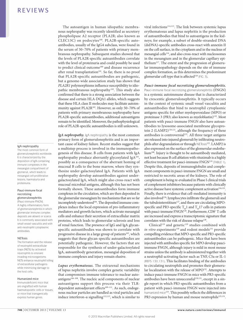
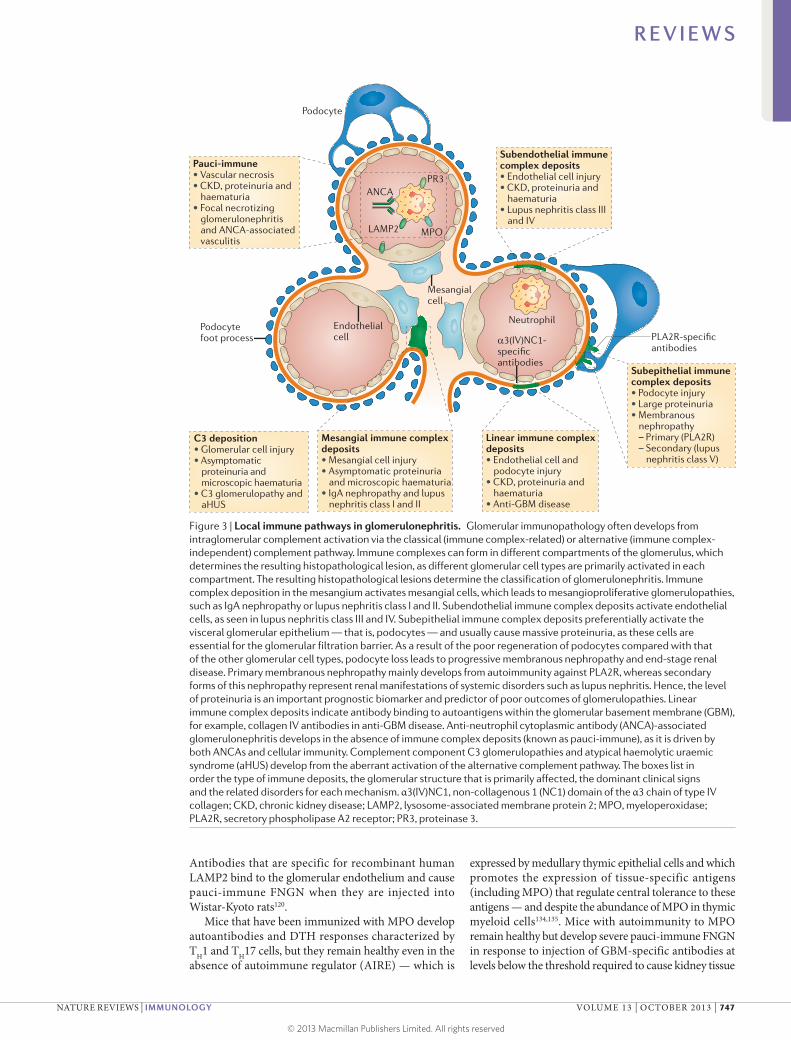
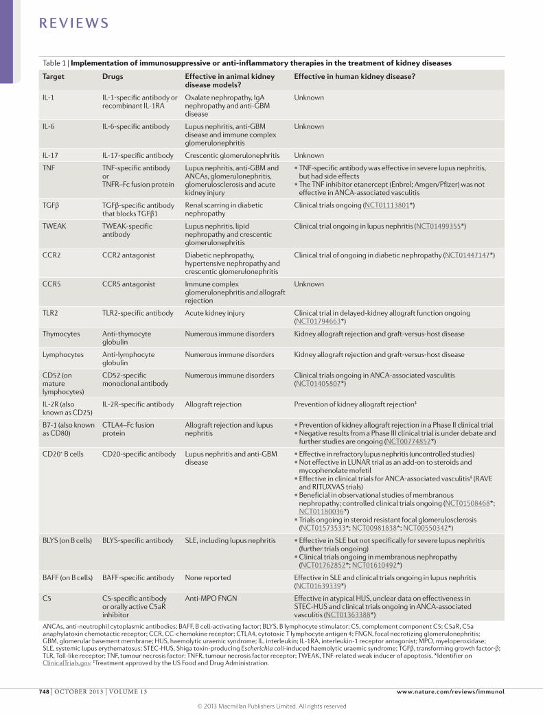


![u r n a l of Kidney Journal of Kidney - Longdom...immune system (Figure 1) [19]. There are 3 stages of immunosuppression: induction, maintenance, and treatment of established rejection.](https://static.documents.pub/doc/80x56/5f08f79c7e708231d42497c2/u-r-n-a-l-of-kidney-journal-of-kidney-longdom-immune-system-figure-1-19.jpg)


