Materials Science and Engineering C 32 (2012) 105–111
Contents lists available at SciVerse ScienceDirect
Materials Science and Engineering C
j ourna l homepage: www.e lsev ie r .com/ locate /msec
The importance of orientation in proton transport of a polymer film based on anoriented self-organized columnar liquid-crystalline polyether
Bartosz Tylkowski a, Nuria Castelao a, Marta Giamberini a,⁎, Ricard Garcia-Valls a,José Antonio Reina b, Tània Gumí a
a Departament d'Enginyeria Química, Universitat Rovira i Virgili, Av. Països Catalans, 26, E-43007, Tarragona, Spainb Departament de Química Analítica i Química Orgànica, Universitat Rovira i Virgili, Carrer Marcel.lí Domingo s/n, E-43007, Tarragona, Spain
We prepared membranes based on a liquid-crystalline side-chain polyether obtained by chemical modifica-tion of commercial poly(epichlorohydrin) (PECH) with dendrons. This polymer exhibited a columnar struc-ture, which could form an ion channel in the inner part. The columns were successfully oriented by takingadvantage of surface interactions between the polymer and hydrophilic substrates, as confirmed by X-ray dif-fraction analysis (XRD), environmental scanning electron microscopy (ESEM) and optical microscopy be-tween crossed polars (POM). Column orientation was found to be crucial for effective transport: theoriented membranes exhibited proton transport comparable to that of Nafion® N117 and no water uptake.An increase in sodium ion concentration in the feed phase suggested a proton/cation antiport. On the contrar-y, no proton transport was detected on unoriented membranes based on the same liquid-crystalline side-chain polyether or on unmodified PECH.
Nature was the first to take advantage of proton transport: mostbiochemical reactions are very sensitive to pH changes, and protontransport plays a crucial role in cell pH stabilization [1]. In biologicalsystems, information can be transferred via alkaline metal ions insome cases. Nevertheless, all processes that imply energy conversionfrom one form to another involve protonation and deprotonation re-actions mediated by proton conductivity: this is the case, for instance,in ATP formation during photosynthesis [2]. Proton conductivity alsoplays a key role in the production of electricity in hydrogen fuel cells[3,4]. As a consequence, proton transport and transfer phenomenahave been the object of extensive research from rather differentpoints of view by materials scientists, chemists, physicists and biolo-gists [5,6]. In the late 1960s, the need for cation-conducting separatormaterials for industrial chlorine–alkali electrolysis encouraged thedevelopment of chemically resistant cation-exchange membranes,which also exhibited good proton transport properties in their hy-drated protonic form. Over the past three decades, most research inthe field of proton conductivity has been undertaken by the materialsscience community, mainly for the development of new proton-conducting materials to be used in electrochemical cells (e.g. fuelcells, batteries, sensors). Perfluorosulfonic acid (PFSA) membranes,
34 977559621.berini).
rights reserved.
such as Nafion® (marketed by DuPont), have aroused great interestin recent years for their proton-conducting properties [7]. Theachievement of optimum performance for these materials requiresdetailed knowledge of chemical microstructure and nanoscale mor-phology. In particular, it is essential to control properties such as pro-ton conductivity, water management, relative affinity of methanoland water in direct methanol fuel cells (DMFCs), mechanical, thermaland oxidative stability, etc. This is a challenge for Nafion® materials,in which the possible chemical variations are quite limited; further-more, PFSA membranes are expensive. Another serious drawback ofmembranes of this sort is their environmental inadaptability. Newtypes of membranes based on different concepts are therefore beingdeveloped, but Nafion® remains the benchmark material againstwhich most results are compared. In recent years, more than 200 pat-ents and papers have been published on the preparation of newproton-conducting membranes [8–12]. An alternative approach is todesign materials containing ion transport channels, in which thechannels localize the permeation path and simultaneously protectthe transport process against the environment, like an ion-transporting molecular cable [13,14]. Very recently, two types ofone-dimensional ion-conductive polymer films containing ion nano-channels were prepared by means of photo-polymerization of thealigned columnar liquid crystals of a fan-shaped imidazolium saltwith acrylate groups at the periphery [15]. In this columnar structure,the ionic portion self-assembles in the inner part of the column; an-isotropic ionic conductivities were observed for the oriented filmsfixed by photo-polymerization. Very recently, an approach based on
106 B. Tylkowski et al. / Materials Science and Engineering C 32 (2012) 105–111
an inverse-ion-conducting cable was reported [16]. In this paper, anoriented columnar hexaphenylbenzene-based material, containingphosphonic acid groups at the periphery, was synthesized and provedto have good proton conductivity, independent of temperature. Thiscolumnar structure contains a proton-conducting periphery and aninsulating core; in this case, proton conductivity is not a water-based diffusion process, as in common amorphous polymers used todate.
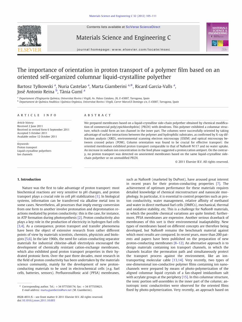
In this study, we prepared membranes capable of transportingprotons based on a side-chain liquid-crystalline polyether, whichwe synthesized ourselves. As we reported in a previous paper [17],this polymer self-assembles into a columnar structure (Fig. 1), dueto an exo-recognition of the side-chain dendrons. In the resultingstructure, the polyether main chain forms a channel in the innerpart of the columns, while the hydrophobic side-chain dendrons lieon the outer part. The presence of the polar ether linkages in theinner channel favors the interaction with proton and other cations,in the same way as crown ethers would do [18]. For this reason, theinner polyether chain could work as an ion channel. The orientationof the columns, which is crucial to getting efficient transport, wasachieved by means of a very simple procedure: casting the polymericchloroform solution in the presence of water. The prepared mem-branes exhibited a proton permeability comparable to that ofNafion® due to the presence of the oriented channels.
2. Experimental
2.1. Characterizations
X-ray diffraction (XRD) measurements were made using a Bruker-AXS D8-Discover diffractometer equipped with parallel incident
Fig. 1. Structure of polymer A and its sel
beam (Göbel Mirror), vertical θ–θ goniometer, XYZ motorized stageand general area diffraction detector system (GADDS). Sampleswere placed directly or mounted on MYLAR film. An X-ray beamwas collimated with a 100 μm collimator for optimum resolution intransmission mode. The X-ray diffractometer was operated at 40 kVand 40 mA to generate CuKα radiation. The GADDS detector was30×30 cm2 with a 1024×1024 pixel CCD sensor. The detector wasequipped with a small-angle X-ray scattering (SAXS) attachmentthat encloses the secondary beam in an He atmosphere to minimizeair scattering at low angles. We collected one frame (2D XRD pattern)per sample covering 0.5–9.5° 2θ at a distance of 30 cm from the sam-ple to the detector. The exposure time was 1800 s per frame and theframe was χ-integrated to generate the conventional 2θ versus inten-sity diffractogram.
The thickness of the membranes was measured using a microme-ter with a sensitivity of 2 μm. The measurements were carried out atvarious points, and the membranes were found to have constantthickness.
The cross-sections and surface morphologies of the polymericmembranes were characterized by environmental scanning electronmicroscopy (ESEM) (Quanta 600, FEI). Cross-sections were preparedby fracturing the membranes in liquid nitrogen. When SEM experi-ments were performed in high vacuum, samples were coated with agold layer before observation.
Optical microscopy between crossed polars was performed atroom temperature with an Axiolab Zeiss optical microscope.
The surface morphology of the oriented membranes was detectedby atomic force microscopy (AFM) (Pico+, 5500, Agilent Technolo-gies). The surface area of the topographical images was 2×2 μm.
Thermogravimetric analyses (TGA) on polymer A membraneswere carried out between 25 and 600 °C on a Mettler Toledo TGA/
f-assembly into columnar structure.
107B. Tylkowski et al. / Materials Science and Engineering C 32 (2012) 105–111
SDTA851e device under a nitrogen atmosphere. The scan rate was10 °C/min.
Swelling experiments were performed by soaking the membranesat room temperature in 1.0 M NaCl aqueous solution and monitoringthe change in membrane weight versus time for 6 h.
Na+ concentration was determined by inductively coupled plasmamass spectrometry (ICP-MS) (Thermo Elemental, Mod. Xseries II, Bre-men, Germany); each sample was analyzed three times and ultrapurecommercial standard solutionswere used (Merck, Darmstadt, Germanyand J.T. Baker, North Kingstown, RI, USA). Sc, Y, Ir and Rh were used asinternal standards.
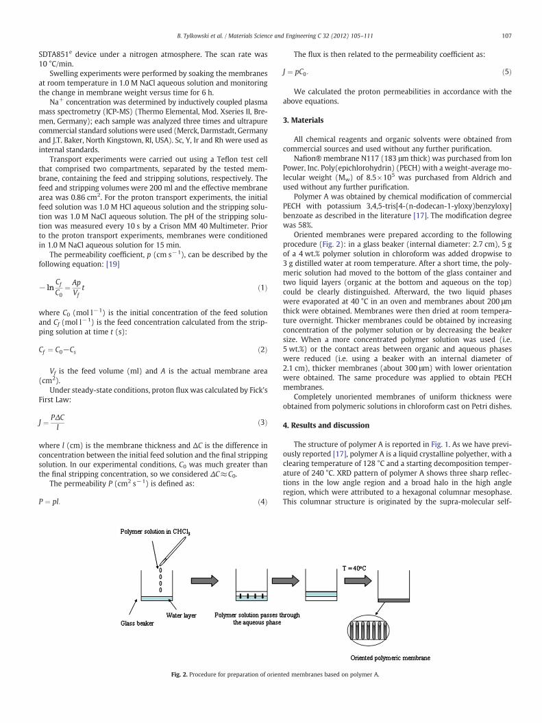
Transport experiments were carried out using a Teflon test cellthat comprised two compartments, separated by the tested mem-brane, containing the feed and stripping solutions, respectively. Thefeed and stripping volumes were 200 ml and the effective membranearea was 0.86 cm2. For the proton transport experiments, the initialfeed solution was 1.0 M HCl aqueous solution and the stripping solu-tion was 1.0 M NaCl aqueous solution. The pH of the stripping solu-tion was measured every 10 s by a Crison MM 40 Multimeter. Priorto the proton transport experiments, membranes were conditionedin 1.0 M NaCl aqueous solution for 15 min.
The permeability coefficient, p (cm s−1), can be described by thefollowing equation: [19]
− lnCf
C0¼ Ap
Vft ð1Þ
where C0 (mol l−1) is the initial concentration of the feed solutionand Cf (mol l−1) is the feed concentration calculated from the strip-ping solution at time t (s):
Cf ¼ C0−Cs ð2Þ
Vf is the feed volume (ml) and A is the actual membrane area(cm2).
Under steady-state conditions, proton flux was calculated by Fick'sFirst Law:
J ¼ PΔCl
ð3Þ
where l (cm) is the membrane thickness and ΔC is the difference inconcentration between the initial feed solution and the final strippingsolution. In our experimental conditions, C0 was much greater thanthe final stripping concentration, so we considered ΔC≈C0.
The permeability P (cm2 s−1) is defined as:
P ¼ pl: ð4Þ
Fig. 2. Procedure for preparation of orien
The flux is then related to the permeability coefficient as:
J ¼ pC0: ð5Þ
We calculated the proton permeabilities in accordance with theabove equations.
3. Materials
All chemical reagents and organic solvents were obtained fromcommercial sources and used without any further purification.
Nafion® membrane N117 (183 μm thick) was purchased from IonPower, Inc. Poly(epichlorohydrin) (PECH) with a weight-average mo-lecular weight (Mw) of 8.5×10⁵ was purchased from Aldrich andused without any further purification.
Polymer A was obtained by chemical modification of commercialPECH with potassium 3,4,5-tris[4-(n-dodecan-1-yloxy)benzyloxy]benzoate as described in the literature [17]. The modification degreewas 58%.
Oriented membranes were prepared according to the followingprocedure (Fig. 2): in a glass beaker (internal diameter: 2.7 cm), 5 gof a 4 wt.% polymer solution in chloroform was added dropwise to3 g distilled water at room temperature. After a short time, the poly-meric solution had moved to the bottom of the glass container andtwo liquid layers (organic at the bottom and aqueous on the top)could be clearly distinguished. Afterward, the two liquid phaseswere evaporated at 40 °C in an oven and membranes about 200 μmthick were obtained. Membranes were then dried at room tempera-ture overnight. Thicker membranes could be obtained by increasingconcentration of the polymer solution or by decreasing the beakersize. When a more concentrated polymer solution was used (i.e.5 wt.%) or the contact areas between organic and aqueous phaseswere reduced (i.e. using a beaker with an internal diameter of2.1 cm), thicker membranes (about 300 μm) with lower orientationwere obtained. The same procedure was applied to obtain PECHmembranes.
Completely unoriented membranes of uniform thickness wereobtained from polymeric solutions in chloroform cast on Petri dishes.
4. Results and discussion
The structure of polymer A is reported in Fig. 1. As we have previ-ously reported [17], polymer A is a liquid crystalline polyether, with aclearing temperature of 128 °C and a starting decomposition temper-ature of 240 °C. XRD pattern of polymer A shows three sharp reflec-tions in the low angle region and a broad halo in the high angleregion, which were attributed to a hexagonal columnar mesophase.This columnar structure is originated by the supra-molecular self-
Fig. 4. Oriented film of polymer A obtained from evaporation of the chloroform/watersystem.
108 B. Tylkowski et al. / Materials Science and Engineering C 32 (2012) 105–111
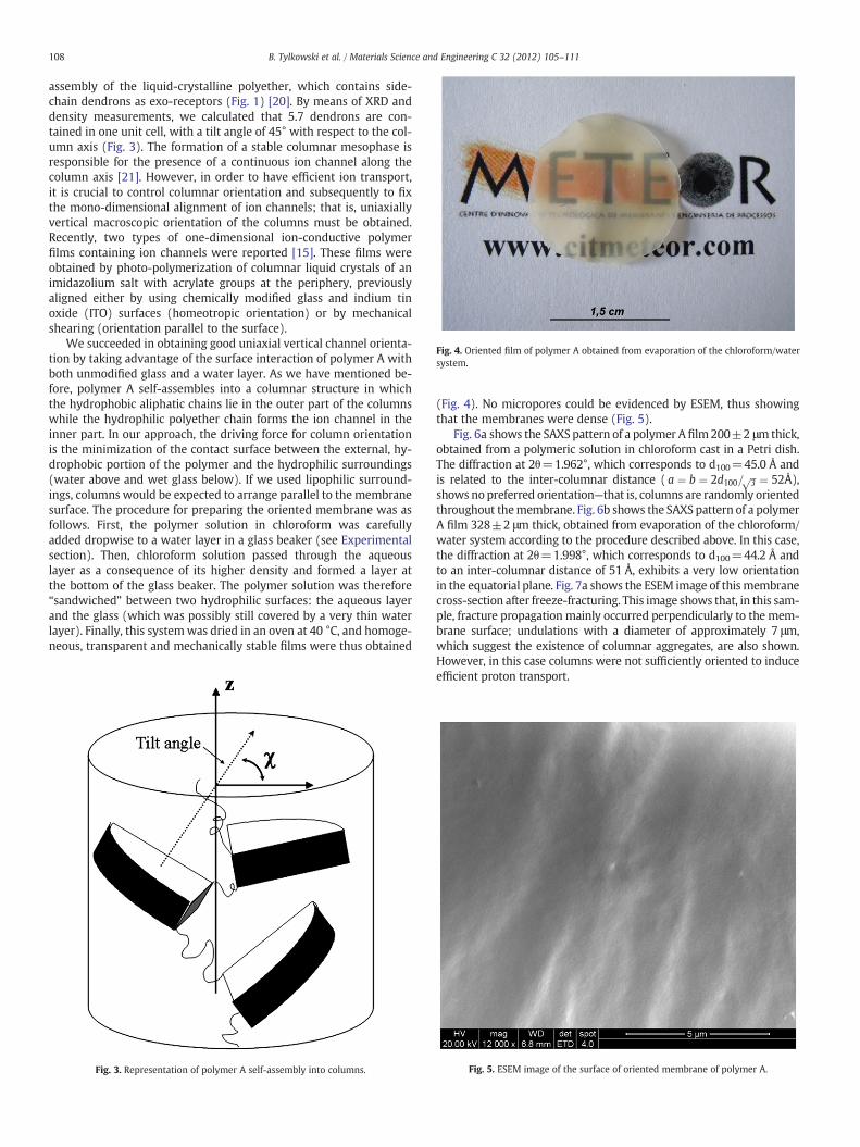
assembly of the liquid-crystalline polyether, which contains side-chain dendrons as exo-receptors (Fig. 1) [20]. By means of XRD anddensity measurements, we calculated that 5.7 dendrons are con-tained in one unit cell, with a tilt angle of 45° with respect to the col-umn axis (Fig. 3). The formation of a stable columnar mesophase isresponsible for the presence of a continuous ion channel along thecolumn axis [21]. However, in order to have efficient ion transport,it is crucial to control columnar orientation and subsequently to fixthe mono-dimensional alignment of ion channels; that is, uniaxiallyvertical macroscopic orientation of the columns must be obtained.Recently, two types of one-dimensional ion-conductive polymerfilms containing ion channels were reported [15]. These films wereobtained by photo-polymerization of columnar liquid crystals of animidazolium salt with acrylate groups at the periphery, previouslyaligned either by using chemically modified glass and indium tinoxide (ITO) surfaces (homeotropic orientation) or by mechanicalshearing (orientation parallel to the surface).
We succeeded in obtaining good uniaxial vertical channel orienta-tion by taking advantage of the surface interaction of polymer A withboth unmodified glass and a water layer. As we have mentioned be-fore, polymer A self-assembles into a columnar structure in whichthe hydrophobic aliphatic chains lie in the outer part of the columnswhile the hydrophilic polyether chain forms the ion channel in theinner part. In our approach, the driving force for column orientationis the minimization of the contact surface between the external, hy-drophobic portion of the polymer and the hydrophilic surroundings(water above and wet glass below). If we used lipophilic surround-ings, columns would be expected to arrange parallel to the membranesurface. The procedure for preparing the oriented membrane was asfollows. First, the polymer solution in chloroform was carefullyadded dropwise to a water layer in a glass beaker (see Experimentalsection). Then, chloroform solution passed through the aqueouslayer as a consequence of its higher density and formed a layer atthe bottom of the glass beaker. The polymer solution was therefore“sandwiched” between two hydrophilic surfaces: the aqueous layerand the glass (which was possibly still covered by a very thin waterlayer). Finally, this systemwas dried in an oven at 40 °C, and homoge-neous, transparent and mechanically stable films were thus obtained
Fig. 3. Representation of polymer A self-assembly into columns.
(Fig. 4). No micropores could be evidenced by ESEM, thus showingthat the membranes were dense (Fig. 5).
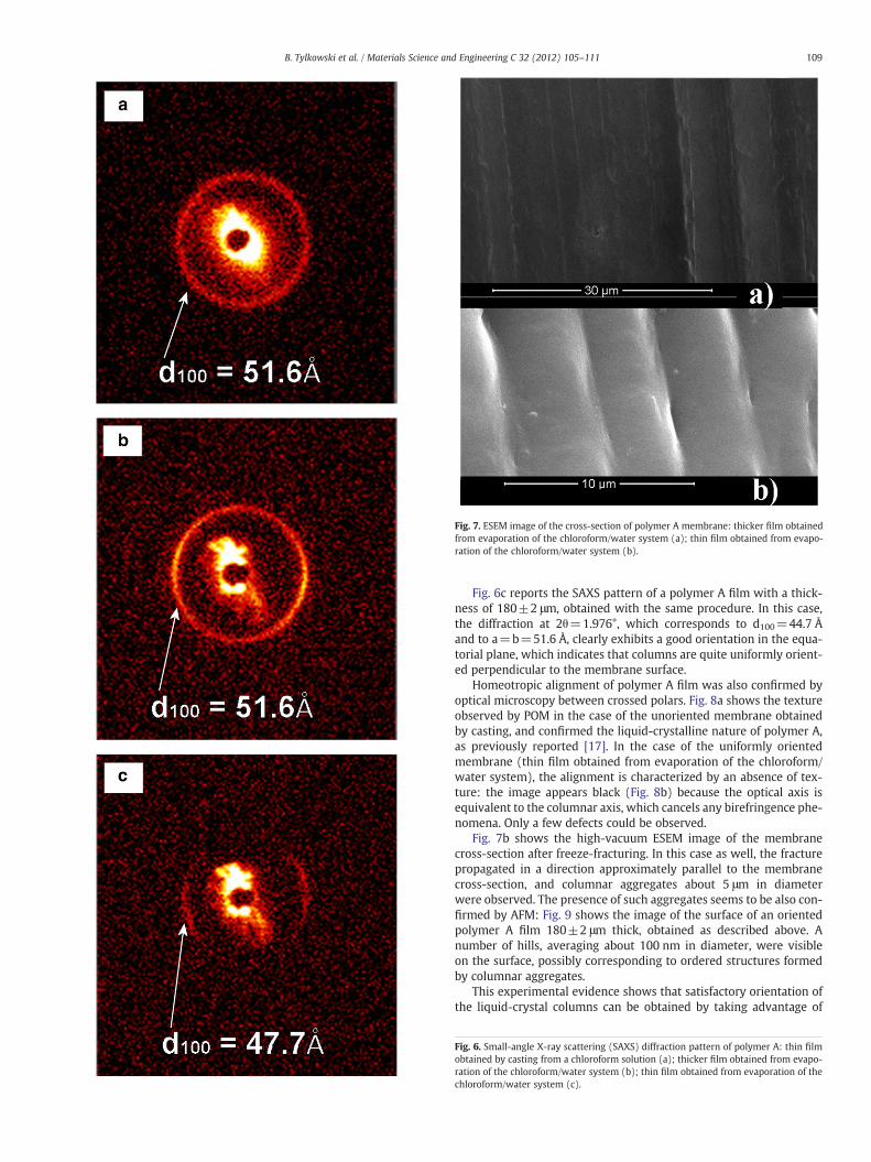
Fig. 6a shows the SAXS pattern of a polymer A film 200±2 μmthick,obtained from a polymeric solution in chloroform cast in a Petri dish.The diffraction at 2θ=1.962°, which corresponds to d100=45.0 Å andis related to the inter-columnar distance ( a ¼ b ¼ 2d100= ffiffiffi
3p ¼ 52Å),
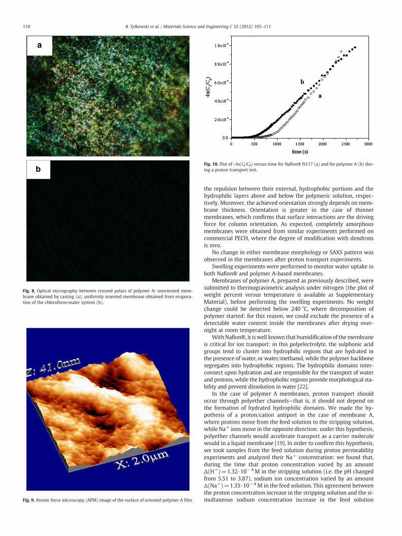
shows no preferred orientation—that is, columns are randomly orientedthroughout themembrane. Fig. 6b shows the SAXS pattern of a polymerA film 328±2 μm thick, obtained from evaporation of the chloroform/water system according to the procedure described above. In this case,the diffraction at 2θ=1.998°, which corresponds to d100=44.2 Å andto an inter-columnar distance of 51 Å, exhibits a very low orientationin the equatorial plane. Fig. 7a shows the ESEM image of this membranecross-section after freeze-fracturing. This image shows that, in this sam-ple, fracture propagation mainly occurred perpendicularly to the mem-brane surface; undulations with a diameter of approximately 7 μm,which suggest the existence of columnar aggregates, are also shown.However, in this case columns were not sufficiently oriented to induceefficient proton transport.
Fig. 5. ESEM image of the surface of oriented membrane of polymer A.
Fig. 7. ESEM image of the cross-section of polymer A membrane: thicker film obtainedfrom evaporation of the chloroform/water system (a); thin film obtained from evapo-ration of the chloroform/water system (b).
109B. Tylkowski et al. / Materials Science and Engineering C 32 (2012) 105–111
Fig. 6c reports the SAXS pattern of a polymer A film with a thick-ness of 180±2 μm, obtained with the same procedure. In this case,the diffraction at 2θ=1.976°, which corresponds to d100=44.7 Åand to a=b=51.6 Å, clearly exhibits a good orientation in the equa-torial plane, which indicates that columns are quite uniformly orient-ed perpendicular to the membrane surface.
Homeotropic alignment of polymer A film was also confirmed byoptical microscopy between crossed polars. Fig. 8a shows the textureobserved by POM in the case of the unoriented membrane obtainedby casting, and confirmed the liquid-crystalline nature of polymer A,as previously reported [17]. In the case of the uniformly orientedmembrane (thin film obtained from evaporation of the chloroform/water system), the alignment is characterized by an absence of tex-ture: the image appears black (Fig. 8b) because the optical axis isequivalent to the columnar axis, which cancels any birefringence phe-nomena. Only a few defects could be observed.
Fig. 7b shows the high-vacuum ESEM image of the membranecross-section after freeze-fracturing. In this case as well, the fracturepropagated in a direction approximately parallel to the membranecross-section, and columnar aggregates about 5 μm in diameterwere observed. The presence of such aggregates seems to be also con-firmed by AFM: Fig. 9 shows the image of the surface of an orientedpolymer A film 180±2 μm thick, obtained as described above. Anumber of hills, averaging about 100 nm in diameter, were visibleon the surface, possibly corresponding to ordered structures formedby columnar aggregates.
This experimental evidence shows that satisfactory orientation ofthe liquid-crystal columns can be obtained by taking advantage of
Fig. 6. Small-angle X-ray scattering (SAXS) diffraction pattern of polymer A: thin filmobtained by casting from a chloroform solution (a); thicker film obtained from evapo-ration of the chloroform/water system (b); thin film obtained from evaporation of thechloroform/water system (c).
Fig. 8. Optical micrography between crossed polars of polymer A: unoriented mem-brane obtained by casting (a); uniformly oriented membrane obtained from evapora-tion of the chloroform/water system (b).
Fig. 9. Atomic force microscopy (AFM) image of the surface of oriented polymer A film.
Fig. 10. Plot of –ln(Cf/C0) versus time for Nafion® N117 (a) and for polymer A (b) dur-ing a proton transport test.
110 B. Tylkowski et al. / Materials Science and Engineering C 32 (2012) 105–111
the repulsion between their external, hydrophobic portions and thehydrophilic layers above and below the polymeric solution, respec-tively. Moreover, the achieved orientation strongly depends on mem-brane thickness. Orientation is greater in the case of thinnermembranes, which confirms that surface interactions are the drivingforce for column orientation. As expected, completely amorphousmembranes were obtained from similar experiments performed oncommercial PECH, where the degree of modification with dendronsis zero.
No change in either membrane morphology or SAXS pattern wasobserved in the membranes after proton transport experiments.
Swelling experiments were performed to monitor water uptake inboth Nafion® and polymer A-based membranes.
Membranes of polymer A, prepared as previously described, weresubmitted to thermogravimetric analysis under nitrogen (the plot ofweight percent versus temperature is available as SupplementaryMaterial), before performing the swelling experiments. No weightchange could be detected below 240 °C, where decomposition ofpolymer started: for this reason, we could exclude the presence of adetectable water content inside the membranes after drying over-night at room temperature.
WithNafion®, it iswell known that humidification of themembraneis critical for ion transport: in this polyelectrolyte, the sulphonic acidgroups tend to cluster into hydrophilic regions that are hydrated inthe presence of water, or water/methanol, while the polymer backbonesegregates into hydrophobic regions. The hydrophilic domains inter-connect upon hydration and are responsible for the transport of waterand protons, while the hydrophobic regions providemorphological sta-bility and prevent dissolution in water [22].
In the case of polymer A membranes, proton transport shouldoccur through polyether channels—that is, it should not depend onthe formation of hydrated hydrophilic domains. We made the hy-pothesis of a proton/cation antiport in the case of membrane A,where protons move from the feed solution to the stripping solution,while Na+ ions move in the opposite direction: under this hypothesis,polyether channels would accelerate transport as a carrier moleculewould in a liquid membrane [19]. In order to confirm this hypothesis,we took samples from the feed solution during proton permeabilityexperiments and analyzed their Na+ concentration: we found that,during the time that proton concentration varied by an amountΔ(H+)=1.32·10−4 M in the stripping solution (i.e. the pH changedfrom 5.51 to 3.87), sodium ion concentration varied by an amountΔ(Na+)=1.35·10−4 M in the feed solution. This agreement betweenthe proton concentration increase in the stripping solution and the si-multaneous sodium concentration increase in the feed solution
111B. Tylkowski et al. / Materials Science and Engineering C 32 (2012) 105–111
suggests that proton/cation antiport mechanism can be reasonablyconsidered for the membrane based on polymer A.
WhenNafion®N117membranes about 200 μm thickwere soaked in1 M NaCl aqueous solution at room temperature, their weight changedfor the first 15 min and then reached a plateau value corresponding to6% water content, in agreement with commercial specifications. In con-trast, when polymer A membranes about 200 μm thick were soaked,no weight change could be detected, even after 6 h of soaking underthe same conditions.
Proton transport tests were performed on Nafion® as well as onpolymer A and PECH membranes of similar thicknesses. Fig. 10shows a typical plot of –ln(Cf/C0) versus time for Nafion® N117 (a)and for polymer A (b). In both cases, the plot exhibits two differentregions: the first one, which was shorter for polymer A, correspondsto proton absorption and diffusion across the membrane; once themembranes were saturated by protons, the mechanism becamehopping-dominated. One has to keep into account that Nafion®N117 membrane is thicker, with respect to polymer A membrane,due to its swelling in 1.0 M NaCl: this could justify the longer timeneeded for proton diffusion across the membrane. From the slope ofthe second region, which was quite similar for both materials, we cal-culated proton permeability; Table 1 shows the results. The valuefound for Nafion® N117 (2.59·10−6 cm2 s−1) was used as a referencestandard in the evaluation of proton permeability of the other material.Table 1 indicates that polymer A, obtained from chemical modificationof commercial PECH with dendrons, has remarkable proton permeabil-ity, around 2·10−6 cm2 s−1 (comparable to that of Nafion® N117).However, for the unmodified PECH, and for unoriented polymer Amembranes, the pH variation lay within the experimental error(±0.1) and therefore no transport could be detected at all on a reason-able time scale. This evidence, together with the results of the swellingexperiments, confirms that efficient proton transport occurred in poly-mer A due to the presence of oriented channels in the membrane.
5. Conclusions
In this paper, orientedmembranes based on a novel liquid-crystallinecolumnar polyether (polymer A)were prepared. Satisfactory orientationof the polymer was achieved by sandwiching the polymeric solution be-tween awater layer and awet glass layer to induce unfavourable surfaceinteractions between the outer, hydrophobic portion of the columns andtheir surroundings. This orientationwas detected by XRD, and ESEM and
AFM revealed the presence of oriented columnar aggregates. UnlikeNafion® N117, the prepared membranes did not exhibit weight changeafter soaking in 1 M NaCl aqueous solution at room temperature evenafter 6 h. The presence of oriented channels in the polymeric membraneresulted in remarkable proton permeability, around 2·10−6 cm2 s−1,comparable to that of Nafion® N117. Na+ concentration was deter-mined in the feed solution during proton permeability experimentsand compared with the simultaneous pH decrease in the stripping solu-tion: the agreement between the data suggested that proton/cation anti-port can be reasonably considered in the transport mechanism ofmembrane A.
Supplementary materials related to this article can be found onlineat doi:10.1016/j.msec.2011.10.003.
Acknowledgments
Financial support fromMAT2008-00456/MAT (Ministerio de Cienciae Innovación) is gratefully acknowledged. The authors are also gratefulto Dr. Francesc Guirado for help with the XRD experiments.
References
[1] R.J.P. Williams, Annu. Rev. Biophys. Biophys. Chem. 17 (1988) 71–97.[2] D. Voet, J.G. Voet, Biochemistry, 3rd, John Wiley & Sons, New York, 2004.[3] J. Larminie, A. Dicks, Fuel Cell Systems Explained, 2nd ed., Wiley, England, 2003.[4] Polymer Membranes for Fuel Cells, Springer, New York, 2009.[5] L.A. Weiss, N. Sakai, B. Ghebremariam, C. Ni, S. Matile, J. Am. Chem. Soc. 119
(1997) 12142–12149.[6] G. Brunklaus, S. Schauff, D. Markova, M. Klapper, K. Müllen, H.-W. Spiess, J. Phys.
Chem. B 113 (2009) 6674–6681.[7] K.A. Mauritz, R.B. Moore, Chem. Rev. 104 (2004) 4535–4585.[8] F.G. Wilhelm, I.G.M. Pünt, N.F.A.v.d. Vegt, H. Strathmann, M. Wessling, J. Membr.
Sci. 199 (2002) 167–176.[9] K. Miyatake, Y. Chikashige, E. Higuchi, M. Watanabe, J. Am. Chem. Soc. 129 (2007)
3879–3887.[10] Y. Li, R. Jin, Z. Wang, Z. Cui, W. Xing, L. Gao, J. Polym. Sci. Polym. Chem. 45 (2007)
222–231.[11] G. Alberti, U. Costantino, M. Casciola, S. Ferroni, L. Massinelli, P. Staiti, Solid State
Ionics 145 (2001) 249–255.[12] A. Ainla, D. Brandell, Solid State Ionics 178 (2007) 581–585.[13] V. Percec, G. Johansson, J. Heck, G. Ungar, S.V.J. Batty, J. Chem. Soc. Perkin Trans. 1
(1993) 1411–1420.[14] U. Beginn, G. Zipp, M. Möller, Adv. Mater. 12 (2000) 510–513.[15] M. Yoshio, T. Kagata, K. Hoshino, T. Mukai, H. Ohno, T. Kato, J. Am. Chem. Soc. 128
(2006) 5570–5577.[16] L. Jiménez-García, A. Kaltbeitzel, W. Pisula, J.S. Gutmann, M. Klapper, K. Müllen,
Angew. Chem. Int. Ed. 48 (2009) 9951–9953.[17] J.C. Ronda, J.A. Reina, M. Giamberini, J. Polym. Sci. A Polym. Chem. 42 (2004)
326–340.[18] L.M. Dulyea, T.M. Fyles, G.D. Robertson, J. Membr. Sci. 34 (1987) 87–108.[19] M. Mulder, Basic Principles of Membrane Technology, 2nd, Kluwer Academic
Publishers, The Netherlands, 2003.[20] V. Percec, D. Schlueter, J.C. Ronda, G. Johansson, G. Ungar, J.P. Zhou, Macromole-
cules 29 (1996) 1464.[21] V. Percec, D. Schlueter, G. Ungar, S.Z.D. Cheng, A. Zhang, Macromolecules 31