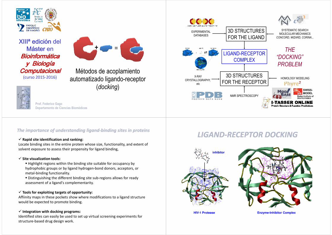
XIIIª edición XIIIª edición del Máster Máster en Bioinformática Bioinformática Bioinformática Bioinformática Bioinformática Bioinformática Bioinformática Bioinformática Métodos de acoplamiento automatizado ligando-receptor (docking) Prof. Federico Gago Departamento de Ciencias Biomédicas y y y y y Biología Biología Biología Biología Biología Biología Biología Biología Computacional Computacional Computacional Computacional Computacional Computacional Computacional Computacional (curso 2015-2016) LIGAND-RECEPTOR COMPLEX THE “DOCKING” EXPERIMENTAL DATABASES 3D STRUCTURES FOR THE LIGAND SYSTEMATIC SEARCH MOLECULAR MECHANICS CONCORD, WIZARD, CORINA... COMPLEX “DOCKING” PROBLEM X-RAY CRYSTALLOGRAPHY, etc 3D STRUCTURES FOR THE RECEPTOR HOMOLOGY MODELING NMR SPECTROSCOPY SWISS- MODEL The importance of understanding The importance of understanding ligand ligand-binding sites in proteins binding sites in proteins Rapid site identification and ranking: Locate binding sites in the entire protein whose size, functionality, and extent of solvent exposure to assess their propensity for ligand binding, Site visualization tools: Highlight regions within the binding site suitable for occupancy by hydrophobic groups or by ligand hydrogen-bond donors, acceptors, or metal-binding functionality. Distinguishing the different binding site sub-regions allows for ready Distinguishing the different binding site sub-regions allows for ready assessment of a ligand's complementarity. Tools for exploiting targets of opportunity: Affiinity maps in these pockets show where modifications to a ligand structure would be expected to promote binding. Integration with docking programs: Identified sites can easily be used to set up virtual screening experiments for structure-based drug design work. LIGAND LIGAND-RECEPTOR DOCKING RECEPTOR DOCKING inhibitor HIV-1 Protease Enzyme-Inhibitor Complex
The importance of understanding The importance of understanding ligandligand--binding sites in proteinsbinding sites in proteins
� Rapid site identification and ranking:
Locate binding sites in the entire protein whose size, functionality, and extent of
solvent exposure to assess their propensity for ligand binding,
� Site visualization tools:
� Highlight regions within the binding site suitable for occupancy by
hydrophobic groups or by ligand hydrogen-bond donors, acceptors, or
metal-binding functionality.
� Distinguishing the different binding site sub-regions allows for ready � Distinguishing the different binding site sub-regions allows for ready
assessment of a ligand's complementarity.
� Tools for exploiting targets of opportunity:
Affiinity maps in these pockets show where modifications to a ligand structure
would be expected to promote binding.
� Integration with docking programs:
Identified sites can easily be used to set up virtual screening experiments for
structure-based drug design work.
LIGANDLIGAND--RECEPTOR DOCKINGRECEPTOR DOCKING
inhibitor
HIV-1 Protease Enzyme-Inhibitor Complex
Validated Targets
Combinatorial Chemistry Libraries+
=
Ligand-Receptor Complexes
THE “DOCKING”
PROBLEM
Computed Atlas of Surface Topography of proteins
CASTp: A Server for
• GPSSpyMOL: Global Protein Surface Survey Plugin for PyMOL
A Server for
Identification of
Protein Pockets &
Cavities
• Identifies all pockets and cavities.
• Measures the volume and area analytically.
http://sts.bioengr.uic.edu/castp/
A software tool for analysis and visualization of
tunnels and channels in protein structures
http://www.caver.cz/
Tunnels are void pathways leading from a cavity buried in a protein core to the
surrounding solvent.
A channel leads through the protein structure and has both ends open to the
surrounding solvent.
� SITE/LIGAND REPRESENTATION(treatment of H atoms?)
“THE DOCKING PROBLEM”“THE DOCKING PROBLEM”
� JUXTAPOSITION OF THE LIGAND AND� JUXTAPOSITION OF THE LIGAND ANDSITE FRAMES OF REFERENCE
(docking engine)
� EVALUATION OF COMPLEMENTARITY(scoring functions)
AIM: To obtain the lowest free energy structure(s) for the receptor-ligand complex.
http://biophysics.cs.vt.edu/H++/
H++ is an automated system that computes pK values of ionizable groups in macromolecules and adds missing hydrogen atoms according to the specified pH of the environment. Given a (PDB) structure file on input, H++ outputs the completed structure in several common formats (PDB, PQR, AMBER inpcrd/prmtop) and provides a set of tools useful for analysis of electrostatic-related molecular properties.
http://pdb2pqr-1.wustl.edu/pdb2pqr/
http://propka.ki.ku.dk/
What, then, does the method of receptor fit offer for a future in which the structure and function of macromolecules will be understood and where doctors may have direct access to the full genetic code of every patient?
� One should be able to design novel drugs of very high affinity for known target sites,
� Systems analysis of a chosen biochemical pathway will enable the most appropriate target site to be identified,
� Sequence variation may be exploited to improve specificity, because systematic differences of protein sequence can often be detected near ligand binding sites.
� All receptors are different until proved identical.
These tentative forecasts point toward:
� a new generation of more potent, specific, effective therapeutic agents with less toxicity, reduced side effects, and fewer aberrant responses, which is what people and society at large are seeking.people and society at large are seeking.
� more costly research, which is the price that must be paid.
One last conclusion seems very probable. Mountaineersclimb because the mountains are there and offerthem a worthwhile challenge, and scientists will try todesign drugs to fit receptors for similar reasons.
15th courseTHREE DIMENSIONAL MOLECULAR STRUCTURE AND DRUG ACTION
Erice (1 −11 June, 1989)
Trimethoprim (TMP), a widely used antibacterial
drug, is a potent inhibitor of bacterial DHFRs but a
much weaker inhibitor of the vertebrate enzymes (e.g., IC50 values against Escherichia coli and human
enzyme are, respectively, 5 x 10⁻⁹ M and 3 x 10⁻⁴ M).
To provide information on the action of this drug
FEBS Lett. 126(1):49-52 (1981)
To provide information on the action of this drug
at the molecular level, we have determined the
structure of the binary complex of E. coli (strain
RT500) form I DHFR with TMP and compared it
with that of the complex of DHFR with
methotrexate (MTX), a drug which binds tightly
to both bacterial and vertebrate DHFR. The
structure of our TMP-enzyme complex differs
from that in [Science 1977; 197, 452-455] of an
MTX-enzyme complex from a different strain
(MB1428) of E. coli. The amino acid sequences of
the two enzymes are currently thought to differ at
3 positions.
trimethoprim binding site of E. coli DHFR binding site for compound 4 in E. coli DHFRDHFR
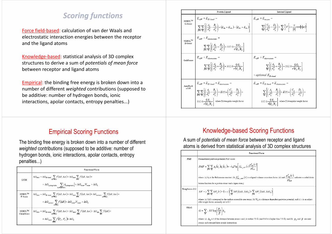
The binding free energy is broken down into a number of different weighted contributions (supposed to be additive: number of hydrogen bonds, ionic interactions, apolar contacts, entropy penalties...)
Knowledge-based Scoring FunctionsA sum of potentials of mean force between receptor and ligand atoms is derived from statistical analysis of 3D complex structures
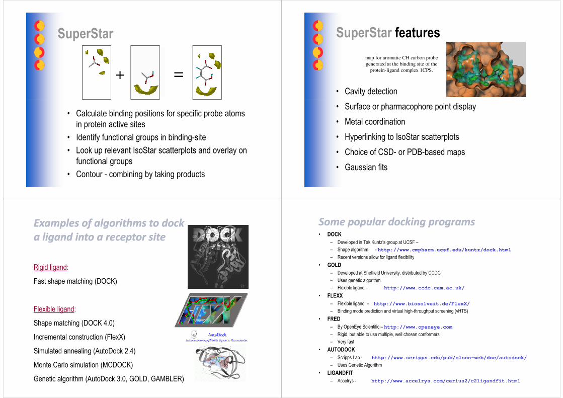
SuperStar
+ =
• Calculate binding positions for specific probe atoms in protein active sites
• Identify functional groups in binding-site
• Look up relevant IsoStar scatterplots and overlay on functional groups
• Contour - combining by taking products
SuperStar features
• Cavity detection
map for aromatic CH carbon probe
generated at the binding site of the
protein-ligand complex 1CPS.
• Surface or pharmacophore point display
• Metal coordination
• Hyperlinking to IsoStar scatterplots
• Choice of CSD- or PDB-based maps
• Gaussian fits
ExamplesExamples of of algorithmsalgorithms to dockto dock
a a ligandligand intointo a receptor a receptor sitesite
Rigid ligand:
Fast shape matching (DOCK)
Flexible ligand:
Shape matching (DOCK 4.0)
Incremental construction (FlexX)
Simulated annealing (AutoDock 2.4)
Monte Carlo simulation (MCDOCK)
Genetic algorithm (AutoDock 3.0, GOLD, GAMBLER)
SomeSome popular dpopular dockingocking programsprograms• DOCK
"A Geometric Approach to Macromolecule-Ligand Interactions"I. D. Kuntz, J. M. Blaney, S. J. Oatley, R. Langridge, T. E. FerrinJ. Mol. Biol. 161, 269-288 (1982)
"Using Shape Complementarity as an Initial Screen in Designing Ligands for a Receptor Binding Site of Known Three-Dimensional Structure"
PROGRAM DOCKPROGRAM DOCK
Receptor Binding Site of Known Three-Dimensional Structure"R. L. DesJarlais, R. P. Sheridan, G. L. Seibel, J. S. Dixon, I. D. Kuntz, R. VenkataraghavanJ. Med. Chem. 31, 722-729 (1988)
"Automated Docking with Grid-Based Energy Evaluation"E. C. Meng, B. K. Soichet, I. D. KuntzJ. Comp. Chem. 13, 505-524 (1991)
RECEPTOR COORDINATES
SITE CHARACTERIZATION GRID CALCULATION
MS molecular “dot” surfaceSPHGEN negative image of site
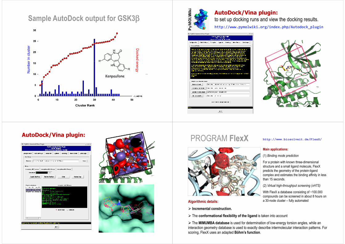
For a protein with known three-dimensional structure and a small ligand molecule, FlexX predicts the geometry of the protein-ligand complex and estimates the binding affinity in less than 15 seconds.
(2) Virtual high-throughput screening (vHTS)
With FlexX a database consisting of ~100.000 compounds can be screened in about 8 hours on a 30-node cluster – fully automated Algorithmic details:
� Incremental construction.
� The conformational flexibility of the ligand is taken into account
� The MIMUMBA database is used for determination of low-energy torsion angles, while an interaction geometry database is used to exactly describe intermolecular interaction patterns. For scoring, FlexX uses an adapted Böhm’s function.
FLEXX: Incremental construction
COO-
O
HN
C
COO-
H
NH2
Ligand fragments
CH3N
N
N
NH2N
H
• Select rigid portion as the base fragment
• Dock the base fragment into the receptor site, optimizing steric and electrostatic interactions.
• Sequentially add the remaining ligand fragments.
( )+×∆+∆=∆ rotrot NGGG0
( )
( )∑
∑+θ∆∆∆+
+θ∆∆∆+
,RfG
,RfG
ion
hb
Loss of entropy during ligand binding
Hydrogen bonds between neutral atoms
Ion bridges and ionic hydrogen bonds
FLEXX: Evaluation of the interaction energy
( )
( )∑
∑
∑
θ∆∆∆+
+θ∆∆∆+
,RfG
,RfG
lipo
aro
ion
Interactions between aromatics
Lipophilic contacts (mainly van der Waals)
( ) 10 ≤θ∆∆≤ ,Rf Geometry penalty function
Program GOLD
• Product of a collaboration between the University of Sheffield, GlaxoSmithKline plc and CCDC
• Uses a genetic algorithm for optimization
• Can output multiple solutions (i.e. output multiple final population members)
• Fitness function combination of four elements:– protein-ligand hydrogen bond energy (external H-bond)
– protein-ligand van der Waals (vdw) energy (external vdw)
– ligand internal vdw energy (internal vdw)
– ligand torsional strain energy (internal torsion)
Genetic Algorithms
• Create a “population” of possible solutions, encoded as “chromosomes”
• Use “fitness function” to score solutions• Use “fitness function” to score solutions
• Good solutions are combined together (“crossover”) and altered (“mutation”) to provide new solutions
• The process repeats until the population “converges” on a solution
How GAs Work
A way is found of encoding possible solutions into a bitstring (chromosome), and of specifying the 'goodness' of a chromosome (fitness function)
1. Initialize a population of chromosomes1. Initialize a population of chromosomes1. Initialize a population of chromosomes1. Initialize a population of chromosomes1. Initialize a population of chromosomes1. Initialize a population of chromosomes1. Initialize a population of chromosomes1. Initialize a population of chromosomes
2. Evaluate the fitness of each chromosome2. Evaluate the fitness of each chromosome2. Evaluate the fitness of each chromosome2. Evaluate the fitness of each chromosome
3. Create new chromosomes from the current population3. Create new chromosomes from the current population3. Create new chromosomes from the current population3. Create new chromosomes from the current population
4. Delete population members to make room for new ones4. Delete population members to make room for new ones4. Delete population members to make room for new ones4. Delete population members to make room for new ones
5. Evaluate the new chromosomes and put them in 5. Evaluate the new chromosomes and put them in 5. Evaluate the new chromosomes and put them in 5. Evaluate the new chromosomes and put them in populationpopulationpopulationpopulation
6. If we want to keep going, go back to step 36. If we want to keep going, go back to step 36. If we want to keep going, go back to step 36. If we want to keep going, go back to step 3
Genetic Algorithms
• For our purpose, we can encode rotation and translation of a molecule, and bond torsion angles in a chromosome, e.g.:
TX TY TZ RX RY RZ τ1 τ2
where we have 3 translation values (T), 3 rotation values(R) and as many torsion angles (τ) as the molecule has rotatable bonds
• Initially, our population will be initialized with random values, e.g.
τ2oτ1
oRZoRY
oRXoTZTYTX
Genetic Algorithms
C7
C6
C5
C4
C3
C2
C1
9826224612-0.2-0.34.4
751552781145.6-2.70.3
183412625194.94.15.8
21631214128027-3.6-2.9-2.2
2983122611433.12.9-8.7
14412614923197-4.61.32.8
1141312281261304.5-1.6-3.2
Fitness Function
• Used to score chromosomes to determine “goodness”
• For our purposes, we are concerned with how well the molecule in a particular orientation binds to the protein
• So a fitness function for a docking GA might be a combination of • So a fitness function for a docking GA might be a combination of the following elements:
– Energy (binding, potential)
– Number and strength of hydrogen bonds formed
– Hydrophobic effects
– Electrostatic effects
Fitness Function
• To score a chromosome, the GA will place the molecule inside the protein using the given translation, rotation and torsion parameters, and the fitness function will calculate the score based on an analysis of the joint 3D calculate the score based on an analysis of the joint 3D structure
Fitness function scoring of population
• Initially, our population will be initialized with random values, e.g.
τ2o Scoreτ1
oRZoRY
oRXoTZTYTX
98
75
18
216
29
144
114
C7
C6
C5
C4
C3
C2
C1
0.6126224612-0.2-0.34.4
0.781552781145.6-2.70.3
0.323412625194.94.15.8
0.0431214128027-3.6-2.9-2.2
0.8783122611433.12.9-8.7
0.9512614923197-4.61.32.8
0.421312281261304.5-1.6-3.2
Create new population members
• Initially, our population will be initialized with random values, e.g.
τ2o Scoreτ1
oRZoRY
oRXoTZTYTX
98
75
18
216
29
144
114
C7
C6
C5
C4
C3
C2
C1
0.6126224612-0.2-0.34.4
0.781552781145.6-2.70.3
0.323412625194.94.15.8
0.0431214128027-3.6-2.9-2.2
0.7783122611433.12.9-8.7
0.6512614923197-4.61.32.8
0.421312281261304.5-1.6-3.2
Crossover
29
144
C3
C2
0.7783122611433.12.9-8.7
0.6512614923197-4.61.32.8
144C8 831226197-4.61.3-8.7
Mutation
134C2 0.65126149221107-4.60.32.8
134C9 126149221107-4.60.32.8
Score new chromosomes
29
144
114
τ2o
C3
C2
C1
0.7783122611433.12.9-8.7
0.6512614923197-4.61.32.8
0.421312281261304.5-1.6-3.2
Scoreτ1oRZ
oRYoRX
oTZTYTX
0.619826224612-0.2-0.34.4C7
0.83144831226197-4.61.3-8.7C8
134
75
18
216
C9
C6
C5
C4
0.56126149221107-4.60.32.8
0.781552781145.6-2.70.3
0.323412625194.94.15.8
0.0431214128027-3.6-2.9-2.2
Delete poor chromosomes
216
29
144
114
τ2o
C4
C3
C2
C1
0.0431214128027-3.6-2.9-2.2
0.7783122611433.12.9-8.7
0.6512614923197-4.61.32.8
0.421312281261304.5-1.6-3.2
Scoreτ1oRZ
oRYoRX
oTZTYTX
0.619826224612-0.2-0.34.4C7
0.83144831226197-4.61.3-8.7C8
134
75
18
216
C9
C6
C5
C4
0.56126149221107-4.60.32.8
0.781552781145.6-2.70.3
0.323412625194.94.15.8
0.0431214128027-3.6-2.9-2.2
Sample GOLD output
GMP into RNaseT1
Program FRED (OpenEye Scientific Software)• Docking is exhaustive
Unlike most docking programs FRED does not use stochastic sampling to dock ligand. Rather it begins with the set of all possible orientations (to a resolution of one Angstrom, by default) of each conformer near the receptor site and selects the docked position of the ligand from this set.
• SpeedFRED typically docks from 7 to 15 conformers per second on a single PIII-800Mhz CPU.
• Multi-processorFRED fully supports PVM (Parallel Virtual Machine) on Linux and SGI platforms. This
• Multi-processorFRED fully supports PVM (Parallel Virtual Machine) on Linux and SGI platforms. This allows FRED to take advantage of multiple processors on multiple machines while still returning a single centralized set of output.
• Multiple scoring fuctionsFRED currently supports Chemscore, PLP, ScreenScore and Gaussian shape scoring. Scoring with ZAP (a Poisson-Boltzmann solver).
• Alternative docking positions for ligandsFRED returns alternative docked poses for each ligand as well as the top scoring ligand.
• Graphical receptor site preparation (with VIDA)While FRED is fully functional as a command line program, the graphics program VIDA has a FRED wizard which can be used to set up the receptor site for Fred.
Program GLIDE (Grid-based Ligand Docking with Energetics)
� Funnel: site point search → diameter test → subset test → greedy score → refinement → grid-based energy optimization → GlideScore.
� Approximates a complete systematic search of the conformational, orientational, and positional space of the docked ligand.
� Hierarchical filters, including a rough scoring function that recognizes hydrophobic and polar contacts, dramatically narrow the search spacehydrophobic and polar contacts, dramatically narrow the search space
� Torsionally flexible energy optimization on an OPLS-AA nonbondedpotential grid for a few hundred surviving candidate poses.
� The very best candidates are further refined via a Monte Carlo sampling of pose conformation.
� A modified ChemScore (Eldridge et al. 1997) that combines empirical and force-field-based terms.
� Validation: 282 complexes, new ligand conformation, the top-ranked pose: 50%<1 Å, ~33% >2 Å.
Docking hierarchy / Funnel: Definition of core and rotamer groups:
Glide Fragment Library
Set of 441 unique small fragments (1-7 ionization/tautomer variants; 6-37 atoms; MW range 32-
226) derived from molecules in the medicinal chemistry literature. The set includes a total of 667
fragments with accessible low energy ionization and tautomeric states and metal and state
penalties for each compound from Epik. These can be used for fragment docking, core hopping,
lead optimization, de novo design, etc.
Program eHITS (SimBioSys Inc.)
• Accurate: validation test runs demonstrate that eHiTS can reproduce X-ray structures with very high accuracy (low RMSD). Not limited to local energy minima dihedral angle samples.
• Fast: million compound libraries can be screened in special VHTS mode in a matter of hours on a Linux cluster. Exhaustive flexible ligand docking can also be performed on a single CPU under three minutes.
• Easy to use, fully automated: automatic pocket detection on the protein surface, automatic assignment of partial charges to atoms, consideration of alternative hydrogen protonation states, etc.
• Customizable scoring function: parameters and weights of all scoring components can be adjusted in a human readable, well documented configuration file.
• Parallel execution: built-in support for SMP (e.g. SGI Origin) and distributed (e.g. Linux clusters) architectures, and also grid computing.
• Output postprocessing: hierarchical file structure and clustering utility.
� Is there any relationship between docking and rankingaccuracies?
� Will docking/scoring combinations provide better results in terms of hit rates? If so, which ones?
Some important questions....
in terms of hit rates? If so, which ones?
� Does “consensus scoring” from two or three independent scoring lists outperform single scoring?
� Will it be possible to find a universal scoring function?
Combined use of 3 docking algorithms (Dock, FlexX, Gold) with 7 scoring functions (Dock, FlexX, Gold, Pmf, Chemscore, Fresno, Score) for screening a 1000-compound library against two different protein targets, thymidine kinase (TK) and the ligand-binding domain of the estrogen receptor R subtype (ERR).
A specific database comprising 990 random and 10 known ligands was specifically created for each target.
Results of the virtual screening examined in terms of:Results of the virtual screening examined in terms of:
(i) docking accuracy (rmsd to known solutions),
(ii) scoring accuracy (prediction of the absolute binding free energy),
(iii) “consensus” versus single scoring,
(iv) discrimination of active from random compounds,
(v) hit rates and enrichment factors among the top scorers.
C. Bissantz, G. Folkers & D. Rognan - J. Med. Chem. 43, 4759-4767 (2000)
Docking method
ligand DOCK FlexX GOLD
deoxythymidine 0.82 0.78 0.72
5-iododeoxyuridine 9.33 1.03 0.77
5-iodouracil-anhydrohexitol 1.16 0.88 0.63
dhbt (not publicly available) 2.02 3.65 0.93
6-(3-hydroxy-propyl-thymine) 1.02 4.18 0.49
6-[6-hydroxymethy-5-methyl-
Doc
king
acc
urac
yev
iatio
ns (n
on h
ydro
gen
atom
s, in
Å) f
rom
the
X-ra
ypo
se]
(top
solu
tion
of e
ach
dock
ing
tool
)
6-[6-hydroxymethy-5-methyl-
2,4-dioxo-hexahydro-pyrimidin-
5-yl-methyl]-5-methyl-1H-
pyrimidin-2,4-dione
9.62 13.30 2.33
(North)-methanocarbathymidine 7.56 1.11 1.19
aciclovir 3.08 2.71 2.74
ganciclovir 3.01 6.07 3.11
penciclovir 4.10 5.96 3.01
Doc
king
acc
urac
y[R
ms
devi
atio
ns (n
on h
ydro
gen
atom
s, in
Å) f
rom
the
(top
solu
tion
of e
ach
dock
ing
tool
)
Only one set of protein (TK) coordinates used: PDB code 1KIM
Misdocked complexes can be categorized as soft and hard failures
Soft failures: the search algorithm is unable to locate the global energy minimum corresponding to the crystal structure but this conformation, after minimization with the force field chosen, yields a lower energy than that of the lowest energy found in the docking simulations
Hard failures: the global energy minimum corresponds to a misdocked structure, i.e. the method is unable to reproduce the differences in relative energies of alternate binding modes
lowest energy found in the docking simulations
DOCK FlexX
Ranking
(position in the scoring list)
3 independent docking posesGOLD
Comparison of the three docking methods each with its
best performing scoring function (TK ligands)
Ranking versus rms deviations from X-ray pose for TK ligands screened with the three best docking/scoring
combinations
C. Bissantz, G. Folkers & D. Rognan
J. Med. Chem. 43, 4759-4767 (2000)
DOCK
FlexX
% = percentages of the total number of ligands for which a docking solution was found
Dock: 10 true hits, 774 random
FlexX: 10 true hits, 488 random ligands
Only partial discrimination of true hits from random ligands
FlexX
GOLD
FlexX: 10 true hits, 488 random ligands
Gold: 10 true hits, 927 random ligands
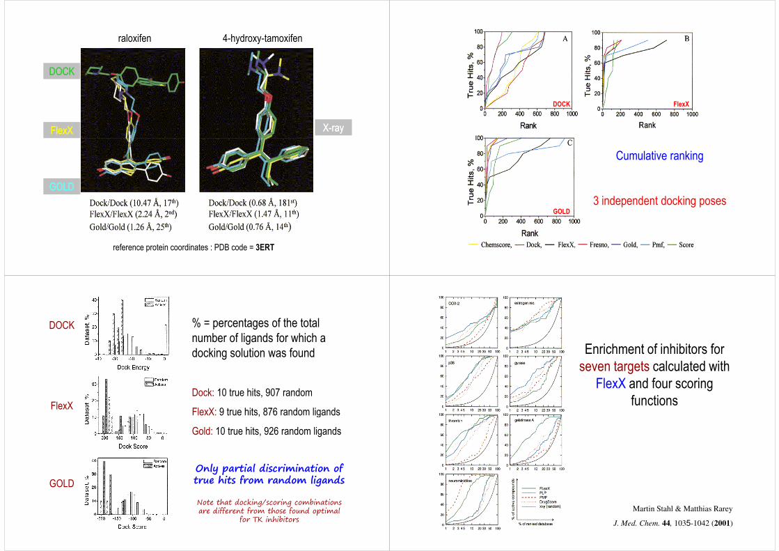
raloxifen 4-hydroxy-tamoxifen
DOCKDOCK
FlexXFlexX XX--rayray
reference protein coordinates : PDB code = 3ERT
GOLDGOLD
DOCK FlexX
Cumulative ranking
(position in the scoring list)
3 independent docking posesGOLD
% = percentages of the total number of ligands for which a docking solution was found
Dock: 10 true hits, 907 random
FlexX: 9 true hits, 876 random ligands
DOCK
FlexX FlexX: 9 true hits, 876 random ligands
Gold: 10 true hits, 926 random ligands
GOLD
Only partial discrimination of true hits from random ligands
Note that docking/scoring combinations are different from those found optimal
for TK inhibitors
Enrichment of inhibitors for seven targets calculated with
FlexX and four scoring functions
Martin Stahl & Matthias Rarey
J. Med. Chem. 44, 1035-1042 (2001)
Consensus scoring: Comparison of the FlexX and PLP scoring functionswith the FlexX-PLP combination ScreenScore.
For each target,For each target,For each target,For each target, the left column of the triplet shows FlexX results, the middlethe left column of the triplet shows FlexX results, the middlethe left column of the triplet shows FlexX results, the middlethe left column of the triplet shows FlexX results, the middle column column column column PLP results, and the right column results calculatedPLP results, and the right column results calculatedPLP results, and the right column results calculatedPLP results, and the right column results calculated with ScreenScore.with ScreenScore.with ScreenScore.with ScreenScore.
Consensus scoringOnly those compounds are regarded that receive high
ranks with two or more scoring functions
considerable reduction of false positives
(“enriched hit-rate”)
P. S. Charifson, J. J. Corkery, M. A. Murcko & W. P.
Walters J. Med. Chem. 42, 5100-5109 (1999)
17 pairs of complexes of the same protein bound to 2 related ligands /
Molecular mechanics (AMBER) and statistical potentials (PMF)
Exhaustive enumeration of all possible docking solutions
Reconstruction of the shape of the energy landscape (coverage-error plots)
Calculation of physico-chemical descriptors
Quantitative evaluation of successQuantitative evaluation of success
Linear discriminant analysis
Physical origin of failures/successes
Desolvation effects
Directional effects of hydrogen bondsDispersive interactions
C. Pérez & A. R. Ortiz J. Med. Chem. 44, 3768-3785 (2001)