ORIGINAL PAPER The inflammatory cellular constituents of foetal and infant leptomeninges: a survey of hospital-based autopsies without trauma Esther Jack & Neil K. Fennelly & Terri Haddix Received: 3 November 2013 /Accepted: 16 December 2013 # The Author(s) 2014. This article is published with open access at Springerlink.com Abstract Objectives Notwithstanding the lack of definitive evidence from studies conducted to date, inflammatory infiltrates and iron depo- sition in the leptomeninges are routinely used as forensic markers of traumatic brain injury. We investigated the presence of these forensic markers of trauma in neonates and infants, with the objective of determining their suitability for use in forensic cases. Methods Leptomeninges derived from non-traumatic deaths were studied. Thirty-three cases were divided into groups 1 and 2, according to set age groups. Inflammatory cells and iron in these groups were quantified. Results CD45, CD68 and CD163 positive inflammatory cells were identified in the leptomeninges of sections of the cere- bellum, brain stem and cortex of all 33 cases of non-traumatic infant deaths surveyed in this study. There were no significant differences between the two groups. Iron was found in the leptomeninges in several cases, even those without recent haemorrhage. Overall within the two subgroups, the numbers of inflammatory cells and iron containing cells were not significantly different. Conclusion These findings demonstrate that inflammatory cells and iron in the leptomeninges can be found in natural and non-traumatic conditions. Further, two cases with no reported neuropathology demonstrated the presence of inflammatory cells and iron. Thus, cautious interpretation of the presence of inflammatory cells and iron containing cells in forensic paediatric cases is recommended. Keywords Leptomeninges . Infants . Inflammatory cells . Trauma . Shaken baby syndrome Introduction The leptomeninges are composed of the pia and arachnoid mater connected by strands termed arachnoid trabeculae. In the young, the leptomeninges are clear but become gradually thickened with age [1]. The leptomeninges and dura mater have been traditionally thought to contain few, if any, inflammatory cells, and any increase in cellularity is potentially equated to a pathologic condition, including inflicted trauma [1, 2]. At pres- ent, we do not know what constitutes ‘normal’ cellularity of infant leptomeninges and if inflammatory or iron containing cells should be present at all in non-forensic settings. However, when indeed present under suspicious circumstances, they are often linked to inflicted trauma, such as in cases of the ‘shaken baby syndrome’ [3]. In order to recognise and characterise the pathologic findings in infant brains, it is important to have an understanding of the normal constituents of the various intracranial compartments. While some studies in the past, largely in rodent pups [4], have sought to evaluate and characterise the leptomeningeal cellular constituents, up until now, a rigorous analysis of the inflamma- tory cellular constituents of the leptomeninges has not been performed in human late-foetal and infant brains. This charac- terisation will serve as a baseline for comparison with brains of similarly aged children in forensic settings. Therefore, in addition to determining the inflammatory cellular composition and presence of iron in foetal and infant leptomeninges associated with natural disease processes and E. Jack (*) Our Lady of Lourdes Hospital, Drogheda, Ireland e-mail: [email protected]E. Jack e-mail: [email protected]N. K. Fennelly Beaumont Hospital, Dublin 9, Ireland e-mail: [email protected]T. Haddix Forensic Analytical Sciences, Hayward, CA, USA Childs Nerv Syst DOI 10.1007/s00381-013-2348-5
Transcript
ORIGINAL PAPER
The inflammatory cellular constituents of foetal and infantleptomeninges: a survey of hospital-basedautopsies without trauma
Esther Jack & Neil K. Fennelly & Terri Haddix
Received: 3 November 2013 /Accepted: 16 December 2013# The Author(s) 2014. This article is published with open access at Springerlink.com
AbstractObjectives Notwithstanding the lack of definitive evidence fromstudies conducted to date, inflammatory infiltrates and iron depo-sition in the leptomeninges are routinely used as forensic markersof traumatic brain injury. We investigated the presence of theseforensic markers of trauma in neonates and infants, with theobjective of determining their suitability for use in forensic cases.Methods Leptomeninges derived from non-traumatic deathswere studied. Thirty-three cases were divided into groups 1and 2, according to set age groups. Inflammatory cells andiron in these groups were quantified.Results CD45, CD68 and CD163 positive inflammatory cellswere identified in the leptomeninges of sections of the cere-bellum, brain stem and cortex of all 33 cases of non-traumaticinfant deaths surveyed in this study. There were no significantdifferences between the two groups. Iron was found in theleptomeninges in several cases, even those without recenthaemorrhage. Overall within the two subgroups, the numbersof inflammatory cells and iron containing cells were notsignificantly different.Conclusion These findings demonstrate that inflammatorycells and iron in the leptomeninges can be found in naturaland non-traumatic conditions. Further, two cases with noreported neuropathology demonstrated the presence of
inflammatory cells and iron. Thus, cautious interpretation ofthe presence of inflammatory cells and iron containing cells inforensic paediatric cases is recommended.
The leptomeninges are composed of the pia and arachnoidmater connected by strands termed arachnoid trabeculae. Inthe young, the leptomeninges are clear but become graduallythickened with age [1]. The leptomeninges and dura mater havebeen traditionally thought to contain few, if any, inflammatorycells, and any increase in cellularity is potentially equated to apathologic condition, including inflicted trauma [1, 2]. At pres-ent, we do not know what constitutes ‘normal’ cellularity ofinfant leptomeninges and if inflammatory or iron containingcells should be present at all in non-forensic settings. However,when indeed present under suspicious circumstances, they areoften linked to inflicted trauma, such as in cases of the ‘shakenbaby syndrome’ [3].
In order to recognise and characterise the pathologic findingsin infant brains, it is important to have an understanding of thenormal constituents of the various intracranial compartments.While some studies in the past, largely in rodent pups [4], havesought to evaluate and characterise the leptomeningeal cellularconstituents, up until now, a rigorous analysis of the inflamma-tory cellular constituents of the leptomeninges has not beenperformed in human late-foetal and infant brains. This charac-terisation will serve as a baseline for comparison with brains ofsimilarly aged children in forensic settings.
Therefore, in addition to determining the inflammatorycellular composition and presence of iron in foetal and infantleptomeninges associated with natural disease processes and
E. Jack (*)Our Lady of Lourdes Hospital, Drogheda, Irelande-mail: [email protected]
N. K. FennellyBeaumont Hospital, Dublin 9, Irelande-mail: [email protected]
T. HaddixForensic Analytical Sciences, Hayward, CA, USA
Childs Nerv SystDOI 10.1007/s00381-013-2348-5
in the absence of physical trauma beyond that accompanyingvaginal birth, this study aims to formulate a basis of compar-ison of leptomeningeal cellular constituents in forensic set-tings, based on rigorous histological analyses of hospital-derived autopsies.
Materials and methods
Subject selection
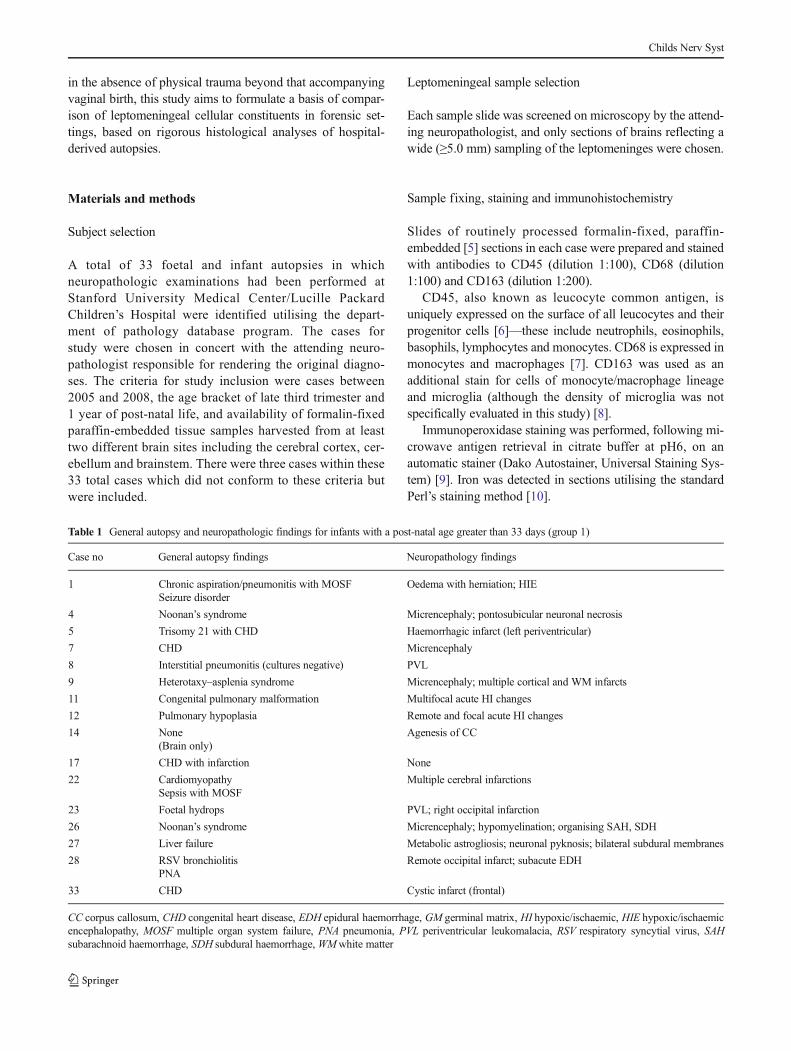
A total of 33 foetal and infant autopsies in whichneuropathologic examinations had been performed atStanford University Medical Center/Lucille PackardChildren’s Hospital were identified utilising the depart-ment of pathology database program. The cases forstudy were chosen in concert with the attending neuro-pathologist responsible for rendering the original diagno-ses. The criteria for study inclusion were cases between2005 and 2008, the age bracket of late third trimester and1 year of post-natal life, and availability of formalin-fixedparaffin-embedded tissue samples harvested from at leasttwo different brain sites including the cerebral cortex, cer-ebellum and brainstem. There were three cases within these33 total cases which did not conform to these criteria butwere included.
Leptomeningeal sample selection
Each sample slide was screened on microscopy by the attend-ing neuropathologist, and only sections of brains reflecting awide (≥5.0 mm) sampling of the leptomeninges were chosen.
Sample fixing, staining and immunohistochemistry
Slides of routinely processed formalin-fixed, paraffin-embedded [5] sections in each case were prepared and stainedwith antibodies to CD45 (dilution 1:100), CD68 (dilution1:100) and CD163 (dilution 1:200).
CD45, also known as leucocyte common antigen, isuniquely expressed on the surface of all leucocytes and theirprogenitor cells [6]—these include neutrophils, eosinophils,basophils, lymphocytes and monocytes. CD68 is expressed inmonocytes and macrophages [7]. CD163 was used as anadditional stain for cells of monocyte/macrophage lineageand microglia (although the density of microglia was notspecifically evaluated in this study) [8].
Immunoperoxidase staining was performed, following mi-crowave antigen retrieval in citrate buffer at pH6, on anautomatic stainer (Dako Autostainer, Universal Staining Sys-tem) [9]. Iron was detected in sections utilising the standardPerl’s staining method [10].
Table 1 General autopsy and neuropathologic findings for infants with a post-natal age greater than 33 days (group 1)
Case no General autopsy findings Neuropathology findings
1 Chronic aspiration/pneumonitis with MOSF Oedema with herniation; HIESeizure disorder
CC corpus callosum, CHD congenital heart disease, EDH epidural haemorrhage, GM germinal matrix, HI hypoxic/ischaemic, HIE hypoxic/ischaemicencephalopathy, MOSF multiple organ system failure, PNA pneumonia, PVL periventricular leukomalacia, RSV respiratory syncytial virus, SAHsubarachnoid haemorrhage, SDH subdural haemorrhage, WMwhite matter
Childs Nerv Syst
Examination of samples
In order to reduce inter-observer variation, the number of vari-ously immunoreactive or iron containing cells was quantified bya single observer and representative slides reviewed for accuracyby a second observer. At a microscopic magnification of 20x,immunoreactive cells within leptomeninges were counted andrecorded.
As the length of leptomeninges evaluated varied betweenslides, the length of leptomeninges scored was measured inmillimetres and results recorded as immunoreactive cells/millimetre. Only leptomeninges on gyral surfaces were scoredas it was impossible to accurately measure the length of theleptomeninges in the sulci.
Analysis of results
The samples were divided into infants who died beyond 33post-natal days up to 1 year (group 1) and newborns whosurvived up to 33 days (group 2)—these represent pre- andpost-natal leptomeninges [11].
The mean number of CD45, CD68 and CD163 positiveimmunoreactive cells/millimetre, occurring in the cerebel-lum, cortex and brain stem, was calculated for groups 1and 2, respectively. Iron was recorded as being eitherpresent or absent. To test for significant differences be-tween the numbers of immunoreactive cells between groups1 and 2, an unpaired, two-tailed student’s T test wasemployed.
Table 2 General autopsy andneuropathologic findings for foe-tuses and infants with a post-gesta-tional age up to 33 days (group 2)
BG basal ganglia, CC corpuscallosum, CHD congenital heartdisease, GM germinal matrix, HIhypoxic/ischaemic, HIE hypoxic/ischaemic encephalopathy, HTNhypertension, IV intraventricular,MOSFmultiple organ systemfailure, NEC necrotizing entero-colitis, PNA pneumonia, PVLperiventricular leukomalacia, SAsubarachnoid, SAH subarachnoidhaemorrhage, SDH subduralhaemorrhage, WMwhite matter,PSNN pontosubicular neuronalnecrosis
Caseno.
General autopsy findings Neuropathologic findings
2 CHD None
3 CHD Micrencephaly;
Arhinencephaly;
Neuronal necrosis, subiculum
6 Harlequin ichthyosis None
10 Diaphragmatic hernia
Omphalocoele
PSNN
13 PNA (Strep) Metabolic encephalopathy;
Hydrocephalus;
Partial agenesis of CC
15 CHD Thoracic meningomyelocoele;
Hydrocephalus; possible partial agenesis of CC
16 CHD Organising SDH
18 Cardiomyopathy Acute HI change (focal);
DiGeorge syndrome PSNN;
Remote dural haemorrhage
19 Pulmonary HTN PSNN
20 Foetal hydrops GM haemorrhage extending to SA;
Diffuse HIE
21 Tetralogy of Fallot PSNN; BG infarct
24 CHD Focal SAH;
Small WM haemorrhages;
PSNN
25 CHD PSNN;
Focal HI, cerebellum;
PVL;
Parietal infarct;
Organising SDH, IVH, SAH
29 None (brain only) Diffuse HIE
30 NEC (MOSF) Meningoencephalitis;
PSNN
31 Multiple congenital abnormalities includingCHD
GM haemorrhage with IVH and SAH; PSNN;
Arhinencephaly
32 Pulmonary hypoplasia/HTN GM haemorrhage with IVand SAH; PSNN
Childs Nerv Syst
Results
Demographics
Of the 33 cases, 16 were male and 17 female. Thirteen wereborn via vaginal delivery and 19 via Caesarean section. Themode of delivery of one case was not available. Sixteen casesinvolved infants who died beyond the post-natal age of 33 days(group 1), and 17 cases represented either foetuses or new-borns who survived up to 33 post-natal days (group 2). Onechild (number 7) who survived to 16 months of age wasincluded in group 1. There were two cases (numbers 25 and30) involving foetuses in the 26th and 28th weeks of gestationwhich were included in group 2.
The general autopsy and neuropathology findings of bothgroups overlapped, and these included congestive heart fail-ure, Noonan’s syndrome, micrencephaly and pontosubicularneuronal necrosis (Tables 1 and 2).
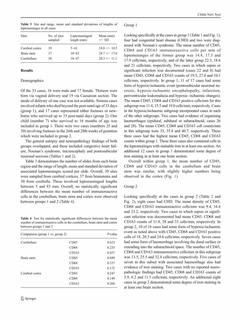
Table 3 demonstrates the number of slides from each brainregion and the range of length, mean and standard deviation ofassociated leptomeninges scored per slide. Overall, 39 siteswere sampled from cerebral cortices, 37 from brainstems and30 from cerebella. These involved leptomeningeal lengthsbetween 5 and 83 mm. Overall, no statistically significantdifferences between the mean number of immunoreactivecells in the cerebellum, brain stem and cortex were observedbetween groups 1 and 2 (Table 4).
Group 1
Looking specifically at the cases in group 1 (Table 1 and Fig. 1),four had congenital heart disease (CHD) and two were diag-nosed with Noonan’s syndrome. The mean number of CD45,CD68 and CD163 immunoreactive cells per mm ofleptomeninges of the former group was 14.4, 17.5 and17.9 cells/mm, respectively, and of the latter group 22.3, 18.6and 21 cells/mm, respectively. Two cases in which sepsis orsignificant infection was documented (cases 22 and 8) hadmean CD45, CD68 and CD163 counts of 19.3, 27.4 and 34.1cells/mm, respectively. In group 1, 11 of 17 cases had someform of hypoxic/ischaemic event (pontosubicular neuronal ne-crosis, hypoxic-ischaemic encephalopathy, infarction,periventricular leukomalacia and hypoxic–ischaemic changes).The mean CD45, CD68 and CD163 positive cells/mm for thissubgroup was 11.4, 15.5 and 19.0 cells/mm, respectively. Casesin the hypoxic/ischaemic subgroup incorporated cases in eachof the other subgroups. Two cases had evidence of organisinghaemorrhages (epidural, subdural or subarachnoid; cases 26and 28). The mean CD45, CD68 and CD163 cell counts/mmin this subgroup were 33, 35.8 and 40.7, respectively. Thesethree cases had the highest mean CD45, CD68 and CD163counts within group 1. These three cases also contained cells inthe leptomeninges with stainable iron in at least one section. Anadditional 12 cases in group 1 demonstrated some degree ofiron staining in at least one brain section.
Overall within group 1, the mean number of CD45,CD68 and CD163 cells in the cerebellum and brainstem was similar, with slightly higher numbers beingobserved in the cortex (Fig. 1)
Group 2
Looking specifically at the cases in group 2 (Table 2 andFig. 2), eight cases had CHD. The mean density of CD45,CD68 and CD163 immunoreactive cells/mm was 9.4, 16.6and 23.2, respectively. Two cases in which sepsis or signifi-cant infection was documented had mean CD45, CD68 andCD163 counts of 31.8, 28 and 33 cells/mm, respectively. Ingroup 2, 10 of 16 cases had some form of hypoxic/ischaemicevent as noted above with CD45, CD68 and CD163 positivecells of 18, 20.5 and 24.6 cells/mm, respectively. Seven caseshad some form of haemorrhage involving the dural surface orextending into the subarachnoid space. The number of CD45,CD68 and CD163 immunoreactive cells/mm in this subgroupwas 13.5, 25.5 and 32.4 cells/mm, respectively. Five cases ofseven in this subset with associated haemorrhage also hadevidence of iron staining. Two cases with no reported neuro-pathologic findings had CD45, CD68 and CD163 counts of5.9, 6.2 and 11.5 cells/mm, respectively. An additional eightcases in group 2 demonstrated some degree of iron staining inat least one brain section.
Table 3 Site and range, mean and standard deviations of lengths ofleptomeninges in all cases
Sites No. of sitessampled
Leptomeningeallength (mm)
Mean (mm)+/− SD
Cerebral cortex 39 5−41 18.8 +/− 10.5
Brain stem 37 10−83 28.7 +/− 17.0
Cerebellum 30 10−47 20.3 +/− 11.1
Table 4 Test for statistically significant differences between the meannumber of immunoreactive cells in the cerebellum, brain stem and cortexbetween groups 1 and 2
Comparison (group 1 vs. group 2) P-value
Cerebellum CD45 0.623
CD68 0.235
CD163 0.457
Brain stem CD45 0.689
CD68 0.337
CD163 0.172
Cerebral cortex CD45 0.247
CD68 0.149
CD163 0.204
Childs Nerv Syst
Overall within group 2, the mean number of CD45, CD68and CD163 was similar across the locations sampled (Fig. 2).
Cases without reported neuropathology
In three cases within both groups without neuropathologic ab-normalities, all classes of inflammatory cells were found in theleptomeninges (case 17 of group 1; cases 2 and 6 of group 2).
Iron findings
Of the 19 cases in which Caesarean sections were performed,16 had positive iron findings, whereas 8 cases of the 13
vaginal births were positive for iron. Four cases of each modeof delivery reported haemorrhage-related neuropathologic di-agnoses. Fifteen cases of Caesarean section births were asso-ciated with some form of hypoxic/ischaemic event in contrastto seven cases involving vaginal births.
Discussion
In the current study, we found the presence of inflammatorycells in the leptomeninges, both overall and when segregatedinto two groups by age (foetal and early post-natal vs. infantsbeyond 33 days post-natal life) and by anatomic and
Fig. 1 Mean immunoreactivecells/mm in cerebral cortex,brainstem and cerebellum (group 1)
Fig. 2 Mean immunoreactivecells/mm in cerebral cortex,brainstem and cerebellum (group 2)
Childs Nerv Syst
neuropathologic conditions (Fig. 3). A notable finding is thateven in foetuses and infants with no neuropathologic abnor-malities, inflammatory cells and occasionally iron was identi-fied in the leptomeninges. This is in contrast to the widely heldbelief that the leptomeninges should be largely devoid ofinflammatory cells and iron in children with no reportedneuropathology [12]. In addition, there was a positive corre-lation between the mean number of CD163 positive immuno-reactive cells in cases with EDH, SDH or SAH pooled fromboth groups 1 and 2, when compared to those without thesehemorrhages (mean immunoreactive CD163 cells/mm 30.34vs. 15.12; p=0.022). Interestingly, this correlation was notdetected for the number of CD45 or CD68 positive cells andprobably reflects the higher sensitivity of CD163 as a markerfor monocytic cells (Fig. 4).
Caesarean section deliveries and vaginal births were asso-ciated with a variety of anatomic and neuropathologic diag-noses, with hypoxic/ischaemic events commonly found inboth modes of delivery. Both modes of delivery were alsoassociated with iron deposition in the leptomeninges andhaemorrhage-related neuropathology (Fig. 5).
Accordingly, it appears that the presence of iron in theleptomeninges does not necessarily equate to traumatic haem-orrhage but may be found in completely naturally occurringprocesses and occurs irrespective of the mode of delivery [2].There also seemed to be no recurring pattern allowing us toassociate the presence of iron to a single anatomic or neuro-pathologic diagnosis.
There are multiple mechanisms which allow the brain tosense inflammatory signals from systemic circulation [13],including interactions with circulating molecules in areas inthe brain devoid of the brain–brain barrier. Microglial cells
Fig. 4 CD68 (panel a) and CD163 (panel b) immunostained sections(600×) from same case depicted in Fig. 3. In each panel, cerebral cortex ison the right side. In panel a, portions of two vessels (yellow stars)containing CD68 immunoreactive cells are also seen
Fig. 5 Example of iron staining in the leptomeninges (600×) in a differ-ent case than shown in Figs. 3 and 4. The leptomeninges are diagonallyoriented downward from left to right and contain a large number of cellswith an appropriate granular intracellular blue reaction product. In theupper right corner are some cells with non-specific staining
Fig. 3 Representative case with CD45 immunostain (600×). Cells in theleptomeninges demonstrating appropriate positive staining are indicatedwith red arrows, and the cells and other constituents demonstrating non-specific staining are indicated with blunt black arrows. A vascular lumencontaining some cells with appropriate staining is indicated by a yellow star.These intravascular cells were not included in counts. The brown blushelsewhere within the vessel represents additional non-specific staining
Childs Nerv Syst
seem to migrate from the germinal matrix to the corticallayers. Early migration of microglia from the ventricular andmeningeal surfaces has been observed as early as 4.5 weeks ofgestation in humans [13].
The function of resting microglia under normal conditionsis unclear. In pathologic conditions, these microglial cells arerapidly activated and proliferate. Animal studies have demon-strated that inflammation, either introduced systemically orwithin the brain, causes microglial activation along with cy-tokine release [14]. Accordingly, there is evidence to suggestthat infection distant from the brain may damage developingfoetal brain [13]. The activation of neuroinflammatory re-sponses may also sensitise the brain to the damaging effectsof other insults, such as hypoxia/ischaemia, and amplify theeffects of the latter [14].
Thus, there are multiple possible reasons to account for thepresence of inflammatory cells within the leptomeninges earlyin gestation in humans outside of inflicted trauma.
Conclusion
We conclude that all patients with various natural diseaseprocesses in this hospital-based population had measureablenumbers of CD45, CD68 and CD163 immunoreactive cells(and a subset had iron containing cells) in the leptomeningesoverall and when segregated by age and anatomic and neuro-pathologic diagnoses. Although we studied a larger number ofcases than the dura study by Croft et al. [2], our numbers oftotal cases are relatively small, particularly in consideration ofthe varied anatomic and neuropathologic diagnoses. Our caseswere derived only from hospital autopsies, and these observa-tions require comparison to actual forensic cases involvingboth traumatic injuries and natural disease processes. Clear-ly, the presence of inflammatory cells and iron in theleptomeninges can occur commonly, and in significant num-bers, in non-traumatic neuropathologic conditions. As thisstudy evaluated only non-forensic cases, we did not define athreshold level of inflammatory or iron containing cells abovewhich a non-natural disease process (including inflicted inju-ry) would be implicated. Thus, these findings support therecommendation of cautious interpretation of the findings ofleptomeningeal inflammation and iron in forensic cases [3].
Acknowledgments We would like to thank the Department of Pathol-ogy and Laboratory of Neuropathology, Stanford University, Stanford,CA, USA.
Open Access This article is distributed under the terms of the CreativeCommons Attribution License which permits any use, distribution, andreproduction in any medium, provided the original author(s) and thesource are credited.
References
1. Sternberg SS (1992) Histology for pathologists, 2nd edn.Raven, New York, pp 164–167, Chapter 6, Pia-arachnoid(Leptomeninges)
2. Croft PR, Reichard RR (2009) Microscopic examination of grosslyunremarkable pediatric dura mater. Am J Forensic Med Pathol 30(1):10–13
3. Squier W (2011) The “shaken baby” syndrome: pathology andmechanisms. Acta Neuropathol 122(5):519–542
4. Perry VH, Hume DA, Gordon S (1985) Immunohistochemical local-ization of macrophages and microglia in the adult and developingmouse brain. Neuroscience 15(2):313–326
5. Nowacek JM, Kiernan JA. (2010) Ch 16: Fixation and TissueProcessing. Special Stains and H&E
6. Altin JG, Sloan EK (1997) The role of CD45 and CD45-associatedmolecules in T cell activation. Immunol Cell Biol 75(5):430–445
7. Tanaka Y, Matsuwaki T, Yamanouchi K, Nishihara M. (2012)Exacerbated Inflammatory Responses Related to ActivatedMicroglia after Traumatic Brain Injury in Progranulin-DeficientMice. Neuroscience
8. Lau SK, Chu PG, Weiss LM (2004) CD163: a specific marker ofmacrophages in paraffin-embedded tissue samples. Am J Clin Pathol122(5):794–801
9. Kumar GL, Rudbeck L. (2009) Ch 8: Demasking of Antigens.Immunohistochemistry Guidebook, 5th ed
10. AFIPManual of Histological StainingMethods (1968) 3rd ed., Ed. L.Luna: New York: McGraw-Hill Pub 19
11. Squier W, Lindberg E, Mack J, Darby S (2009) Demonstration offluid channels in human dura and their relationship to age andintradural bleeding. Childs Nerv Syst 25(8):925–931
12. Esiri M, Morris C (1990) Immunocytochemical study of macro-phages and microglial cells and extracellular matrix components inhuman CNS disease. J Neurol Sci 101:59–72
13. Malaeb S, Dammann O (2009) Fetal inflammatory responseand brain injury in the preterm newborn. J Child Neurol24(9):1119–1126
14. Chew LJ, Takanohashi A, Bell M (2006) Microglia and inflamma-tion: impact on developmental brain injuries. Ment Retard DevDisabil Res Rev 12(2):105–112