Page 1
Entropy 2012, 14, 1399-1442; doi:10.3390/e14081399
entropy ISSN 1099-4300
www.mdpi.com/journal/entropy
Review
The Initial Common Pathway of Inflammation, Disease, and Sudden Death
Robert M. Davidson 1,* and Stephanie Seneff 2
1 Internal Medicine Group Practice, PhyNet, Inc., Longview, TX 75604, USA 2 Computer Science and Artificial Intelligence Laboratory, MIT, Cambridge, MA 02140, USA;
E-Mail: [email protected]
* Author to whom correspondence should be addressed; E-Mail: [email protected] ;
Tel.: +1-903-235-0731.
Received: 29 June 2012; in revised form: 19 July 2012 / Accepted: 20 July 2012 /
Published: 2 August 2012
Abstract: In reviewing the literature pertaining to interfacial water, colloidal stability, and
cell membrane function, we are led to propose that a cascade of events that begins with
acute exogenous surfactant-induced interfacial water stress can explain the etiology of
sudden death syndrome (SDS), as well as many other diseases associated with modern
times. A systemic lowering of serum zeta potential mediated by exogenous cationic
surfactant administration is the common underlying pathophysiology. The cascade leads to
subsequent inflammation, serum sickness, thrombohemorrhagic phenomena, colloidal
instability, and ultimately even death. We propose that a sufficient precondition for sudden
death is lowered bioavailability of certain endogenous sterol sulfates, sulfated glycolipids,
and sulfated glycosaminoglycans, which are essential in maintaining biological equipose,
energy metabolism, membrane function, and thermodynamic stability in living organisms.
Our literature review provides the basis for the presentation of a novel hypothesis as to the
origin of endogenous bio-sulfates which involves energy transduction from sunlight. Our
hypothesis is amply supported by a growing body of data showing that parenteral
administration of substances that lower serum zeta potential results in kosmotropic cationic
and/or chaotropic anionic interfacial water stress, and the resulting cascade.
Keywords: inflammation; serum sickness; colloidal instability; interfacial water stress;
bio-sulfates; Shwartzman phenomena; sudden death syndrome
OPEN ACCESS
Page 2
Entropy 2012, 14 1400
PACS Codes: 87.15, 87.19, 87.19.ly, 87.50.cf
Glossary of Terms
Anaphylaxis a severe, rapidly progressing, life-threatening, generalized allergic reaction. Biological equipoise a stable, non-equilibrium, dissipative system synonymous with life.
Cholesterol sulfate (Ch-S)
quantitatively the most important known sterol sulfate in human plasma where it regulates the activity of the serine proteases, in cell membranes where it has a stabilizing role, and in platelet membranes where it supports platelet adhesion.
Coherence domain (CD)
a water CD is a collection of liquid water molecules which oscillate in unison in tune with a self-trapped electromagnetic field at a well-defined frequency. The coherent oscillations produce an ensemble of quasi-free electrons, able to collect noise energy from the environment and transform it into high-grade coherent energy in the form of electron vortices. This high-grade energy may then activate biomolecules resonating with the water CD.
Colloidal instability
a property attributed to a colloidal suspension that develops when stabilizing repulsive steric and electrostatic forces between colliding particles are insufficient to prevent their natural tendency to aggregate into masses large enough to precipitate.
Colloidal suspension a colloid that has a continuous liquid phase in which a solid is suspended in a liquid, e.g., our flowing blood.
Exclusion zone (EZ)
a glass-like, gel phase consisting of water CDs resonating in-phase, adjacent to hydrophilic surfaces, several hundred micrometers wide which excludes colloidal particles and various solutes as a consequence of water molecules re-orienting to produce a more ordered structure, which then excludes the particles.
Exogenous interfacial water stress (EIWS)
a property of interfacial water—interfacial tension—which destabilizes enzymes, protein structure, and cell membranes.
Glycosaminoglycans
a group of high molecular weight linear polysaccharides constructed with various disaccharide repeating units usually occurring in proteoglycans, including the chondroitin sulfates, dermatan sulfates, heparan sulfate and heparin, keratan sulfates, and hyaluronic acid, with the primary configurations containing an amino sugar and a uronic acid.
Hofmeister series
the Hofmeister series or lyotropic series is a classification of ions in order of their ability to change water structure. A scale can be established wherein: kosmotropic ions or nonionic kosmotropes stabilize proteins and hydrophobic aggregates in solution and reduce the solubility of hydrophobes, and chaotropic ions or nonionic chaotropes unfold proteins, destabilize hydrophobic aggregates and increase the solubility of hydrophobes.
Page 3
Entropy 2012, 14 1401
Interfacial tension
a measure of the cohesive (excess) energy present at an interface arising from the imbalance of forces between molecules at an interface (gas/liquid, liquid/liquid, gas/solid, liquid/solid). The excess energy is called surface free energy and can be quantified as a measurement of energy/area, i.e., the energy required to increase the surface area of the interface by a unit amount.
Jones-Ray effect the observation of a minimum in the surface tension at very low ionic concentrations (<1 mM).
Kinetic Terahertz Absorption (KITA)
KITA monitors the changing THz electric field pulse shape on the picosecond time scale Δt, as a chemical reaction proceeds on a longer time scale t, and has been applied to measure the changing protein-hydration-water dynamics.
Shwartzman reaction or Shwartzman phenomenon occurs when a “preparatory”, i.e., injection of bacterial filtrates, is followed after a proper time interval by intravenous “provocation” with the same or some similar material.
Surface tension
the cohesive forces among liquid molecules responsible for the surface free energy at a gas liquid interface is produced by the attraction between the molecules being directed away from the surface as surface molecules are more attracted to the molecules within the liquid than they are to molecules of the gas at the surface.
Surfactant in principle, anything can be called a surfactant that affects the surface tension of a liquid, the interfacial tension between two liquids, or that between a liquid and a solid.
Thrombohemorrhagic phenomenon (THP)
a change characterized by thrombosis and hemorrhage.
Zeta potential (ZP) a measure of the net charge density of a particle.
1. Introduction
An extensive review of the colloid and interface science literature has led us to conclude that the
very earliest events in inflammation, disease, and sudden death are purely biophysical. We now
strongly believe that biophysical properties underlie the origin, development, and perpetuation of life.
The evidence also supports our view that inflammation, disease, and sudden death rest on purely
biophysical events that provoke a branching, cascade-like, chain of reactions, which typically begins in
our vascular system. The literature amply supports the concept that our vascular system, including the
molecular and humoral immune defense and lymphatic systems, acts as though it is a single organ [1].
Both microvascular disease and endothelial dysfunction share a common initial pathophysiology.
A better understanding of these earliest of biophysical events will enable a more rational approach to
dealing with many of today’s idiopathic diseases. These diseases, are likely to be pluricausal [2] and
highly stereotyped [3] in their clinical presentations, often with substantial overlap of symptoms.
In this review, we hope to highlight, summarize, and call attention to some of the many historical
milestones we have relied on to recommend areas of further research in the diagnosis, treatment, and
prevention of disease. We believe that the development of sustainable health paradigms of tomorrow
will require a multidisciplinary approach today. This approach will, at times, require each of us to look
to the literature found in diverse fields of endeavor, for better understanding, particularly in areas in
Page 4
Entropy 2012, 14 1402
which we were not formally trained. We have purposefully chosen to limit the scope of this review to
simply trying to prospectively identify, at least in broad terms, potentially fruitful areas of future
research which might have meaningful impact on the public health. The references cited in support of
this review are intended to give the reader a place to begin. Lead references are cited upon which to
base further inquiry.
We will begin this review by discussing the definition and clinical characteristics of a syndrome of
events, the net outcome of which is all too often, the sudden death of the victim. We will follow with a
delineation of some of the underlying colloid and interface science which can reasonably be implicated
in the pathophysiology of sudden death. We will present the preclinical research which we believe is
most relevant to understanding sudden death. We will give examples of ongoing clinical research
which are the progeny of colloid and interface science. We will present a novel hypothesis as to the
origin of endogenous bio-sulfates, and discuss their central role in the establishment and maintenance
of our biomembranes. We focus on the glycocalyx layers upon which rests the interfacial stability and
function of our biomembranes in health. We will detail the role of the nitric oxide synthase family of
enzymes, which, we argue, plays an essential role in energy capture and energy transduction, in
support of the integrity of our biomembranes. We will introduce and define the concept of exogenous
interfacial water stress (EIWS) and review several clinically-relevant sources of EIWS. Throughout
this review we will endeavor to point out the necessary, central role of interfacial water as an essential
reactant, mediator, and effector molecule in our bodies. Since water is the most abundant molecule on
Earth and in our bodies, and the third most abundant molecule in the Universe (after H and CO), and
because the vast majority of water in our bodies is interfacial water, it is in no way unreasonable to
conclude that life without water is an impossibility.
In the recent medical literature, SDS (Sudden Death Syndrome) is used as an umbrella term for a
number of conditions that can lead to a sudden death. Two distinct forms of SDS have been identified:
Sudden Infant Death Syndrome (SIDS) and Sudden Adult Death Syndrome (SADS). In this paper, we
expressly define Sudden Adult Death Syndrome (SADS) as non-traumatic, non-violent, and
unexpected occurrences resulting from unknown cause. We strongly believe that defining SADS as
Sudden Arrhythmia Death Syndrome or as Sudden Cardiac Death is misleading and unfortunate
because such a definition (a) infers a cardiac etiology, when the etiology is often unknown; (b) infers a
six hour or less time frame, when the time frame is also unknown; and (c) limits by inference any
open-minded investigation as to the actual cause to solely cardiac etiologies, when other etiologies
may well exist.
Multiple hypotheses have been proposed as the pathophysiologic mechanisms responsible for SIDS.
However, none have been proven. The triple-risk model of SIDS proposes that the cause of SIDS is
multifactorial, and that the sudden death of an infant may occur when a predisposed infant in an
unstable period of homeostatic control is exposed to triggering factors [4]. Examples of various
proposed etiologies include the QT interval hypothesis, the apnea hypothesis, neuroconvulsive,
anaphylactic, thrombohemorrhagic, infectious, inflammatory, genetic, e.g., Brugada’s syndrome [5],
and brainstem etiologies [6]. SIDS is a diagnosis of exclusion used to describe the sudden and
unexpected death of an infant when no other plausible cause can be found [7]. Factors such as maternal
smoking, prone sleeping, infection, lack of breast feeding and overheating have all been associated
Page 5
Entropy 2012, 14 1403
with SIDS mortality [8]. Several theories have been proposed to explain the sudden deaths in this age
group; however, the mechanisms responsible for SIDS remain poorly understood [6,9–15].
Colloid and interface science literature supports the novel view that all instances of SDS share an
initial common pathophysiology which is purely biophysical. Hofmeister (1888) ordered anions
according to their ability to precipitate globular proteins from water [16]. Setschenow (1889)
established an empirical law linking the solubility of a protein with cosolute (salt) concentration [17].
Heydweiller (1910) discovered that salt dissolved in water increased the surface tension of the
solution-air interface [18]. Langmuir (1917) was the first to attempt a theoretical explanation of the
physical mechanism behind the increase in surface tension produced by electrolytes [19]. More
recently, Dér et al. instead of focusing on air-water surface tension, used protein-water interfacial
tension as a general description of the free energy changes associated with salt-induced changes of
protein solubility and conformation [20,21]. In general terms, the interfacial tension is determined by
the cohesion and adhesion free energies within and between phases separated by the interface,
respectively. Fluctuations in protein conformation are linked to interfacial tension and protein
structural stability [22–25]. The fluctuation-dissipation theorem [22] was first proven by Callen and
Welton in 1951. This theorem describes how dissipative forces and fluctuating random forces are
connected. According to Grassia in 2000, when a large particle moves through a sea of small particles,
on the microscale, all particle collisions are elastic. However, on the macroscale, where only the large
particle is properly resolved, dissipative forces and fluctuating random forces are observed [23]. Many
working proteins (functional proteins such as enzymes) oscillate between “open” and “closed”
conformations, which also implies water-exposed surface area changes [20,21]. Hofmeister ion-dependent
fluctuations at the interface are implied. Hofmeister effects can be rationalized by considering the
contribution of interfacial energy to the total free energy change of the protein. A general relationship
exists between salt concentration and protein-water interfacial tension. Protein-water interfacial tension
plays a key role in protein structure and dynamics. An essential role for correct protein folding can be
gleaned from the observation that the unfolded protein response is a common stressor leading to
cellular apoptosis.
Rosina has reported the temperature dependence of blood surface tension of healthy subjects [26].
There is now ample literature [27–39] to support the conclusion that many of the surfactants,
administered parenterally to humans in vaccines, such as aluminum, mercury, and polysorbate 80 (in
the acidic pH range), raise blood surface tension, leading to an increase in surface tension of
intracellular, extracellular, and interstitial water, resulting in a pathology that we will refer to
henceforth as “water stress”. Throughout this paper, we will refer to water stress as a property of
interfacial water—interfacial tension—which destabilizes enzymes, protein structure, and cell
membranes [20,21,40–49].
Several compelling examples of water stress as applied to humans can now be cited [26,37,38,44,45,47–57].
Polycation-induced agglutination and sensitization of red blood cells has been demonstrated by several
investigators [47–49,58–67]. In 1985, Coakley et al. demonstrated a regular periodicity of cell-cell
contact points by both light microscopy and transmission electron micrography of polylysine
agglutinated erythrocytes [44,49,68–70]. Serum surface tension was reported by Enoki Yoshisuke to
be transiently elevated after provocation injection for the Shwartzman reaction [71]. Absolom has data
showing that the surface tension of erythrocytes obtained from untreated cardiac arrhythmia patients
Page 6
Entropy 2012, 14 1404
was higher than that of treated patients [45]. Kratochvil and Hrncir demonstrated that the level of
surface tension of the blood marks changes occurring in various disease conditions such as rheumatoid
arthritis, proliferative glomerulonephritis, and some tumors of the central nervous system or the
urogenital tracts [72]. Kazakov et al. applied dynamic surface tensiometry of serum and cerebrospinal
fluid for diagnosis and monitoring of treatment in patients with rheumatic, neurological and
oncological diseases [57]. Esitashvili and Msuknishvili observed an increase in blood surface tension
during acute myocardial infarction in humans [54]. Electrostatic interactions and interfacial charge
regulation of biomembranes is a focus of intense ongoing research [65–67,73–81]. Fiszer-Kierzkowska
recently suggested that cationic liposomes may not be suitable vehicles for gene transfection, given
that they produced apoptosis and aggregation of misfolded proteins and/or fluidity changes of cellular
membranes [82].
Recently, experiments with yeast and in cardiac cells by Lloyd et al. has revealed astonishing
parallels and similarities in their dynamic biochemical organization, despite being separated by
1.2 billion years of evolution according to the prevailing orthodoxy [83]. This is not the first time that
oscillatory fluctuations of characteristic periodicity have been noted in living tissue. Fluctuating
phenomena of this nature have been studied for almost a decade. They are likely to be mesoscopic,
supramolecular manifestations of the fluctuation-dissipation theorem [22].
Literature review provides strong support for our conclusion that the serial parenteral administration
of both polycationic and non-ionic surfactants is causing cumulative, synergistic, and systemic interfacial
water stress. Non-ionic surfactants, e.g., Triton 100 and Tween 80, found in many of today’s marketed
vaccines, can induce apoptotic cell death [36,84], hemolysis [32,33,38], and meningoencephalitis [85].
Aluminum hydroxide gels have been used as vaccine adjuvants for many years. There is a well-known
relationship between hemolytic activity and adsorption capacity of aluminum hydroxide adjuvants [28].
Intraperitoneal exposure to aluminum sulfate in rats increases blood viscosity and red blood cell
aggregation [86]. A link between aluminum and the pathogenesis of Alzheimer's disease was recently
proposed [87]. Aluminum hydroxide injections have been shown to lead to motor deficits and motor
neuron degeneration in mice [88]. Aluminum-treated mice showed significantly increased apoptosis of
motor neurons and increases in reactive astrocytes and microglial proliferation within the spinal cord
and cortex. Morin stain detected the presence of aluminum in the cytoplasm of motor neurons with
some neurons also testing positive for the presence of hyper-phosphorylated tau protein, a pathological
hallmark of various neurological diseases, including Alzheimer's disease and frontotemporal dementia.
Aluminum (Al), the most commonly used vaccine adjuvant, is a demonstrated neurotoxin and a strong
immune stimulator. Hence, adjuvant Al has the potential to induce neuroimmune disorders. Al in
vaccines may also be a contributing factor in autism spectrum disorder (ASD) [89–93].
More than a century ago, in 1858, the Russian investigator Botkin first described what later became
known as “erythrocyte agglutination thrombi” [94]. In 1894, Sanarelli first observed a condition which
later became known as the generalized Sanarelli-Shwartzman phenomenon (SSP-G) [95], after further
clarification by Schwartzman in 1928 [3,96]. There is ample evidence in the literature that this is an
appropriate model for serial exogenous surfactant administration, as in scheduled vaccination programs,
and it may constitute a “preparatory” or “sensitizing” or “priming” event. A final “provocation” or
“challenge” or “shocking” event may induce a chain of reactions or a branching (avalanche-like)
Page 7
Entropy 2012, 14 1405
cascade of events [97–102], equated with the Sanarelli-Shwartzman Phenomenon, whether localized or
generalized [3,97,98,103,104].
Thrombohemorrhagic phenomena (THP), also either localized (THP-L) or generalized (THP-G),
are manifestations of the nonspecific or generalized stress adaptation syndrome [105]. In 1981, there
was a proposal for a univisceral or single organ third type of Shwartzman reaction [106]. Sometimes,
after the inflammatory cascade is “induced”, a new equilibrium is achieved, albeit in a more susceptible
state for further subsequent thrombohemorrhagic events, which are sometimes fatal, depending
critically upon such factors as the blood ζ-potential (zeta potential, to be abbreviated herein as ZP) and
surface tension, in part controlled by bio-sulfate levels. Whether this is truly a “new equilibrium” or a
“stable, non-equilibrium” or an “unstable, non-equilibrium” is uncertain to the authors, in light of
Bauer, Voeikov and Del Giudice, and Morowitz and Smith [101,102,107]. Our literature review
supports the view that human blood ZP, serum surface tension, and systemic bio-sulfate levels
[76,108–113] should be employed clinically as biomarkers for risk of SDS. Such biomarkers might
thereby guide future preventive health strategies.
In this paper, we introduce a novel hypothesis to explain sudden death, relating impaired sulfur
metabolism to serum colloidal instability. We argue that individuals with such impairment are
vulnerable to an acute reaction to exposure to toxins that would otherwise be efficiently expelled from
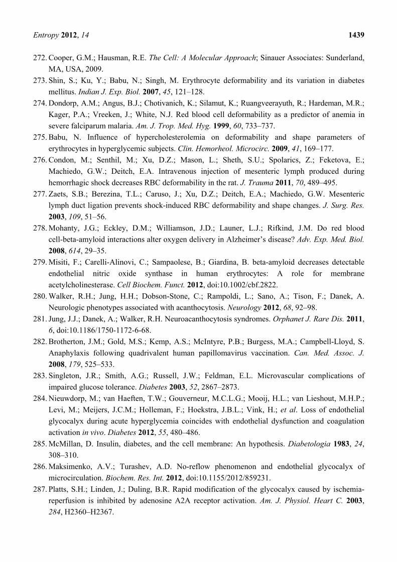
the body. Cholesterol sulfate (Ch-S), whose structure is depicted in Figure 1, sulfated
glycosaminoglycans (sGAGs), and sulfated glycolipids play an essential role in maintaining the in vivo
serum ZP in all living organisms. Any process which acutely lowers the ZP, such as the introduction of
polycationic surfactant stress into the vascular system, has the potential to trigger SDS. Mediated by
water, this manifests hemodynamically as increased viscosity, decreased RBC and vascular
distensibility, increased vascular permeability, acutely deranged ionic gradients [ion channelopathy],
colloidal instability, abnormal blood flow dynamics, impaired tissue oxygenation and oxygen delivery,
thrombohemorrhagic phenomena, microvascular ischemia, cellular anoxia, infarctions, cell necrosis,
and death.
While we believe that everyone is vulnerable to SDS, some are more vulnerable than others. A child’s
prenatal (via the mother) and postnatal nutritional status are important determinants of risk [114].
Adequate blood Ch-S sources are essential to health, e.g., adequate endogenous and exogenous
cholesterol [114–117] and sulfur [114,118], as well as certain minerals and cofactors, e.g., zinc and
tetrahydrobiopterin (BH4). Sunlight exposure to the skin is essential in the production of Ch-S (as well as
its derivative, vitamin D3-sulfate), and excessive use of sun block will interfere with this process [114]. We
propose that lowered levels of endogenous sterol sulfates, sulfated glycolipids, and sulfated GAGs are
a sufficient risk factor for sudden death, in the context of cationic surfactant water stress. These
biologically active molecules are essential for maintaining biological equipose, energy metabolism,
membrane function, and thermodynamic stability in living organisms.
Page 8
Entropy 2012, 14 1406
Figure 1. Cholesterol sulfate.
This review calls attention to the very earliest perturbations to our living internal biological milieu
that we believe are common to all pathways to the sudden death syndrome. These involve cationic
kosmotropic or anionic chaotropic surfactant induction of interfacial water stress, lowering of ZP,
colloidal instability, membrane instability, cellular dysfunction, and electrokinetic, hemorrheologic,
and hemodynamic derangements. We propose that the induction of interfacial water stress is mediated
by exogenous dietary and environmental exposures, often in the form of polycationic surfactants, some
of which are parenterally administered, hence iatrogenic. We further propose that the toxicity of these
stressors is both cumulative and synergistic.
A pioneering study [119] by Gruebele and Havenith in 2008, provided an important new
technology, Kinetic Terahertz Absorption (KITA) spectroscopy, which was employed to measure the
changing protein-hydration-water dynamics during the fast refolding of ubiquitin. KITA has been
shown to be generally applicable to studies of water hydration dynamics and protein folding. These
studies [119–125] have revealed that solvent dynamics are coupled to secondary structure formation of
the protein. Terahertz (THz) spectroscopy has provided experimental evidence that collective long-
range dynamics are a key factor of biomolecular hydration [121]. We suggest that KITA studies of
water hydration dynamics may provide additional direct empiric support for our concept of interfacial
water stress [IWS]. For example, when a polyanionic osmolyte (sodium citrate) was added to an
aqueous solution, long-range collective water dynamics were enhanced [126]. We suggest that the
observed long-range collective water dynamics enhancement likely occurred via concomitant raising
of the ZP and lowering of IWS.
2. Results
In this paper, we propose that the biophysical effects of pro-inflammatory cationic surfactants on
cell membrane function, mediated by water, provide the provocation that induces SDS. Under our
hypothesis, polycationic surfactants are proinflammatory agonists. The downstream anti-inflammatory
counter-regulatory effectors, which balance out the hyper-permeable state, are hydrogen sulfide (H2S),
inorganic sulfate, and the bio-sulfates. The restoration of basal permeability and/or promotion of
enhanced barrier integrity are based on adequate sunlight exposure and adequate dietary sulfur,
cholesterol, and zinc. Recent studies by Chen and Mehta and Kleinbongard demonstrated that human
erythrocytes possess an active and functional eNOS that is located within the plasma membrane
[127,128]. It is with these background studies clearly in mind that we are led to introduce here a key
Page 9
Entropy 2012, 14 1407
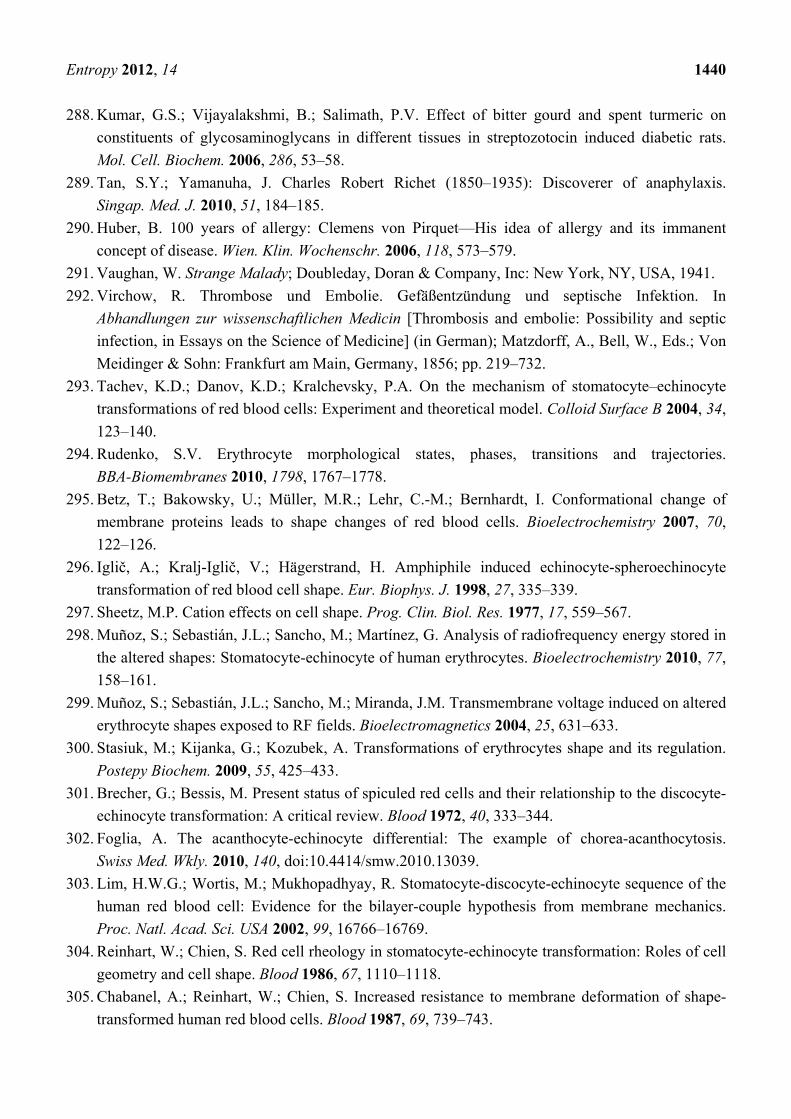
novelty, the “Seneff hypothesis,” the concept that membrane bound endothelial and erythrocytic nitric
oxide synthase (eNOS), in the presence of caveolin-1, and sunlight, oxidizes H2S to sulfate [114]. See
Figure 2. To wit, the zinc-tetrathiolate complex, situated in a cavity formed between the two
monomers of the eNOS dimer, is the proposed site of endogenous sulfate biosynthesis, with
superoxide being provided for the reaction as a consequence of sunlight exposure to the flavins, FMN
and FAD. We propose further that Ch-S, sulfated glycolipids, and sulfated GAGs form as subsequent
reaction products.
Figure 2. Schematic depiction of membrane bound Nitric Oxide Synthase Producing Sulfate.
We thus propose that eNOS, in addition to its role in producing small amounts of nitric oxide (NO),
has a more significant but heretofore overlooked role as a major supplier of sulfate to the extracellular
matrix proteins throughout the body. We propose here the novel hypothesis that eNOS is a dual-purpose
enzyme, and that, in many cells, its main purpose might be to produce sulfate (an anionic kosmotrope)
rather than nitrate (an anionic chaotrope), the ultimate product from nitric oxide. This makes both
intuitive and biophysical sense in light of the apparent alignment between cellular Ch-S production and
eNOS localization, and the positions of nitrate and sulfate within the Hofmeister series [16]. Under
pathological conditions, when excess calcium enters the cell due to stressors, eNOS detaches from the
membrane and switches to nitrate synthesis, in order to compensate for the kosmotropic cation
(calcium) that is replacing the chaotropic cation (potassium). Aluminum, which is added as an
adjuvant to many vaccines, is a much stronger kosmotrope than calcium, and hence has an even more
dramatic effect in dislodging eNOS and disabling sulfate synthesis.
Eukaryotic cells have a characteristic negative surface charge established by anionic integral or
peripheral plasma membrane components [129]. At physiologic pH in health, both the blood serum ZP
and pH are high and the cellular elements of our blood are dispersed and electronegative [130]. The
ionic buffering of our blood by inorganic sulfate and the bio-sulfates (sulfatides and sulfamates)—
sterol sulfates, sulfated glycolipids, and sulfated GAGs—is essential in maintaining the ZP of our
blood [131]. Any event which sufficiently lowers the concentration of sulfate results in colloidal
instability of the many macromolecules and cells suspended in our blood.
Page 10
Entropy 2012, 14 1408
Below a certain threshold of ZP-lowering, “salting out” of various macromolecules and cells will
occur [132]. Such a phase-transition can be fatal. Platelets release thrombin and thromboplastin to
restore hemostasis via clot formation. Albumin provides electrostatic stability to our blood and buffers
the effect of exogenous cations and polyelectrolytes [133]. With age, our blood albumin levels decline.
This decline shifts the balance between blood clotting and blood dispersion towards clotting. Both
mercury and aluminum bind strongly to cysteines in serum albumin in the blood stream [134,135]. The
absorption of aluminum onto serum albumin has a profound effect on ZP [134], driving it even to
positive values at physiologic pH with sufficient concentrations of aluminum hydroxide. It is believed
that much of the mercury that is filtered into the proximal tubular lumen in the glomerulus of the
kidney is present primarily as a conjugate of albumin, bound to the sulfhydryl group of a cysteine
molecule [135]. Thus, positively-charged mercuric Hg2+ salts bound to serum albumin would be
expected to cause a similar effect as aluminum on serum ZP.
2.1. Stress Induced Breathing Patterns Following Vaccination
“Stress”, according to Hans Selye, is “the sum of all nonspecific changes caused by function,
damage, or the rate of wear and tear in the body. In simple terms: the common results of exposure to
anything” [2]. We propose that interfacial water stress [44] triggers the non-specific stress adaptation
syndrome, resulting in the stereotyped, biophysically-determined phenomena, e.g., THP-G, and
SSP-G, so well-described by Selye [3] and others, which are ultimately responsible for the “pluricausal
diseases”. All stressors have the potential to produce the characteristic manifestations of an “alarm
reaction”. Alarm reactions are not due to distinctive actions but to the stressor property that they
share—the ability to induce interfacial water stress [44].
Stress-induced respiratory pattern changes have been reported in asthma [136], pulmonary
microembolism [137], pulmonary inflammation [137], septicemia [138,139], myocardial infarction [140],
brainstem infarction [141], preeclampsia [142,143], eclampsia [135], and anaphylactic shock [144].
Emotions and stress are known to change the respiratory pattern [136,145,146]. The breathing control
centre of the brain is in the respiratory centre located in the lower part of the brain stem called the
medulla oblongata.
According to Dr Viera Scheibner, SIDS researchers refer to all the events where a child is breathing
very shallowly, but not dying, as “false alarms” with regard to SIDS. Instead of deeming such events
as insignificant, Dr Scheibner used a computerized breathing monitor to study them, recording the
babies’ breathing longitudinally over weeks on end. She maintains that overnight six to eight hour
studies, often used in SIDS research, are very misleading [11–15]. Through non-stop hour by hour
recording of babies’ breathing for up to 5 1/2 months, both apneas (pauses in breathing) and hypopneas
(a stress-induced shallow, low volume breathing pattern) can be demonstrated, all of which showed
increased stress patterns after vaccinations. The time frame for these stress patterns has been described
by Dr Scheibner as the “critical days” [12]. According to Dr Scheibner, the pattern of breathing
of babies after vaccinations shows an alarm reaction within one to two days, which may be
biphasic, followed by the stage of resistance around day 5 to 7, and finally the stage of exhaustion
around day 16 [15,147].
Page 11
Entropy 2012, 14 1409
2.2. Hofmeister Effect
Hofmeister [16,24,148] showed that neutral salts varied in their effect on the solubility of proteins.
One group of salts could be ranked according to their efficiency at precipitating proteins, while a
second group could be ranked according to their efficiency at solubilizing proteins. The Hofmeister
ionic sequence has been thought of as ranging from stabilizing “kosmotropes” to disruptive
“chaotropes.” The structure-making (kosmotrope) and structure-breaking (chaotrope) influence of ions
on the hydration water has been basically understood as arising from a balance between the water-water
and ion-water interactions, which vary considerably with the charge density on the solute surface.
Different salts have different efficiencies in salting-out proteins, while some salts have no effect. Most
importantly, the effectiveness of the anions and cations seems to assume a particular specific order.
The Hofmeister series has been speculated to reflect different ordering powers of ions on the
surrounding water molecules. There is ample evidence supporting the importance of hydration effects
beyond the first hydration shell [40,43,149–157]. The structure and dynamics of interfacial water
molecules are different from those in the bulk and exhibit specific ion effects [40,43,158–163].
2.3. Serum Albumin and Zeta Potential
One of the main functions of serum albumin is to control colloidal stability in the blood [133]. The
hypoalbuminemia of aging and the hypoalbuminemia of end stage renal disease potentiate coagulation
by cationic electrolytes and/or polyelectrolytes. Cells and complex molecules suspended in the blood
avoid agglomeration through a negative charge field maintained in the immediate surrounding space.
The rate at which a charged particle suspended in a medium will travel in an applied electric field is an
important measure of colloidal stability in the medium, and is associated in physics with Zeta Potential
(ZP) [130]. A high negative value for ZP is essential for maintaining blood as a colloidal suspension [164].
In 2010, Tigrek and Barnes (p. 35) defined ZP more specifically as “the electrical potential drop from
the particle surface across the bound fluid, to the interface where the liquid begins to flow under the
shear stress. Stated another way, the ‘zeta potential’ is the potential at the surface boundary between
the stationary fluid and the liquid that is moving with the particle” [165].
2.4. Origin of the Surface Charge
Most particles in an aqueous colloidal suspension carry an electric charge. There are many origins
of this surface charge depending upon the nature of the particle and its surrounding medium. Some of
the more important mechanisms include ionization of surface groups, differential loss of ions, and
adsorption of charged species. Surfactant ions may be specifically adsorbed onto the surface of a
particle. Anionic surfactants would lead to a negatively charged surface, whereas cationic surfactants
would lead to a positively charged surface. Most cells of eukaryotic origin have a net negative surface
charge from anionic plasma membrane components [129,130]. This charge distribution is thought to be
important in the movement of various macromolecules across cell membranes.
Page 12
Entropy 2012, 14 1410
2.5. Surfactant-Induced Interfacial Water Stress
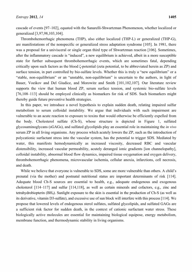
Riddick found a minimum in the ZP enhancement at low concentrations, e.g., 100 parts per million,
of potassium sulfate, in an anionic colloidal suspension of Minusil [132]. See Figure 3. Jones and Ray
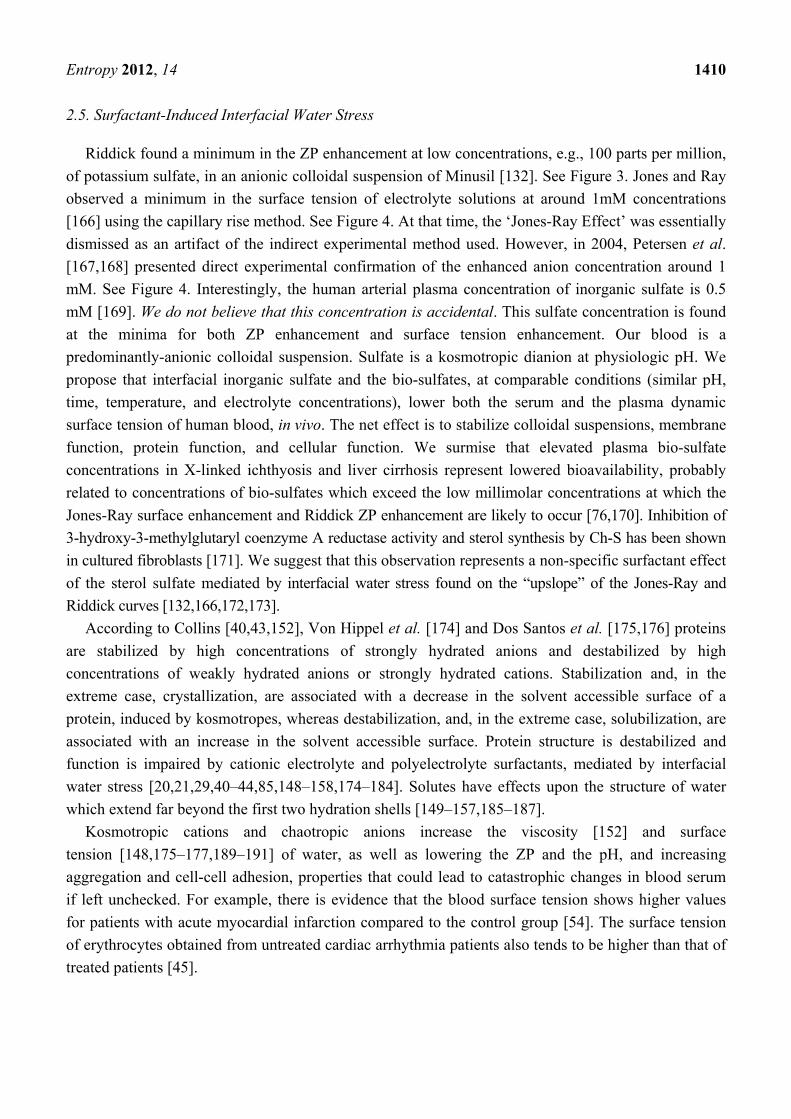
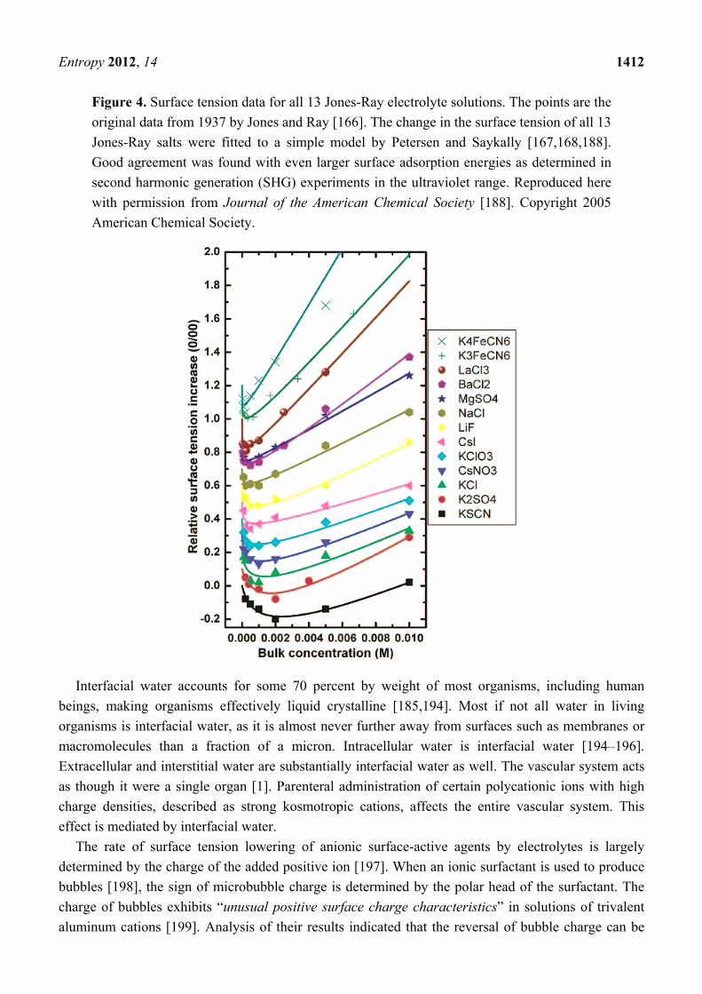
observed a minimum in the surface tension of electrolyte solutions at around 1mM concentrations
[166] using the capillary rise method. See Figure 4. At that time, the ‘Jones-Ray Effect’ was essentially
dismissed as an artifact of the indirect experimental method used. However, in 2004, Petersen et al.
[167,168] presented direct experimental confirmation of the enhanced anion concentration around 1
mM. See Figure 4. Interestingly, the human arterial plasma concentration of inorganic sulfate is 0.5
mM [169]. We do not believe that this concentration is accidental. This sulfate concentration is found
at the minima for both ZP enhancement and surface tension enhancement. Our blood is a
predominantly-anionic colloidal suspension. Sulfate is a kosmotropic dianion at physiologic pH. We
propose that interfacial inorganic sulfate and the bio-sulfates, at comparable conditions (similar pH,
time, temperature, and electrolyte concentrations), lower both the serum and the plasma dynamic
surface tension of human blood, in vivo. The net effect is to stabilize colloidal suspensions, membrane
function, protein function, and cellular function. We surmise that elevated plasma bio-sulfate
concentrations in X-linked ichthyosis and liver cirrhosis represent lowered bioavailability, probably
related to concentrations of bio-sulfates which exceed the low millimolar concentrations at which the
Jones-Ray surface enhancement and Riddick ZP enhancement are likely to occur [76,170]. Inhibition of
3-hydroxy-3-methylglutaryl coenzyme A reductase activity and sterol synthesis by Ch-S has been shown
in cultured fibroblasts [171]. We suggest that this observation represents a non-specific surfactant effect
of the sterol sulfate mediated by interfacial water stress found on the “upslope” of the Jones-Ray and
Riddick curves [132,166,172,173].
According to Collins [40,43,152], Von Hippel et al. [174] and Dos Santos et al. [175,176] proteins
are stabilized by high concentrations of strongly hydrated anions and destabilized by high
concentrations of weakly hydrated anions or strongly hydrated cations. Stabilization and, in the
extreme case, crystallization, are associated with a decrease in the solvent accessible surface of a
protein, induced by kosmotropes, whereas destabilization, and, in the extreme case, solubilization, are
associated with an increase in the solvent accessible surface. Protein structure is destabilized and
function is impaired by cationic electrolyte and polyelectrolyte surfactants, mediated by interfacial
water stress [20,21,29,40–44,85,148–158,174–184]. Solutes have effects upon the structure of water
which extend far beyond the first two hydration shells [149–157,185–187].
Kosmotropic cations and chaotropic anions increase the viscosity [152] and surface
tension [148,175–177,189–191] of water, as well as lowering the ZP and the pH, and increasing
aggregation and cell-cell adhesion, properties that could lead to catastrophic changes in blood serum
if left unchecked. For example, there is evidence that the blood surface tension shows higher values
for patients with acute myocardial infarction compared to the control group [54]. The surface tension
of erythrocytes obtained from untreated cardiac arrhythmia patients also tends to be higher than that of
treated patients [45].
Page 13
Entropy 2012, 14 1411
Figure 3. Zeta potential data for various electrolytes in an anionic colloidal suspension of
100 ppm Minusil [132]. Data were originally published per Thomas M. Riddick (1968)
in [132] and are reproduced here with permission of Zeta-Meter, Inc. (Staunton, VA,
USA).
Wu et al. reported linkage between ZP and electron donicity of charged polar surfaces [192].
Plurivalent counterions are well-known flocculents, making the polar surfaces more
hydrophobic. Yoshisuke [71], and others [97,98,193], have shown that the Shwartzman reaction
induces thrombus formation and hemorrhagic necrosis [71]. How the molecular and humoral
immune responses are converted into mechanical and physical phenomena in the circulation
remains unclear. It is the authors’ opinion, that neither humoral nor cellular immune responses
are fast enough to explain the rapidity in which SDS has been reported to occur, unless there has
been previous sensitization. Even the substantial mobility of Ch-S, attributable to its amphiphilic
character, is not likely sufficient to completely explain the connectedness and rapidity of the
reaction in SDS. Rather, we believe that the branching (avalanche-like) chain of reactions
leading to SDS can only be explained by invoking the direct involvement and intermediacy of
interfacial water. The provocation in SDS is the inducement of interfacial water stress by
exogenous environmental “stressors”. Sometimes the exogenous interfacial water stress (EIWS)
is of sufficiently great degree that prior sensitization is not a necessary prerequisite to SDS.
Serum surface tension is transiently elevated after provocation injection for the Shwartzman
reaction, and the coefficient of foaming is simultaneously reduced at the site of the hemorrhage
caused by the sensitization injection [71]. In addition, it was found that the interface viscosity of
serum and vascular endothelium is greater when interfaced with sensitized endothelium than
with normal endothelium. We hypothesize that these vascular changes may be due to a
breakdown of the sulfated glycocalyx following the sensitizing event.
Page 14
Entropy 2012, 14 1412
Figure 4. Surface tension data for all 13 Jones-Ray electrolyte solutions. The points are the
original data from 1937 by Jones and Ray [166]. The change in the surface tension of all 13
Jones-Ray salts were fitted to a simple model by Petersen and Saykally [167,168,188].
Good agreement was found with even larger surface adsorption energies as determined in
second harmonic generation (SHG) experiments in the ultraviolet range. Reproduced here
with permission from Journal of the American Chemical Society [188]. Copyright 2005
American Chemical Society.
Interfacial water accounts for some 70 percent by weight of most organisms, including human
beings, making organisms effectively liquid crystalline [185,194]. Most if not all water in living
organisms is interfacial water, as it is almost never further away from surfaces such as membranes or
macromolecules than a fraction of a micron. Intracellular water is interfacial water [194–196].
Extracellular and interstitial water are substantially interfacial water as well. The vascular system acts
as though it were a single organ [1]. Parenteral administration of certain polycationic ions with high
charge densities, described as strong kosmotropic cations, affects the entire vascular system. This
effect is mediated by interfacial water.
The rate of surface tension lowering of anionic surface-active agents by electrolytes is largely
determined by the charge of the added positive ion [197]. When an ionic surfactant is used to produce
bubbles [198], the sign of microbubble charge is determined by the polar head of the surfactant. The
charge of bubbles exhibits “unusual positive surface charge characteristics” in solutions of trivalent
aluminum cations [199]. Analysis of their results indicated that the reversal of bubble charge can be
Page 15
Entropy 2012, 14 1413
attributed to specific adsorption of Al3+ and its hydroxo complexes at the gas-liquid interface in the
low pH range and to precipitation of aluminum hydroxide in the intermediate pH range. In the
presence of nonionic surfactants, bubbles can be charged either positively or negatively depending on
the pH, and the isoelectric points appear to be related to the oxygen-to-carbon ratio of the surfactant
molecule [198].
In 1996, Weissenborn studied the surface tension of aqueous solutions of simple inorganic
electrolytes [189]. Results were analyzed in terms of surface tension/electrolyte concentration gradients
and this parameter was found to correlate with the entropies of ion hydration, Jones-Dole viscosity
coefficients and dissolved oxygen gradients. The concentration of salt in our bodies corresponds to the
minimum required for optimal prevention of bubble coalescence [200–202]. As small bubbles are
much less harmful than large bubbles, this fact is crucial [203–205]. pH sensitive phase transitions have
been suggested as mechanisms for cellular action [159]. The “autothixotropy” of water is thought to
possibly play an important role in proton transfer in living beings [206]. Solute-free “exclusion zones,”
a general feature of water adjacent to hydrophilic surfaces, were first reported four decades ago [207]
by Green et al. pH measurements show an extreme drop of pH immediately beyond the exclusion
zone, often to less than pH 3 [154–161]. Variation in the size of the exclusion zone with charge, pH,
and solutes, reported by Zheng and Pollack in 2003, is consistent with a water-structuring hypothesis
[208] which may involve as many as 106 solvent layers. We suggest that KITA studies may enable
further experimental validation of the formation of such massive Exclusion Zones [154].
2.6. Zeta Potential and Cardiovascular Disease
There is a close relationship between inflammation, intravascular coagulation, and cardiovascular
disease. We propose that the underlying reason is that all three processes are driven by the colloidal
instability of the blood associated with abnormally low ZP. We propose that the earliest events in the
inflammatory process are characterized by ZP lowering, increased water stress, cell membrane
dysfunction [29,156,157], and hemostatic [3] and immune derangement [3,209–214]. Inflammation
and serum sickness can be thought of as an unstable dispersion state of our blood, which has an
increased tendency to aggregate, flocculate, gelate, hemorrhage, and coagulate [3,209–214]. This
unstable state is manifest clinically as acute coronary syndromes, transient cerebral ischemic events,
myocardial infarctions [211,212] and cerebrovascular accidents [211,215]. It is known that static
surface tension of blood (49–50 Din/cm2) is normally lower than the surface tension of water or
normal saline solution (approx. 72 Din/cm2). An increase in blood surface tension during acute
myocardial infarction has been observed in humans [54], a difference that was significant (p < 0.05). It
was suggested that the increasing surface tension of blood results in rheological disturbances leading to
heart failure during acute myocardial infarction.
Sherman (1981) has argued through simple physical considerations based on Laplace’s equation of
capillarity, that blood will flow out of capillaries into both the arteries and the veins when the surface
tension is too high in the capillary, due to the inverse relationship between the size of the vessel and
the pressure [216]. “Critical closure” is a term which describes the phenomenon of flow cessation in the
presence of a positive perfusion pressure gradient. Sherman’s use of the term “critical closure”, may be
slightly misleading because “closure” seems to imply a “collapse” of capillary lumen. It might be more
Page 16
Entropy 2012, 14 1414
accurate to say irreversible “critically-arrested capillary flow”, especially if RBCs, proteins, and bodily
humors become “trapped” within the capillary lumen, as is likely to occur, for example, in the “no
reflow” phenomenon. Perfusion is strongly dependent on interfacial tension at the microvascular level
because capillary interfacial tension, if high enough, can not only reduce capillary flow, but also lead to
flow reversal, and eventually to emptying of the capillary and collapse or arresting of flow. The
suggestion was made that factors affecting the surface tension of blood to decrease the interfacial
tension between blood and endothelium may be clinically beneficial [216].
The term microvascular disease refers to the damage that occurs to the smallest blood vessels
throughout the body, including the vital organs (e.g., heart, brain, kidneys, liver). It usually affects the
whole body to some degree. The small blood vessels of the eye, the kidney and of the sheaths around
the nerves, are often at great risk in diabetes, i.e., risk for the development of diabetic retinopathy,
nephropathy, neuropathy. Interestingly, cardiac syndrome X, sometimes referred to as microvascular
angina, often has associated findings of systemic microvascular dysfunction [217] to strongly suggest
that microvascular dysfunction is a systemic malady. While vascular complications of diabetes are often
attributed to loss of nitric-oxide-mediated vasodilation, we suggest an alternative pathophysiology:
impaired microvascular perfusion due to (a) decreased fluidity and deformability of the RBC
membrane related to bio-sulfate deficiency in the endothelial glycocalyx layer (EGL); and
(b) increased capillary endothelial interfacial tension. That is, we suggest that the vascular defect in
diabetes is more a problem with elevated capillary endothelial interfacial tension [216] and poorly
deformable RBCs, than it is a problem with loss of nitric-oxide mediated vasodilation.
The respiratory and auditory centers [218,219] in the brainstem are vulnerable to microvascular
ischemic stress. So too is the pancreas [220–225]. Watershed and terminal vascular distributions are
particularly susceptible to microvascular ischemic stress [223–225]. These vascular distributions would
be predicted to be highly susceptible to pathologic inflammatory stimulation and thrombohemorrhagic
phenomena [3] induced by zeta potential-lowering and interfacial water stress-inducing properties
of cationic kosmotropic electrolytes and polyelectrolyte surfactants. Dr Mohammed Al-Bayati’s
histopathologic analyses of SIDS [220–222] and so-called Shaken Baby Syndrome (SBS) victims [226] are
most informative. Al-Bayati’s work provides strong support for the view that microvascular ischemia plays
a central role in the pathophysiology of SIDS and, by inference, all SDS events [220–222]. Surfactant-
induced water stress [29,40,43,45,54,174,178,211,212], especially that associated with polycationic
surfactants, is an important determinant of risk. Any exogenous food, chemical, or biological exposure
which lowers blood pH and ZP is also a risk factor. Risk factors for SDS are synergistic and
cumulative. Expressed most simply, anything that perturbs the ZP toward less negative values and/or
induces cationic kosmotropic or anionic chaotropic water stress represents a step in the direction of
enzyme inhibition, protein dysfunction, cellular dysfunction, flocculation, gelation, coagulation,
microvascular ischemia, cellular anoxia, infarction, and death. Transcytosis, both endocytosis and
exocytosis, membrane potentials [219,227], and ion channels are all profoundly disrupted by
polycationic surfactants [31,89,228–231]. A very relevant example is aluminum (Al3+), a kosmotropic
trivalent cation, which is a potent and irreversible blocker of voltage activated calcium channels in
mammalian neurons [31,228]. Ca-ATPase, protein kinase C and calmodulin (CaM) are biological
systems known to be disrupted by aluminum [37,87,178,232]. Another very relevant example is
mercury (Hg2+), a kosmotropic divalent cation. It has been shown in infant monkeys that the
Page 17
Entropy 2012, 14 1415
ethylmercury in thimerosal is more readily stored as inorganic mercury (Hg2+) in the brain than is
orally-delivered ethylmercury and that inorganic mercury tends to linger longer in the tissues [231].
Thus, the sudden death syndrome can now be defined as an acute disruption of the colloidal
stability of the vascular system, which triggers a cascade of events [97–100,229,230] leading to death,
whenever compensatory mechanisms to maintain colloidal stability of the blood are insufficient. If
compensatory mechanisms are sufficient, the cascade can instead lead to a new equilibrium.
Mucopolysaccharides, also known as glycosaminoglycans (GAGs), contribute to the inflammatory
state of the Shwartzman phenomenon [233–236]. Upon exposure to systemic stress, increases in
sulfomucopolysaccharide incorporation occur throughout the body, and this is designated as the
“universal nonspecific mesenchymal reaction” [3].
Examples of sudden death syndrome from the medical literature support our hypothesis, including
thrombohemorrhagic phenomena (THP), such as anaphylaxis [229,230,237–239], disseminated
intravascular coagulation (DIC) [240,241], HELLP syndrome [242], acute liver necrosis [243],
Waterhouse–Friderichsen’s syndrome, hemolytic uremic anemia, idiopathic pulmonary hemorrhage [244],
acute pancreatitis [245], acute pituitary necrosis [246], pseudomembranous colitis, thrombotic
thrombocytopenic purpura (TTP), Sanarelli-Shwartzman phenomenon (SSP), Henoch-Schonlein
purpura (HSP), eclampsia [238], serum sickness, hemolytic anemia, preeclampsia, and stillbirths [247].
2.7. The Role of Bio-sulfates in Maintaining Cell Membrane Function
Adequate endogenous and dietary cholesterol is essential in maintaining proper cell membrane
function [114–117], as is adequate endogenous and dietary sulfur [114,118]. The surface charge of
RBCs plays a significant role in cell-to-cell interactions [248]. We propose that it is primarily the
negatively-charged sulfate head-groups imparted to RBC and endothelial cell membranes that are
responsible for both their net and specific surface charge [130]. We propose that this is one of the
mechanisms by which cell membranes are able to store energy [249–252]. The amphiphilic property of
Ch-S endows it with extremely facile, dynamic intercellular and intracellular mobility. We further
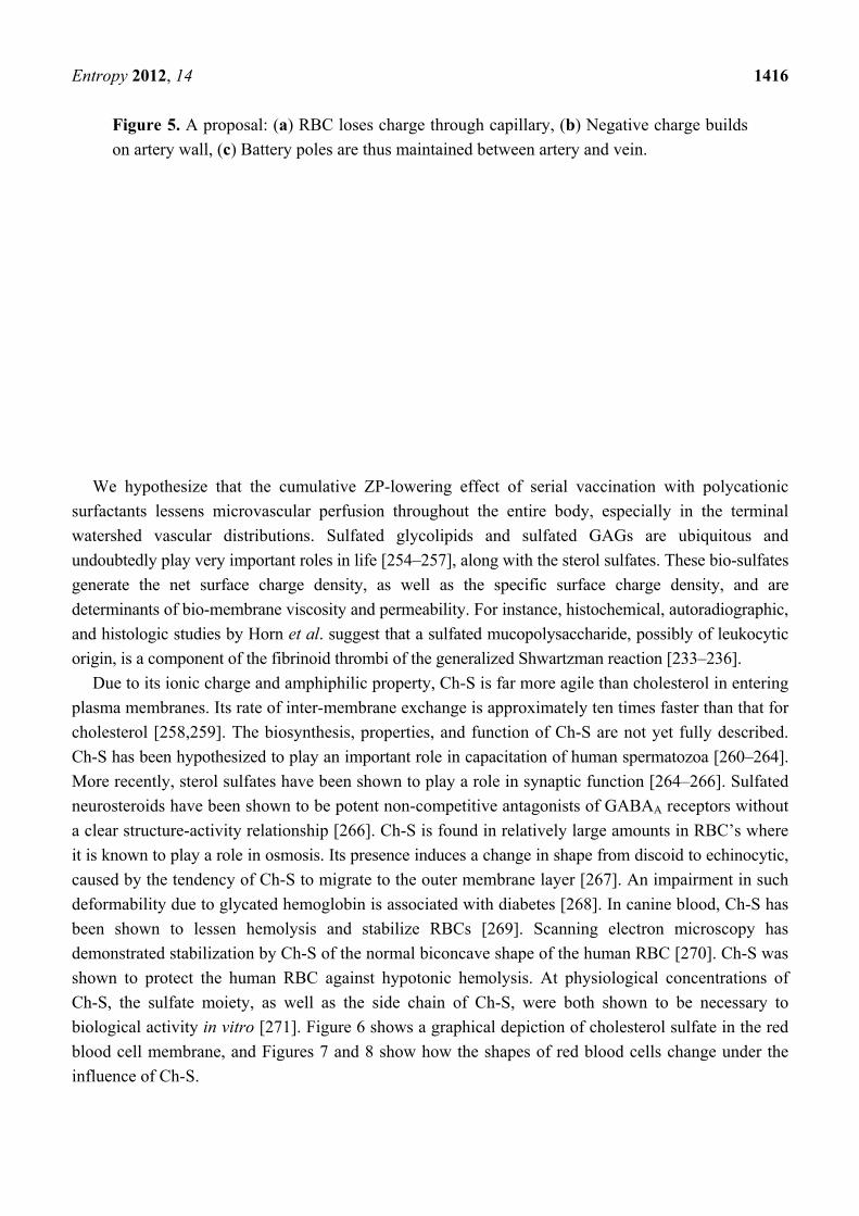
suggest the novel concept that RBCs may actually discharge negative charge by unloading sulfate onto
the endothelial wall, as illustrated in Figure 5. In part due to their higher concentration of CO2, veins
are more acidic than arteries, which suggests that there is an electric field that would propel negatively
charged particles in the capillaries towards the veins. This process would be renewable as the RBCs
travel through superficial veins, regenerating their supply of Ch-S through a process catalyzed by
sunlight exposure [114,116,253].
The electrostatic charge-charge repulsion of the negatively-charged Ch-S and GAG head-groups in
the outer membrane are the primary determinants of viscosity of membrane lipids in both eukaryotes
and prokaryotes. The ZP is of critical importance to maintenance of membrane viscosity in all cells.
Page 18
Entropy 2012, 14 1416
Figure 5. A proposal: (a) RBC loses charge through capillary, (b) Negative charge builds
on artery wall, (c) Battery poles are thus maintained between artery and vein.
We hypothesize that the cumulative ZP-lowering effect of serial vaccination with polycationic
surfactants lessens microvascular perfusion throughout the entire body, especially in the terminal
watershed vascular distributions. Sulfated glycolipids and sulfated GAGs are ubiquitous and
undoubtedly play very important roles in life [254–257], along with the sterol sulfates. These bio-sulfates
generate the net surface charge density, as well as the specific surface charge density, and are
determinants of bio-membrane viscosity and permeability. For instance, histochemical, autoradiographic,
and histologic studies by Horn et al. suggest that a sulfated mucopolysaccharide, possibly of leukocytic
origin, is a component of the fibrinoid thrombi of the generalized Shwartzman reaction [233–236].
Due to its ionic charge and amphiphilic property, Ch-S is far more agile than cholesterol in entering
plasma membranes. Its rate of inter-membrane exchange is approximately ten times faster than that for
cholesterol [258,259]. The biosynthesis, properties, and function of Ch-S are not yet fully described.
Ch-S has been hypothesized to play an important role in capacitation of human spermatozoa [260–264].
More recently, sterol sulfates have been shown to play a role in synaptic function [264–266]. Sulfated
neurosteroids have been shown to be potent non-competitive antagonists of GABAA receptors without
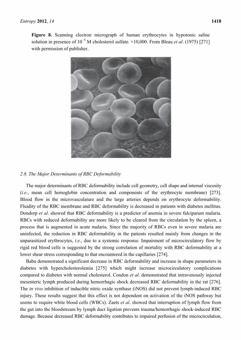
a clear structure-activity relationship [266]. Ch-S is found in relatively large amounts in RBC’s where
it is known to play a role in osmosis. Its presence induces a change in shape from discoid to echinocytic,
caused by the tendency of Ch-S to migrate to the outer membrane layer [267]. An impairment in such
deformability due to glycated hemoglobin is associated with diabetes [268]. In canine blood, Ch-S has
been shown to lessen hemolysis and stabilize RBCs [269]. Scanning electron microscopy has
demonstrated stabilization by Ch-S of the normal biconcave shape of the human RBC [270]. Ch-S was
shown to protect the human RBC against hypotonic hemolysis. At physiological concentrations of
Ch-S, the sulfate moiety, as well as the side chain of Ch-S, were both shown to be necessary to
biological activity in vitro [271]. Figure 6 shows a graphical depiction of cholesterol sulfate in the red
blood cell membrane, and Figures 7 and 8 show how the shapes of red blood cells change under the
influence of Ch-S.
Page 19
Entropy 2012, 14 1417
Figure 6. Graphical depiction of cholesterol sulfate in the red blood cell membrane.
Adapted from Cooper and Hausman: The Cell: A Molecular Approach, Fifth Edition, [272]
by permission of the publisher.
Figure 7. (a) Scanning electron micrograph of human erythrocytes in hypotonic saline
solution. ×10,000; (b) Scanning electron micrograph of human erythrocytes in hypotonic
saline solution. ×20,000. Scans were originally published by Bleau et al. [271] and are
republished here with permission of the publisher.
Page 20
Entropy 2012, 14 1418
Figure 8. Scanning electron micrograph of human erythrocytes in hypotonic saline
solution in presence of 10−5 M cholesterol sulfate. ×10,000. From Bleau et al. (1975) [271]
with permission of publisher.
2.8. The Major Determinants of RBC Deformability
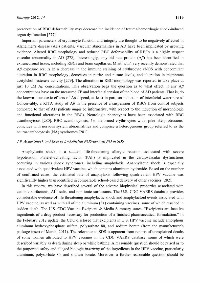
The major determinants of RBC deformability include cell geometry, cell shape and internal viscosity
(i.e., mean cell hemoglobin concentration and components of the erythrocyte membrane) [273].
Blood flow in the microvasculature and the large arteries depends on erythrocyte deformability.
Fluidity of the RBC membrane and RBC deformability is decreased in patients with diabetes mellitus.
Dondorp et al. showed that RBC deformability is a predictor of anemia in severe falciparum malaria.
RBCs with reduced deformability are more likely to be cleared from the circulation by the spleen, a
process that is augmented in acute malaria. Since the majority of RBCs even in severe malaria are
uninfected, the reduction in RBC deformability in the patients resulted mainly from changes in the
unparasitized erythrocytes, i.e., due to a systemic response. Impairment of microcirculatory flow by
rigid red blood cells is suggested by the strong correlation of mortality with RBC deformability at a
lower shear stress corresponding to that encountered in the capillaries [274].
Babu demonstrated a significant decrease in RBC deformability and increase in shape parameters in
diabetes with hypercholesterolemia [275] which might increase microcirculatory complications
compared to diabetes with normal cholesterol. Condon et al. demonstrated that intravenously injected
mesenteric lymph produced during hemorrhagic shock decreased RBC deformability in the rat [276].
The in vivo inhibition of inducible nitric oxide synthase (iNOS) did not prevent lymph-induced RBC
injury. These results suggest that this effect is not dependent on activation of the iNOS pathway but
seems to require white blood cells (WBCs). Zaets et al. showed that interruption of lymph flow from
the gut into the bloodstream by lymph duct ligation prevents trauma/hemorrhagic shock-induced RBC
damage. Because decreased RBC deformability contributes to impaired perfusion of the microcirculation,
Page 21
Entropy 2012, 14 1419
preservation of RBC deformability may decrease the incidence of trauma/hemorrhagic shock-induced
organ dysfunction [277].
Important parameters of erythrocyte function and integrity are thought to be negatively affected in
Alzheimer’s disease (AD) patients. Vascular abnormalities in AD have been implicated by growing
evidence. Altered RBC morphology and reduced RBC deformability of RBCs is a highly suspect
vascular abnormality in AD [278]. Interestingly, amyloid beta protein (Aβ) has been identified in
extraneuronal tissue, including RBCs and brain capillaries. Misiti et al. very recently demonstrated that
Aβ exposure results in a decrease in the immune staining of erythrocyte eNOS with concomitant
alteration in RBC morphology, decreases in nitrite and nitrate levels, and alteration in membrane
acetylcholinesterase activity [279]. The alteration in RBC morphology was reported to take place at
just 10 μM Aβ concentrations. This observation begs the question as to what effect, if any Aβ
concentrations have on the measured ZP and interfacial tension of the blood of AD patients. That is, do
the known neurotoxic effects of Aβ depend, at least in part, on induction of interfacial water stress?
Conceivably, a KITA study of Aβ in the presence of a suspension of RBCs from control subjects
compared to that of AD patients might be informative, with respect to the induction of morphologic
and functional alterations in the RBCs. Neurologic phenotypes have been associated with RBC
acanthocytosis [280]. RBC acanthocytosis, i.e., deformed erythrocytes with spike-like protrusions,
coincides with nervous system abnormalities and comprise a heterogeneous group referred to as the
neuroacanthocytosis (NA) syndromes [281]. 2.9. Acute Shock and Role of Endothelial NOS-derived NO in SDS
Anaphylactic shock is a sudden, life-threatening allergic reaction associated with severe
hypotension. Platelet-activating factor (PAF) is implicated in the cardiovascular dysfunctions
occurring in various shock syndromes, including anaphylaxis. Anaphylactic shock is especially
associated with quadrivalent HPV vaccine, which contains aluminum hydroxide. Based on the number
of confirmed cases, the estimated rate of anaphylaxis following quadrivalent HPV vaccine was
significantly higher than identified in comparable school-based delivery of other vaccines [282].
In this review, we have described several of the adverse biophysical properties associated with
cationic surfactants, Al3+ salts, and non-ionic surfactants. The U.S. CDC VAERS database provides
considerable evidence of life threatening anaphylactic shock and anaphylactoid events associated with
HPV vaccine, as well as with all of the aluminum (3+) containing vaccines, some of which resulted in
sudden death. The U.S. CDC Vaccine Excipient & Media Summary states, “Excipients are inactive
ingredients of a drug product necessary for production of a finished pharmaceutical formulation.” In
the February 2012 update, the CDC disclosed that excipients in U.S. HPV vaccine include amorphous
aluminum hydroxyphosphate sulfate, polysorbate 80, and sodium borate (from the manufacturer’s
package insert of March, 2011). The relevance to SDS is apparent from reports of unexplained deaths
of some women attributed to HPV vaccines in the CDC VAERS database, some of which were
described variably as death during sleep or while bathing. A reasonable question should be raised as to
the purported safety and alleged biologic inactivity of the ingredients in the HPV vaccine, particularly
aluminum, polysorbate 80, and sodium borate. Moreover, a further reasonable question should be
Page 22
Entropy 2012, 14 1420
raised as to the purported safety and alleged biologic inactivity of all of the aluminum and polysorbate
80 containing vaccines. This question may be further explored by analysis of the CDC VAERS database.
For example, CDC VAERS report ID: 337242 states “my daughter had her 3rd GARDASIL
vaccine in Sept. She was a very healthy young lady, she went to take a shower and died. Autopsy
report states undermined [undetermined] death. There was no sign of trauma to the body to indicate a
fall. She had pointed the shower head away from her and she got down on her knees and put her head
on the edge of the tub and passed away.”
Excessive production of the vasodilator NO causes inflammatory hypotension and shock. It had
been generally accepted that transcriptionally regulated NOS (iNOS) was responsible for the NO
synthesis. However, Cauwels et al. found that anaphylactic shock depends on PI3K (phosphatidylinositol
3 kinase) and eNOS-derived NO. In two different models of active systemic anaphylaxis, either eNOS
deficiency or inhibition of eNOS, PI3K, or Akt provided complete protection. Thus, in contrast to the
unsubstantiated paradigm that only excessive iNOS-derived NO underlies cardiovascular collapse in
shock, their data strongly supported the unexpected concept that eNOS-derived NO is the principal
vasodilator in anaphylactic shock [229]. Duran et al. have proposed a putative mechanism by which
eNOS-derived NO stimulates increased microvascular permeability [230].
In the experiment which demonstrated that eNOS was responsible for NO production [225], the
conditions inducing anaphylactic shock in mice included aluminum hydroxide adjuvant, as does HPV
vaccine. We propose that eNOS switches from sulfate to nitrate production under such conditions,
after detaching from the membrane at the caveolae. The surfactant aluminium, a highly kosmotropic
cation, binds CaM with a much greater affinity than does calcium. Calcium, upon binding to CaM,
causes eNOS to detach from the membrane, and, following phosphorylation, to produce nitric oxide.
We hypothesize that eNOS produces sulfate only when it is attached to the membrane, so that, in this
way, aluminum interferes with eNOS’ ability to produce sulfate. Through subsequent systemic
depletion of sulfate, this leads inexorably to SDS.
Dianionic inorganic SO42− raises the ZP and provides the putative obligatory precursor (sulfate)
for the bio-sulfates (sulfamates and sulfatides)—sterol sulfates, sulfated glycolipids, and sulfated
GAGs-monoanionic (1-) at physiologic pH—which also raise the ZP. The bio-sulfates (sulfatides and
sulfamates) are essential in maintaining the ZP, viscosity, permeability barriers of the vascular system,
ion channel function, and transcytosis of nutrients and metabolites of our cells. Sulfate is also
responsible for binding to cationic toxins like mercury [98] and aluminum and expelling them through
the kidneys. Such action would however also lead to a further reduction in the bioavailability of
sulfate. X-linked icthyosis and liver cirrhosis are examples of two clinical conditions in which plasma
bio-sulfate levels are elevated, but unavailable, biologically [76,170].
The formation of sulfate by eNOS results in an immediate increase (to more negative values) in the
ZP [132], and thus the disruption of this process would lead to further induction of water stress. Within
the sheltered environment of the caveolae, within lipid rafts of endothelial, erythrocytic, and neuronal
cell membranes, sulfotransferases (SULTs) synthesize Ch-S and sulfated sphingolipids. Any dietary,
environmental, or iatrogenic (e.g., parenteral) event that impairs sulfate synthesis by endothelial NOS
(eNOS) or neuronal NOS (nNOS), also impairs bio-sulfate synthesis, thereby immediately altering the
junctional proteins between endothelial cells and increasing microvascular permeability. The clinical
signs and symptoms of the metabolic syndrome, microvascular ischemia, and endothelial dysfunction
Page 23
Entropy 2012, 14 1421
are often seen together in diabetic patients [283], who are highly-prone to multisystemic, end-organ
damage involving the retina, kidneys, nervous system, and vascular system. Shedding of the EGL
during acute hyperglycemia has been shown to coincide with endothelial dysfunction, including
increased permeability of the endothelium, increased reactive oxygen species (ROS), reduced nitric
oxide (NO) synthesis, and coagulation activation in vivo [272]. It is entirely possible that the
underlying, shared, pathophysiology is elevated microvascular interfacial tension [216,284–287], as
has been discussed previously. We surmise that an EGL which is depleted in sulfate content has
elevated microvascular interfacial tension, a key risk factor for insulin resistance [283,285]. Cationic
surfactants generally associate with GAGs, e.g., the EGL of the vascular system [249,250,284–288],
disabling their protection against water stress. The process of expelling cationic surfactants via the
kidney necessarily further depletes the sulfate supply. The deficit in intramembrane sterol sulfate and
sulfated GAGs results in decreased intramembrane viscosity, decreased intramembrane deformability,
and increased intramembrane permeability, all of which compromise the stability of the blood
colloidal system.
3. Discussion
The terms “anaphylaxis” and “allergy” were created by Charles Richet in 1901 and Clemens von
Pirquet in 1906, respectively. When Charles Richet attempted to vaccinate dogs to jellyfish (Physalia)
poison, he provoked a violent reaction that quickly killed the dogs following a second injection. These
results were subsequently replicated with the tentacles of sea anemone (Actinia eqnina). Remarkably,
certain of the dogs experienced no apparent ill effects and survived when they received only a single
injection. However, when a repeat injection was given three to four weeks after the first sensitizing (or
preparatory) dose, the animals immediately showed serious symptoms of shock: vomiting, bloody
diarrhea, dyspnea, incontinence, hypotension, syncope, unconsciousness, asphyxia and death, within
15 to 30 minutes. For this reaction, Richet used a Latin term ana-phylaxis or anti-protection, because
the outcome was the opposite from the protection that the vaccine was supposed to provide. In further
experiments with numerous other species, including cats, rabbits and horses, Richet showed that
anaphylaxis is a universal immune system response [237–239,289]. In 1907, Richet demonstrated what
is known as passive anaphylaxis. He also established a relationship between leukocytosis and
anaphylaxis. He concluded his Nobel Prize lecture in 1913, stating that “anaphylaxis is an universal
defence [sic] mechanism against the penetration of heterogenous substances in the blood, whence they
can not be eliminated.”
At the close of the 1800s, a similar anaphylactic phenomenon, serum sickness (“Serum-
Krankheit”), was a common outcome in children subjected to injections of the first mass preserved,
hypodermically delivered injections of sera for scarlet fever, tetanus and diphtheria. The symptoms
observed included urticaria, erythema, pangs of pain, itching, and in the worst cases near-syncope,
with nausea, vomiting, hyperthermia, edema over the whole skin area and general urticaria. Viennese
pediatrician Clemens von Pirquet introduced the Latin derived term “allergy” in 1906, to better
describe this “altered reactivity” to the sera [290]. With it he wanted to describe in general a change in
reactivity of the organism, namely in time, quality and quantity. Prior to advent of vaccination, mass
allergy such as serum sickness was unknown. According to allergist Warren Vaughan, “serum disease,
Page 24
Entropy 2012, 14 1422
as this is called, is a man-made malady. If we had no curative serums and if there were no such thing
as a hypodermic syringe with which to introduce the material under the skin, there would be no serum
disease.” Hence, serum sickness is iatrogenic disease [290,291].
Based on extensive review of the scientific literature, we propose in this paper that any exogenous
substance that lowers the ZP, and/or introduces polycationic kosmotropic interfacial water stress,
and/or lowers the bio-sulfate level, increases the likelihood of sudden death, by triggering a cascade of
events in the pathogenesis of inflammation, allergy, infection, thrombosis, hemorrhage, ischemia,
infarction, anaphylaxis, disease, and death. Inflammation, allergy, anaphylaxis, and serum sickness
should be redefined to reflect this reality. A novel hypothesis as to the etiology of sudden death
syndrome is presented which looks specifically at the very earliest events in the pathophysiology
of SDS. In most instances, introduction of polycationic surfactants into our bloodstream causes
acute interfacial water stress, lowering of ZP, lowering of pH, elevation of viscosity, and
electrohemorheologic—hemodynamic derangement. This triggers a cascade of immunologic and
hemostatic events, leading inexorably to tissue hypoperfusion, cellular anoxia, seizures, arrhythmias,
infarctions, cardiovascular collapse, and death [97–100,209,229,230,237–239].
A biophysically-based disruption of blood flow seems likely to be involved in SDS. Seizure activity
or cardiac arrhythmias, sometimes both, often accompanies SDS. Shock, either cardiogenic or
anaphylactic, followed rapidly by cardiovascular collapse, e.g., with froth at mouth, is often the initial
manifestation of SDS. Rudolph Virchow originally proposed, in 1856, that vascular events are a
common pathophysiology for deaths and disease [292]. More recently, postmortem studies [220–222]
of SIDS victims commonly use the word “ischemic,” as in “ischemic changes,” on histopathology
analysis. Thrombohemorrhagic phenomena, e.g., disseminated intravascular coagulation (DIC),
thrombotic thrombocytopenic purpura (TTP), hemolytic, inflammatory, and anaphylactic events, have
all been associated with SDS. Thus, in the authors’ opinion, an acute vascular ischemic etiology is
strongly favored as the central common pathophysiology of SDS. We propose that the initial events in
SDS are universally vascular events. These hemorrheologic events trigger a cascade of immunologic
and hemostatic consequences, leading rapidly to death. They are “sparked” by exogenous polycationic
surfactant-induced water stress. They are biophysical in origin and have profound hemodynamic
consequences.
We hypothesize that acute lowering of the ZP can result in an acute increase in endothelial cell
permeability and acute alteration in RBC morphology from dispersed biconcave discs to adherent rouleaux
formations [248], echinocyte, stomatocyte, acanthocyte, and spherocyte transformations [113,293–306],
with consequent elevation in blood viscosity, elevated resistance to microvascular blood flow,
diminished oxygen transport, and diminished oxygen delivery. CO2 overload is a secondary inevitable
consequence of the acute colloidal instability of our blood induced by the ZP-lowering, polycationic
surfactant stress. Acute colloidal instability of flowing blood impairs perfusion to the brainstem
respiratory and auditory centers. Flowing blood has electrokinetic, hemodynamic, and hemorheologic
properties which are substantially affected by polycationic surfactant water stress. Our entire vascular
system, including the lymph, and all body fluids in the humoral system, reacts to water stress like a
single organ system [1] at mesoscopic scales [194–196,307,308] in a quantum coherent manner.
Exogenous surfactant-induced interfacial surface tension, i.e., “water stress,” is the biophysical
basis [1–3] for the generalized Sanarelli-Shwartzman phenomenon, generalized thrombohemorrhagic
Page 25
Entropy 2012, 14 1423
phenomena, the nonspecific generalized stress adaptation syndrome, the systemic inflammatory
response syndrome, immune cell activation, macropinocytosis [73,304–306], vesiculation [77],
adhesion [63,64,78], engulfment [45,46,256], envelopment [309], fusion [74,77,78,172,309–311],
podokinesis [312–315], transcellular diapedesis [316] and nutritive endocytosis [317–320]. These
highly-stereotyped supramolecular (mesoscopic), quantum coherent, biological processes are examples
of the biophysical properties which motivate homeopathy.
ZP-lowering immediately lessens microvascular perfusion and increases interfacial water stress
throughout the entire vascular system, especially in the terminal watershed vascular distributions. This
explains sudden death from snake bites [321,322], vaccines [323–333], and cocaine [215], in otherwise
healthy people, as examples. Depending on their surfactant property and their position within the
Hofmeister series, environmental biological or chemical intoxicants of virtually any type can provide
the EIWS to induce SDS. Certain dietary deficiencies, e.g., sulfur and zinc, and lack of adequate
sunlight are likely to predispose to SDS. Bio-sulfate deficiency, in particular, is very likely to lower the
threshold for an exogenous interfacial water “stressor” to induce the branching (avalanche-like) chain
of reactions we have referred to as SDS. Our basal endogenous interfacial water tone is likely to be
closely-related to bio-sulfate levels. Inorganic sulfate and the bio-sulfates—Ch-S, sulfated GAGs, and
sulfated glycolipids—play an essential role in preserving and maintaining our ZP. It is appropriate that
they be strongly considered for use as biomarkers of disease, including autoimmune, neurodegenerative,
and oncologic disease [334]. Both bio-sulfate levels and measurement of EIWS may ultimately prove
to be useful in diagnosing Shoenfeld’s syndrome [335], the autoimmune (autoinflammatory) syndrome
induced by adjuvant (ASIA). Sunlight exposure to the skin is essential to the maintenance of the
supply of Ch-S to the blood system and to the tissues, mediated by eNOS.
4. Conclusions
We have reviewed the colloid and interface science literature as a basis for proposing a new
hypothesis as to the cause of the Sudden Death Syndrome. We have defined the concept of surfactant-
induced interfacial water stress and conclude that the initial events in the inflammatory cascade are
purely biophysical. We have detailed the role of the bio-sulfates in maintaining cell membrane
function and the role of endothelial Nitric Oxide Synthase-derived Nitric Oxide in acute shock and the
Sudden Death Syndrome.
Acknowledgments
The authors are grateful to Glyn Wainwright, Paul Maher, Laurie Lentz-Marino, and Ann Lauritzen
for thoughtful comments on the preparation of this manuscript. The authors have no financial
disclosures to make.
References
1. Bechamp, A. The Blood and Its Third Element; Metropolis Inc: New York, NY, USA, 2002.
2. Selye, H. In Vivo: The Case for Supramolecular Biology; Liveright Publishing Corporation:
New York, NY, USA, 1967.
Page 26
Entropy 2012, 14 1424
3. Selye, H. Thrombohemorrhagic Phenomena; Charles, C., Ed.; Thomas: Springfield, IL, USA, 1966.
4. Filiano, J.J.; Kinney, H.C. A perspective on neuropathologic findings in victims of the sudden
infant death syndrome: The triple-risk model. Biol. Neonate 1994, 65, 194–197.
5. Vatta, M.; Dumaine, R.; Varghese, G.; Richard, T.A.; Shimizu, W.; Aihara, N.; Nademanee, K.;
Brugada, R.; Brugada, J.; Veerakul, G.; et al. Genetic and biophysical basis of sudden
unexplained nocturnal death syndrome (SUNDS), a disease allelic to Brugada syndrome.
Hum. Mol. Genet. 2002, 11, 337–345.
6. Kinney, H.C. Brainstem mechanisms underlying the sudden infant death syndrome: Evidence
from human pathologic studies. Dev. Psychobiol. 2009, 51, 223–233.
7. Krous, H.F.; Beckwith, J.B.; Byard, R.W.; Rognum, T.O.; Bajanowski, T.; Corey, T.; Cutz, E.;
Hanzlick, R.; Keens, T.G.; Mitchell, E.A. Sudden infant death syndrome and unclassified sudden
infant deaths: A definitional and diagnostic approach. Pediatrics 2004, 114, 234–238.
8. Blood-Siegfried, J.; Bowers, M.T.; Lorimer, M. Is shock a key element in the pathology of sudden
infant death syndrome (SIDS)? Biol. Res. Nurs. 2009, 11, 187–194.
9. Buttram, H. Shaken baby syndrome or vaccine-induced encephalitis? Med. Sentin. 2001, 6,
83–89.
10. Mage, D.T.; Donner, M. A unifying theory for SIDS. Int. J. Pediatr. 2009, doi:10.1155/2009/
368270.
11. Scheibner, V. Adverse effects of adjuvants in vaccines. Nexus 2000, 8, 37–40.
12. Scheibner, V. Vaccination: One Hundred Years of Orthodox Research Shows That Vaccines
Represent a Medical Assault on the Immune System; New Atlantean Press: Santa Fe, NM, USA,
1993.
13. Scheibner, V. Shaken baby syndrome: The vaccination link. Nexus Magazine 1998, 5, 35–38,
continued on page 87.
14. Scheibner, V. Adverse effects of adjuvants in vaccines. Nexus Magazine 2001, 8, 37–40, 41–44,
continued on page 84.
15. Scheibner, V. Dynamics of critical days as part of the dynamics of non-specific stress syndrome
discovered during monitoring with Cotwatch Breathing Monitor. J. Australas. Coll. Nutr.
Environ. Med. 2004, 23, 1–5.
16. Hofmeister, F. Naunyn-Schmiedebergs Zur Lehre von der Wirkung der Salze (in German).
Arch. Pharmacol. 1888, 24, 247–260.
17. Setschenow, J. Über die Konstitution der Salzlösungen auf Grund ihres Verhaltens zu
Kohlensäure (in German). Z. Phys. Chem. 1889, 4, 117–125.
18. Heydweiller, A. Über physikalische Eigenschaften von Lösungen in ihrem Zusammenhang. II.
Oberflächenspannung und elektrisches Leitvermögen wässeriger Salzlösungen (in German).
Annalen der Physik 1910, 4, 145–185.
19. Langmuir, I. The constitution and fundamental properties of solids and liquids. II. Liquids.
J. Am. Chem. Soc. 1917, 39, 1848–1906.
20. Dér, A.; Kelemen, L.; Fabian, L.; Taneva, S.G.; Fodor, E.; Pali, T.; Cupane, A.; Cacace, M.G.;
Ramsden, J.J. Interfacial water structure controls protein conformation. J. Phys. Chem. B 2007,
111, 5344–5350.
Page 27
Entropy 2012, 14 1425
21. Dér, A. Salts, Interfacial water and protein conformation. Biotechnol. Biotechnol. Equip. 2008,
22, 629–633.
22. Callen, H.B.; Welton, T.A. Irreversibility and generalized noise. Phys. Rev. 1951, 83, 34–40.
23. Grassia, P. Dissipation, fluctuations, and conservation laws. Am. J. Phys. 2000, 69, 113–119.
24. Neagu, A.; Neagu, M.; Dér, A. Fluctuations and the Hofmeister effect. Biophys. J. 2001, 81,
1285–1294.
25. Dér, A.; Ramsden, J.J. Evidence for loosening of a protein mechanism. Naturwissenschaften
1998, 85, 353–355.
26. Rosina, J.; Kvasnak, E.; Suta, D.; Kolarova, H.; Malek, J.; Krajci, L. Temperature dependence of
blood surface tension. Physiol. Res./Acad. Sci. Bohemoslov. 2007, 56, S93–S98.
27. Coates, E.L.; Li, A.; Nattie, E.E. Widespread sites of brain stem ventilatory chemoreceptors.
J. App. Physiol. 1993, 75, 5–14.
28. Kato, H.; Shibano, M.; Saito, T.; Yamaguchi, J.; Yoshihara, S.; Goto, N. Relationship between
hemolytic activity and adsorption capacity of aluminum hydroxide and calcium phosphate as
immunological adjuvants for biologicals. Microbiol. Immunol. 1994, 38, 543–548.
29. Lupidi, G.; Angeletti, M.; Eleuteri, A. M.; Fioretti, E.; Marini, S.; Gioia, M.; Coletta, M.
Aluminum modulation of proteolytic activities. Coordin. Chem. Rev. 2002, 228, 263–269.
30. Peeters, B.W.; Cheung, K.S.; Vossen, J.M.; Coenen, A.M. Some solvents for antiepileptics have
proepileptic potencies in the WAG/Rij rat model for absence epilepsy. Brain Res. Bull. 1992, 29,
515–517.
31. Platt, B.; Busselberg, D. Actions of aluminum on voltage-activated calcium channel currents.
Cell. Mol. Neurobiol. 1994, 14, 819–829.
32. Preté, P.S.C.; Gomes, K.; Malheiros, S.V.P.; Meirelles, N.C.; de Paula, E. Solubilization of
human erythrocyte membranes by non-ionic surfactants of the polyoxyethylene alkyl ethers series.
Biophys. Chem. 2002, 97, 45–54.
33. Preté, P.S.C.; Malheiros, S.V.P.; Meirelles, N.C.; de Paula, E. Quantitative assessment of human
erythrocyte membrane solubilization by Triton X-100. Biophys. Chem. 2002, 97, 1–5.
34. Shafer, T.J.; Mundy, W.R.; Tilson, H.A. Aluminum decreases muscarinic, adrenergic, and
metabotropic receptor-stimulated phosphoinositide hydrolysis in hippocampal and cortical slices
from rat brain. Brain Res. 1993, 629, 133–140.
35. Shaw, C.A.; Petrik, M.S. Aluminum hydroxide injections lead to motor deficits and motor neuron
degeneration. J. Inorg. Biochem. 2009, 103, 1555–1562.
36. Strupp, W.; Weidinger, G.; Scheller, C.; Ehret, R.; Ohnimus, H.; Girschick, H.; Tas, P.;
Flory, E.; Heinkelein, M.; Jassoy, C. Treatment of cells with detergent activates caspases and
induces apoptotic cell death. J. Membr. Biol. 2000, 175, 181–189.
37. Weis, C.; Haug, A. Aluminum-induced conformational changes in calmodulin alter the dynamics
of interaction with melittin. Arch. Biochem. Biophys. 1987, 254, 304–312.
38. Joshi, M.; Pathak, S.; Sharma, S.; Patravale, V. Design and in vivo pharmacodynamic evaluation
of nanostructured lipid carriers for parenteral delivery of artemether: Nanoject. Int. J. Pharm.
2008, 364, 119–126.
Page 28
Entropy 2012, 14 1426
39. Goto, N.; Kato, H.; Maeyama, J.; Eto, K.; Yoshihara, S. Studies on the toxicities of aluminium
hydroxide and calcium phosphate as immunological adjuvants for vaccines. Vaccine 1993, 11,
914–918.
40. Collins, K.D. Ions from the Hofmeister series and osmolytes: Effects on proteins in solution and
in the crystallization process. Methods 2004, 34, 300–311.
41. Ortiz, V.; Nielsen, S.O.; Klein, M.L.; Discher, D.E. Unfolding a linker between helical repeats.
J. Mol. Biol. 2005, 349, 638–647.
42. Sah, H.; Bahl, Y. Effects of aqueous phase composition upon protein destabilization at
water/organic solvent interface. J. Control. Release 2005, 106, 51–61.
43. Washabaugh, M.W.; Collins, K.D. The systematic characterization by aqueous column
chromatography of solutes which affect protein stability. J. Biol. Chem. 1986, 261, 12477–12485.
44. Gallez, D.; Coakley, W.T. Interfacial instability at cell membranes. Prog. Biophys. Mol. Biol.
1986, 48, 155–199.
45. Absolom, D.R. Measurement of Surface Properties of Phagocytes, Bacteria, and Other Particles.
In Methods in Enzymology; di Sabato, G., Everse, J., Eds.; Academic Press: Waltham, MA, USA,
1986; Volume 132, pp. 16–95.
46. Absolom, D.R.; Zingg, W.; Neumann, A.W. Measurement of contact angles on biological and
other highly hydrated surfaces. J. Colloid Interface Sci. 1986, 112, 599–601.
47. Nevo, A.; de Vries, A.; Katchalsky, A. Interaction of basic polyamino acids with the red blood
cell. I. Combination of polylysine with single cells. Biochim. Biophys. Acta 1955, 17, 536–547.
48. Karchalsky, A.; Danon, D.; Nevo, A.; de Vries, A. Interaction of basic polyelectrolytes with the
red blood cell II. Agglutination of red blood cells by polymeric bases. Biochim. Biophys. Acta
1959, 33, 120–138.
49. Coakley, W.T.; Hewison, L.A.; Tilley, D. Interfacial instability and the agglutination of
erythrocytes by polylysine. Eur. Biophys. J. 1985, 13, 123–130.
50. Kratochvil, A.; Hrncir, E. Correlation between the blood surface tension and the activity of some
enzymes. Phys. Res./Acad. Sci. Bohemoslov. 2001, 50, 433–437.
51. Kratochvil, A.; Hrncir, E. Correlations between the cerebrospinal fluid surface tension value and
1. Concentration of total proteins 2. Number of cell elements. Gen. Physiol. Biophys. 2002, 21,
47–53.
52. Krishnan, A.; Cha, P.; Liu, Y.H.; Allara, D.; Vogler, E.A. Interfacial energetics of blood plasma
and serum adsorption to a hydrophobic self-assembled monolayer surface. Biomaterials 2006, 27,
3187–3194.
53. Krishnan, A.; Wilson, A.; Sturgeon, J.; Siedlecki, C.A.; Vogler, E.A. Liquid-vapor interfacial
tension of blood plasma, serum and purified protein constituents thereof. Biomaterials 2005, 26,
3445–3453.
54. Esitashvili, T.; Msuknishvili, M. Increase of blood surface tension during acute myocardial
infarction. In Proceedings of 8th World Congress on Heart Failure, Internation Academy of
Cardiology, Washington, DC, USA, 16 July 2002.
55. Surface tensiometry in rheumatology. In Studies in Interface Science; Kazakov, V.N.,
Sinyachenko, O.V., Fainerman, V.B., Pison, U., Miller, R., Eds.; Elsevier: Waltham, MA, USA,
2000; Volume 8, Chapter 5, pp. 191–244.
Page 29
Entropy 2012, 14 1427
56. Beccerica, E.; Piergiacomi, G.; Curatola, G.; Ferretti, G. Changes of lymphocyte membrane
fluidity in rheumatoid arthritis: A fluorescence polarisation study. Ann. Rheum. Dis. 1988, 47,
472–477.
57. Kazakov, V.N.; Vozianov, A.F.; Sinyachenko, O.V.; Trukhin, D.V.; Kovalchuk, V.I.; Pison, U.
Studies on the application of dynamic surface tensiometry of serum and cerebrospinal liquid for
diagnostics and monitoring of treatment in patients who have rheumatic, neurological or
oncological diseases. Adv. Colloid Interface 2000, 86, 1–38.
58. Weinstein, M.B.; Joensuu, O.I.; Duffy, P.; Bennett, B. Technical difficulties in labeling
erythrocytes with 99mTc: Identification of agglutinating substance. J. Nucl. Med. 1971, 12,
183–185.
59. Lin, M.S.; MacGregor, R.D., Jr.; Yano, Y. Ionic aluminum (3) in generator eluate as an
erythrocyte-agglutinating agent. J. Nucl. Med. 1971, 12, 297–299.
60. Jandl, J.H.; Simmons, R.L. The agglutination and sensitization of red cells by metallic cations:
Interactions between multivalent metals and the red-cell membrane. Br. J. Haematol. 1957, 3,
19–38.
61. Hsu, J.-P.; Kuo, Y.-C.; Chang, Y.-I. Electrostatic interaction between a charge-regulated particle
and a solid surface in electrolyte solution: Effect of cationic electrolytes. Colloid Polym. Sci.
1994, 272, 946–954.
62. Chang, Y.-I.; Hsieh, C.-Y. The effect of cationic electrolytes on the electrophoretic properties of
bacterial cells. Colloid Surface 1991, 53, 21–31.
63. Chang, Y.-I. The effect of cationic electrolytes on the adhesion of cells. Biotechnol. Adv. 1993,
11, 711–724.
64. Chang, Y.-I.; Hsu, J.-P. Effect of multivalent cations on the adhesion rate of cellular surfaces
bearing ionizable groups. Colloid Surface A 1995, 96, 155–163.
65. Hong, Y.; Brown, D.G. Electrostatic behavior of the charge-regulated bacterial cell surface.
Langmuir 2008, 24, 5003–5009.
66. Hartvig, R.A.; van de Weert, M.; Østergaard, J.; Jorgensen, L.; Jensen, H. Protein adsorption at
charged surfaces: The role of electrostatic interactions and interfacial charge regulation. Langmuir
2011, 27, 2634–2643.
67. Hartvig, R.A.; van de Weert, M.; Østergaard, J.; Jorgensen, L.; Jensen, H. Formation of dielectric
layers and charge regulation in protein adsorption at biomimetic interfaces. Langmuir 2011, 28,
1804–1815.
68. Thomas, N.E.; Coakley, W.T.; Akay, G. The lateral separation of contacts on erythrocytes
agglutinated by polylysine. Cell Biophys. 1992, 20, 125–147.
69. Coakley, W.T.; Gallez, D.; de Souza, E.R.; Gauci, H. Ionic strength dependence of localized
contact formation between membranes: Nonlinear theory and experiment. Biophys. J. 1999, 77,
817–828.
70. Thomas, N.E.; Coakley, W.T. Localized contact formation by erythrocyte membranes:
Electrostatic effects. Biophys. J. 1995, 69, 1387–1401.
71. Yoshisuke, E. The studies on the effects of gelatin and its related substances. Available online:
http://www.ps-corp.co.jp/column/thesis/n001_e.html (accessed on 24 July 2012).
Page 30
Entropy 2012, 14 1428
72. Kratochvil, A.; Hrncir, E. Surface tension of body fluids in pathophysiology and diagnosis (in
Czech). Prakt. Lek. 1999, 79, 258–259.
73. Damm, E.M.; Pelkmans, L.; Kartenbeck, J.; Mezzacasa, A.; Kurzchalia, T.; Helenius, A. Clathrin-
and caveolin-1-independent endocytosis: Entry of simian virus 40 into cells devoid of caveolae.
J. Cell Biol. 2005, 168, 477–488.
74. Roy, S.M.; Sarkar, M. Membrane fusion induced by small molecules and ions. J. Lipids 2011,
doi:10.1155/2011/528784.
75. Korte, S.; Wiesinger, A.; Straeter, A.S.; Peters, W.; Oberleithner, H.; Kusche-Vihrog, K. Firewall
function of the endothelial glycocalyx in the regulation of sodium homeostasis. Pflug. Arch. Eur.
J. Phy. 2012, 463, 269–278.
76. Tamasawa, N.; Tamasawa, A.; Takebe, K. Higher levels of plasma cholesterol sulfate in patients
with liver cirrhosis and hypercholesterolemia. Lipids 1993, 28, 833–836.
77. Tanaka, T.; Yamazaki, M. Membrane fusion of giant unilamellar vesicles of neutral phospholipid
membranes induced by La3+. Langmuir 2004, 20, 5160–5164.
78. Sun, Y.; Lee, C.-C.; Huang, H.W. Adhesion and merging of lipid bilayers: A method for
measuring the free energy of Adhesion and Hemifusion. Biophys. J. 2011, 100, 987–995.
79. Ginsburg, I. The role of bacteriolysis in the pathophysiology of inflammation, infection and
post-infectious sequelae. APMIS 2002, 110, 753–770.
80. Jinendra, B.; Tamaki, K.; Kuroki, S.; Vassileva, M.; Yoshida, S.; Tsenkova, R. Near infrared
spectroscopy and aquaphotomics: Novel approach for rapid in vivo diagnosis of virus infected
soybean. Biochem. Biophys. Res. Commun. 2010, 397, 685–690.
81. Perera, R.; Riley, C.; Isaac, G.; Hopf-Jannasch, A.S.; Moore, R.J.; Weitz, K.W.; Pasa-Tolic, L.;
Metz, T.O.; Adamec, J.; Kuhn, R.J. Dengue virus infection perturbs lipid homeostasis in infected
mosquito cells. PLoS Pathog. 2012, 8, e1002584.
82. Fiszer-Kierzkowska, A.; Vydra, N.; Wysocka-Wycisk, A.; Kronekova, Z.; Jarzab, M.; Lisowska, K.;
Krawczyk, Z. Liposome-based DNA carriers may induce cellular stress response and change gene
expression pattern in transfected cells. BMC Mol. Biol. 2011, 12, doi:10.1186/1471-2199-12-27.
83. Lloyd, D.; Cortassa, S.; O’Rourke, B.; Aon, M.A. What yeast and cardiomyocytes share:
Ultradian oscillatory redox mechanisms of cellular coherence and survival. Integr. Biol. (Camb)
2012, 4, 65–74.
84. Yang, Y.-W.; Wu, C.-A.; Morrow, W.J.W. Cell death induced by vaccine adjuvants containing
surfactants. Vaccine 2004, 22, 1524–1536.
85. Gilman, S.; Koller, M.; Black, R.S.; Jenkins, L.; Griffith, S.G.; Fox, N.C.; Eisner, L.; Kirby, L.;
Rovira, M.B.; Forette, F.; et al. Clinical effects of Abeta immunization (AN1792) in patients with
AD in an interrupted trial. Neurology 2005, 64, 1553–1562.
86. Turgut, S.; Bor-Kucukatay, M.; Emmungil, G.; Atsak, P.; Turgut, G. The effects of low dose
aluminum on hemorheological and hematological parameters in rats. Arch. Toxicol. 2007, 81,
11–17.
87. Kawahara, M.; Kato-Negishi, M. Link between aluminum and the pathogenesis of alzheimer’s
disease: The integration of the aluminum and amyloid cascade hypotheses. Int. J. Alzheimer’s Dis.
2011, 2011, doi:10.4061/2011/276393.
Page 31
Entropy 2012, 14 1429
88. Shaw, C.A.; Petrik, M.S. Aluminum hydroxide injections lead to motor deficits and motor neuron
degeneration. J. Inorg. Biochem. 2009, 103, 1555–1562.
89. Tomljenovic, L.; Shaw, C.A. Do aluminum vaccine adjuvants contribute to the rising prevalence
of autism? J. Inorg. Biochem. 2011, 105, 1489–1499.
90. Lemire, J.; Appanna, V.D. Aluminum toxicity and astrocyte dysfunction: A metabolic link to
neurological disorders. J. Inorg. Biochem. 2011, 105, 1513–1517.
91. Gardener, H.; Spiegelman, D.; Buka, S.L. Perinatal and neonatal risk factors for autism: A
comprehensive meta-analysis. Pediatrics 2011, 128, 344–355.
92. Oller, J.W.; Oller, S.D. Autism: The Diagnosis, Treatment, & Etiology of the Undeniable
Epidemic; Jones & Bartlett Learning: Sudbury, MA, USA, 2010.
93. Olmsted, D.; Blaxill, M. The Age of Autism: Mercury, Medicine, and a Man-Made Epidemic;
St. Martin’s Press: New York, NY, USA, 2010.
94. Botkin, S.P. Über die Wirkung der Salze auf die circulirenden rothen Blutkörperchen [On the
action of salts on living blood-corpuscles] (in German). Virchows Arch. Path. Anat. 1858, 15,
173–176.
95. Sanarelli, J. Etudes sur la fievre typhoide experimentale. Ann. Inst. Pasteur (Paris) 1894, 8,
193–230.
96. Hjort, P.; Rapaport, S. The Shwartzman reaction: Pathogenetic mechanisms and clinical
manifestations. Annu. Rev. Med. 1965, 16, 135–168.
97. Brozna, J.P. Shwartzman reaction. Semin. Thromb. Hemost. 1990, 16, 326–332.
98. Lipinski, B.; Jeljaszewicz, J. A hypothesis for the pathogenesis of the generalized Shwartzman
reaction. J. Infect. Dis. 1969, 120, 160–168.
99. Levy, G.A.; Schwartz, B.S.; Curtiss, L.K.; Edgington, T.S. Regulatory roles of T mu and T
gamma cells in the collaborative cellular initiation of the extrinsic coagulation pathway by
bacterial lipopolysaccharide. J. Clin. Invest. 1985, 76, 548–555.
100. Berg, D.J.; Kuhn, R.; Rajewsky, K.; Muller, W.; Menon, S.; Davidson, N.; Grunig, G.; Rennick, D.
Interleukin-10 is a central regulator of the response to LPS in murine models of endotoxic shock
and the Shwartzman reaction but not endotoxin tolerance. J. Clin. Invest. 1995, 96, 2339–2347.
101. Bauer, E. Theoretical Biology; VIEM Publishing House: Moscow-Leningrad, Russia, 1935.
102. Voeikov, V.; del Giudice, E. Water Respiration-The Basis of the Living State. Water 2009, 1,
52–75.
103. Shiga, J.; Mori, W. A light- and electron-microscopic study of the so-called uni-visceral
Shwartzman reaction. Acta Pathol. Japon. 1980, 30, 705–712.
104. Watanabe, T.; Tanaka, K. Electron microscopic observations of the kidney in the generalized
Shwartzman reaction. Virchows Arch. A 1977, 374, 183–196.
105. Selye, H. Confusion and controversy in the stress field. J. Hum. Stress 1975, 1, 37–44.
106. Mori, W. The Shwartzman reaction: A review including clinical manifestations and proposal for a
univisceral or single organ third type. Histopathology 1981, 5, 113–126.
107. Morowitz, H.; Smith, E. Energy flow and the organization of life: Essays and commentaries.
Complexity 2007, 13, 51–59.
Page 32
Entropy 2012, 14 1430
108. Van der Kraan, P.M.; de Vries, B.J.; Vitters, E.L.; van den Berg, W.B.; van de Putte, L.B. The
effect of low sulfate concentrations on the glycosaminoglycan synthesis in anatomically intact
articular cartilage of the mouse. J. Orthop. Res. 1989, 7, 645–653.
109. Nimni, M.E.; Han, B.; Cordoba, F. Are we getting enough sulfur in our diet? Nutr. Metab. 2007,
4, doi:10.1186/1743-7075-4-24.
110. Cole, D.E.; Evrovski, J. The clinical chemistry of inorganic sulfate. Crit. Rev. Clin. Lab. Sci.
2000, 37, 299–344.
111. Hoffer, L.J.; Hamadeh, M.J.; Robitaille, L.; Norwich, K.H. Human sulfate kinetics. Am. J.
Physiol. 2005, 289, R1372–R1380.
112. Markovich, D. Physiological roles and regulation of mammalian sulfate transporters. Physiol. Rev.
2001, 81, 1499–1533.
113. Bleau, G. Biochimie du Sulfate de Cholestérol (in French); Université de Montréal: Montreal, QC,
Canada, 1972.
114. Seneff, S.; Davidson, R.; Mascitelli, L. Might cholesterol sulfate deficiency contribute to the
development of autistic spectrum disorder? Med. Hypotheses 2011, 78, 213–217.
115. Seneff, S.; Wainwright, G.; Mascitelli, L. Is the metabolic syndrome caused by a high fructose,
and relatively low fat, low cholesterol diet? Arch. Med. Sci. 2010, 7, 8–20.
116. Seneff, S.; Wainwright, G.; Mascitelli, L. Nutrition and Alzheimer’s disease: The detrimental role
of a high carbohydrate diet. Eur. J. Int. Med. 2011, 22, 134–140.
117. Wainwright, G.; Mascitelli, L.; Goldstein, M. Cholesterol-lowering therapy and cell membranes.
Stable plaque at the expense of unstable membranes? Arch. Med. Sci. 2009, 5, 289–295.
118. Seneff, S. Could sulfur deficiency be a contributing factor in obesity, heart disease, alzheimer’s
and chronic fatigue syndrome? 2010. Available online: http://people.csail.mit.edu/seneff/
sulfur_obesity_alzheimers_muscle_wasting.html (accessed on 24 July 2012).
119. Kim, S.J.; Born, B.; Havenith, M.; Gruebele, M. Real-time detection of protein-water dynamics
upon protein folding by terahertz absorption spectroscopy. Angew. Chem. Int. Ed. 2008, 47,
6486–6489.
120. Heugen, U.; Schwaab, G.; Brundermann, E.; Heyden, M.; Yu, X.; Leitner, D.M.; Havenith, M.
Solute-induced retardation of water dynamics probed directly by terahertz spectroscopy.
Proc. Natl. Acad. Sci. USA 2006, 103, 12301–12306.
121. Heyden, M.; Ebbinghaus, S.; Havenith, M. Terahertz Spectroscopy as a Tool to Study Hydration
Dynamics. In Encyclopedia of Analytical Chemistry; John Wiley & Sons, Ltd: New York, NY,
USA, 2006; pp. 1–19.
122. Heyden, M.; Sun, J.; Funkner, S.; Mathias, G.; Forbert, H.; Havenith, M.; Marx, D. Dissecting the
THz spectrum of liquid water from first principles via correlations in time and space.
Proc. Natl. Acad. Sci. USA 2010, 107, 12068–12073.
123. Luong, T.Q.; Verma, P.K.; Mitra, R.K.; Havenith, M. Do hydration dynamics follow the
structural perturbation during thermal denaturation of a protein: A terahertz absorption study.
Biophys. J. 2011, 101, 925–933.
124. Grossman, M.; Born, B.; Heyden, M.; Tworowski, D.; Fields, G.B.; Sagi, I.; Havenith, M.
Correlated structural kinetics and retarded solvent dynamics at the metalloprotease active site.
Nat. Struct. Mol. Biol. 2011, 18, 1102–1108.
Page 33
Entropy 2012, 14 1431
125. Ebbinghaus, S.; Meister, K.; Born, B.; DeVries, A.L.; Gruebele, M.; Havenith, M. Antifreeze
glycoprotein activity correlates with long-range protein-water dynamics. J. Am. Chem. Soc. 2010,
132, 12210–12211.
126. Havenith, M. Watching the dance of water in the hydration shell of ions and biomolecules in the
THz frequency range. Keynote Lecture, 86th Am. Chem. Soc. Colloid & Surface Science
Symposium, Johns-Hopkins University, 11 June 2012.
127. Chen, L.Y.; Mehta, J.L. Evidence for the presence of L-arginine-nitric oxide pathway in human
red blood cells: Relevance in the effects of red blood cells on platelet function. J. Cardiovasc.
Pharm. 1998, 32, 57–61.
128. Kleinbongard, P.; Schulz, R.; Rassaf, T.; Lauer, T.; Dejam, A.; Jax, T.; Kumara, I.; Gharini, P.;
Kabanova, S.; Ozuyaman, B.; et al. Red blood cells express a functional endothelial nitric oxide
synthase. Blood 2006, 107, 2943–2951.
129. Revel, J.; Ito, S. The Surface Components of Cells; Prentice Hall: Englewood Cliffs, NJ, USA,
1967.
130. Seaman, G.V. The surface chemistry of the erythrocyte and thrombocyte membrane. J. Supramol.
Str. Cell 1973, 1, 437–447.
131. Conrad, H.E. Inhibition of Heparan Sulfate Synthesis by Chlorate. Proteoglycan Protocols in
Methods in Molecular Biology; Iozzo, R.V., Ed.; Humana Press Inc: Clifton, NJ, USA, 2001;
Volume 171, pp. 325–328.
132. Riddick, T. Control of Colloid Stability through Zeta Potential (with a Closing Chapter on Its
Relationship to Cardiovascular Disease); Livingston Pub. Co.: Wynnewood, PA, USA, 1968.
133. Carter, D.; Ho, J. Structure of serum albumin. In Advances in Protein Chemistry; Academic Press:
San Diego, CA, USA, 1994; Volume 45, Chapter 4.
134. Rezwan, K.; Meier, L.; Rezwan, M. UV-Vis Measurements. Langmuir 2004, 20, 10055–10061.
135. Zalups, R.K. Molecular interactions with mercury in the kidney. Pharmacol. Rev. 2000, 52, 113–143.
136. Ritz, T.; Simon, E.; Trueba, A.F. Stress-induced respiratory pattern changes in asthma.
Psychosom. Med. 2011, 73, 514–521.
137. Guz, A.; Trenchard, D. The mechanism of tachypnoea in pulmonary microembolism and
pulmonary inflammation. Acta Neurobiol. Exp. 1973, 33, 9–14.
138. Abraham, E.; Shoemaker, W.C.; Bland, R.D.; Cobo, J.C. Sequential cardiorespiratory patterns in
septic shock. Crit. Care Med. 1983, 11, 799–803.
139. Okrent, D.G.; Abraham, E.; Winston, D. Cardiorespiratory patterns in viral septicemia. Am. J.
Med. 1987, 83, 681–686.
140. Nikki, P.; Tahvanainen, J.; Rasanen, J.; Makelainen, A. Ventilatory pattern in respiratory failure
arising from acute myocardial infarction. II. PtcO2 and PtcCO2 compared to Pao2 and PaCO2
during IMV4 vs. IPPV12 and PEEP0 vs. PEEP10. Crit. Care Med. 1982, 10, 79–81.
141. Lee, M.C.; Klassen, A.C.; Heaney, L.M.; Resch, J.A. Respiratory rate and pattern disturbances in
acute brain stem infarction. Stroke 1976, 7, 382–385.
142. Connolly, G.; Razak, A.R.; Hayanga, A.; Russell, A.; McKenna, P.; McNicholas, W.T.
Inspiratory flow limitation during sleep in pre-eclampsia: Comparison with normal pregnant and
nonpregnant women. Eur. Respir. J. 2001, 18, 672–676.
Page 34
Entropy 2012, 14 1432
143. Tukur, J. The use of magnesium sulphate for the treatment of severe pre-eclampsia and eclampsia.
Ann. Afr. Med. 2009, 8, 76–80.
144. Vetander, M.; Helander, D.; Lindquist, C.; Hedlin, G.; Alfvén, T.; Östblom, E.; Nilsson, C.;
Lilja, G.; Wickman, M. Classification of anaphylaxis and utility of the EAACI Taskforce position
paper on Anaphylaxis in Children. Pediatr. Allergy Immunol. 2011, 22, 369–373.
145. Bloch, S.; Lemeignan, M.; Aguilera, N. Specific respiratory patterns distinguish among human
basic emotions. Int. J. Psychophysiol. 1991, 11, 141–154.
146. Boiten, F.A.; Frijda, N.H.; Wientjes, C.J. Emotions and respiratory patterns: Review and critical
analysis. Int. J. Psychophysiol. 1994, 17, 103–128.
147. Selye, H. The Stress of Life; McGill University Press: Montreal, QC, Canada, 1978.
148. Kunz, W.; Lo Nostro, P.; Ninham, B.W. The present state of affairs with Hofmeister effects.
Curr. Opin. Colloid Interface 2004, 9, 1–18.
149. Chaplin, M.F. Information Exchange Within Intracellular Water. In Water and the Cell; Pollack, G.,
Cameron, I., Wheatley, D., Eds.; Springer: Berlin, Germany, 2006; pp. 113–123.
150. Chaplin, M.F. Water in biological recognition processes. In Wiley Encyclopedia of Chemical
Biology; John Wiley & Sons: New York, NY, USA, 2008; pp. 1–8.
151. Chaplin, M.F. A proposal for the structuring of water. Biophys. Chem. 2000, 83, 211–221.
152. Collins, K.D. Charge density-dependent strength of hydration and biological structure. Biophys. J.
1997, 72, 65–76.
153. Ho, M. The Rainbow and the Worm, the Physics of Organisms, 3rd ed; World Scientific:
Singapore, Singapore, 1993.
154. Ho, M. Water forms massive exclusion zones. Sci. Soc. Ser. 2004, 23, 50–51.
155. Ho, M. Cooperative and coherent water. Sci. Soc. Ser. 2010, 48, 6–9.
156. Ho, M. Quantum coherent water and life. Sci. Soc. Ser. 2011, 51, 26–29.
157. Ho, M. Quantum coherent water, non-thermal EMF effects, and homeopathy. Sci. Soc. Ser. 2011,
51, 30–33.
158. Zhang, Y.; Cremer, P. The inverse and direct Hofmeister series for lysozyme. Proc. Natl. Acad.
Sci. USA 2009, 106, 15249–15253.
159. Pollack, G. Cells, Gels and the Engines of Life: A New, Unifying Approach to Cell Function;
Ebner & Sons: Seattle, WA, USA, 2001.
160. Pollack, G.H.; Clegg, J. Unexpected Linkage Between Unstirred Layers, Exclusion Zones, and
Water Phase Transitions in Cell Biology; Pollack, G.H., Chin, W.-C., Eds.; Springer: Berlin,
Germany, 2008; pp. 143–152.
161. Pollack, G.H.; Figueroa, X.; Zhao, Q. Molecules, water, and radiant energy: New clues for the
origin of life. Int. J. Mol. Sci. 2009, 10, 1419–1429.
162. Ling, G.N. Life at the Cell and Below-Cell Level: The Hidden History of a Functional Revolution
in Biology; Pacific Press: Melville, NY, USA, 2001.
163. Wiggins, P. Life depends upon two kinds of water. PLoS One 2008, 3, e1406.
164. Horan, F.E.; Hirsch, F.G.; Wood, L.A.; Wright, I.S. Surface effects on blood-clotting components
as determined by Zeta-Potentials. J. Clin. Invest. 1950, 29, 202–211.
Page 35
Entropy 2012, 14 1433
165. Tigrek, S.; Barnes, F. Water structures and effects of electric and magnetic fields. In Non-Thermal
Effects and Mechanisms of Interaction between Electromagnetic Fields and Living Matter: An
ICEMS Monograph; Giuliani, L., Soffritti, M., Eds.; Fidenza: Bologna, Italy, 2010; pp. 25–50.
166. Jones, G.; Ray, J. J. Am. Chem. Soc. 1937, 59, 187–198.
167. Petersen, P.B.; Johnson, J.C.; Knutsen, K.P.; Saykally, R.J. Direct experimental validation of the
Jones–Ray effect. Chem. Phys. Lett. 2004, 397, 46–50.
168. Petersen, P.B.; Saykally, R.J. On the nature of ions at the liquid water surface. Annu. Rev. Phys.
Chem. 2006, 57, 333–364.
169. Vander, A.; Sherman, J.; Luciano, D. Human Physiology: The Mechanisms of Body Function,
4th ed.; McGraw-Hill: New York, NY, USA, 1985.
170. Kent, L.; Emerton, J.; Bhadravathi, V.; Weisblatt, E.; Pasco, G.; Willatt, L.R.; McMahon, R.;
Yates, J.R. X-linked ichthyosis (steroid sulfatase deficiency) is associated with increased risk of
attention deficit hyperactivity disorder, autism and social communication deficits. J. Med. Genet.
2008, 45, 519–524.
171. Williams, M.L.; Hughes-Fulford, M.; Elias, P.M. Inhibition of 3-hydroxy-3-methylglutaryl
coenzyme A reductase activity and sterol synthesis by cholesterol sulfate in cultured fibroblasts.
Biochim. Biophys. Acta 1985, 845, 349–357.
172. Cheetham, J.J.; Epand, R.M.; Andrews, M.; Flanagan, T.D. Cholesterol sulfate inhibits the fusion
of Sendai virus to biological and model membranes. J. Biol. Chem. 1990, 265, 12404–12409.
173. Smondyrev, A.M.; Berkowitz, M.L. Molecular dynamics simulation of dipalmitoylphosphatidylcholine
membrane with cholesterol sulfate. Biophys. J. 2000, 78, 1672–1680.
174. Von Hippel, P.; Schleich, T. Structure and Stability of Biological Macromolecules; Timashev, S.,
Fasman, G., Eds.; Marcel Dekker: New York, NY, USA, 1969.
175. Dos Santos, A.P.; Diehl, A.; Levin, Y. Surface tensions, surface potentials, and the Hofmeister
series of electrolyte solutions. Langmuir 2010, 26, 10778–10783.
176. Dos Santos, A.P.; Levin, Y. Ion specificity and the theory of stability of colloidal suspensions.
Phys. Rev. Lett. 2011, 106, doi:10.1103/PhysRevLett.106.167801.
177. Breslow, R.; Guo, T. Surface tension measurements show that chaotropic salting-in denaturants
are not just water-structure breakers. Proc. Natl. Acad. Sci. USA 1990, 87, 167–169.
178. Richardt, G.; Federolf, G.; Habermann, E. The interaction of aluminum and other metal ions with
calcium-calmodulin-dependent phosphodiesterase. Arch. Toxicol. 1985, 57, 257–259.
179. Ahmad, F. Free energy changes in denaturation of ribonuclease A by mixed denaturants. Effects
of combinations of guanidine hydrochloride and one of the denaturants lithium bromide, lithium
chloride, and sodium bromide. J. Biol. Chem. 1984, 259, 4183–4186.
180. Timasheff, S.N. Water as ligand: Preferential binding and exclusion of denaturants in protein
unfolding. Biochemistry-US 1992, 31, 9857–9864.
181. Vidanovic, D.; Milic Askrabic, J.; Stankovic, M.; Poprzen, V. Effects of nonionic surfactants on
the physical stability of immunoglobulin G in aqueous solution during mechanical agitation.
Pharmazie 2003, 58, 399–404.
182. Klahn, M.; Lim, G.S.; Seduraman, A.; Wu, P. On the different roles of anions and cations in the
solvation of enzymes in ionic liquids. Phys. Chem. Chem. Phys. 2011, 13, 1649–1662.
Page 36
Entropy 2012, 14 1434
183. Klahn, M.; Lim, G.S.; Wu, P. How ion properties determine the stability of a lipase enzyme in
ionic liquids: A molecular dynamics study. Phys. Chem. Chem. Phys. 2011, 13, 18647–18660.
184. Laage, D.; Hynes, J.T. Reorientional dynamics of water molecules in anionic hydration shells.
Proc. Natl. Acad. Sci. USA 2007, 104, 11167–11172.
185. Ho, M. Positive electricity zaps through water chains. ISIS Report [Online] 2005. Available
online: http://www.i-sis.org.uk/full/PEZTWCFull.php (accessed on 24 July 2012).
186. Dill, K.A.; Truskett, T.M.; Vlachy, V.; Hribar-Lee, B. Modeling water, the hydrophobic effect,
and ion solvation. Annu. Rev. Biophys. Biom. 2005, 34, 173–199.
187. Tielrooij, K.J.; Garcia-Araez, N.; Bonn, M.; Bakker, H.J. Cooperativity in ion hydration. Science
2010, 328, 1006–1009.
188. Petersen, P.B.; Saykally, R.J. Adsorption of ions to the surface of dilute electrolyte solutions: The
Jones-Ray effect revisited. J. Am. Chem. Soc. 2005, 127, 15446–15452.
189. Weissenborn, P.K.; Pugh, R.J. Surface tension of aqueous solutions of electrolytes: Relationship
with ion hydration, oxygen solubility, and bubble coalescence. J. Colloid Interface Sci. 1996, 184,
550–563.
190. Lo Nostro, P.; Ninham, B.W.; Milani, S.; Lo Nostro, A.; Pesavento, G.; Baglioni, P. Hofmeister
effects in supramolecular and biological systems. Biophys. Chem. 2006, 124, 208–213.
191. Lo Nostro, P.; Ninham, B.W. Hofmeister phenomena: An update on ion specificity in biology.
Chem. Rev. 2012, 112, 2286–2322.
192. Wu, W.; Giese, R.F., Jr.; van Oss, C.J. Linkage between ζ-potential and electron donicity of
charged polar surfaces 2. Repeptization of flocculation caused by plurivalent counterions by
means of complexing agents. Colloid Surface A 1994, 89, 253–262.
193. Nakajima, Y. Shwartzman reaction in the brain induced by fractions of Fusobacterium
necrophorum and Escherichia coli lipopolysaccharide in rabbits. Acta Pathol. Japon. 1988, 38,
541–547.
194. Del Giudice, E.; Spinetti, P.R.; Tedeschi, A. Water dynamics at the root of metamorphosis in
living organisms. Water 2010, 2, 566–586.
195. Del Giudice, E.; Fuchs, E.; Vitiello, G. Collective molecular dynamics of a floating water bridge.
Water 2010, 2, 69–82.
196. Del Giudice, E.; Giuliani, L. Coherence in water and the kT problem in living matter. In Non-
Thermal Effects and Mechanisms of Interaction Between Electromagnetic Fields and Living
Matter: An ICEMS Monograph; Giuliani, L., Soffritti, M., Eds.; Fidenza: Bologna, Italy, 2010;
pp. 7–24.
197. Burcik, E.J.; Vaughn, C.R. The effect of pH on the rate of surface tension lowering. J. Colloid
Sci. 1951, 6, 522–527.
198. Yoon, R.-H.; Yordan, J.L. Zeta-potential measurements on microbubbles generated using various
surfactants. J. Colloid Interface Sci. 1986, 113, 430–438.
199. Li, C.; Somasundaran, P. Reversal of bubble charge in multivalent inorganic salt solutions—
Effect of aluminum. J. Colloid Interface Sci. 1992, 148, 587–591.
200. Jin, F.; Li, J.; Ye, X.; Wu, C. Effects of pH and ionic strength on the stability of nanobubbles in
aqueous solutions of α-Cyclodextrin. J. Phys. Chem. B 2007, 111, 11745–11749.
Page 37
Entropy 2012, 14 1435
201. Jin, F.; Ye, X.; Wu, C. Observation of kinetic and structural scalings during slow coalescence of
nanobubbles in an aqueous solution. J. Phys. Chem. B 2007, 111, 13143–13146.
202. Craig, V.S.J.; Ninham, B.W.; Pashley, R.M. Effect of electrolytes on bubble coalescence. Nature
1993, 364, 317–319.
203. Henry, C.L.; Craig, V.S. The link between ion specific bubble coalescence and Hofmeister effects
is the partitioning of ions within the interface. Langmuir 2010, 26, 6478–6483.
204. Karakashev, S.I.; Nguyen, P.T.; Tsekov, R.; Hampton, M.A.; Nguyen, A.V. Anomalous ion
effects on rupture and lifetime of aqueous foam films formed from monovalent salt solutions up to
saturation concentration. Langmuir 2008, 24, 11587–11591.
205. Nickolov, Z.S.; Miller, J.D. Water structure in aqueous solutions of alkali halide salts: FTIR
spectroscopy of the OD stretching band. J. Colloid Interface Sci. 2005, 287, 572–580.
206. Verdel, N.; Jerman, I.; Bukovec, P. The “autothixotropic” phenomenon of water and its role in
proton transfer. Int. J. Mol. Sci. 2011, 12, 7481–7494.
207. Green, K.; Otori, T. Direct measurements of membrane unstirred layers. J. Physiol. 1970, 207,
93–102.
208. Zheng, J.-M.; Pollack, G.H. Long-range forces extending from polymer-gel surfaces. Phys. Rev. E
2003, 68, e031408.
209. Levy, G.; Edgington, T. Lymphocyte cooperation is required for amplification of macrophage
procoagulant activity. J. Exp. Med. 1980, 151, 1232–1244.
210. Liuba, P.; Aburawi, E.H.; Pesonen, E.; Andersson, S.; Truedsson, L.; Yla-Herttuala, S.;
Holmberg, L. Residual adverse changes in arterial endothelial function and LDL oxidation after a
mild systemic inflammation induced by influenza vaccination. Ann. Med. 2007, 39, 392–399.
211. Bhakdi, S.; Lackner, K.; Doerr, H.W. Possible hidden hazards of mass vaccination against new
influenza A/H1N1: Have the cardiovascular risks been adequately weighed? Med. Microbiol.
Immunol. 2009, 198, 205–209.
212. Chan, J.; Knutsen, S.F.; Blix, G.G.; Lee, J.W.; Fraser, G.E, Water, other fluids, and fatal coronary
heart disease: The Adventist Health Study. Am. J. Epidemiol. 2002, 155, 827–833.
213. Monsalvo, A.C.; Batalle, J.P.; Lopez, M.F.; Krause, J.C.; Klemenc, J.; Hernandez, J.Z.; Maskin, B.;
Bugna, J.; Rubinstein, C.; Aguilar, L.; et al. Severe pandemic 2009 H1N1 influenza disease due to
pathogenic immune complexes. Nat. Med. 2011, 17, 195–199.
214. Reik, L., Jr. Disseminated vasculomyelinopathy: An immune complex disease. Ann. Neurol.
1980, 7, 291–296.
215. Lucena, J.; Blanco, M.; Jurado, C.; Rico, A.; Salguero, M.; Vazquez, R.; Thiene, G.; Basso, C.
Cocaine-related sudden death: A prospective investigation in south-west Spain. Eur. Heart J.
2010, 31, 318–329.
216. Sherman, I.A. Interfacial tension effects in the microvasculature. Microvasc. Res. 1981, 22,
296–307.
217. Botker, H.; Sonne, H.; Sorensen, K. Frequency of systemic microvascular dysfunction in
syndrome X and in variant angina. Am. J. Cardiol. 1996, 78, 182–186.
218. Gardener, H.; Spiegelman, D.; Buka, S.L. Perinatal and neonatal risk factors for autism: A
comprehensive meta-analysis. Pediatrics 2011, 128, 344–355.
Page 38
Entropy 2012, 14 1436
219. Simon, E.N. Auditory system damage and anoxic birth. Arch. Pediatr. Adolesc. Med. 2007, 161,
1106–1107.
220. Al-Bayati, M. Analysis of causes that led to Baby Robert’s respiratory arrest and death in August
of 2000. Med. Veritas 2004, 1, 179–200.
221. Al-Bayati, M. Analysis of causes that led to Evyn Vaugn’s respiratory arrest, intracranial and
retinal bleeding, and death. Med. Veritas 2009, 6, 1937–1958.
222. Al-Bayati, M. Analysis of causes that led to baby Ron James Douglas’ cardiopulmonary arrest,
bleeding (intracranial, retinal, and pulmonary), and rib fracture. Med. Veritas 2009, 6,
1959–1976.
223. Sakorafas, G.H.; Tsiotos, G.G.; Sarr, M.G. Ischemia/reperfusion-induced pancreatitis. Dig. Surg.
2000, 17, 3–14.
224. Stys, P.K. Anoxic and ischemic injury of myelinated axons in CNS white matter: From
mechanistic concepts to therapeutics. J. Cereb. Blood Flow Metab. 1998, 18, 2–25.
225. Zhou, Z.G.; Chen, Y.D. Influencing factors of pancreatic microcirculatory impairment in acute
panceatitis. World J. Gastroenterol. 2002, 8, 406–412.
226. Al-Bayati, M. Shaken baby syndrome or medical malpractice? Med. Veritas 2004, 1, 117–129.
227. Ho, M. Membrane potential rules. ISIS Report [Online] 2011. Available online:
http://www.i-sis.org.uk/Membrane_potential_rules.php (accessed online 24 July 2012).
228. Busselberg, D.; Platt, B.; Haas, H.L.; Carpenter, D.O. Voltage gated calcium channel currents of
rat dorsal root ganglion (DRG) cells are blocked by Al3+. Brain Res.1993, 622, 163–168.
229. Cauwels, A.; Janssen, B.; Buys, E.; Sips, P.; Brouckaert, P. Anaphylactic shock depends on PI3K
and eNOS-derived NO. J. Clin. Invest. 2006, 116, 2244–2251.
230. Duran, W.N.; Breslin, J.W.; Sanchez, F.A. The NO cascade, eNOS location, and microvascular
permeability. Cardiovasc. Res. 2010, 87, 254–261.
231. Mashhoodi, T.; Zahedi-Asl, S.; Sarkaki, A. Inhibitory effect of aluminum on kcl and
phenylephrine induced contraction in isolated rat aorta. Acta Medica Iranica 2004, 42, 379–382.
232. Burbacher, T.M.; Shen, D.D.; Liberato, N.; Grant, K.S.; Cernichiari, E.; Clarkson, T. Comparison
of blood and brain mercury levels in infant monkeys exposed to methylmercury or vaccines
containing thimerosal. Environ. Health Perspect. 2005, 113, 1015–1021.
233. Horn, R.G. Evidence for participation of granulocytes in the pathogenesis of the generalized
Shwartzman reaction: A review. J. Infect. Dis. 1973, 128, S134–S143.
234. Horn, R.G.; Spicer, S.S. Sulfated mucopolysaccharide and basic protein in certain granules of
circulating heterophils of rabbits during endotoxin-induced leukocytosis. Am. J. Pathol. 1964, 44,
905–919.
235. Horn, R.G.; Spicer, S.S. Sulfated mucopolysaccharide in fibrinoid glomerular occlusions of the
generalized Shwartzman reaction. Am. J. Pathol. 1965, 46, 197–213.
236. Berenson, G.S.; Dalferes, E.R. Metabolism of acid mucopolysaccharides in the Shwartzman
phenomenon. J. Exp. Med. 1960, 112, 713–724.
237. Portier, P.; Richet, C. De l’action anaphylactique de certains venins [The anaphylactic reaction to
certain venoms] (in French). C R Soc. Biol. 1902, 2, 170.
238. Peavy, R.D.; Metcalfe, D.D. Understanding the mechanisms of anaphylaxis. Curr. Opin. Allergy
Clin. 2008, 8, 310–315.
Page 39
Entropy 2012, 14 1437
239. Khan, B.Q.; Kemp, S.F. Pathophysiology of anaphylaxis. Curr. Opin. Allergy Clin. 2011, 11,
319–325.
240. McKay, D.G. Progress in disseminated intravascular coagulation. Calif. Med. 1969, 111,
186–199.
241. Letsky, E.A. Disseminated intravascular coagulation. Best Pract. Res. Clin. Obstet. 2001, 15,
623–644.
242. Davidson, R.; Barron, B.; White, P.; Fraire, A. Diagnosis by radiocolloid imaging of postpartum
hepatic necrosis in the syndrome of hemolysis, elevated liver enzymes, and low platelets.
Clin. Nucl. Med. 1992, 17, 322–324.
243. Davidson, R.; Dhekne, R.; Moore, W. Indium-111 WBC Scan in acute toxic centrilobular hepatic
necrosis. Clin. Nucl. Med. 1989, 14, 877–880.
244. Weir, J.M. The vascular processes in the Shwartzman phenomenon as observed in pulmonary
reactions. J. Immunol. 1938, 34, 75–90.
245. Parmar, M.S. Pancreatic necrosis associated with preeclampsia-eclampsia. JOP: J. Pancreas
2004, 5, 101–104.
246. McKay, D.G.; Merrill, S.J.; Weiner, A.E.; Hertig, A.T.; Reid, D.E. The pathologic anatomy of
eclampsia, bilateral renal cortical necrosis, pituitary necrosis, and other acute fatal complications
of pregnancy, and its possible relationship to the generalized Shwartzman phenomenon. Am. J.
Obstet. Gynecol. 1953, 66, 507–539.
247. Scott, J.S. Blood coagulation failure in obstetrics; effects of dextran and plasma. Br. Med. J. 1955,
2, 290–292.
248. Antonova, N.; Riha, P.; Ivanov, I. Experimental evaluation of mechanical and electrical properties
of RBC suspensions under flow. Role of RBC aggregating agent. Clin. Hemorheol. Microcirc.
2010, 45, 253–261.
249. Tarbell, J.M.; Pahakis, M.Y. Mechanotransduction and the glycocalyx. J. Intern. Med. 2006, 259,
339–350.
250. Weinbaum, S.; Tarbell, J.M.; Damiano, E.R. The structure and function of the endothelial
glycocalyx layer. Ann. Rev. Biomed. Eng. 2007, 9, 121–167.
251. Henrich, M.; Gruss, M.; Weigand, M.A. Sepsis-induced degradation of endothelial glycocalix.
Sci. World J. 2010, 10, 917–923.
252. Lennon, F.E.; Singleton, P.A. Hyaluronan regulation of vascular integrity. Am. J. Cardiovasc.
Dis. 2011, 1, 200–213.
253. Higashi, Y.; Fuda, H.; Yanai, H.; Lee, Y.; Fukushige, T.; Kanzaki, T.; Strott, C.A. Expression of
cholesterol sulfotransferase (SULT2B1b) in human skin and primary cultures of human epidermal
keratinocytes. J. Invest. Dermatol. 2004, 122, 1207–1213.
254. Bortagary, V.; Aramburu, R.; Barrios, L.; Ojeda, P.; Puerto, G.; Rodriguez-Ithurralde, D.
Embryotoxicity and teratogenesis in zebrafish embryos exposed in vitro to glyphosate-type
herbicides. J. Dev. Toxicol. 2010, Available online: http://drithurralde.wordpress.com/2010/06/
(accessed on 24 July 2012).
255. Yu, R.K.; Nakatani, Y.; Yanagisawa, M. The role of glycosphingolipid metabolism in the
developing brain. J. Lipid Res. 2009, 50, S440–S445.
Page 40
Entropy 2012, 14 1438
256. Brodland, G.W. The Differential Interfacial Tension Hypothesis (DITH): A comprehensive theory
for the self-rearrangement of embryonic cells and tissues. J. Biomech. Eng. 2002, 124, 188–197.
257. Schoetz, E. Dynamics and Mechanics of Zebrafish Embryonic Tissues; Der Fakultat fur Physik
der Technischen Universitat Dresden: Dresden, Germany, 2007.
258. Rodrigueza, W.V.; Wheeler, J.J.; Klimuk, S.K.; Kitson, C.N.; Hope, M.J. Transbilayer movement
and net flux of cholesterol and cholesterol sulfate between liposomal membranes. Biochemistry
1995, 34, 6208–6217.
259. Strott, C.A.; Higashi, Y. Cholesterol sulfate in human physiology: What’s it all about? J. Lipid
Res. 2003, 44, 1268–1278.
260. Langlais, J.; Zollinger, M.; Plante, L.; Chapdelaine, A.; Bleau, G.; Roberts, K.D. Localization of
cholesteryl sulfate in human spermatozoa in support of a hypothesis for the mechanism of
capacitation. Proc. Natl. Acad. Sci. USA 1981, 78, 7266–7270.
261. Roberts, K.D. Sterol sulfates in the epididymis; synthesis and possible function in the
reproductive process. J. Steroid Biochem. 1987, 27, 337–341.
262. Gadella, B.M.; Tsai, P.S.; Boerke, A.; Brewis, I.A. Sperm head membrane reorganisation during
capacitation. Int. J. Dev. Biol. 2008, 52, 473–480.
263. Tulsiani, D.R.P.; Abou-Haila, A. Is sperm capacitation analogous to early phases of
Ca2+-triggered membrane fusion in somatic cells and viruses? Bioessays 2004, 26, 281–290.
264. Zitranski, N.; Borth, H.; Ackermann, F.; Meyer, D.; Viewig, L.; Breit, A.; Gudermann, T.;
Boekhoff, I. The “acrosomal synapse”: Subcellular organization by lipid rafts and scaffolding
proteins exhibits high similarities in neurons and mammalian spermatozoa. Commun. Integr. Biol.
2010, 3, 513–521.
265. Mennerick, S.; Lamberta, M.; Shu, H.J.; Hogins, J.; Wang, C.; Covey, D.F.; Eisenman, L.N.;
Zorumski, C.F. Effects on membrane capacitance of steroids with antagonist properties at
GABAA receptors. Biophys. J. 2008, 95, 176–185.
266. Chisari, M.; Wu, K.; Zorumski, C.F.; Mennerick, S. Hydrophobic anions potently and
uncompetitively antagonize GABA(A) receptor function in the absence of a conventional binding
site. Br. J. Pharmacol. 2011, 164, 667–680.
267. Przybylska, M.; Faber, M.; Zaborowski, A.; Świe’toslawski, J.; Bryszewska, M. Morphological
changes of human erythrocytes induced by cholesterol sulphate. Clin. Biochem. 1998, 31, 73–79.
268. Cignarelli, M.; Damato, A.; Cospite, M.R.; Guastamacchia, E.; Nardelli, G.M.; Giorgino, R.
Erythrocyte cholesterol and red blood cells deformability in diabetes mellitus. Boll Soc. Ital. Biol.
Sper. 1982, 58, 1115–1118.
269. Lalumiere, G.; Longpre, J.; Trudel, J.; Chapdelaine, A.; Roberts, K.D. Cholesterol sulfate. II.
Studies on its metabolism and possible function in canine blood. Biochim. Biophys. Acta 1975,
394, 120–128.
270. Bleau, G.; Bodley, F.H.; Longpre, J.; Chapdelaine, A.; Roberts, K.D. Cholesterol sulfate. I.
Occurrence and possible biological function as an amphipathic lipid in the membrane of the
human erythrocyte. Biochim. Biophys. Acta 1974, 352, 1–9.
271. Bleau, G.; Lalumiure, G.; Chapdelaine, A.; Roberts, K. Red cell surface structure. Stabilization by
cholesterol sulfate as evidenced by scanning electron microscopy. Biochim. Biophys. Acta 1975,
375, 220–223.
Page 41
Entropy 2012, 14 1439
272. Cooper, G.M.; Hausman, R.E. The Cell: A Molecular Approach; Sinauer Associates: Sunderland,
MA, USA, 2009.
273. Shin, S.; Ku, Y.; Babu, N.; Singh, M. Erythrocyte deformability and its variation in diabetes
mellitus. Indian J. Exp. Biol. 2007, 45, 121–128.
274. Dondorp, A.M.; Angus, B.J.; Chotivanich, K.; Silamut, K.; Ruangveerayuth, R.; Hardeman, M.R.;
Kager, P.A.; Vreeken, J.; White, N.J. Red blood cell deformability as a predictor of anemia in
severe falciparum malaria. Am. J. Trop. Med. Hyg. 1999, 60, 733–737.
275. Babu, N. Influence of hypercholesterolemia on deformability and shape parameters of
erythrocytes in hyperglycemic subjects. Clin. Hemorheol. Microcirc. 2009, 41, 169–177.
276. Condon, M.; Senthil, M.; Xu, D.Z.; Mason, L.; Sheth, S.U.; Spolarics, Z.; Feketova, E.;
Machiedo, G.W.; Deitch, E.A. Intravenous injection of mesenteric lymph produced during
hemorrhagic shock decreases RBC deformability in the rat. J. Trauma 2011, 70, 489–495.
277. Zaets, S.B.; Berezina, T.L.; Caruso, J.; Xu, D.Z.; Deitch, E.A.; Machiedo, G.W. Mesenteric
lymph duct ligation prevents shock-induced RBC deformability and shape changes. J. Surg. Res.
2003, 109, 51–56.
278. Mohanty, J.G.; Eckley, D.M.; Williamson, J.D.; Launer, L.J.; Rifkind, J.M. Do red blood
cell-beta-amyloid interactions alter oxygen delivery in Alzheimer’s disease? Adv. Exp. Med. Biol.
2008, 614, 29–35.
279. Misiti, F.; Carelli-Alinovi, C.; Sampaolese, B.; Giardina, B. beta-amyloid decreases detectable
endothelial nitric oxide synthase in human erythrocytes: A role for membrane
acetylcholinesterase. Cell Biochem. Funct. 2012, doi:10.1002/cbf.2822.
280. Walker, R.H.; Jung, H.H.; Dobson-Stone, C.; Rampoldi, L.; Sano, A.; Tison, F.; Danek, A.
Neurologic phenotypes associated with acanthocytosis. Neurology 2012, 68, 92–98.
281. Jung, J.J.; Danek, A.; Walker, R.H. Neuroacanthocytosis syndromes. Orphanet J. Rare Dis. 2011,
6, doi:10.1186/1750-1172-6-68.
282. Brotherton, J.M.; Gold, M.S.; Kemp, A.S.; McIntyre, P.B.; Burgess, M.A.; Campbell-Lloyd, S.
Anaphylaxis following quadrivalent human papillomavirus vaccination. Can. Med. Assoc. J.
2008, 179, 525–533.
283. Singleton, J.R.; Smith, A.G.; Russell, J.W.; Feldman, E.L. Microvascular complications of
impaired glucose tolerance. Diabetes 2003, 52, 2867–2873.
284. Nieuwdorp, M.; van Haeften, T.W.; Gouverneur, M.C.L.G.; Mooij, H.L.; van Lieshout, M.H.P.;
Levi, M.; Meijers, J.C.M.; Holleman, F.; Hoekstra, J.B.L.; Vink, H.; et al. Loss of endothelial
glycocalyx during acute hyperglycemia coincides with endothelial dysfunction and coagulation
activation in vivo. Diabetes 2012, 55, 480–486.
285. McMillan, D. Insulin, diabetes, and the cell membrane: An hypothesis. Diabetologia 1983, 24,
308–310.
286. Maksimenko, A.V.; Turashev, A.D. No-reflow phenomenon and endothelial glycocalyx of
microcirculation. Biochem. Res. Int. 2012, doi:10.1155/2012/859231.
287. Platts, S.H.; Linden, J.; Duling, B.R. Rapid modification of the glycocalyx caused by ischemia-
reperfusion is inhibited by adenosine A2A receptor activation. Am. J. Physiol. Heart C. 2003,
284, H2360–H2367.
Page 42
Entropy 2012, 14 1440
288. Kumar, G.S.; Vijayalakshmi, B.; Salimath, P.V. Effect of bitter gourd and spent turmeric on
constituents of glycosaminoglycans in different tissues in streptozotocin induced diabetic rats.
Mol. Cell. Biochem. 2006, 286, 53–58.
289. Tan, S.Y.; Yamanuha, J. Charles Robert Richet (1850–1935): Discoverer of anaphylaxis.
Singap. Med. J. 2010, 51, 184–185.
290. Huber, B. 100 years of allergy: Clemens von Pirquet—His idea of allergy and its immanent
concept of disease. Wien. Klin. Wochenschr. 2006, 118, 573–579.
291. Vaughan, W. Strange Malady; Doubleday, Doran & Company, Inc: New York, NY, USA, 1941.
292. Virchow, R. Thrombose und Embolie. Gefäßentzündung und septische Infektion. In
Abhandlungen zur wissenschaftlichen Medicin [Thrombosis and embolie: Possibility and septic
infection, in Essays on the Science of Medicine] (in German); Matzdorff, A., Bell, W., Eds.; Von
Meidinger & Sohn: Frankfurt am Main, Germany, 1856; pp. 219–732.
293. Tachev, K.D.; Danov, K.D.; Kralchevsky, P.A. On the mechanism of stomatocyte–echinocyte
transformations of red blood cells: Experiment and theoretical model. Colloid Surface B 2004, 34,
123–140.
294. Rudenko, S.V. Erythrocyte morphological states, phases, transitions and trajectories.
BBA-Biomembranes 2010, 1798, 1767–1778.
295. Betz, T.; Bakowsky, U.; Müller, M.R.; Lehr, C.-M.; Bernhardt, I. Conformational change of
membrane proteins leads to shape changes of red blood cells. Bioelectrochemistry 2007, 70,
122–126.
296. Iglič, A.; Kralj-Iglič, V.; Hägerstrand, H. Amphiphile induced echinocyte-spheroechinocyte
transformation of red blood cell shape. Eur. Biophys. J. 1998, 27, 335–339.
297. Sheetz, M.P. Cation effects on cell shape. Prog. Clin. Biol. Res. 1977, 17, 559–567.
298. Muñoz, S.; Sebastián, J.L.; Sancho, M.; Martínez, G. Analysis of radiofrequency energy stored in
the altered shapes: Stomatocyte-echinocyte of human erythrocytes. Bioelectrochemistry 2010, 77,
158–161.
299. Muñoz, S.; Sebastián, J.L.; Sancho, M.; Miranda, J.M. Transmembrane voltage induced on altered
erythrocyte shapes exposed to RF fields. Bioelectromagnetics 2004, 25, 631–633.
300. Stasiuk, M.; Kijanka, G.; Kozubek, A. Transformations of erythrocytes shape and its regulation.
Postepy Biochem. 2009, 55, 425–433.
301. Brecher, G.; Bessis, M. Present status of spiculed red cells and their relationship to the discocyte-
echinocyte transformation: A critical review. Blood 1972, 40, 333–344.
302. Foglia, A. The acanthocyte-echinocyte differential: The example of chorea-acanthocytosis.
Swiss Med. Wkly. 2010, 140, doi:10.4414/smw.2010.13039.
303. Lim, H.W.G.; Wortis, M.; Mukhopadhyay, R. Stomatocyte-discocyte-echinocyte sequence of the
human red blood cell: Evidence for the bilayer-couple hypothesis from membrane mechanics.
Proc. Natl. Acad. Sci. USA 2002, 99, 16766–16769.
304. Reinhart, W.; Chien, S. Red cell rheology in stomatocyte-echinocyte transformation: Roles of cell
geometry and cell shape. Blood 1986, 67, 1110–1118.
305. Chabanel, A.; Reinhart, W.; Chien, S. Increased resistance to membrane deformation of shape-
transformed human red blood cells. Blood 1987, 69, 739–743.
Page 43
Entropy 2012, 14 1441
306. Hagerstrand, H.; Mrowczynska, L.; Salzer, U.; Prohaska, R.; Michelsen, K.A.; Kralj-Iglic, V.;
Iglic, A. Curvature-dependent lateral distribution of raft markers in the human erythrocyte
membrane. Mol. Membr. Biol. 2006, 23, 277–288.
307. Del Giudice, E.; Vitiello, G. Influence of gravity on the collective molecular dynamics of liquid
water: The case of the floating water bridge. Water 2011, 2, 133–141.
308. Szent-Gyorgyi, A. Introduction to Supramolecular Biology; Academic Press: New York, NY,
USA, 1960.
309. Wayne Brodland, G.; Chen, H.H. The mechanics of cell sorting and envelopment. J. Biomech.
2000, 33, 845–851.
310. Knutton, S. The mechanism of virus-induced cell fusion. Micron (1969) 1978, 9, 133–154.
311. Väänänen, P.; Gahmberg, C.G.; Kääriäinen, L. Fusion of Semliki forest virus with red cell
membranes. Virology 1981, 110, 366–374.
312. Noiri, E.; Lee, E.; Testa, J.; Quigley, J.; Colflesh, D.; Keese, C.R.; Giaever, I.; Goligorsky, M.S.
Podokinesis in endothelial cell migration: Role of nitric oxide. Am. J. Physiol. 1998, 274,
C236–C244.
313. Stuehr, D.J.; Santolini, J.; Wang, Z.Q.; Wei, C.C.; Adak, S. Update on mechanism and catalytic
regulation in the NO synthases. J. Biol. Chem. 2004, 279, 36167–36170.
314. Whited, C.A.; Warren, J.J.; Lavoie, K.D.; Weinert, E.E.; Agapie, T.; Winkler, J.R.; Gray, H.B.
Gating NO release from nitric oxide synthase. J. Am. Chem. Soc. 2011, 134, 27–30.
315. Gorren, A.C.; Mayer, B. Nitric-oxide synthase: A cytochrome P450 family foster child.
Biochim. Biophys. Acta 2007, 1770, 432–445.
316. Carman, C.V. Mechanisms for transcellular diapedesis: Probing and pathfinding by invadosome-
like protrusions’. J. Cell Sci. 2009, 122, 3025–3035.
317. McNeil, P.L. Mechanisms of nutritive endocytosis. I. Phagocytic versatility and cellular
recognition in Chlorohydra digestive cells, a scanning electron microscope study. J. Cell Sci.
1981, 49, 311–339.
318. Kruth, H.S. Receptor-independent fluid-phase pinocytosis mechanisms for induction of foam cell
formation with native low-density lipoprotein particles. Curr. Opin. Lipidol. 2011, 22, 386–393.
319. Plihtari, R.; Kovanen, P.T.; Öörni, K. Acidity increases the uptake of native LDL by human
monocyte-derived macrophages. Atherosclerosis 2011, 217, 401–406.
320. Gould, N.S.; Min, E.; Day, B.J. Macropinocytosis of extracellular glutathione ameliorates tumor
necrosis factor alpha release in activated macrophages. PLoS One 2011, 6, e25704.
321. Gawarammana, I.; Mendis, S.; Jeganathan, K. Acute ischemic strokes due to bites by Daboia
russelii in Sri Lanka-first authenticated case series. Toxicon 2009, 54, 421–428.
322. Seneviratne, U.; Dissanayake, S. Neurological manifestations of snake bite in Sri Lanka.
J. Postgrad. Med. 2002, 48, 275–278; discussion 278–279.
323. Kuhnert, R.; Hecker, H.; Poethko-Muller, C.; Schlaud, M.; Vennemann, M.; Whitaker, H.J.;
Farrington, C.P. A modified self-controlled case series method to examine association between
multidose vaccinations and death. Stat. Med. 2011, 30, 666–677.
324. Ottaviani, G.; Matturri, L.; Mingrone, R.; Lavezzi, A.M. Hypoplasia and neuronal immaturity of
the hypoglossal nucleus in sudden infant death. J. Clin. Pathol. 2006, 59, 497–500.
Page 44
Entropy 2012, 14 1442
325. D'Errico, S.; Neri, M.; Riezzo, I.; Rossi, G.; Pomara, C.; Turillazzi, E.; Fineschi, V. Beta-tryptase
and quantitative mast-cell increase in a sudden infant death following hexavalent immunization.
Forensic Sci. Int. 2008, 179, e25–e29.
326. Ottaviani, G.; Lavezzi, A.M.; Matturri, L. Sudden infant death syndrome (SIDS) shortly after
hexavalent vaccination: Another pathology in suspected SIDS? Virchows Arch. 2006, 448,
100–104.
327. Ottaviani, G.; Lavezzi, A.M.; Matturri, L. Fibromuscular hyperplasia of the pulmonary artery in
sudden infant and perinatal unexpected death. Cardiovasc. Pathol. 2009, 18, 223–230.
328. Jaster, J.H.; Ottaviani, G.; Matturri, L.; Lavezzi, A.M.; Zamecnik, J.; Smith, T.W. Sudden
unexpected death related to medullary brain lesions. Am. J. Forensic Med. Pathol. 2008, 29,
371–374.
329. Schmitt, H.J.; Siegrist, C.A.; Salmaso, S.; Law, B.; Booy, R.; Zinka, B. Unexplained cases of
sudden infant death shortly after hexavalent vaccination. Vaccine 2006, 24, 5781–5782; author
reply 5785–5786.
330. Traversa, G.; Spila-Alegiani, S.; Bianchi, C.; Ciofi degli Atti, M.; Frova, L.; Massari, M.;
Raschetti, R.; Salmaso, S.; Scalia Tomba, G. Sudden unexpected deaths and vaccinations during
the first two years of life in Italy: A case series study. PLoS One 2011, 6, e16363.
331. von Kries, R.; Toschke, A.M.; Strassburger, K.; Kundi, M.; Kalies, H.; Nennstiel, U.; Jorch, G.;
Rosenbauer, J.; Giani, G. Sudden and unexpected deaths after the administration of hexavalent
vaccines (diphtheria, tetanus, pertussis, poliomyelitis, hepatitis B, Haemophilius influenzae type
B): Is there a signal? Eur. J. Pediatr. 2005, 164, 61–69.
332. Zinka, B.; Rauch, E.; Buettner, A.; Rueff, F.; Penning, R. Unexplained cases of sudden infant
death shortly after hexavalent vaccination. Vaccine 2006, 24, 5779–5780.
333. Miller, N.Z.; Goldman, G.S. Infant mortality rates regressed against number of vaccine doses
routinely given: Is there a biochemical or synergistic toxicity? Hum. Exp. Toxicol. 2011, 30,
1420–1428.
334. Eberlin, L.S.; Dill, A.L.; Costa, A.B.; Ifa, D.R.; Cheng, L.; Masterson, T.; Koch, M.; Ratliff, T.L.;
Cooks, R.G. Cholesterol sulfate imaging in human prostate cancer tissue by desorption
electrospray ionization mass spectrometry. Anal. Chem. 2010, 82, 3430–3434.
335. Israeli, E. Gulf War syndrome as a part of the autoimmune (autoinflammatory) syndrome induced
by adjuvant (ASIA). Lupus 2012, 21, 190–194.
© 2012 by the authors; licensee MDPI, Basel, Switzerland. This article is an open access article
distributed under the terms and conditions of the Creative Commons Attribution license
(http://creativecommons.org/licenses/by/3.0/).