48
| Date post: | 15-Jul-2015 |
| Category: |
Education |
| Upload: | eslam-massoud |
| View: | 25 times |
| Download: | 0 times |

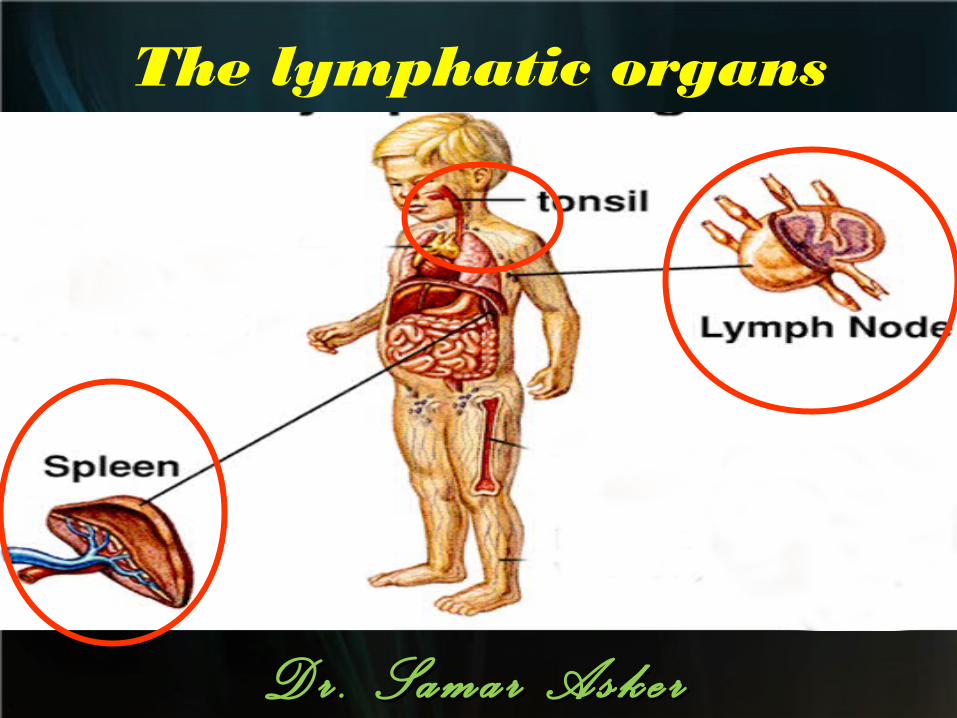
The lymphatic organs
D r . S a m a r A s k e rD r . S a m a r A s k e r
LYMPH NODE
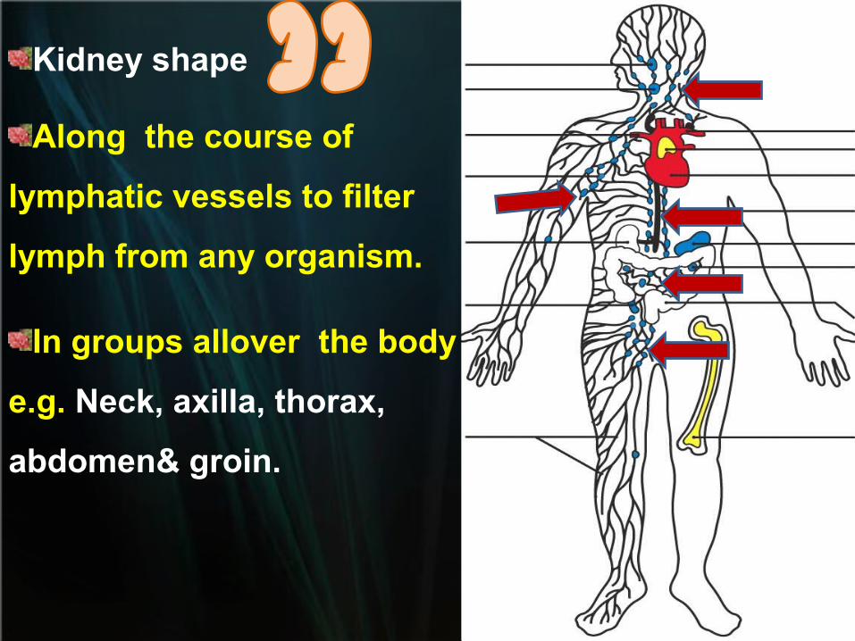
Kidney shape
Along the course of
lymphatic vessels to filter
lymph from any organism.
In groups allover the body
e.g. Neck, axilla, thorax,
abdomen& groin.
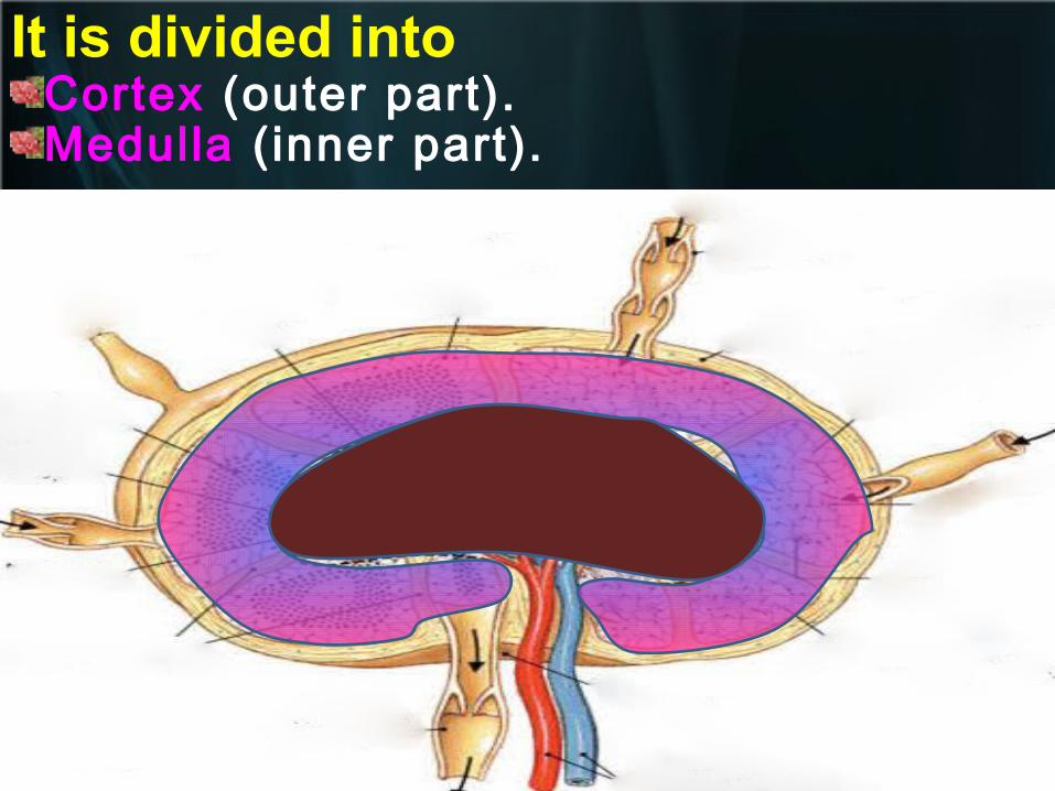
It is divided intoCortex (outer part).Medulla (inner part).
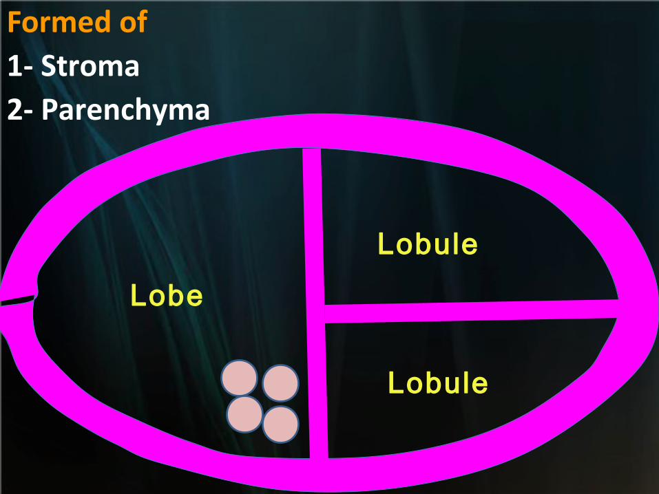
Formed of 1- Stroma 2- Parenchyma
Lobe
Lobule
Lobule
StromaStroma
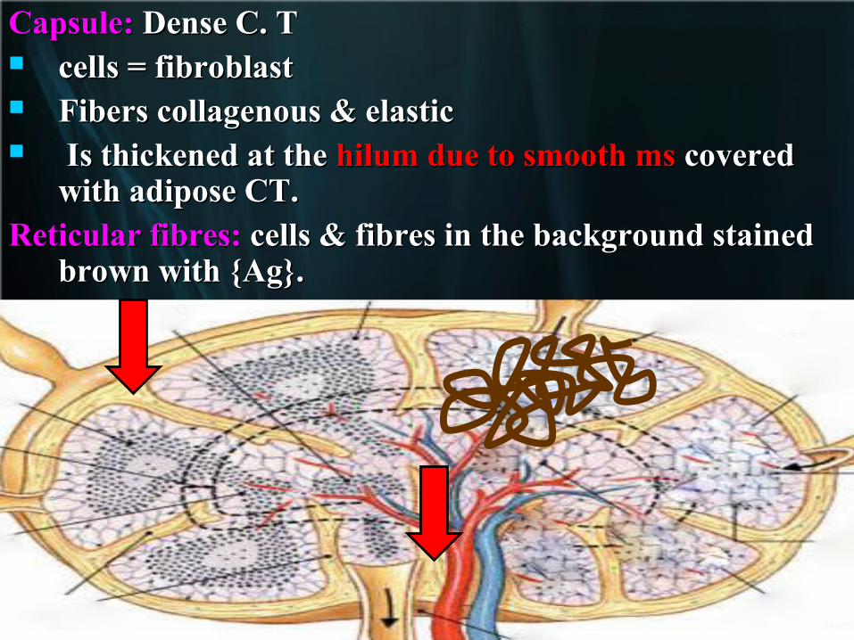
Capsule:Capsule: Dense C. T Dense C. T cells = fibroblast cells = fibroblast Fibers collagenous & elastic Fibers collagenous & elastic Is thickened at the Is thickened at the hilum due to smooth ms hilum due to smooth ms covered covered
with adipose CT.with adipose CT.Reticular fibres: Reticular fibres: cells & fibres in the background stained cells & fibres in the background stained
brown with {Ag}. brown with {Ag}.
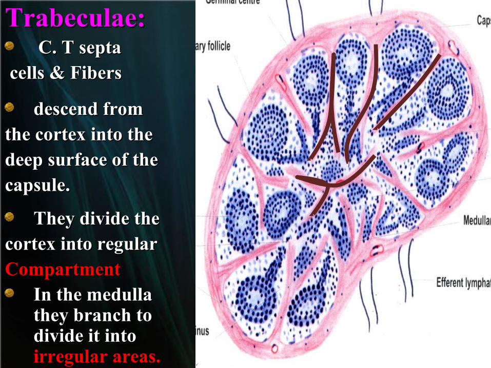
Trabeculae:Trabeculae: C. T septaC. T septa
cells & Fiberscells & Fibers
descend fromdescend fromthe cortex into thethe cortex into thedeep surface of thedeep surface of thecapsule. capsule.
They divide theThey divide thecortex into regularcortex into regularCompartmentCompartment
In the medulla In the medulla they branch to they branch to divide it into divide it into irregular areas. irregular areas.
ParENcHYMParENcHYMaa
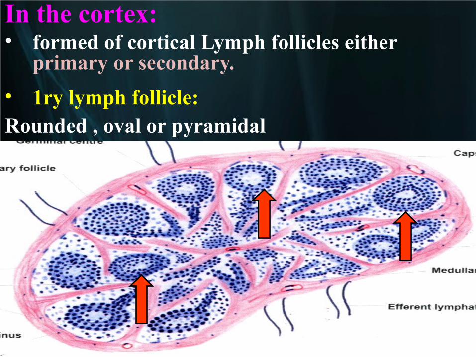
In the cortex: • formed of cortical Lymph follicles either
primary or secondary.
• 1ry lymph follicle:Rounded , oval or pyramidal
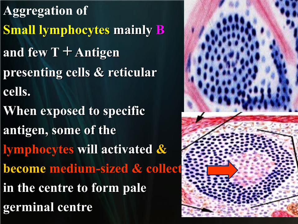
Aggregation of
Small lymphocytes lymphocytes mainly mainly BB
and few T and few T ++ Antigen Antigen
presenting cells & reticularpresenting cells & reticular
cells.cells.
When exposed to specificWhen exposed to specific
antigen, some of theantigen, some of the
lymphocyteslymphocytes will activated will activated & &
become become medium-sized & collect medium-sized & collect
in the centre to form palein the centre to form pale
germinal centregerminal centre
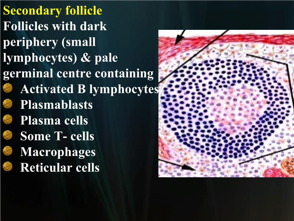
Secondary follicleFollicles with darkperiphery (smalllymphocytes) & palegerminal centre containing
Activated B lymphocytesPlasmablastsPlasma cellsSome T- cellsMacrophagesReticular cells
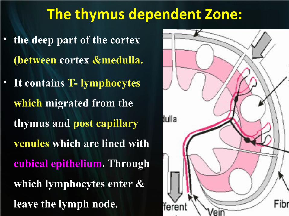
The thymus dependent Zone:
• the deep part of the cortex
(between cortex &medulla.
• It contains T- lymphocytes
which migrated from the
thymus and post capillary
venules which are lined with
cubical epithelium. Through
which lymphocytes enter &
leave the lymph node.
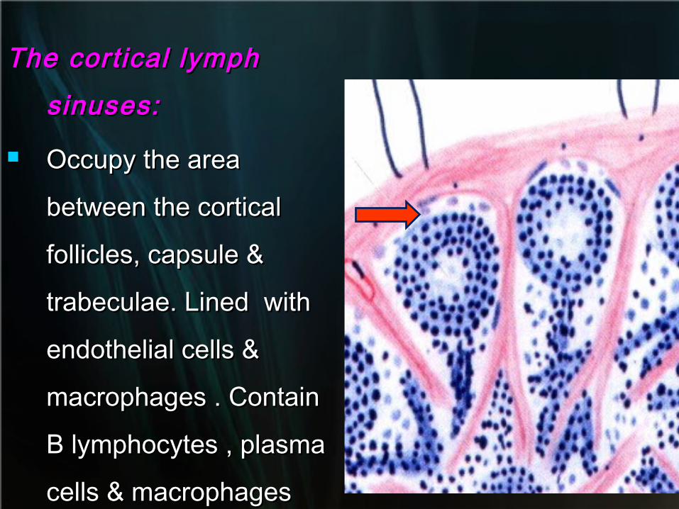
The cortical lymph The cortical lymph
sinuses:sinuses:
Occupy the area Occupy the area
between the cortical between the cortical
follicles, capsule & follicles, capsule &
trabeculae. Lined with trabeculae. Lined with
endothelial cells & endothelial cells &
macrophages . Contain macrophages . Contain
B lymphocytes , plasma B lymphocytes , plasma
cells & macrophagescells & macrophages
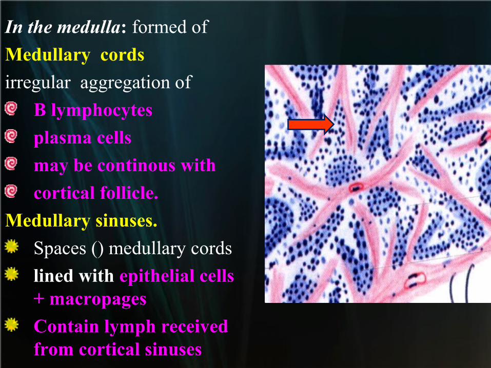
In the medulla: formed of
Medullary cords
irregular aggregation of
B lymphocytes
plasma cells
may be continous with
cortical follicle.
Medullary sinuses.
Spaces () medullary cords
lined with epithelial cells + macropages
Contain lymph received from cortical sinuses
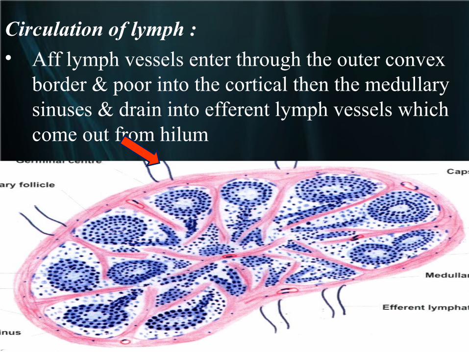
Circulation of lymph :
• Aff lymph vessels enter through the outer convex border & poor into the cortical then the medullary sinuses & drain into efferent lymph vessels which come out from hilum
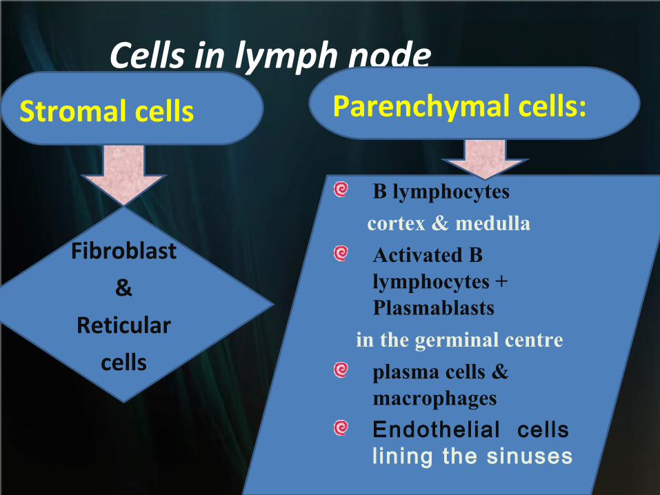
Cells in lymph nodeStromal cells Parenchymal cells:
Fibroblast
&
Reticular
cells
B lymphocytes
cortex & medulla
Activated B lymphocytes + Plasmablasts
in the germinal centre
plasma cells & macrophages
Endothelial cells l ining the sinuses
Function oF Lymph
node
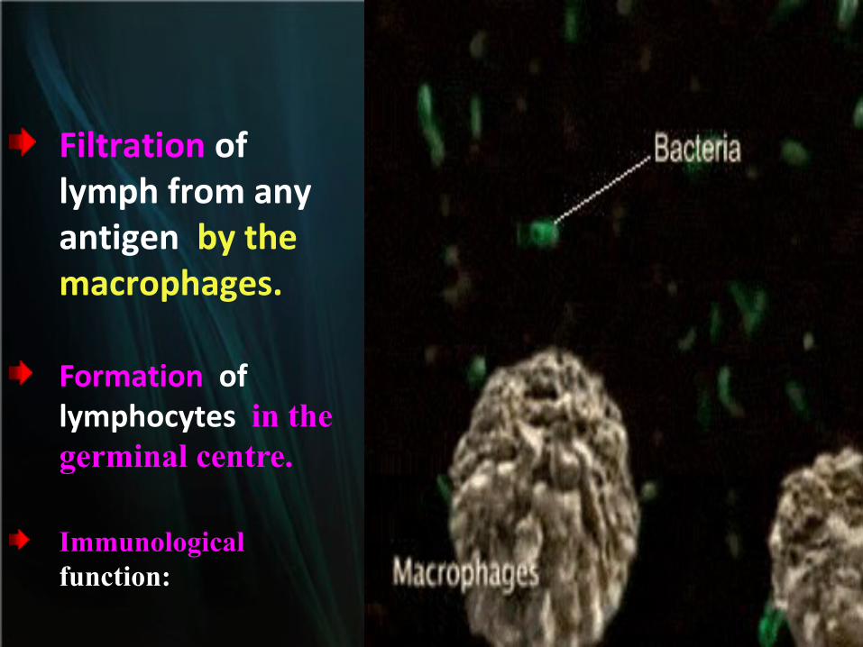
Filtration of lymph from any antigen by the macrophages.
Formation of lymphocytes in the germinal centre.
Immunological function:
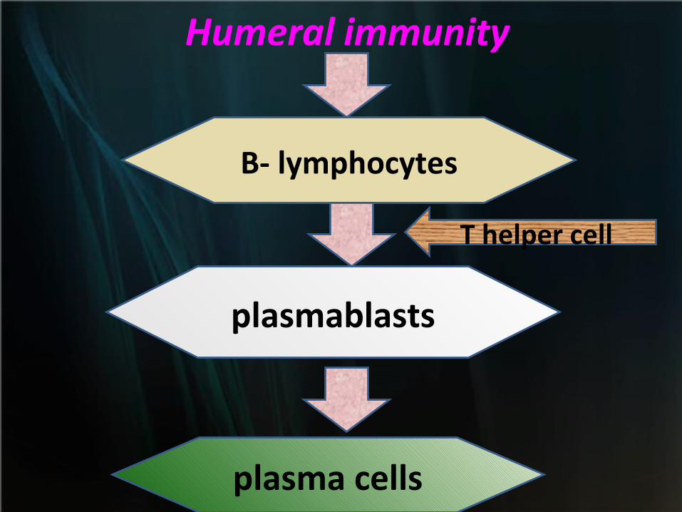
B- lymphocytes
plasmablasts
plasma cells
Humeral immunity
T helper cell
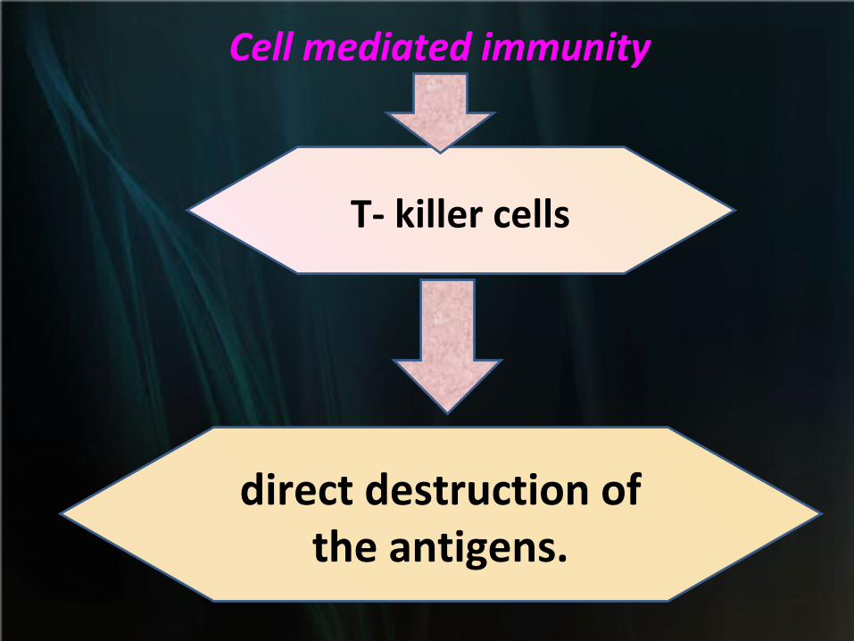
Cell mediated immunity
T- killer cells
direct destruction of the antigens.
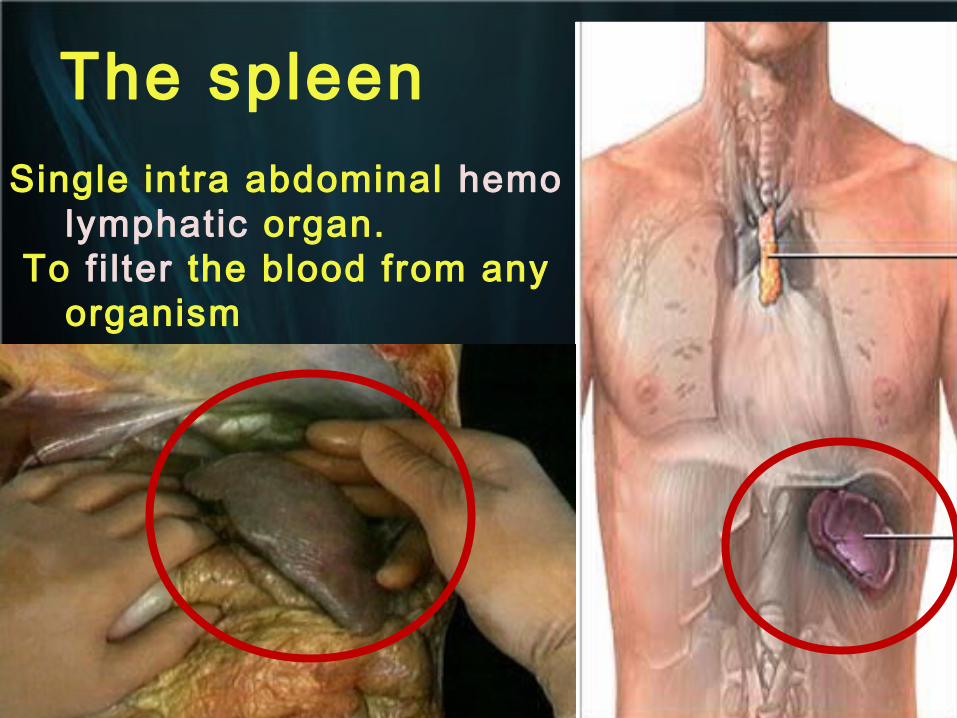
The spleenSingle intra abdominal hemo
lymphatic organ. To fi l ter the blood from any
organism
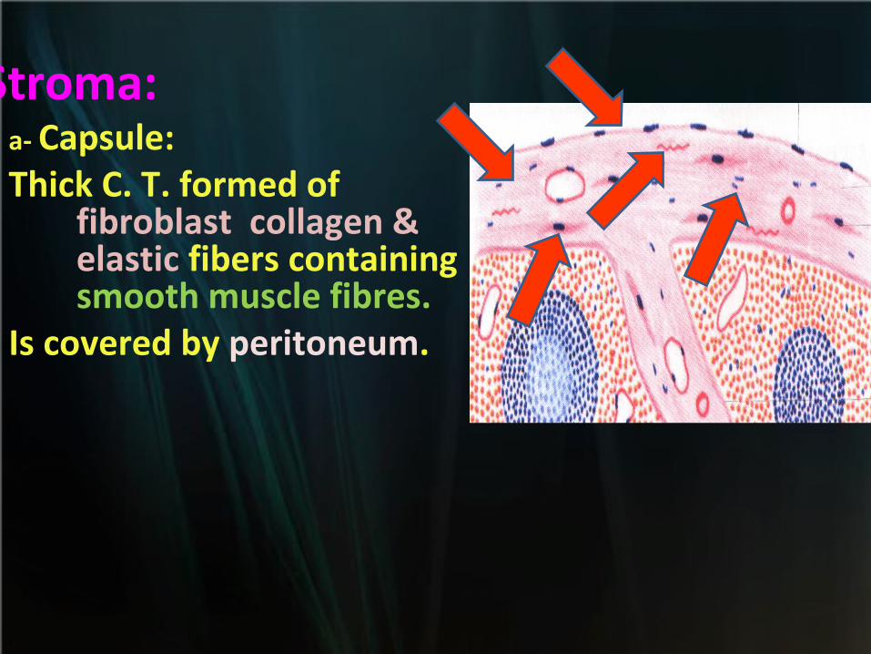
a- Capsule: Thick C. T. formed of
fibroblast collagen & elastic fibers containing smooth muscle fibres.
Is covered by peritoneum.
Stroma:
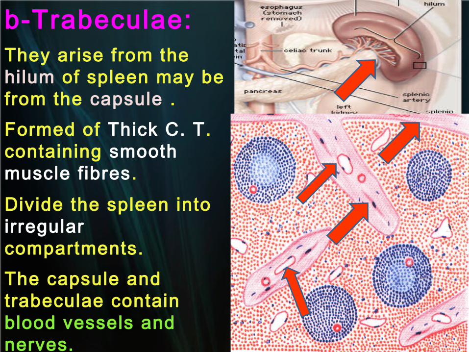
b-Trabeculae:They arise from the hilum of spleen may be from the capsule .Formed of Thick C. T. containing smooth muscle fibres.
Divide the spleen into irregular compartments.The capsule and trabeculae contain blood vessels and nerves.
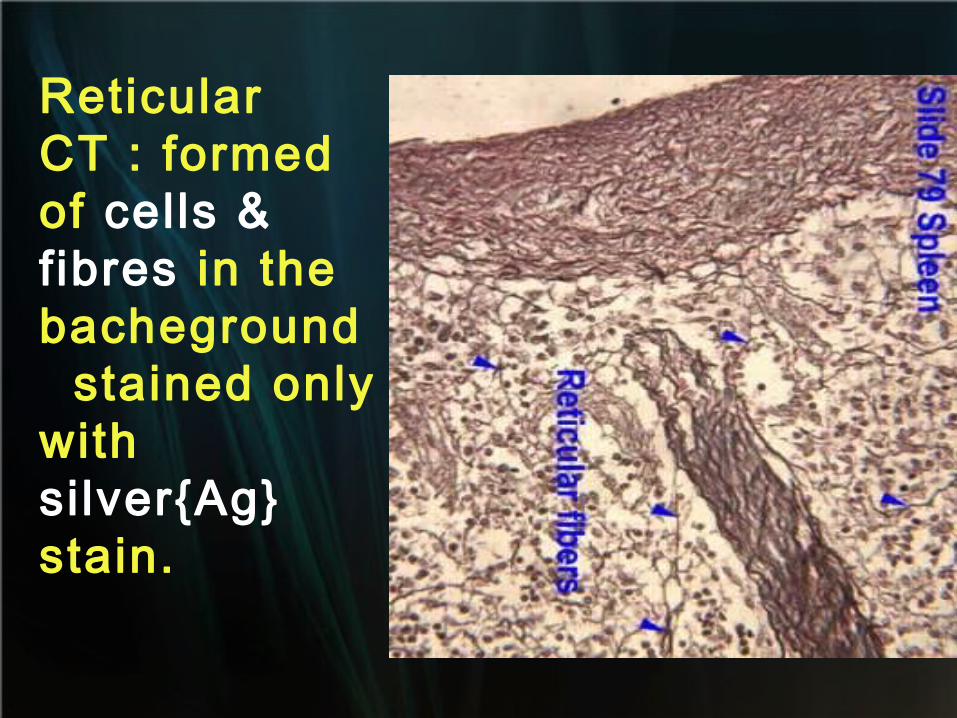
Reticular CT : formed of cells & fibres in the bacheground stained only with si lver{Ag} stain.
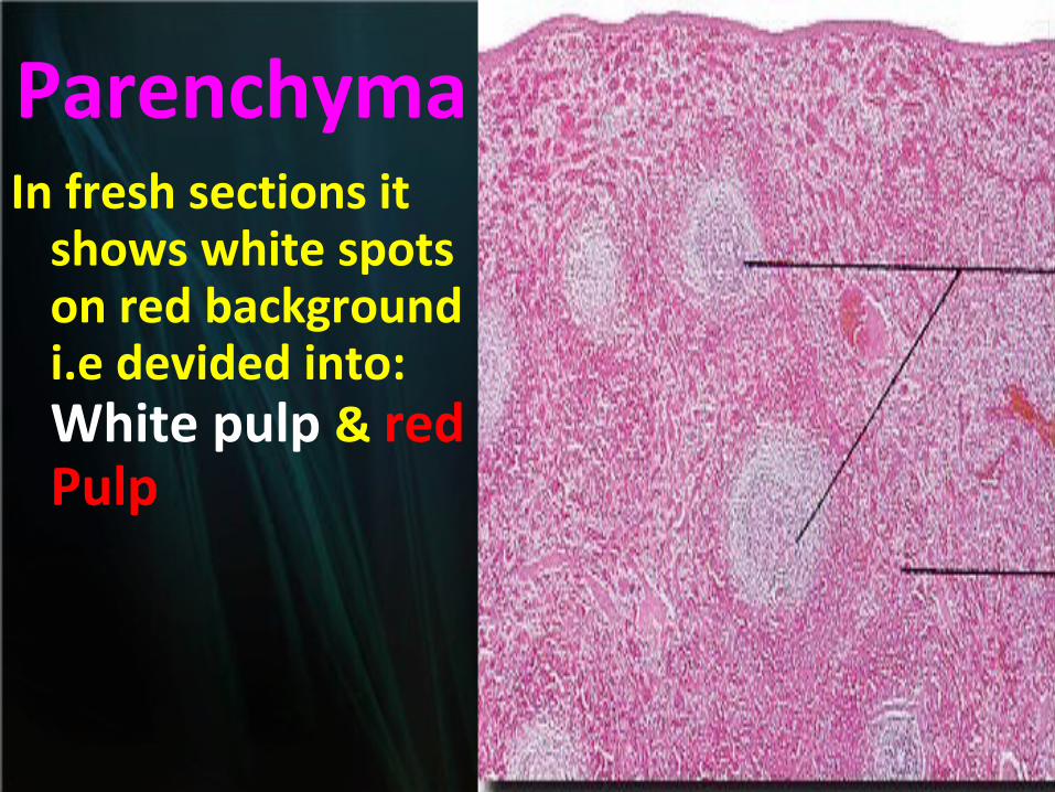
ParenchymaIn fresh sections it
shows white spots on red background i.e devided into: White pulp & red Pulp
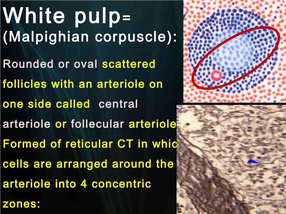
White pulp= (Malpighian corpuscle):
Rounded or oval scattered
foll icles with an arteriole on
one side called central
arteriole or follecular arteriole.
Formed of reticular CT in which
cells are arranged around the
arteriole into 4 concentric
zones:
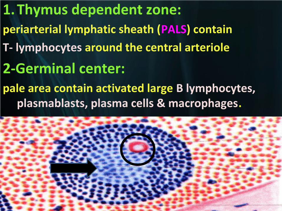
1. Thymus dependent zone: periarterial lymphatic sheath (PALS) contain
T- lymphocytes around the central arteriole
2-Germinal center: pale area contain activated large B lymphocytes,
plasmablasts, plasma cells & macrophages.
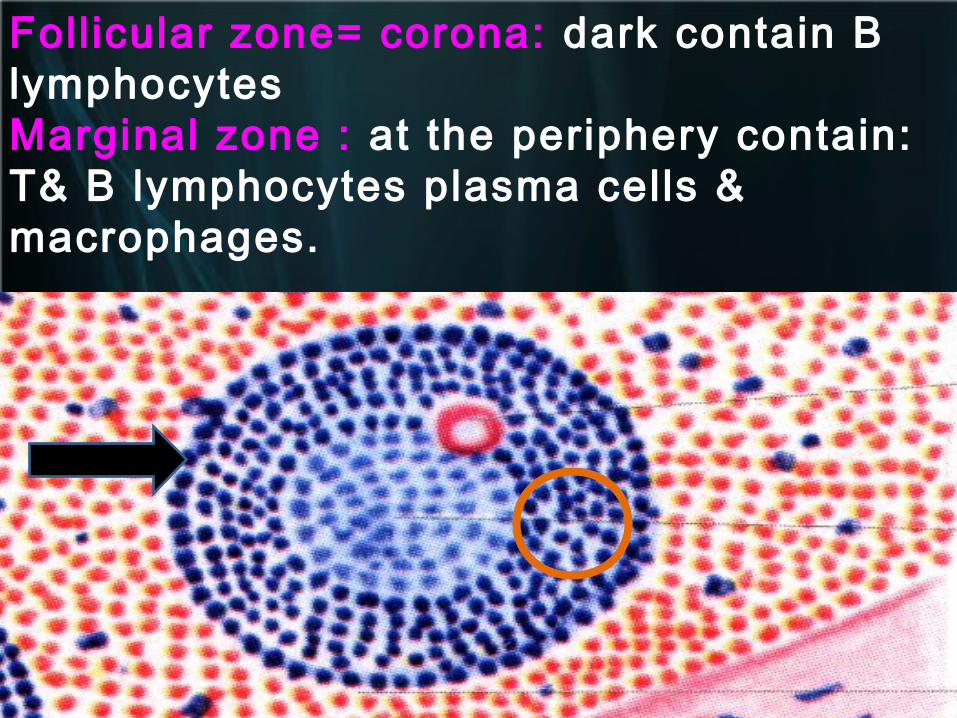
Foll icular zone= corona: dark contain B lymphocytesMarginal zone : at the periphery contain: T& B lymphocytes plasma cells & macrophages.
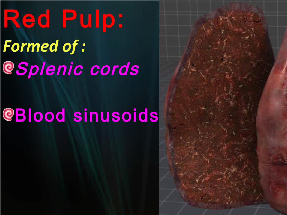
Red Pulp:Formed of :
Splenic cords
Blood sinusoids.
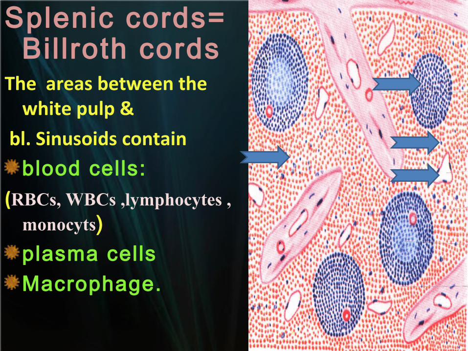
Splenic cords= Bil lroth cords
The areas between the white pulp &
bl. Sinusoids contain
blood cells:
(RBCs, WBCs ,lymphocytes , monocyts)plasma cellsMacrophage.
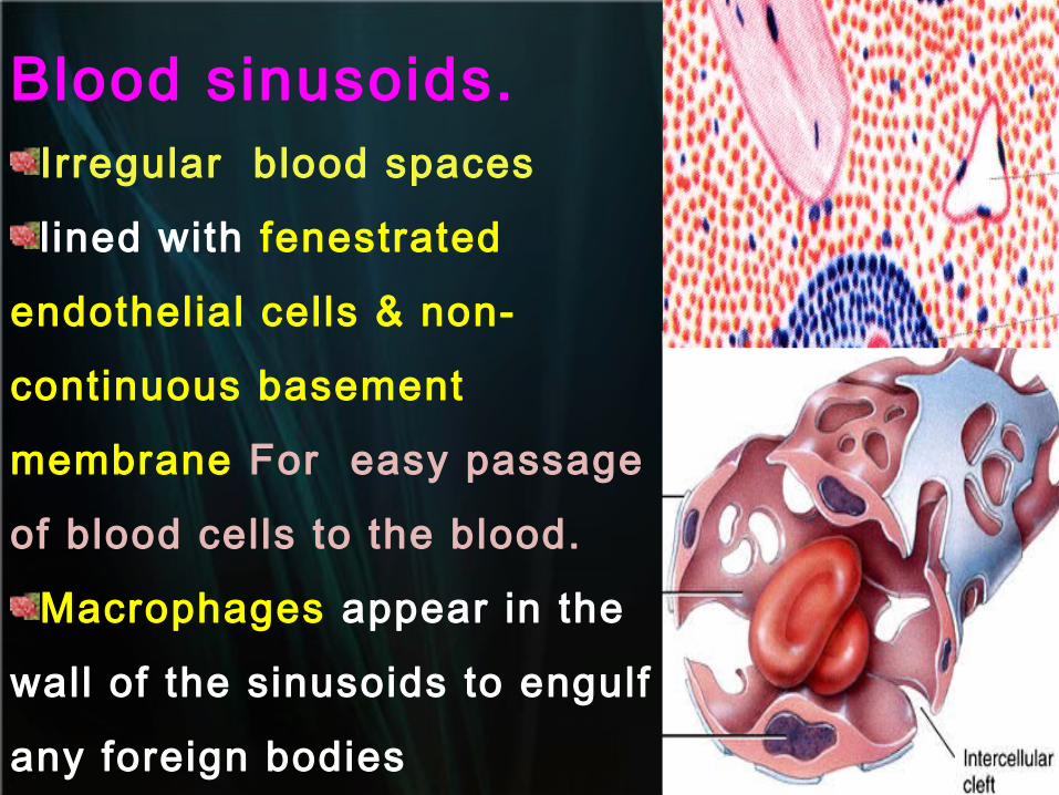
Blood sinusoids. Irregular blood spaces
lined with fenestrated
endothelial cells & non-
continuous basement
membrane For easy passage
of blood cells to the blood.
Macrophages appear in the
wall of the sinusoids to engulf
any foreign bodies
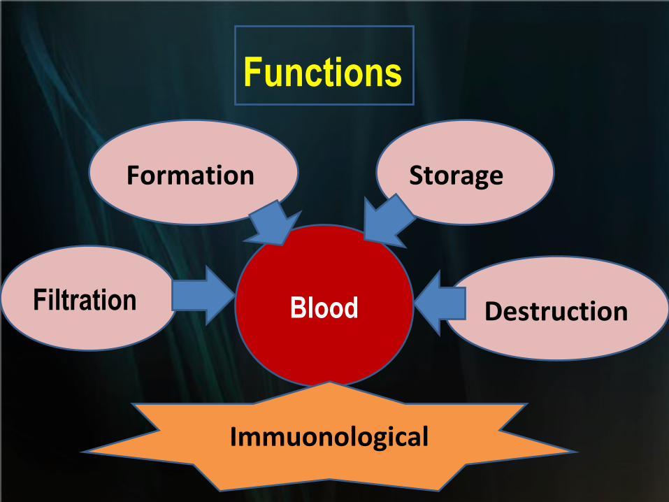
Filtration
Formation Storage
DestructionBlood
Functions
Immuonological
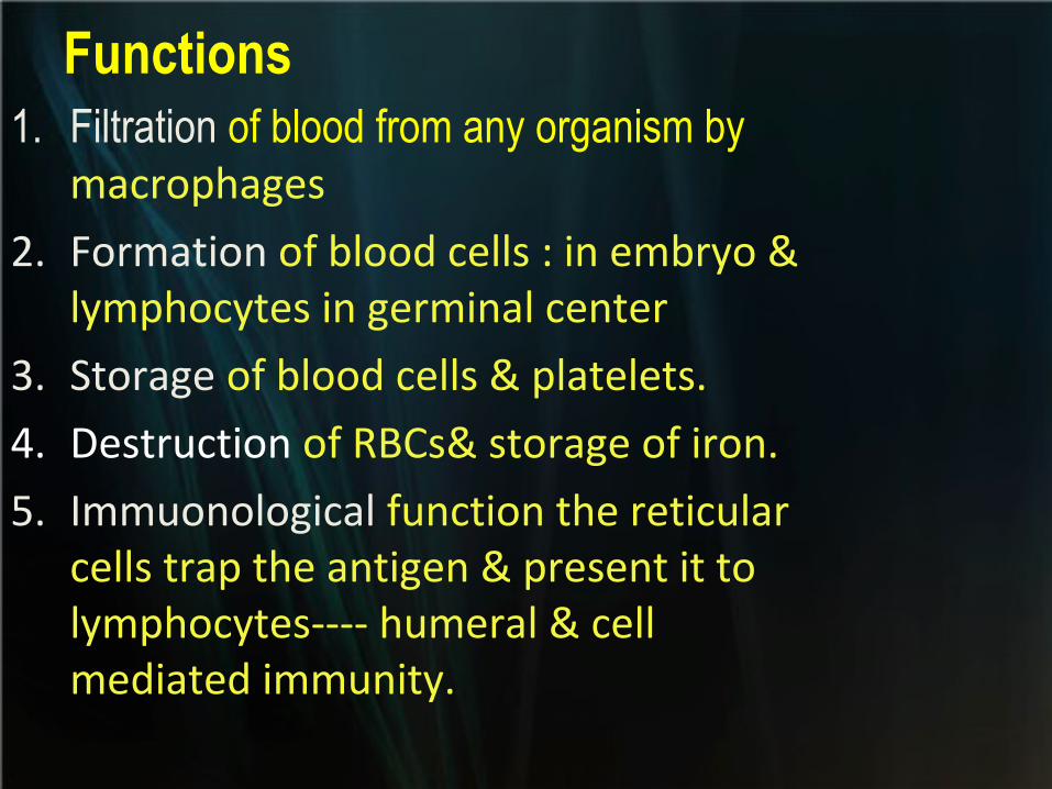
Functions1. Filtration of blood from any organism by
macrophages
2. Formation of blood cells : in embryo & lymphocytes in germinal center
3. Storage of blood cells & platelets.
4. Destruction of RBCs& storage of iron.
5. Immuonological function the reticular cells trap the antigen & present it to lymphocytes---- humeral & cell mediated immunity.
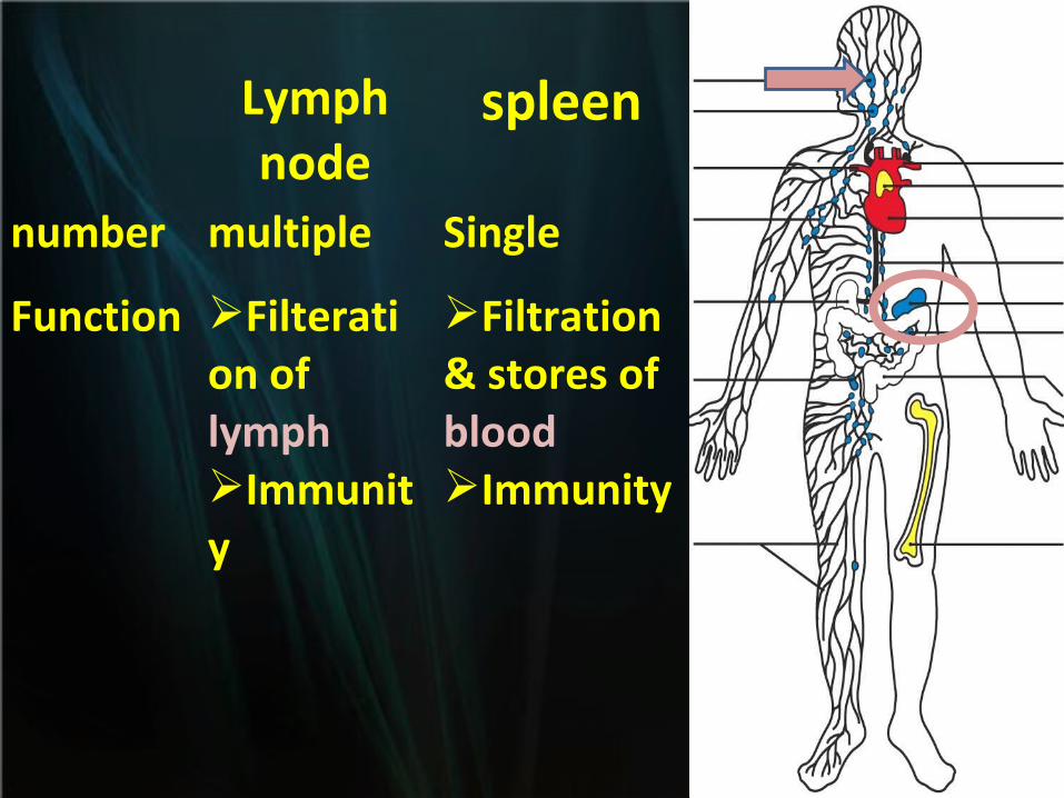
Lymph node
spleen
number multiple Single
Function Filteration of lymphImmunity
Filtration& stores of blood Immunity
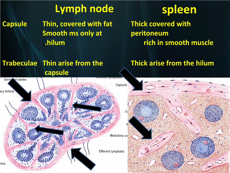
Lymph node spleenCapsule Thin, covered with fat
Smooth ms only at hilum.,
Thick covered with peritoneum
rich in smooth muscle
Trabeculae Thin arise from the capsule
Thick arise from the hilum
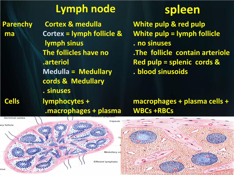
Lymph node spleenParenchyma
Cortex & medulla Cortex = lymph follicle & lymph sinus
The follicles have no arteriol.
Medulla = Medullary cords & Medullary
sinuses.
White pulp & red pulpWhite pulp = lymph follicle
no sinuses. The follicle contain arteriole.
Red pulp = splenic cords & blood sinusoids.
Cells lymphocytes + macrophages + plasma.
macrophages + plasma cells + WBCs +RBCs
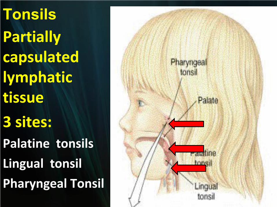
Tonsils
Partially capsulated lymphatic tissue
3 sites:Palatine tonsils
Lingual tonsil
Pharyngeal Tonsil
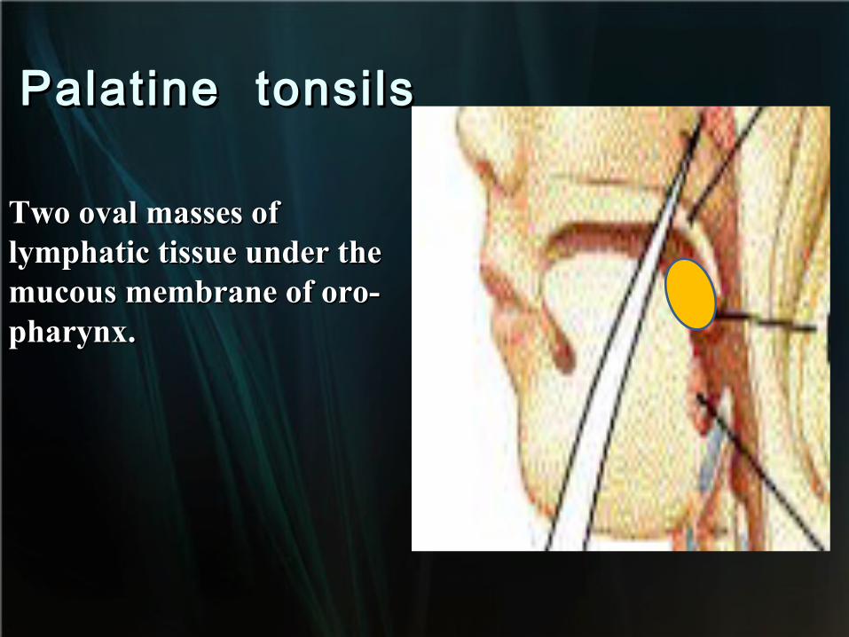
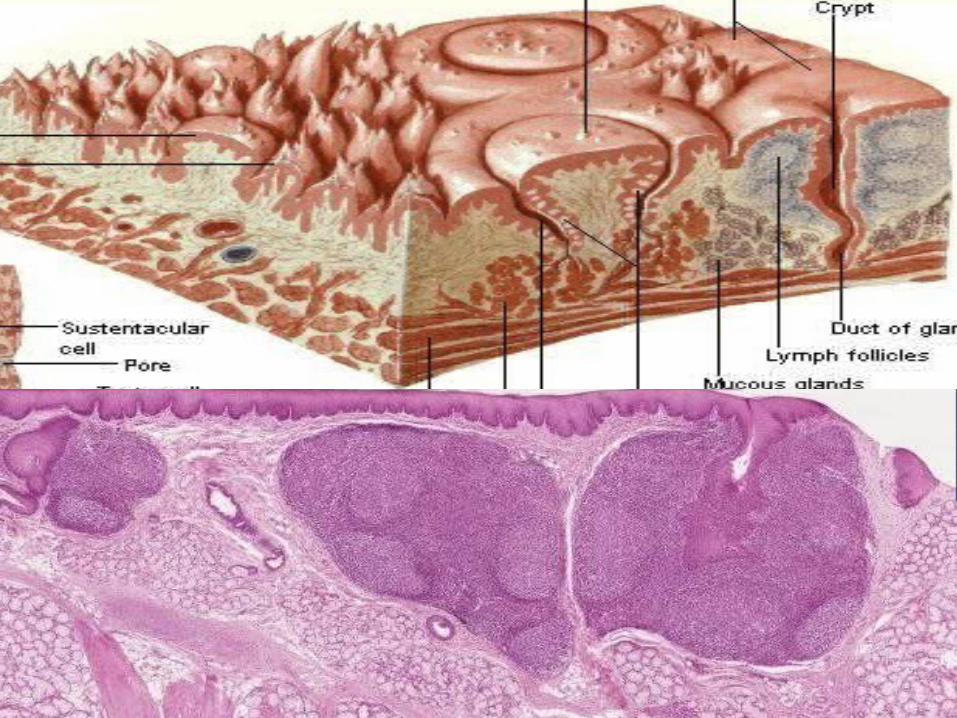
Palatine tonsilsPalatine tonsils
Two oval masses of Two oval masses of lymphatic tissue under the lymphatic tissue under the mucous membrane of oro-mucous membrane of oro-pharynx. pharynx.
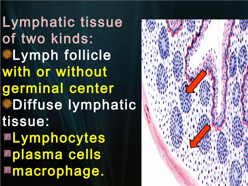
Lymphatic t issue of two kinds:
Lymph fol l icle with or without germinal center
Diffuse lymphatic t issue:
Lymphocytesplasma cellsmacrophage.
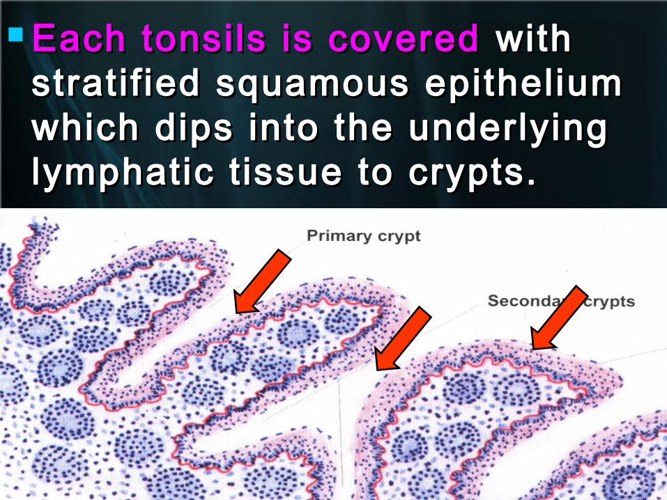
Each tonsils is coveredEach tonsils is covered with with stratif ied squamous epithelium stratif ied squamous epithelium which dips into the underlying which dips into the underlying lymphatic t issue to crypts. lymphatic t issue to crypts.
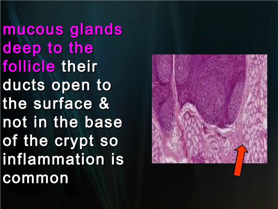
mucous glands mucous glands deep to the deep to the fol l icle fol l icle their their ducts open to ducts open to the surface & the surface & not in the base not in the base of the crypt so of the crypt so inflammation is inflammation is common common
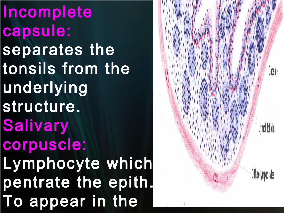
Incomplete capsule:separates the tonsils from the underlying structure.Salivary corpuscle:Lymphocyte which pentrate the epith. To appear in the saliva.
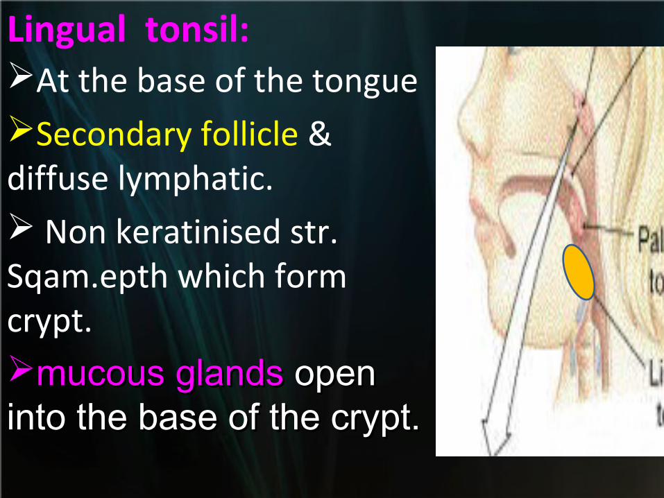
Lingual tonsil:At the base of the tongueSecondary follicle & diffuse lymphatic. Non keratinised str. Sqam.epth which form crypt.mucous glands mucous glands open open into the base of the crypt.into the base of the crypt.
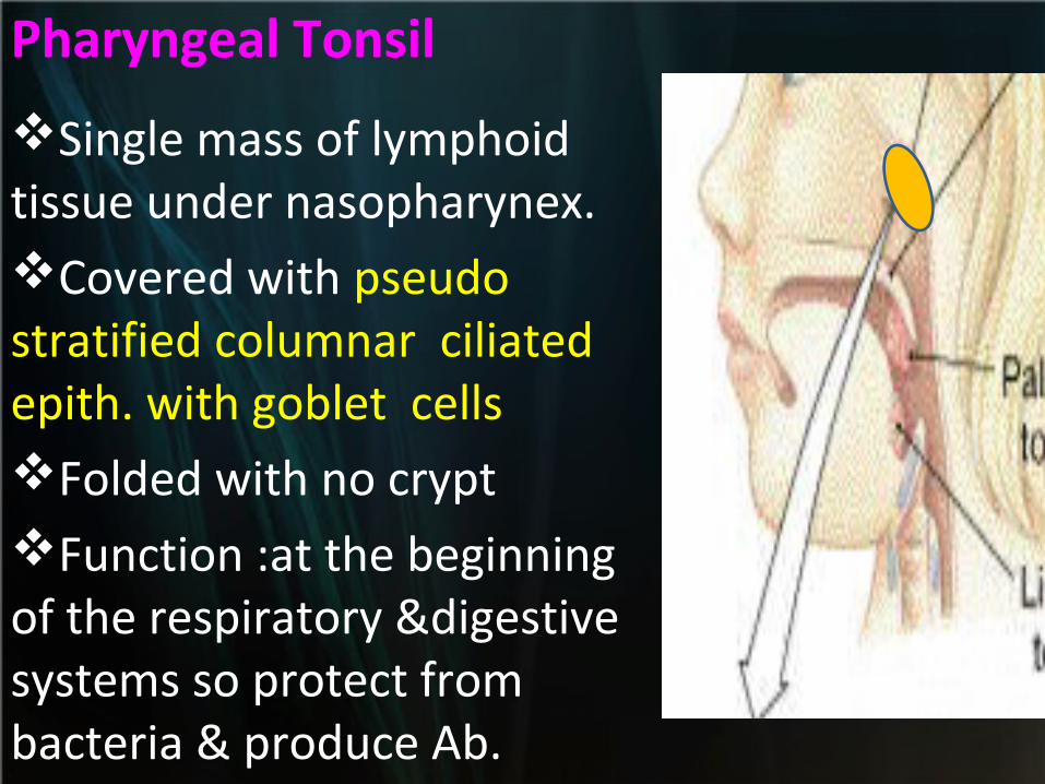
Pharyngeal Tonsil
Single mass of lymphoid tissue under nasopharynex.Covered with pseudo stratified columnar ciliated epith. with goblet cellsFolded with no cryptFunction :at the beginning of the respiratory &digestive systems so protect from bacteria & produce Ab.