Jennifer M. Grants,*,† Lisa T. L. Ying,† Akinori Yoda,‡,1 Charlotte C. You,† Hideyuki Okano,‡,§
Hitoshi Sawa,‡,** and Stefan Taubert*,†,2
*Department of Medical Genetics and †Centre for Molecular Medicine and Therapeutics, Child and Family Research Institute,University of British Columbia, Vancouver, British Columbia, V5Z 4H4, Canada, ‡Division of Neuroanatomy, Osaka University
Graduate School of Medicine, Osaka, 565-0871, Japan, §Department of Physiology, Keio University School of Medicine, Tokyo,108-8345, Japan, and **Multicellular Organization Laboratory, National Institute of Genetics, Mishima, 411-0801, Japan
ORCID ID: 0000-0002-2432-7257 (S.T.)
ABSTRACT Cell signaling pathways that control proliferation and determine cell fates are tightly regulated to prevent developmentalanomalies and cancer. Transcription factors and coregulators are important effectors of signaling pathway output, as they regulatedownstream gene programs. In Caenorhabditis elegans, several subunits of the Mediator transcriptional coregulator complex promoteor inhibit vulva development, but pertinent mechanisms are poorly defined. Here, we show that Mediator’s dissociable cyclin de-pendent kinase 8 (CDK8) module (CKM), consisting of cdk-8, cic-1/Cyclin C, mdt-12/dpy-22, and mdt-13/let-19, is required to inhibitectopic vulval cell fates downstream of the epidermal growth factor receptor (EGFR)-Ras-extracellular signal-regulated kinase (ERK)pathway. cdk-8 inhibits ectopic vulva formation by acting downstream ofmpk-1/ERK, cell autonomously in vulval cells, and in a kinase-dependent manner. We also provide evidence that the CKM acts as a corepressor for the Ets-family transcription factor LIN-1, as cdk-8promotes transcriptional repression by LIN-1. In addition, we find that CKM mutation alters Mediator subunit requirements in vulvadevelopment: the mdt-23/sur-2 subunit, which is required for vulva development in wild-type worms, is dispensable for ectopic vulvaformation in CKM mutants, which instead display hallmarks of unrestrained Mediator tail module activity. We propose a modelwhereby the CKM controls EGFR-Ras-ERK transcriptional output by corepressing LIN-1 and by fine tuning Mediator specificity, thusbalancing transcriptional repression vs. activation in a critical developmental signaling pathway. Collectively, these data offer anexplanation for CKM repression of EGFR signaling output and ectopic vulva formation and provide the first evidence of MediatorCKM-tail module subunit crosstalk in animals.
PRECISE regulation of transcription is required to executedevelopmental programs such as proliferation and cell
fate determination. The Mediator complex (“Mediator”) is a
conserved eukaryotic transcriptional coregulator of RNA poly-merase II (Pol II) transcription (Malik and Roeder 2010; Posset al. 2013). Mediator consists of �30 subunits that assembleinto fourmodules. “Core”Mediator consists of three of the fourmodules: the head and middle modules, which contact Pol II,and the tail module, which serves as a docking site for tran-scription factors. The fourth module, the dissociable cyclin de-pendent kinase 8 (CDK8) kinasemodule (CKM), interactswithtranscription factors, core Mediator, chromatin, and the Pol IImachinery to either repress or activate transcription (Malikand Roeder 2010; Nemet et al. 2014). Whereas many heador middle Mediator subunits are broadly required for Pol II
2Corresponding author: University of British Columbia, Dept. of Medical Genetics, 950W. 28th Ave., Room 3018, Vancouver, BC, Canada V5Z 4H4.E-mail: [email protected]
transcription, tail and CKM subunits regulate specific tran-scriptional programs in animal development or physiology(Malik and Roeder 2010; Nemet et al. 2014).
The CKM consists of enzymatic subunits CDK8 and cyclin C,and structural subunitsMED12andMED13that tether theCKMto core Mediator (Tsai et al. 2013). CKM subunits regulatemany transcriptional programs important for developmentand/or tumorigenesis, often by directly binding to and influ-encing the activity of key transcription factors (e.g., b-catenin,Notch, etc.) (Fryer et al. 2004; Donner et al. 2007; Firesteinet al. 2008; Zhou et al. 2012). Furthermore, in Saccharomycescerevisiae, the CKM regulates the activity of the Mediator tailmodule subunits MED2, MED3, and MED15 (van de Peppelet al. 2005; Gonzalez et al. 2014). However, whether suchintra-Mediator signaling effects occur in metazoans and affecte.g., animal development has not yet been tested.
Several Mediator subunits including at least one CKM sub-unit regulate vulva development in Caenorhabditis elegans(Tuck and Greenwald 1995; Singh and Han 1995; Kwonand Lee 2001; Moghal and Sternberg 2003a). The study ofcell fate specification in the C. elegans vulva has proven a pow-erful way to identify the components and regulatory interac-tions of several evolutionarily conserved signaling pathways(Félix and Barkoulas 2012; Schmid and Hajnal 2015). Thus,this organogenesis event provides an ideal paradigm to studyMediator subunit specificity and cooperation in a metazoan.
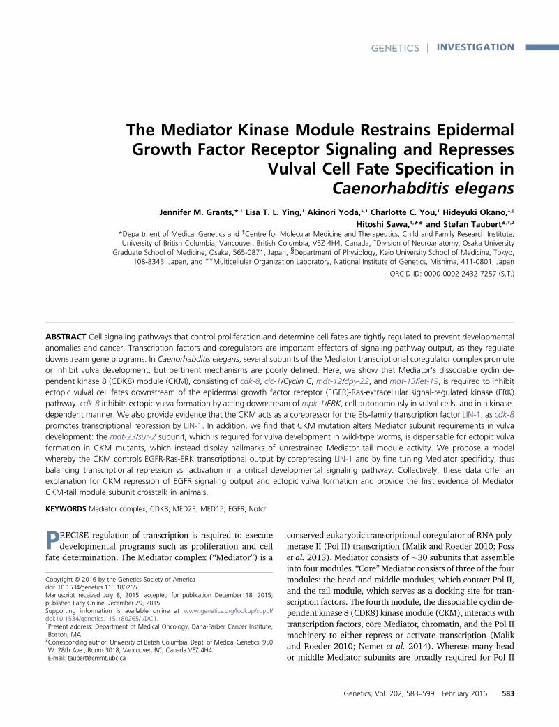
C. elegans vulval organogenesis is induced by epidermalgrowth factor receptor (EGFR) signaling (Moghal and Sternberg2003b), a prominent pathway in animal development thatis frequently activated in human cancers (Normanno et al.2006; Baselga and Swain 2009). The C. elegans vulva de-velops from six ventral vulva precursor cells (VPCs), namedP3.p through P8.p from anterior to posterior (Figure 1).The VPCs form an equivalence group, meaning that all sixcells are able to adopt the primary (1�) vulval cell fate (pro-ducing eight descendants), the secondary (2�) vulval cell fate(producing seven descendants), or the tertiary (3�) non-vulval fate (producing two descendants that fuse with thesurrounding hypodermis). A signaling cell in the somatic go-nad, called the anchor cell, emits a LIN-3/EGF-like ligand inclose proximity to P6.p (Hill and Sternberg 1992); therefore,LET-23/EGFR and the downstream LET-60/Ras, MPK-1/ex-tracellular signal-regulated kinase (ERK) cascade is stronglyactivated in P6.p (Aroian et al. 1990). MPK-1/ERK activationin P6.p modulates the activity of effector transcription factorssuch as the ELK1/Ets-family transcription factor LIN-1 andthe FoxB transcription factor LIN-31, thereby specifying the1� vulval fate in P6.p (Miller et al. 1993; Tan et al. 1998;Jacobs et al. 1998). The neighboring P5.p and P7.p cells arethought to receive a weaker LIN-3/EGF signal from the an-chor cell (Katz et al. 1995) as well as a lateral Notch signalemitted from the 1� cell P6.p, inducing them to adopt a 2�vulval fate (Chen and Greenwald 2004). Located furthestfrom the anchor cell, P3.p, P4.p, and P8.p do not receivesufficient EGF signal, and adopt the 3� nonvulval cell fate(Sternberg and Horvitz 1986). Mutations that enhance or
reduce EGFR or Notch signaling induce ectopic vulval cellfates (multivulva phenotype, Muv) or loss of vulval cell fates(vulvaless phenotype, Vul), respectively (Sternberg and Hor-vitz 1989). These phenotypes are thus powerful indicators ofEGFR and Notch signaling pathway activity.
Transcriptional regulation is important in maintainingappropriate EGFR signaling pathway output (Figure 1). Forexample, transcription factors such as LIN-1/Ets and LIN-31/Forkhead are required to repress 1� cell fate specification inVPCs other than P6.p (Miller et al. 1993; Beitel et al. 1995). Inaddition, multiple chromatin-modifying complexes, encodedby the synthetic multivulva (synMuv) genes, redundantly re-press ectopic lin-3/EGF transcription in the hypodermis andother tissues to inhibit 1� cell fate specification in VPCs otherthan P6.p (Myers and Greenwald 2005; Cui et al. 2006; Safferet al. 2011). Furthermore, theMediator subunitsmdt-23/sur-2, mdt-24/lin-25, and mdt-6 promote vulva development,whereas the CKM subunit mdt-12/dpy-22 inhibits vulva de-velopment in an anchor cell-independent manner (reviewedin Grants et al. 2015); for standardized Mediator subunitnomenclature, please see Bourbon et al. (2004). The mecha-nism by which mdt-23/sur-2 promotes vulva developmenthas been partially elucidated, as it is a critical coactivator ofa target gene downstream of the EGFR signaling pathway, thelag-2 Notch ligand gene (Zhang and Greenwald 2011). Thelin-1/Ets effector transcription factor is similarly required torepress the lag-2 gene (Zhang and Greenwald 2011), raisingthe question of whether and howMediator and LIN-1 interactto control common target genes. The other three Mediatorsubunits implicated in vulva development (mdt-6, mdt-12/dpy-22, and mdt-24/lin-25) interact genetically with compo-nents of the EGFR signaling pathway, but their mode of actionwithin this pathway remain poorly understood.
Here,weusedthevulvaorganogenesisparadigmtostudytherequirements of all four CKM subunits in this process and tointerrogate functional interactions with other transcriptionalregulators, including the synMuv genes, the key transcriptionfactors lin-1/Ets and lin-31/FoxB, and the Mediator subunitmdt-23/sur-2, an essential effector of EGFR signaling output.We show that all four CKM subunits inhibit ectopic vulval cellfates in C. elegans. We demonstrate that the CKM catalytic sub-unit cdk-8 acts downstream of let-23/EGFR and mpk-1/ERK inVPCs, in a kinase-dependent manner. Our data implicate cdk-8as a corepressor for the LIN-1/Ets repressive transcription fac-tor to inhibit EGFR signaling-induced transcription. Further-more, our data indicate that vulval induction in CKM mutantsis independent of the mdt-23/sur-2 coactivator, and insteadrequires the Mediator tail module subunits mdt-15, mdt-27,and mdt-29 for induction of ectopic vulval cell fates.
Materials and Methods
Microarrays and data analysis
Microarray gene expression profiling was performed at theUniversity of California San Francisco SABRE Functional
Genomics Facility. We used Agilent C. elegans (V2) 4x44KGene Expression Microarrays (G2519F-020186) and singlecolor labeling. Total RNA was extracted from developmen-tally synchronized mid-L4 stage worms as assessed by vulvalmorphology [wild-type N2 worms and cdk-8(tm1238) mu-tants], as described (Taubert et al. 2008). RNA quality wasassessed on an Agilent 2100 Bioanalyzer using a Pico Chip(Agilent). RNA was amplified and labeled with Cy3-CTPusing the Agilent low RNA input fluorescent linear amplifica-tion kit. Labeled cRNA was assessed using the NanodropND-100, and equal amounts of Cy3-labeled target were hy-bridized to the microarrays for 14 hr, according to the man-ufacturer’s protocol. Arrays were scanned using the Agilentmicroarray scanner and raw signal intensities were extractedwith Feature Extraction v9.1 software. The dataset was nor-malized using quantile normalization (Bolstad et al. 2003).No background subtraction was performed, and median fea-ture pixel intensity was used as raw signal before normali-zation. All arrays were of good quality and had similarforeground and background signal distributions for bothmessenger RNA (mRNA) and control probes. This suggeststhat quantile normalization is appropriate. To identify dif-ferentially expressed genes, a linear model was fit to thecomparison to estimate the mean M-values and calculatemoderated t-statistic, B-statistic, false discovery rate, andP-value for each gene. Adjusted P-values (AdjP) were producedas described (Holm 1979). All procedures were carried outusing functions in the R package limma in Bioconductor(Gentleman et al. 2004; Smyth 2004). Using this approach,we identified a total of 1860 spots with an AdjP of,0.05 anda fold change of $2 (representing 461 downregulated and829 upregulated genes) (Supporting Information, Table S1).Microarray data have been deposited in Gene ExpressionOmnibus (GSE68520).
Differentially expressed genes were compared to pub-lished gene expression datasets using EASE (Hosack et al.2003). For best comparison to our data, we reanalyzedpublished lin-35 data (Kirienko and Fay 2007) to definea set of genes deregulated twofold or more in L4 larvae,yielding 132 downregulated and 367 upregulatedgenes. We compared this set to our cdk-8 targets andcalculated the significance of the overlap using Fisher’sexact test.
C. elegans strains, culture, and genetic methods
C. elegans strains were cultured as described (Brenner 1974)at 20� or 23�, as indicated. We used nematode growth me-dium (NGM)-lite (0.2% NaCl, 0.4% tryptone, 0.3% KH2PO4,0.05% K2HPO4) agar plates seeded with Escherichia colistrain OP50 unless otherwise indicated.
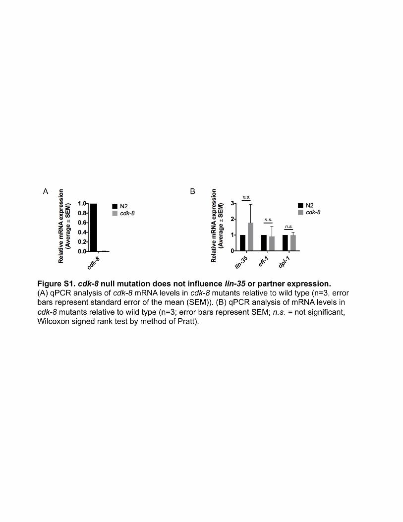
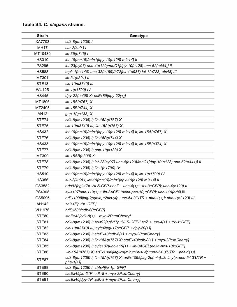
Strains are listed in Table S4. Wild type was Bristol N2.cdk-8(tm1238) and cic-1(tm3740) are likely null alleles thatabolish cdk-8 expression (Figure S1B) and cic-1 function, re-spectively (see also Steimel et al. 2013). For allele details, seewww.wormbase.org.mdt-12/dpy-22mutants were identifiedas Dpy, GFP-negative progeny of rescued dpy-22(os38);osEx89[dpy-22(+)] mothers, and homozygous mdt-13/let-19 mutants were identified as Dpy, GFP-negative progenyof balanced let-19(mn19)/mIn1 mothers.
VPC induction
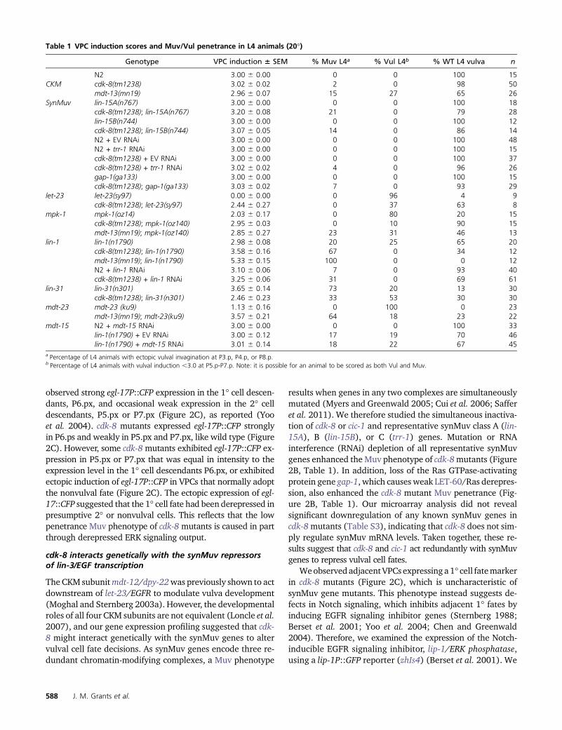
VPC induction was scored as described (Han et al. 1990), insynchronous mid-L4 animals under DIC optics at 31000magnification. In wild-type animals, P5.p–P7.p are inducedto give a VPC induction score of 3.0. In Vul animals, theseVPCs are not fully induced (VPC induction ,3.0); in Muvanimals, P3.p, P4.p, or P.8p are induced (VPC induction.3.0).
Figure 1 Transcriptional regulators in C. elegansvulval induction. The C. elegans vulva is derivedfrom an equivalence group consisting of six vulvaprecursor cells (VPCs), named P3.p through P8.pfrom anterior to posterior. A localized LIN-3/EGFsignal from the anchor cell (AC) in the somaticgonad activates a LET-23/EGFR-LET-60/Ras-MPK-1/ERK signaling cascade strongly in P6.p. ERKactivation in P6.p modulates transcription factoractivity in the nucleus (only LIN-1 is shown herefor simplicity), leading to induction of the 1� cellfate. In P5.p and P7.p, a weak LIN-3/EGF signalcombined with lateral Notch signaling from P6.p(not depicted) instead produces the 2� cell fate. InP3.p, P4.p, and P8.p, the EGFR signaling cascade isnot activated by LIN-3/EGF, and cells adopt thenonvulval 3� cell fate. Transcriptional regulatorsof the EGFR signaling pathway are critical for cor-rect vulval cell fate specification: e.g., the MDT-23/SUR-2 Mediator subunit is a coactivator of EGFRsignaling-induced transcription, the SynMuv core-pressor complexes are required to inhibit ectopic
lin-3/EGF transcription in the hypodermis (Hyp7) surrounding the VPCs, and the MDT-12/DPY-22 Mediator subunit is required to inhibit vulva devel-opment by mechanisms that remain unclear (dashed arrow). The GTPase activating protein GAP-1 that negatively regulates LET-60/Ras activity post-translationally is also shown.
Muv and Vul morphologies have been described (Horvitz andSulston 1980; Sulston and Horvitz 1981). To facilitate scoringa large number of worms to accurately assess low-penetrancephenotypes, Muv phenotype penetrance was scored in syn-chronous day 1 adult animals in a dissection microscope at3200 magnification (mdt-13/let-19 mutants) or 356 magni-fication (all other strains). To corroborate Muv penetrancesscored in adult animals, we also conducted VPC inductionanalysis in L4 animals (see above). To assess Vul phenotypes,both Vul and Muv penetrances were extrapolated from VPCinduction scores: animals were scored as Vul if VPC inductionwas ,3.0 in P5.p–P7.p and were scored as Muv if VPC induc-tion occurred in P3.p, P4.p or P8.p; using these criteria, ani-mals were occasionally scored as simultaneously Vul and Muv.
RNA isolation and quantitative real-time PCR
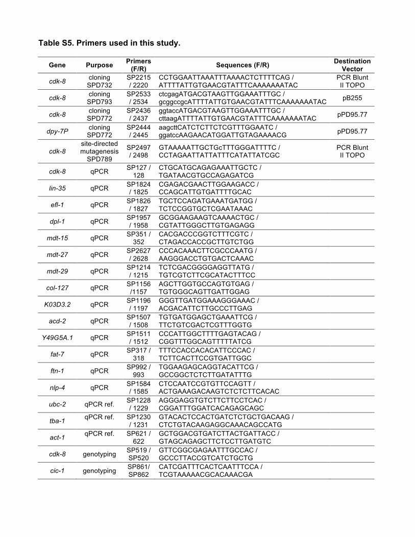
Total RNA was extracted from developmentally synchronizedmid-L4 stage worms as assessed by vulval morphology. RNAisolation and qPCR were performed as described (Goh et al.2014). We used t-tests (two-tailed, equal variance) to calcu-late statistical significance of gene expression changes be-tween mutants (Gaussian distribution). qPCR primers weredesigned with Primer3web (bioinfo.ut.ee/primer3/) and testedon serial cDNA dilutions to analyze PCR efficiency (primer se-quences in Table S5), except lin-3 (analyzed by TaqMan assay,Invitrogen 4448892, assay ID Ce02418781_m1).
Fluorescent reporter analysis
Synchronous worms were imaged using DIC optics and fluo-rescencemicroscopyonaZeissAxioplan2microscope.Analysisoffluorescence intensitywas conductedusing ImageJsoftware,normalizing for cell size and background fluorescence.
Generation of transgenic rescue strains
cdk-8 rescue transgenes (steEx43, 45–47) were generated bygonad microinjection of a mixture of 50 ng/ml rescue plasmid[cdk-8(+), SPD732; lin-31P::cdk-8, SPD793; dpy-7P::cdk-8,SPD772; cdk-8(KD), SPD789; 5 ng/ml pCFJ90[myo-2p::mCherry], and 95 ng/ml pPD95.77 empty vector into N2worms, then selecting transgenic mCherry-positive progeny.These were then crossed to cdk-8 and/or cdk-8; lin-15Amutants(Table S4). Cloning primer sequences are provided (Table S5).
Feeding RNAi knockdown
Feeding RNAi was performed as described (Goh et al. 2014),with the following modifications: synchronous mid-L4 her-maphrodites were allowed to lay eggs at 20� overnight onRNAi plates (Ahringer Library 96-well format; mdt-15: plate74, well C09; lin-1: 94, G02; Vidal Library 96-well format:mdt-27: GHR-11064@H02; mdt-29: GHR-11007@D05; allclones were sequenced to confirm identity; negative controlwas empty vector L4440), after which embryos were isolatedby bleach treatment and transferred to fresh RNAi plates. F1progeny were grown on RNAi plates (20� or 23�) until theyreached the desired developmental stage.
Western blot
Immunoblot using standard lysis, SDS/PAGE and Westernblot techniques was performed, with a-MDT-15 (Taubertet al. 2006) and a-GAPDH (Calbiochem, CB1001) antibodies,as described (Goh et al. 2014).
Data availability
Primer sequences are listed in Table S5. Strains are listedin Table S4 and are available upon request. Gene expres-sion data are available at GEO with the accession numberGSE68520.
Results
cdk-8-dependent transcripts overlap with targets of asynMuv gene
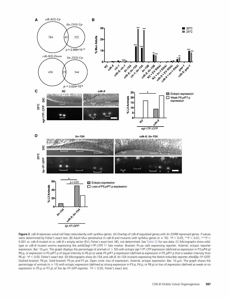
To define the role of the CKM in metazoan development, wecompared transcriptional profiles of developmentally syn-chronized L4 larval stage cdk-8(tm1238) null mutants towild-type N2 worms using microarrays (see Materials andMethods and Figure S1A for cdk-8 mutant information). Wefound that 829 genes were upregulated and 461 genes weredownregulated more than twofold in cdk-8 null mutants,representing �6.7% of all C. elegans genes (Table S1). Toidentify cdk-8-dependent gene programs, we compared ourlists of cdk-8 regulated genes to other gene lists using EASE(Hosack et al. 2003; Engelmann et al. 2011). The top hitamong genes upregulated in cdk-8mutants was a set of genesupregulated in lin-35/Retinoblastoma (RB) synMuv genemu-tants (Kirienko and Fay 2007), and lin-35-repressed genesalso overlapped significantly with genes downregulated incdk-8 mutants (Figure 2A). Importantly, the mRNA levels oflin-35 and the efl-1/dpl-1 transcription factor heterodimerthat is repressed by LIN-35 were not altered in cdk-8mutants(Figure S1B), indicating that cdk-8 does not affect lin-35 tar-get gene expression by altering the abundance of lin-35 or itspartners. Together, these data suggest that cdk-8 could act inparallel to lin-35 as they regulate similar gene sets.
We next investigated whether cdk-8, like mdt-12/dpy-22(Moghal and Sternberg 2003a), also represses vulval induc-tion. Indeed, cdk-8 and cic-1(tm3740) null mutants displayeda low penetrance Muv phenotype, as measured both by VPCinduction analysis in L4 animals (Table 1) and by scoring theoccurrence of ectopic vulval protrusions in adult worms (Fig-ure 2B; see statistical comparisons between L4 and adult Muvscores, Table S2). cdk-8 and cic-1 appeared to function re-dundantly in vulva formation, as cdk-8; cic-1 double mutantsshowed no significant increase in Muv penetrance comparedto either singlemutant (Figure 2B).We then tested if the cdk-8mutant Muv phenotype was associated with ectopic EGFR sig-naling-induced 1� vulval cell fates using an egl-17P::CFP re-porter (arIs92) (Yoo et al. 2004). In wild-type worms, we
Figure 2 cdk-8 represses vulval cell fates redundantly with synMuv genes. (A) Overlap of cdk-8 regulated genes with lin-35/RB repressed genes. P valueswere determined by Fisher’s exact test. (B) Adult Muv penetrance in cdk-8 and mutants with synMuv genes (n $ 76). *P , 0.05, **P , 0.01, ***P ,0.001 vs. cdk-8 mutant or vs. cdk-8 + empty vector (EV), Fisher’s exact test. ND, not determined. See Table S2 for raw data. (C) Micrographs show wild-type or cdk-8 mutant worms expressing the arIs92[egl-17P::CFP] 1� fate marker. Bracket: Pn.px cells expressing reporter. Asterisk: ectopic reporterexpression. Bar: 10 mm. The graph displays the percentage of animals (n. 50) with ectopic egl-17P::CFP expression (defined as expression in P3.p/P4.p/P8.p, or expression in P5.p/P7.p of equal intensity to P6.p) or weak P5.p/P7.p expression (defined as expression in P5.p/P7.p that is weaker intensity thanP6.p). *P , 0.05, Fisher’s exact test. (D) Micrographs show lin-15A and cdk-8; lin-15A mutants expressing the Notch-inducible reporter zhIs4[lip-1P::GFP].Dashed bracket: P6.px. Solid bracket: P5.px and P7.px. Open circle: loss of expression. Asterisk: ectopic expression. Bar: 10 mm. The graph shows thepercentage of animals (n $ 13) with ectopic expression (defined as strong expression in P3.p, P4.p, or P8.p) or loss of expression (defined as weak or noexpression in P5.p or P7.p) of the lip-1P::GFP reporter. *P , 0.05, Fisher’s exact test.
observed strong egl-17P::CFP expression in the 1� cell descen-dants, P6.px, and occasional weak expression in the 2� celldescendants, P5.px or P7.px (Figure 2C), as reported (Yooet al. 2004). cdk-8 mutants expressed egl-17P::CFP stronglyin P6.ps and weakly in P5.px and P7.px, like wild type (Figure2C). However, some cdk-8mutants exhibited egl-17P::CFP ex-pression in P5.px or P7.px that was equal in intensity to theexpression level in the 1� cell descendants P6.px, or exhibitedectopic induction of egl-17P::CFP in VPCs that normally adoptthe nonvulval fate (Figure 2C). The ectopic expression of egl-17::CFP suggested that the 1� cell fate had been derepressed inpresumptive 2� or nonvulval cells. This reflects that the lowpenetrance Muv phenotype of cdk-8 mutants is caused in partthrough derepressed ERK signaling output.
cdk-8 interacts genetically with the synMuv repressorsof lin-3/EGF transcription
The CKM subunitmdt-12/dpy-22was previously shown to actdownstream of let-23/EGFR to modulate vulva development(Moghal and Sternberg 2003a). However, the developmentalroles of all four CKM subunits are not equivalent (Loncle et al.2007), and our gene expression profiling suggested that cdk-8 might interact genetically with the synMuv genes to altervulval cell fate decisions. As synMuv genes encode three re-dundant chromatin-modifying complexes, a Muv phenotype
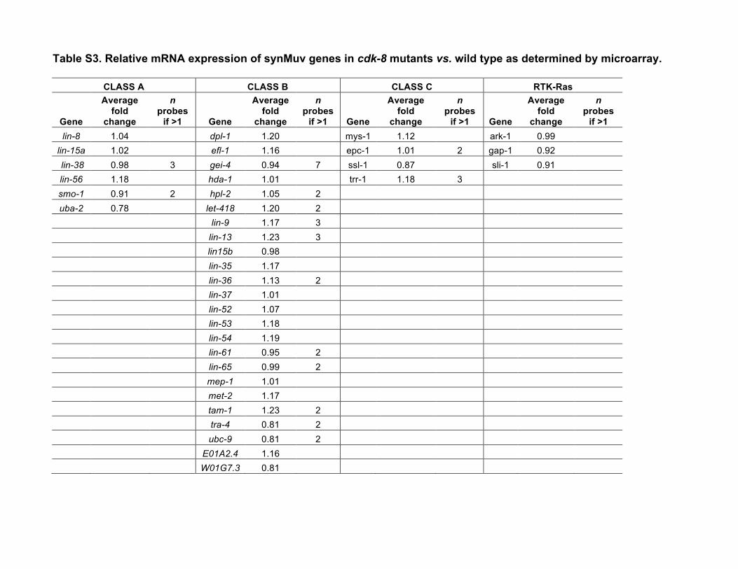
results when genes in any two complexes are simultaneouslymutated (Myers and Greenwald 2005; Cui et al. 2006; Safferet al. 2011). We therefore studied the simultaneous inactiva-tion of cdk-8 or cic-1 and representative synMuv class A (lin-15A), B (lin-15B), or C (trr-1) genes. Mutation or RNAinterference (RNAi) depletion of all representative synMuvgenes enhanced theMuv phenotype of cdk-8mutants (Figure2B, Table 1). In addition, loss of the Ras GTPase-activatingprotein gene gap-1, which causes weak LET-60/Ras derepres-sion, also enhanced the cdk-8 mutant Muv penetrance (Fig-ure 2B, Table 1). Our microarray analysis did not revealsignificant downregulation of any known synMuv genes incdk-8mutants (Table S3), indicating that cdk-8 does not sim-ply regulate synMuv mRNA levels. Taken together, these re-sults suggest that cdk-8 and cic-1 act redundantly with synMuvgenes to repress vulval cell fates.
WeobservedadjacentVPCs expressing a1� cell fatemarkerin cdk-8 mutants (Figure 2C), which is uncharacteristic ofsynMuv gene mutants. This phenotype instead suggests de-fects in Notch signaling, which inhibits adjacent 1� fates byinducing EGFR signaling inhibitor genes (Sternberg 1988;Berset et al. 2001; Yoo et al. 2004; Chen and Greenwald2004). Therefore, we examined the expression of the Notch-inducible EGFR signaling inhibitor, lip-1/ERK phosphatase,using a lip-1P::GFP reporter (zhIs4) (Berset et al. 2001). We
Table 1 VPC induction scores and Muv/Vul penetrance in L4 animals (20�)
Genotype VPC induction 6 SEM % Muv L4a % Vul L4b % WT L4 vulva n
a Percentage of L4 animals with ectopic vulval invagination at P3.p, P4.p, or P8.p.b Percentage of L4 animals with vulval induction ,3.0 at P5.p-P7.p. Note: it is possible for an animal to be scored as both Vul and Muv.
used the sensitized lin-15A mutant background to increasethe frequency of ectopic VPC induction events. lin-15A sin-gle mutants expressed lip-1P::GFP strongly in P5.px andP7.px, but expression was weak or absent in other Pn.pxcells (Figure 2D), as reported for wild-type worms (Bersetet al. 2001). In contrast, some cdk-8; lin-15A mutants loststrong lip-1P::GFP expression in P5.px and P7.px, consistentwith loss of the 2� fate (Figure 2D). Furthermore, some cdk-8;lin-15A mutants ectopically expressed lip-1P::GFP strongly innonvulval P3.px, P4.px, or P8.px, suggesting ectopic 2� fates(Figure 2D). Thus, cdk-8 mutants display hallmarks of bothdown- and upregulated Notch signaling, suggesting thatCDK-8 action on the Notch pathway may occur indirectlyvia the EGFR signaling pathway upstream.
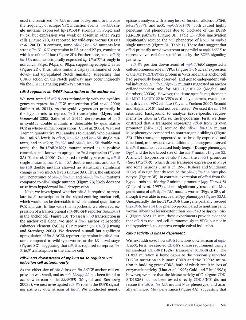
cdk-8 regulates lin-3/EGF transcription in the anchor cell
We next tested if cdk-8 acts redundantly with the synMuvgenes to repress lin-3/EGF transcription (Cui et al. 2006;Saffer et al. 2011). As the synMuv genes act primarily inthe hypodermis to repress lin-3 transcription (Myers andGreenwald 2005; Saffer et al. 2011), derepression of lin-3in synMuv double mutants is detectable by quantitativePCR in whole-animal preparations (Cui et al. 2006). We usedTaqman quantitative PCR analysis to quantify whole-animallin-3 mRNA levels in cdk-8, lin-15A, and lin-15B single mu-tants, and in cdk-8; lin-15A and cdk-8; lin-15B double mu-tants; the lin-15AB(n309) mutant served as a positivecontrol, as it is known to upregulate lin-3 expression (Figure3A) (Cui et al. 2006). Compared to wild-type worms, cdk-8single mutants, cdk-8; lin-15A double mutants, and cdk-8;lin-15B double mutants showed no statistically significantchange in lin-3mRNA levels (Figure 3A). Thus, the enhancedMuv penetrance of cdk-8; lin-15A and cdk-8; lin-15Bmutantscompared to cdk-8 single mutants (Figure 2B) likely does notarise from hypodermal lin-3 derepression.
Next, we investigated whether cdk-8 is required to regu-late lin-3 transcription in the signal-emitting anchor cell,which would not be detectable in whole-animal quantitativePCR analysis. In line with this hypothesis, we observed ex-pression of a transcriptional cdk-8P::GFP reporter (hdEx508)in the anchor cell (Figure 3B). To assess lin-3 transcription inthe anchor cell alone, we used a lin-3 anchor cell-specificenhancer element (ACEL) GFP reporter (syIs107) (Hwangand Sternberg 2004). We detected a small but significantupregulation of lin-3 ACEL reporter expression in cdk-8 mu-tants compared to wild-type worms at the L3 larval stage(Figure 3C), suggesting that cdk-8 is required to repress lin-3/EGF transcription in the anchor cell.
cdk-8 acts downstream of mpk-1/ERK to regulate VPCinduction cell autonomously
As the effect size of cdk-8 loss on lin-3/EGF anchor cell ex-pression was small, and asmdt-12/dpy-22 has been found toact downstream of let-23/EGFR (Moghal and Sternberg2003a), we next investigated cdk-8’s role in the EGFR signal-ing pathway downstream of lin-3. We conducted genetic
epistasis analyses with strong loss-of-function alleles of EGFR,let-23(sy97), and ERK, mpk-1(oz140); both caused highlypenetrant Vul phenotypes due to blockade of the EGFR-Ras-ERK pathway (Figure 3D, Table 1). cdk-8 inactivationsignificantly rescued the Vul phenotype of let-23 or mpk-1single mutants (Figure 3D, Table 1). These data suggest thatcdk-8 primarily acts downstream or parallel tompk-1/ERK torepress vulval cell fate specification by the EGFR signalingpathway.
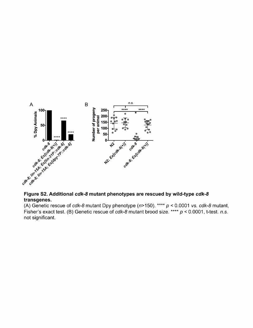
cdk-8’s position downstream of mpk-1/ERK suggested acell-autonomous role in VPCs (Figure 1). Nuclear expressionof theMDT-12/DPY-22 protein in VPCs and in the anchor cellhad previously been observed, and gonad-independent vul-val induction inmdt-12/dpy-22mutants suggested an anchorcell-independent role for MDT-12/DPY-22 (Moghal andSternberg 2003a). However, the tissue-specific requirementsfor MDT-12/DPY-22 in VPCs vs. the hypodermis, two impor-tant drivers of VPC cell fate (Fay and Yochem 2007; Schmidand Hajnal 2015), had not been tested. We used the lin-15Asensitized background to analyze tissue-specific require-ments for cdk-8 in VPCs vs. the hypodermis. First, we dem-onstrated that a transgene expressing cdk-8 from its ownpromoter [cdk-8(+)] rescued the cdk-8; lin-15A mutantMuv phenotype compared to nontransgenic siblings (Figure3E). This transgene appeared to be broadly expressed andfunctional, as it rescued two additional phenotypes observedin cdk-8mutants: decreased body length (Dumpy phenotype,Dpy) and the low brood size of the cdk-8 mutant (Figure S2,A and B). Expression of cdk-8 from the lin-31 promoter(lin-31P::cdk-8), which drives transgene expression in Pn.psand some neurons (Tan et al. 1998; Kishore and Sundaram2002), also significantly rescued the cdk-8; lin-15AMuv phe-notype (Figure 3E). In contrast, expression of cdk-8 from thehypodermis-specific dpy-7minimal promoter (dpy-7P::cdk-8)(Gilleard et al. 1997) did not significantly rescue the Muvpenetrance of cdk-8; lin-15A mutant worms (Figure 3E), al-though it was able to rescue the Dpy phenotype (Figure S2A).Unexpectedly, the lin-31P::cdk-8 transgene partially rescuedthe cdk-8; lin-15ADpy phenotype compared to nontransgenicworms, albeit to a lesser extent than cdk-8(+) or dpy-7P::cdk-8 (Figure S2A). In sum, these experiments provide evidencethat cdk-8 is required cell autonomously in VPCs but not inthe hypodermis to suppress ectopic vulval induction.
cdk-8 activity is kinase dependent
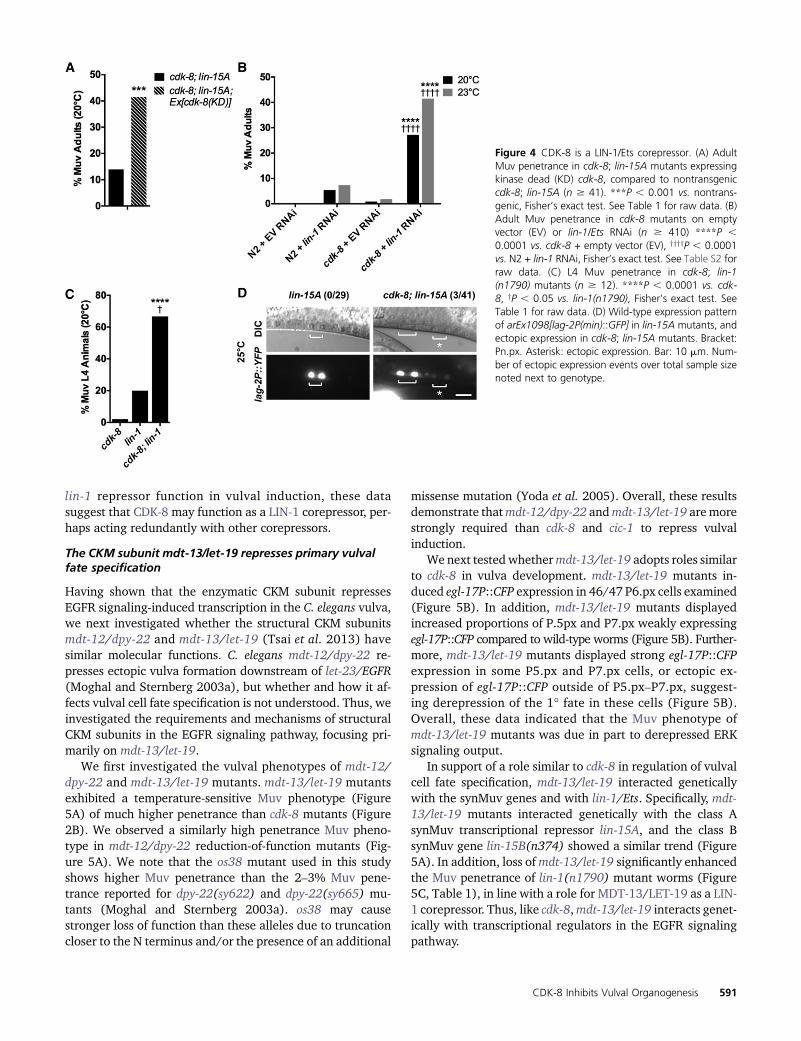
We next addressed how cdk-8 functions downstream ofmpk-1/ERK. First, we studied CDK-8’s kinase requirement using akinase-dead CDK-8(D182A) transgene [CDK-8(KD)]. TheD182A mutation is homologous to the previously reportedD173A mutation in human CDK8 and the D290A muta-tion in budding yeast CDK8, both of which result in loss ofenzymatic activity (Liao et al. 1995; Gold and Rice 1998);however, we note that the kinase activity of C. elegans CDK-8(D182A) has not been tested directly. CDK-8(KD) did notrescue the cdk-8; lin-15A mutant Muv phenotype, and actu-ally enhanced Muv penetrance (Figure 4A), suggesting that
kinase activity is required for transgenic rescue of the Muvphenotype of cdk-8 null mutants.
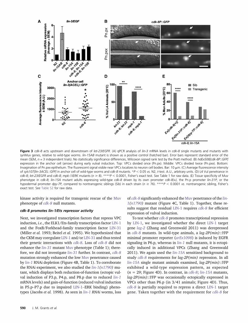
cdk-8 promotes lin-1/Ets repressor activity
Next, we investigated transcription factors that repress VPCinduction, i.e., the ELK1/Ets-family transcription factor LIN-1and the FoxB/Forkhead-family transcription factor LIN-31(Miller et al. 1993; Beitel et al. 1995). We hypothesized thatthe CKMmay coregulate LIN-1 and/or LIN-31 and thus testedtheir genetic interactions with cdk-8. Loss of cdk-8 did notenhance the lin-31 mutant Muv phenotype (Table 1); there-fore, we did not investigate lin-31 further. In contrast, cdk-8mutation strongly enhanced the low Muv penetrance causedby lin-1 RNAi depletion (Figure 4B, Table 1). To corroboratethe RNAi experiment, we also studied the lin-1(n1790) mu-tant, which displays both reduction-of-function (ectopic vul-val induction of P3.p, P4.p, and P8.p due to reduced lin-1mRNA levels) and gain-of-function (reduced vulval inductionin P5.p–P7.p due to impaired LIN-1–ERK binding) pheno-types (Jacobs et al. 1998). As seen in lin-1 RNAi worms, loss
of cdk-8 significantly enhanced theMuv penetrance of the lin-1(n1790) mutant (Figure 4C, Table 1). Together, these re-sults suggest that residual LIN-1 requires cdk-8 for efficientrepression of vulval induction.
To test whether cdk-8 promotes transcriptional repressionby LIN-1, we investigated whether the direct LIN-1 targetgene lag-2 (Zhang and Greenwald 2011) was derepressedin cdk-8 mutants. In wild-type animals, a lag-2P(min)::YFPminimal promoter reporter (arEx1098) is induced by EGFRsignaling in P6.p, whereas in lin-1 null mutants, it is ectopi-cally induced in additional VPCs (Zhang and Greenwald2011). We again used the lin-15A sensitized background tostudy cdk-8 requirements for lag-2P(min) repression. In alllin-15A single mutant animals examined, lag-2P(min)::YFPexhibited a wild-type expression pattern, as expected(n = 29; Figure 4D). In contrast, in cdk-8; lin-15A mutants,lag-2P(min)::YFP was occasionally ectopically expressed inVPCs other than P6.p (in 3/41 animals; Figure 4D). Thus,cdk-8 is partially required to repress a direct LIN-1 targetgene. Taken together with the requirement for cdk-8 for
Figure 3 cdk-8 acts upstream and downstream of let-23/EGFR. (A) qPCR analysis of lin-3 mRNA levels in cdk-8 single mutants and mutants withsynMuv genes, relative to wild-type worms. lin-15AB mutant is shown as a positive control (hatched bar). Error bars represent standard error of themean (SEM, n = 3 independent trials). No statistically significance differences, Wilcoxon signed rank test by the Pratt method. (B) hdEx508[cdk-8P::GFP]expression in the anchor cell (arrow) during early vulval induction. Top: VPCs divided once (Pn.px). Middle: VPCs divided twice (Pn.pxx). Bottom:Invagination of Pn.pxx epithelium. The fluorescent signal visible near VPCs localizes to neuron cell bodies. Bar: 10 mm. (C) Average fluorescence intensityof syIs107[lin-3ACEL::GFP] in anchor cell of wild-type worms and cdk-8 mutants. *P , 0.05 vs. N2, t-test. A.U., arbitrary units. (D) L4 Vul penetrance incdk-8; let-23/EGFR and cdk-8; mpk-1/ERK mutants (n $ 8). ****P , 0.0001, Fisher’s exact test. See Table 1 for raw data. (E) Tissue specificity of Muvphenotype in cdk-8; lin-15A mutant adults expressing wild-type cdk-8 driven by its own promoter cdk-8(+), the Pn.p promoter lin-31P, or thehypodermal promoter dpy-7P, compared to nontransgenic siblings (Sib) in each strain (n $ 76). ****P , 0.0001 vs. nontransgenic sibling, Fisher’sexact test. See Table S2 for raw data.
lin-1 repressor function in vulval induction, these datasuggest that CDK-8 may function as a LIN-1 corepressor, per-haps acting redundantly with other corepressors.
The CKM subunit mdt-13/let-19 represses primary vulvalfate specification
Having shown that the enzymatic CKM subunit repressesEGFR signaling-induced transcription in the C. elegans vulva,we next investigated whether the structural CKM subunitsmdt-12/dpy-22 and mdt-13/let-19 (Tsai et al. 2013) havesimilar molecular functions. C. elegans mdt-12/dpy-22 re-presses ectopic vulva formation downstream of let-23/EGFR(Moghal and Sternberg 2003a), but whether and how it af-fects vulval cell fate specification is not understood. Thus, weinvestigated the requirements and mechanisms of structuralCKM subunits in the EGFR signaling pathway, focusing pri-marily on mdt-13/let-19.
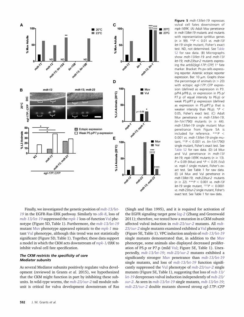
We first investigated the vulval phenotypes of mdt-12/dpy-22 and mdt-13/let-19 mutants. mdt-13/let-19 mutantsexhibited a temperature-sensitive Muv phenotype (Figure5A) of much higher penetrance than cdk-8 mutants (Figure2B). We observed a similarly high penetrance Muv pheno-type in mdt-12/dpy-22 reduction-of-function mutants (Fig-ure 5A). We note that the os38 mutant used in this studyshows higher Muv penetrance than the 2–3% Muv pene-trance reported for dpy-22(sy622) and dpy-22(sy665) mu-tants (Moghal and Sternberg 2003a). os38 may causestronger loss of function than these alleles due to truncationcloser to the N terminus and/or the presence of an additional
missense mutation (Yoda et al. 2005). Overall, these resultsdemonstrate thatmdt-12/dpy-22 andmdt-13/let-19 aremorestrongly required than cdk-8 and cic-1 to repress vulvalinduction.
We next tested whethermdt-13/let-19 adopts roles similarto cdk-8 in vulva development. mdt-13/let-19 mutants in-duced egl-17P::CFP expression in 46/47 P6.px cells examined(Figure 5B). In addition, mdt-13/let-19 mutants displayedincreased proportions of P.5px and P7.px weakly expressingegl-17P::CFP compared to wild-type worms (Figure 5B). Further-more, mdt-13/let-19 mutants displayed strong egl-17P::CFPexpression in some P5.px and P7.px cells, or ectopic ex-pression of egl-17P::CFP outside of P5.px–P7.px, suggest-ing derepression of the 1� fate in these cells (Figure 5B).Overall, these data indicated that the Muv phenotype ofmdt-13/let-19 mutants was due in part to derepressed ERKsignaling output.
In support of a role similar to cdk-8 in regulation of vulvalcell fate specification, mdt-13/let-19 interacted geneticallywith the synMuv genes and with lin-1/Ets. Specifically, mdt-13/let-19 mutants interacted genetically with the class AsynMuv transcriptional repressor lin-15A, and the class BsynMuv gene lin-15B(n374) showed a similar trend (Figure5A). In addition, loss ofmdt-13/let-19 significantly enhancedthe Muv penetrance of lin-1(n1790) mutant worms (Figure5C, Table 1), in line with a role for MDT-13/LET-19 as a LIN-1 corepressor. Thus, like cdk-8,mdt-13/let-19 interacts genet-ically with transcriptional regulators in the EGFR signalingpathway.
Figure 4 CDK-8 is a LIN-1/Ets corepressor. (A) AdultMuv penetrance in cdk-8; lin-15A mutants expressingkinase dead (KD) cdk-8, compared to nontransgeniccdk-8; lin-15A (n $ 41). ***P , 0.001 vs. nontrans-genic, Fisher’s exact test. See Table 1 for raw data. (B)Adult Muv penetrance in cdk-8 mutants on emptyvector (EV) or lin-1/Ets RNAi (n $ 410) ****P ,0.0001 vs. cdk-8 + empty vector (EV), yyyyP , 0.0001vs. N2 + lin-1 RNAi, Fisher’s exact test. See Table S2 forraw data. (C) L4 Muv penetrance in cdk-8; lin-1(n1790) mutants (n $ 12). ****P , 0.0001 vs. cdk-8, yP , 0.05 vs. lin-1(n1790), Fisher’s exact test. SeeTable 1 for raw data. (D) Wild-type expression patternof arEx1098[lag-2P(min)::GFP] in lin-15A mutants, andectopic expression in cdk-8; lin-15A mutants. Bracket:Pn.px. Asterisk: ectopic expression. Bar: 10 mm. Num-ber of ectopic expression events over total sample sizenoted next to genotype.
Finally, we investigated the genetic position ofmdt-13/let-19 in the EGFR-Ras-ERK pathway. Similarly to cdk-8, loss ofmdt-13/let-19 suppressed thempk-1 loss-of-function Vul phe-notype (Figure 5D, Table 1). Furthermore, themdt-13/let-19mutant Muv phenotype appeared epistatic to the mpk-1 mu-tant Vul phenotype, although this trend was not statisticallysignificant (Figure 5D, Table 1). Together, these data supporta model in which the CKM acts downstream ofmpk-1/ERK toinhibit vulval cell fate specification.
The CKM restricts the specificity of coreMediator subunits
As several Mediator subunits positively regulate vulva devel-opment (reviewed in Grants et al. 2015), we hypothesizedthat the CKM might function in part by inhibiting these sub-units. In wild-type worms, themdt-23/sur-2 tail module sub-unit is critical for vulva development downstream of Ras
(Singh and Han 1995), and it is required for activation ofthe EGFR signaling target gene lag-2 (Zhang and Greenwald2011); therefore, we tested how amutation in a CKM subunitaffected vulval induction in mdt-23/sur-2 mutants. All mdt-23/sur-2 single mutants examined exhibited a Vul phenotype(Figure 5E, Table 1). VPC induction analysis ofmdt-13/let-19single mutants demonstrated that, in addition to the Muvphenotype, some animals also displayed decreased prolifer-ation of P5.p or P7.p (mild Vul; Figure 5E, Table 1). Unex-pectedly, mdt-13/let-19; mdt-23/sur-2 mutants exhibited asignificantly stronger Muv penetrance than mdt-13/let-19single mutants, and loss of mdt-13/let-19 function signifi-cantly suppressed the Vul phenotype of mdt-23/sur-2 singlemutants (Figure 5E, Table 1), suggesting that loss ofmdt-13/let-19 derepresses vulval induction independently ofmdt-23/sur-2. As seen inmdt-13/let-19 single mutants, mdt-13/let-19;mdt-23/sur-2 double mutants showed strong egl-17P::CFP
Figure 5 mdt-13/let-19 repressesvulval cell fates downstream ofmpk-1/ERK. (A) Adult Muv penetrancein mdt-13/let-19 mutants and mutantswith representative synMuv genes(n $ 99). **P , 0.01 vs. mdt-13/let-19 single mutant, Fisher’s exacttest. ND, not determined. See TableS2 for raw data. (B) Micrographsshow mdt-13/let-19 and mdt-13/let-19; mdt-23/sur-2 mutants express-ing the arIs92[egl-17P::CFP] 1� fatemarker. Bracket: Pn.px cells express-ing reporter. Asterisk: ectopic reporterexpression. Bar: 10 mm. Graphs showthe percentage of animals (n . 20)with ectopic egl-17P::CFP expres-sion (defined as expression in P3.p/P4.p/P8.p, or expression in P5.p/P7.p of equal intensity to P6.p) orweak P5.p/P7.p expression (definedas expression in P5.p/P7.p that isweaker intensity than P6.p). *P ,0.05, Fisher’s exact test. (C) AdultMuv penetrance in mdt-13/let-19;lin-1(n1790) mutants (n $ 44).mdt-13/let-19 single mutant Muvpenetrance from Figure 5A isincluded for reference. ***P ,0.001 vs. mdt-13/let-19 single mu-tant, yyyP , 0.001 vs. lin-1(n1790)single mutant, Fisher’s exact test. SeeTable S2 for raw data. (D) L4 Muvand Vul penetrance in mdt-13/let-19; mpk-1/ERK mutants (n $ 13).P = 0.09 (Muv) and *P , 0.05 (Vul)vs. mpk-1 single mutant, Fisher’s ex-act test. See Table 1 for raw data.(E) L4 Muv and Vul penetrance inmdt-13/let-19; mdt-23/sur-2 mutants(n $ 22). ***P , 0.001 vs. mdt-13/let-19 single mutant, yyyyP , 0.0001vs. mdt-23/sur-2 single mutant, Fisher’sexact test. See Table 1 for raw data.
expression in some P5.px and P7.px cells, or ectopic expres-sion of egl-17P::CFP outside of P5.px–P7.px, suggesting de-repression of the 1� fate (Figure 5B). Overall, these dataindicated that the Muv phenotype of mdt-13/let-19; mdt-23/sur-2 double mutants was due in part to derepressed ERKsignaling output. Together, these findings indicate that lossof the CKM allows activation of EGFR signaling-driven cellfate specification independently of mdt-23/sur-2 activity.
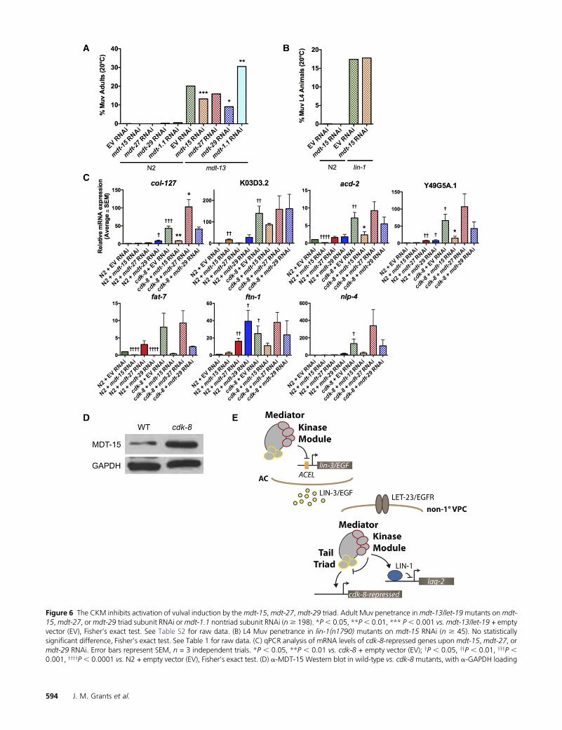
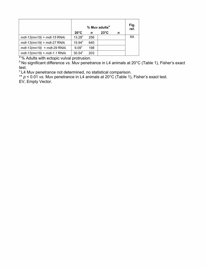
Our results suggested that the CKM might influence Me-diator subunit(s) other than mdt-23/sur-2. As S. cerevisiaeCDK8 inhibits the Mediator tail module triad composed ofMED2, MED3, and MED15 (van de Peppel et al. 2005; Gon-zalez et al. 2014), we hypothesized that their putative C.elegans orthologs MDT-29, MDT-27, and MDT-15 (Bourbon2008), might be targets for CKM inhibition.mdt-15 andmdt-29 knockdown had no effect on vulva formation in wild-typeanimals (i.e., causing neither Muv nor Vul phenotypes anddisplaying normal VPC induction; Table 1), but significantlyreduced the Muv penetrance of mdt-13/let-19 mutants (Fig-ure 6A); mdt-27 RNAi caused a similar trend (Figure 6A).This effect was specific to the tail module triad, as knockdownof the mdt-1.1 tail module subunit in fact increased the Muvpenetrance of mdt-13/let-19 mutants (Figure 6A). Further-more, requirement for the tail module triad in ectopic vulvaformation appeared to be specific to CKMmutants, asmdt-15RNAi had no effect on ectopic vulva formation in lin-1(n1790) mutants (Figure 6B, Table 1). Thus, tail moduletriad activity appears to be derepressed in a CKM mutant,causing aberrant activation of vulval fate specification.
Next,we investigatedwhether theCKMmodifies the targetgene specificity of the triad. We used qPCR to quantify theexpression of cdk-8-repressed genes identified by our micro-array analysis (Table S1) in wild-type worms and cdk-8 mu-tants treated with empty vector (EV), mdt-15, mdt-27, ormdt-29 RNAi. On EV RNAi, seven of nine genes testedwere upregulated in cdk-8 mutants compared to wild-typeworms, as expected (Figure 6C). Upregulation of these cdk-8-repressed genes was strongly attenuated by mdt-15 depletion,whereas mdt-29 knockdown only affected fat-7, and mdt-27depletion caused no significant changes (Figure 6C). Thus,for the genes investigated, induction in cdk-8 mutants ap-pears to specifically require mdt-15, but not the other pre-dicted tail module triad subunits. However, we cannot ruleout unequal RNAi efficiency accounting for these differingrequirements, although RNAi knockdown of all three genesappeared successful, as we observed partial sterility (notshown) consistent with the essential nature of these coreMediator subunits (Fernandez et al. 2005; Sönnichsen et al.2005; Taubert et al. 2006). Notably, only two cdk-8-repressedgenes, acd-2 and fat-7, displayed mdt-15 and/or mdt-29-dependent activation in wild-type worms (Figure 6C). Thus,as seen in the genetic analysis of vulva induction, loss of cdk-8appears to cause unrestrained tail module activity, i.e., mdt-15activates novel target genes when cdk-8 is deleted.
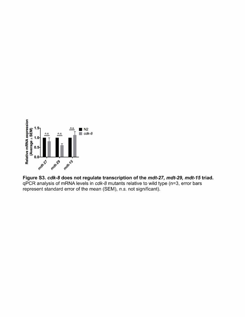
Finally, we investigated the molecular cause of unre-strained mdt-15 activity in CKM mutants. Loss of cdk-8 did
not alter mRNA levels of any triad subunits (Figure S3).Western blot analysis showed elevated MDT-15 protein lev-els in cdk-8 mutants compared to wild type (Figure 6D).Taken together, these results demonstrate that cdk-8 is re-quired for post-transcriptional regulation of MDT-15.
Discussion
EGFR signaling is critical for cell proliferation and cell fatedetermination in animal development. Several Mediator sub-units positively or negatively regulate EGFR signaling-drivendevelopmental processes (reviewed inGrants et al. 2015), butpertinent mechanisms remain incompletely understood.Here, we used the well-characterized vulva development par-adigm in C. elegans to delineate the role of the Mediator CKMmodule. Our results suggest a model whereby the CKM actswithin the vulval precursor cells, in a kinase-dependent man-ner, to fine tune EGFR transcriptional output by modulatingtwo transcriptional regulators: the key downstream tran-scription factor LIN-1/Ets and core Mediator (Figure 6E).This model is based on four key observations: First, we dem-onstrate that the primary site of action for the CKM is in theVPCs, as cdk-8 and mdt-13/let-19 repress vulva formationdownstream of mpk-1/ERK, a key component of the EGFRsignaling cascade inside VPCs, and cdk-8 expression in VPCsis sufficient for this repression. Second, cdk-8 repression ofectopic vulva formation is kinase dependent. Third, the CKMappears to act as a corepressor of the Ets-family transcriptionfactor LIN-1, as loss of cdk-8 or mdt-13/let-19 enhances theectopic vulval induction caused by lin-1 reduction of func-tion, and cdk-8 is required for full repression of a directLIN-1 target promoter. Fourth, ectopic vulva formation inmdt-13/let-19 is independent of the Mediator subunit mdt-23/sur-2, which is critical for EGFR signaling-driven tran-scription and vulval development in wild-type worms (Singhand Han 1995; Zhang and Greenwald 2011); instead, mdt-13/let-19 modulates the specificity of the tail module triadsubunits mdt-15, mdt-29, and mdt-27, preventing aberrantactivation of downstream transcription. By implicating allCKM subunits and by connecting the CKM to lin-1 and to coreMediator, our data substantially expand on the prior findingthat loss of CKM subunitmdt-12 caused ectopic vulva forma-tion by unknown molecular mechanisms (Moghal and Sternberg2003a). Additionally, our genetic and molecular analysisprovide first evidence that CKM-tail module crosstalk, akinto that seen in yeast Mediator (van de Peppel et al. 2005;Gonzalez et al. 2014), occurs in metazoan Mediator, an impor-tant experimental finding as tail module subunit sequence con-servation between species is extremely poor (Bourbon 2008).
The CKM inhibits vulva development in a kinase-dependent manner
We performed unbiased gene expression profiling to definegene programs that depend on cdk-8 in vivo, which revealedthat only 6.7% of C. elegans genes are regulated by cdk-8(Figure 2A, Table S1). This number F agrees with studies in
yeast, wherein CDK8 regulates only 3% of genes (Holstegeet al. 1998). Thus, CDK8 appears to be a gene program-specific transcriptional coregulator across species.
Among cdk-8-dependent genes, we identified a significantoverlap with genes regulated by lin-35/RB, a synMuv tran-scriptional repressor (Figure 2A) (Kirienko and Fay 2007).We note that as synMuv genes act redundantly, lin-35 singlemutants do not exhibit any defects in EGFR signaling or vul-val induction (Myers and Greenwald 2005; Cui et al. 2006;Kirienko and Fay 2007). Thus, the overlap between cdk-8-and lin-35-dependent genes suggested that the CKM and lin-35 cooperate in multiple aspects of C. elegans development.Similarly, in Drosophila, CDK8 and RB act in parallel in theWnt signaling pathway (Morris et al. 2008). Therefore, weexplored whether the CKM and synMuv genes cooperate inthe EGFR signaling pathway to regulate C. elegans vulva de-velopment. Both cdk-8 and mdt-13/let-19 were required torepress C. elegans vulva formation, in a partially redundantmanner with the synMuv genes (Figure 2B and Figure 5A).However, cdk-8 did not act redundantly with the synMuvgenes to repress lin-3/EGF transcription (Figure 3A), suggest-ing that the CKM and the synMuv genes regulate EGFR sig-naling at different junctions, as discussed below.
Comparing the vulval phenotypes of CKM mutants, wefound evidence that cdk-8 and cic-1 act redundantly to repressvulval induction, as a cdk-8; cic-1 double mutant displayedthe same Muv penetrance as cdk-8 or cic-1 single mutants(Figure 2B). In addition, we found that mdt-12/dpy-22 andmdt-13/let-19 were more strongly required to repress vulvadevelopment than cdk-8 or cic-1. In S. cerevisiae Mediator,MED12 and MED13 enable CDK8 and cyclin C docking toMediator (Tsai et al. 2013). Loss of MDT-12/DPY-22 or -13in C. elegansmay similarly disrupt CDK-8 and CIC-1 function,as well as considerably reducing the size of the CKM. Al-though CDK-8’s kinase activity is required to inhibit vulvadevelopment (Figure 4A), this does not rule out the possibil-ity that the CKM also employs kinase-independent stericmechanisms, as observed in other systems (Knuesel et al.2009). Thus, additional kinase-independent mechanismscould account for the stronger requirement for mdt-12/dpy-22 and -13 in vulva development.
The CKM inhibits the primary vulval cell fate
Vulva formation in C. elegans requires both EGFR and Notchsignaling (Félix and Barkoulas 2012), and human CDK8 re-presses Notch signaling-driven transcription by promotingturnover of the Notch intracellular domain (Fryer et al.2004). Therefore, we examined whether the vulval pheno-types in CKMmutants occur due to defects in EGFR signaling,Notch signaling, or both. Using an EGFR-Ras-ERK signaling-induced 1� cell fate reporter, we demonstrated that cdk-8 and
mdt-13/let-19 are required to repress ectopic vulva formationin part by repressing the 1� cell fate (Figure 2C and Figure5B). Using a Notch signaling-induced 2� cell fate reporter, weshowed that cdk-8 is required to represses ectopic 2� fates innonvulval VPCs, as well as to promote the 2� fate in P5.p andP7.p (Figure 2D). However, cdk-8 action in the Notch path-way might occur indirectly in this context. EGFR signaling inP6.p induces expression of Notch ligands, e.g., lag-2, whichpromote the 2� fate in the neighboring cells, P5.p and P7.p(Chen and Greenwald 2004). We observed evidence of pos-sible cell fate transformations from 2� to 1� in P5.p or P7.pin cdk-8 and mdt-13 mutants, as these cells occasionallyexhibited strong expression of the 1� cell fate marker egl-17P::CFP (Figure 2C and Figure 5B), expression of the EGFRsignaling target gene lag-2 (Figure 4D) or loss of the stronglip-1P::GFP expression characteristic of 2� cells (Figure 2D).It is possible that VPCs transformed to the 1� fate could theninduce 2� fates in neighboring VPCs, accounting for our ob-servation of ectopic 2� cells.
CKM subunits have been implicated as regulators of ca-nonical Wnt signaling (Zhang and Emmons 2000; Firesteinet al. 2008; Morris et al. 2008) and cell cycle quiescence(Clayton et al. 2008), processes which also contribute tovulva development. Activation of Wnt signaling can bypassrequirements for let-23/EGFR in vulva development (Gleasonet al. 2002). However, the Muv phenotype of mdt-12/dpy-22mutants is independent of bar-1/b-catenin (Moghal andSternberg 2003b), suggesting that the CKM does not repressvulva development through the canonical Wnt signalingpathway. Deregulation of cell cycle quiescence can expandthe VPC equivalence group, which are competent to formectopic vulvae if presented with the appropriate signals[e.g., lin-12/Notch gain of function employed by Claytonet al. (2008)]. Although CKM subunits are required for VPCcell cycle quiescence (Clayton et al. 2008), this alone is un-likely to account for the ectopic vulvae observed in theseanimals. First, the ectopic vulval invaginations observed incdk-8 andmdt-13/let-19 animals while scoring VPC induction(Table 1) were positioned in the correct location for P3.p, P4.p, and P8.p descendants. Second, ectopic expression of 1�and 2� cell fate markers in cdk-8 and mdt-13/let-19 mutants(Figure 2, C andD, and Figure 5B) suggests that EGFR and/orNotch signaling indeed drives ectopic vulva formation inthese mutants.
The CKM promotes LIN-1/Ets repressor activity
We observed derepression of the lin-3/EGF ACEL in cdk-8mutants (Figure 3C), implicating cdk-8 as a novel repressorof lin-3/EGF transcription in the anchor cell. Albeit interest-ing, genetic epistasis analysis with let-23/EGFR and mpk-1/ERK loss-of-function alleles clearly demonstrated that cdk-8 is
control. Representative immunoblot from one of three trials. (E) Model of CKM inhibition of EGFR-Ras-MAPK signaling-dependent cell fate specificationby repressing lin-3/EGF in the anchor cell (AC), promoting LIN-1 repressor activity (e.g., at lag-2), and inhibiting tail module triad activity (e.g., at cdk-8-repressed genes) in non-1� VPCs (i.e., VPCs other than P6.p).
primarily required downstream ofmpk-1/ERK to repress vul-val induction (Figure 3D). The let-23(sy97)mutant protein isligand insensitive (Aroian and Sternberg 1991; Aroian et al.1994); therefore, weak lin-3/EGF activation in the anchorcell due to loss of cdk-8 cannot account for the vulval pheno-types observed in cdk-8; let-23(sy97) mutants (Figure 3D).Furthermore, epistasis analysis with mpk-1/ERK confirmedthat cdk-8 acts downstream of the core EGFR-Ras-ERK path-way to regulate vulval induction (Figure 3D). In line with aposition downstream ofmpk-1/ERK, we showed that cdk-8 isrequired in VPCs to suppress ectopic vulval induction (Figure3E). A previous report demonstrated that repression of vul-val induction by the CKM subunit mdt-12/dpy-22 is gonadindependent, and thus anchor cell independent, and thatan MDT-12/DPY-22::GFP transgene is expressed in VPCs(Moghal and Sternberg 2003a), supporting a role for theCKM in VPCs.
Downstream of mpk-1/ERK, we found evidence that theCKM promotes LIN-1/Ets-mediated repression of vulval in-duction (Figure 4, B and C, and Figure 5C), and that cdk-8promotes transcriptional repression of a direct LIN-1 target,the lag-2/Notch ligand minimal promoter (Figure 4D). Thelag-2 minimal promoter contains activator and repressor el-ements, VPCact and VPCrep, that cooperatively restrict ex-pression to P6.p (Zhang and Greenwald 2011). On its own,VPCact is sufficient to drive transcription in all VPCs (P3.p–P8.p) in a LIN-3/EGF ligand-independent manner. VPCreprepresses this basal VPCact-driven transcription in VPCs otherthan P6.p, thereby restricting expression of the lag-2minimalpromoter to the 1�-fated VPC. VPCrep contains an Elk1 con-sensus site, which is bound by LIN-1 in vitro (Miley et al.2004), and requires lin-1/Ets for repression of transcriptionin VPCs other than P6.p (Zhang and Greenwald 2011). Ourresults indicate that cdk-8 is partially required for transcrip-tional repression of the lag-2 minimal promoter (Figure 4D),suggesting that the CKM promotes LIN-1-mediated repres-sion at VPCrep. An alternative explanation for the ectopicexpression of the lag-2 minimal promoter observed in cdk-8mutants is that the CKM might inhibit a factor that activatestranscription through VPCact. The transcription factor(s)that acts at VPCact remains poorly defined; however, themdt-23/sur-2Mediator subunit is required for VPCact-driventranscription in P3.p–P8.p (Zhang and Greenwald 2011). Aswe demonstrated that vulval induction in mdt-13/let-19mu-tants does not require mdt-23/sur-2 (Figure 5, B and E), thisimplies that the CKM likely does not inhibit MDT-23/SUR-2activity at VPCact. Overall, our findings suggest that the CKMmay act as a corepressor for LIN-1.
In murine embryonic stem cells, Mediator recruitment isimportant for transcriptional activation by Ets factors, e.g.,Elk1 (Stevens et al. 2002; Balamotis et al. 2009). In thiscontext, activation of Elk1 by ERK phosphorylation promotesbinding to Mediator in a MED23/Sur2-dependent manner(Stevens et al. 2002). Similarly, in a colon cancer cell line,CDK8 promotes transcriptional elongation of serum responseimmediate early genes, which are targeted by multiple tran-
scription factors including Elk1 (Donner et al. 2010). How-ever, the role of Mediator in transcriptional repression by anEts factor has not previously been explored. In the absence ofERK phosphorylation, Ets factors, e.g., LIN-1, can promotetranscriptional repression of target genes (Jacobs et al.1998; Zhang and Greenwald 2011). Although the Sin3A-HDAC-1 corepressor complex has been implicated in an epi-genetic mechanism that attenuates transcriptional activationby ERK-phosphorylated Elk1(Yang et al. 2001), to our knowl-edge, corepressors of Ets factor-mediated transcriptional re-pression have not previously been identified. This reportprovides evidence that the Mediator CKM is required for re-pression by Ets factors, representing an advance in our un-derstanding of Ets factor repressive mechanisms.
Our findings are also of potential clinical interest, as thehuman CKM is implicated in tumorigenesis (Firestein et al.2008; Donner et al. 2010; Mäkinen et al. 2011; Lim et al.2014). Loss of MED12 causes cellular resistance to chemo-therapeutic agents that inhibit activated BRAF, the humanERK kinase kinase (Shalem et al. 2014); this suggests thatMED12 represses EGFR signaling downstream of BRAF, inline with our findings for the C. elegans CKM. Furthermore,recurrent MED12 mutations are implicated in uterine leio-myomas and breast fibroadenomas (Mäkinen et al. 2011;Lim et al. 2014; Mittal et al. 2015), but the pathogenic mech-anisms of these mutations have not been fully elucidated.Investigation of these mutations using the C. elegans vulvadevelopment paradigm may provide insight into their modeof action.
The CKM restrains the core Mediator tail module triad
Epistatic relationships between Mediator subunits have beenidentified in S. cerevisiae (van de Peppel et al. 2005; Gonzalezet al. 2014), but intra-Mediator regulation has not been dem-onstrated in metazoans. Previous studies (reviewed in Grantset al. 2015) and our data show that several core Mediatorsubunits promote C. elegans vulva development, whereasCKM subunits inhibit this process. This suggested that in-tra-Mediator regulation might coordinate gene expressiondownstream of the EGFR-Ras-ERK signaling pathway thatdrives vulva development. Initially, we hypothesized thatthe CKM may oppose mdt-23/sur-2-mediated activation ofEGFR signaling, asmdt-23/sur-2 is required for vulval induc-tion and activation of EGFR signaling-induced transcription,e.g., lag-2 (Singh and Han 1995; Zhang and Greenwald2011). Unexpectedly, loss of mdt-13/let-19 circumventedthe requirement formdt-23/sur-2 in vulval induction (Figure5E). We therefore explored regulatory interactions betweenthe CKM and the metazoan orthologs of S. cerevisiae MED2,MED3, and MED15, which are subject to inhibitory post-translational regulation by CDK8 (van de Peppel et al.2005; Gonzalez et al. 2014). Sequence conservation is weakbetween yeast MED2 andMED3 and their putative metazoanhomologs, MED29 and MED27, respectively (Bourbon2008). Whether MED29 and MED27 function as part of thetail module remains unclear, as structural and biochemical
studies locate these subunits between the head and tail mod-ules (Sato et al. 2003; Tsai et al. 2014). Thus, we were in-trigued to find that vulva formation in a C. elegans CKMmutant required mdt-15, mdt-27, and mdt-29 (Figure 6A).This requirement appeared specific to the tail module triad,as neither mdt-1.1/MED1 nor mdt-23/sur-2 was required forvulval induction in CKM mutants (Figure 5E and Figure 6A).Furthermore, the triad did not appear to be generally re-quired for ectopic vulval induction in animals with a wild-type CKM, as mdt-15 knockdown had no effect on ectopicvulval induction in lin-1(n1790) mutants (Figure 6B). To-gether, these findings suggest that the CKM restrains triadactivity, preventing it from aberrantly activating vulvalinduction.
Gene expression analysis in cdk-8 mutants identified a re-quirement formdt-15, but little or no requirement formdt-27or mdt-29, in transcriptional activation of cdk-8-repressedgenes (Figure 6C). In the S. cerevisiae tail module triad, bothMED3 and MED2 are required for overexpression of CDK8-repressed genes in CDK8 mutants, but the requirement forMED15 has not been tested directly (van de Peppel et al.2005; Gonzalez et al. 2014). These requirements might beexplained by the fact that both MED2 and MED3 are neces-sary to anchor the triad to the tail module (Myers et al. 1999;van de Peppel et al. 2005; Gonzalez et al. 2014). Similarrequirements may not exist in metazoan Mediator, as humanMediator displays more extensive structural contacts be-tween the head and tail modules (Tsai et al. 2014), whichmay result in redundancy for some tail module subunits.
Investigating the regulatory relationship between CDK-8andMDT-15 further, we found that cdk-8 is required for post-transcriptional negative regulation of MDT-15, as MDT-15protein but not mRNA levels increase in cdk-8 mutants (Fig-ure 6D). This regulatory relationship resembles that seen inyeast where the three triad subunits MED2, MED3, andMED15 are negatively regulated post-translationally byCDK8-driven phosphorylation of MED3, promoting ubiqui-tin-proteasome-dependent turnover of all three triad sub-units (Gonzalez et al. 2014). It will be interesting to delineatewhether the metazoan CKM regulates MDT-15 protein levelsdirectly, e.g., by phosphorylation leading to ubiquitin-mediateddegradation, or indirectly through action upon other Mediatorsubunits.
In summary, our findings suggest that the Mediator CKMrepresses EGFR-Ras-ERK signaling-driven cell fate specifica-tion in C. elegans by regulating repressor activity of an Ets-family transcription factor and by promoting specificity ofMediator tail module subunits.
Acknowledgments
We thank all Taubert lab members for critical discussions;Shirley Chen for help with experiments; R. Barbeau, C.Eisley, A. Barczak, and D. Erle from the Sandler AsthmaBasic Research Center Functional Genomics Core Facility(University of California San Francisco) for help with
microarray gene expression profiling; J. Ewbank for EASEanalysis of gene expression arrays; J. Escobar for advice onscoring VPC induction; I. Greenwald for GS5096 arEx1098[lag-2p(min)::YFP]; S. Mitani for cdk-8(tm1238) and cic-1(tm3740) mutants; and D. Moerman and J. Ward for com-ments on the manuscript. Some strains were provided by theCaenorhabditis Genetics Center, which is funded by NationalInstitutes of Health Office of Research Infrastructure Pro-grams (P40 OD010440). This work was supported by grantsfrom Canadian Institute of Health Research (MOP-93713),Natural Sciences and Engineering Research Council of Can-ada (RGPIN 386398-13), Canada Foundation for Innovation(all to S.T.), and Grants-in-Aid for Scientific Research fromthe Ministry of Education, Culture, Sports, Science, andTechnology of Japan (to H.S.). J.M.G. was supported byVanier Canada Graduate Scholarship, Natural Sciences andEngineering Research Council of Canada Canada GraduateScholarship - Masters, Child and Family Research Institute,and University of British Columbia scholarships, and S.T. bya Canada Research Chair. The authors declare no competingfinancial interests.
J.M.G., H.S., and S.T. conceived and designed experiments;J.M.G., L.T.L.Y., A.Y., C.C.Y., H.S., and S.T. performedexperiments; J.M.G., A.Y., H.O., H.S., and S.T. analyzed data;J.M.G., A.Y., H.O., and H.S. contributed reagents/materials/analysis tools; and J.M.G., H.S., and S.T. wrote the paper.
Literature Cited
Aroian, R. V., and P. W. Sternberg, 1991 Multiple functions of let-23, a Caenorhabditis elegans receptor tyrosine kinase gene re-quired for vulval induction. Genetics 128: 251–267.
Aroian, R. V., M. Koga, J. E. Mendel, Y. Ohshima, and P. W. Sternberg,1990 The let-23 gene necessary for Caenorhabditis elegansvulval induction encodes a tyrosine kinase of the EGF receptorsubfamily. Nature 348: 693–699.
Aroian, R. V., G. M. Lesa, and P. W. Sternberg, 1994 Mutations inthe Caenorhabditis elegans let-23 EGFR-like gene define ele-ments important for cell-type specificity and function. EMBOJ. 13: 360–366.
Balamotis, M. A., M. A. Pennella, J. L. Stevens, B. Wasylyk, A. S.Belmont et al., 2009 Complexity in transcription control at theactivation domain-mediator interface. Sci. Signal. 2: ra20.
Baselga, J., and S. M. Swain, 2009 Novel anticancer targets: revisit-ing ERBB2 and discovering ERBB3. Nat. Rev. Cancer 9: 463–475.
Beitel, G. J., S. Tuck, I. Greenwald, and H. R. Horvitz, 1995 TheCaenorhabditis elegans gene lin-1 encodes an ETS-domain pro-tein and defines a branch of the vulval induction pathway. GenesDev. 9: 3149–3162.
Berset, T., E. F. Hoier, G. Battu, S. Canevascini, and A. Hajnal,2001 Notch inhibition of RAS signaling through MAP kinasephosphatase LIP-1 during C. elegans vulval development. Sci-ence 291: 1055–1058.
Bolstad, B. M., R. A. Irizarry, M. Astrand, and T. P. Speed, 2003 Acomparison of normalization methods for high density oligonu-cleotide array data based on variance and bias. Bioinformatics19: 185–193.
Bourbon, H.-M., 2008 Comparative genomics supports a deepevolutionary origin for the large, four-module transcriptionalmediator complex. Nucleic Acids Res. 36: 3993–4008.
Bourbon, H.-M., A. Aguilera, A. Z. Ansari, F. J. Asturias, A. J. Berket al., 2004 A unified nomenclature for protein subunits ofmediator complexes linking transcriptional regulators to RNApolymerase II. Mol. Cell 14: 553–557.
Brenner, S., 1974 The genetics of Caenorhabditis elegans. Genetics77: 71–94.
Chen, N., and I. Greenwald, 2004 The lateral signal for LIN-12/Notch in C. elegans vulval development comprises redundantsecreted and transmembrane DSL proteins. Dev. Cell 6: 183–192.
Clayton, J. E., S. J. L. van den Heuvel, and R. M. Saito,2008 Transcriptional control of cell-cycle quiescence duringC. elegans development. Dev. Biol. 313: 603–613.
Cui, M., J. Chen, T. R. Myers, B. J. Hwang, P. W. Sternberg et al.,2006 SynMuv genes redundantly inhibit lin-3/EGF expressionto prevent inappropriate vulval induction in C. elegans. Dev. Cell10: 667–672.
Donner, A. J., S. Szostek, J. M. Hoover, and J. M. Espinosa,2007 CDK8 is a stimulus-specific positive coregulator of p53target genes. Mol. Cell 27: 121–133.
Donner, A. J., C. C. Ebmeier, D. J. Taatjes, and J. M. Espinosa,2010 CDK8 is a positive regulator of transcriptional elongationwithin the serum response network. Nat. Struct. Mol. Biol. 17:194–201.
Engelmann, I., A. Griffon, L. Tichit, F. Montañana-Sanchis, G. Wanget al., 2011 A comprehensive analysis of gene expressionchanges provoked by bacterial and fungal infection in C. ele-gans. PLoS One 6: e19055.
Fay, D. S., and J. Yochem, 2007 The SynMuv genes of Caenorhab-ditis elegans in vulval development and beyond. Dev. Biol. 306:1–9.
Félix, M.-A., and M. Barkoulas, 2012 Robustness and flexibility innematode vulva development. Trends Genet. TIG 28: 185–195.
Fernandez, A. G., K. C. Gunsalus, J. Huang, L.-S. Chuang, N. Yinget al., 2005 New genes with roles in the C. elegans embryorevealed using RNAi of ovary-enriched ORFeome clones. Ge-nome Res. 15: 250–259.
Firestein, R., A. J. Bass, S. Y. Kim, I. F. Dunn, S. J. Silver et al.,2008 CDK8 is a colorectal cancer oncogene that regulates[bgr]-catenin activity. Nature 455: 547–551.
Fryer, C. J., J. B. White, and K. A. Jones, 2004 Mastermind re-cruits CycC:CDK8 to phosphorylate the Notch ICD and coordi-nate activation with turnover. Mol. Cell 16: 509–520.
Gentleman, R. C., V. J. Carey, D. M. Bates, B. Bolstad, M. Dettlinget al., 2004 Bioconductor: open software development forcomputational biology and bioinformatics. Genome Biol. 5: R80.
Gilleard, J. S., J. D. Barry, and I. L. Johnstone, 1997 cis regulatoryrequirements for hypodermal cell-specific expression of the Cae-norhabditis elegans cuticle collagen gene dpy-7. Mol. Cell. Biol.17: 2301–2311.
Gleason, J. E., H. C. Korswagen, and D. M. Eisenmann,2002 Activation of Wnt signaling bypasses the requirementfor RTK/Ras signaling during C. elegans vulval induction. GenesDev. 16: 1281–1290.
Goh, G. Y. S., K. L. Martelli, K. S. Parhar, A. W. L. Kwong, M. A.Wong et al., 2014 The conserved Mediator subunit MDT-15 isrequired for oxidative stress responses in Caenorhabditis ele-gans. Aging Cell 13: 70–79.
Gold, M. O., and A. P. Rice, 1998 Targeting of CDK8 to a pro-moter-proximal RNA element demonstrates catalysis-dependentactivation of gene expression. Nucleic Acids Res. 26: 3784–3788.
Gonzalez, D., N. Hamidi, R. Del Sol, J. J. Benschop, T. Nancy et al.,2014 Suppression of Mediator is regulated by Cdk8-dependentGrr1 turnover of the Med3 coactivator. Proc. Natl. Acad. Sci. USA111: 2500–2505.
Grants, J. M., G. Y. S. Goh, and S. Taubert, 2015 The Mediatorcomplex of Caenorhabditis elegans: insights into the develop-
mental and physiological roles of a conserved transcriptionalcoregulator. Nucleic Acids Res. 43: 2442–2453.
Han, M., R. V. Aroian, and P. W. Sternberg, 1990 The let-60 locuscontrols the switch between vulval and nonvulval cell fates inCaenorhabditis elegans. Genetics 126: 899–913.
Hill, R. J., and P. W. Sternberg, 1992 The gene lin-3 encodes aninductive signal for vulval development in C. elegans. Nature358: 470–476.
Holm, S., 1979 A simple sequentially rejective multiple test pro-cedure. Scand. J. Stat. 6: 65–70.
Holstege, F. C., E. G. Jennings, J. J. Wyrick, T. I. Lee, C. J. Hengartneret al., 1998 Dissecting the regulatory circuitry of a eukaryoticgenome. Cell 95: 717–728.
Horvitz, H. R., and J. E. Sulston, 1980 Isolation and genetic char-acterization of cell-lineage mutants of the nematode Caenorhab-ditis elegans. Genetics 96: 435–454.
Hosack, D. A., G. Dennis, Jr., B. T. Sherman, H. C. Lane, and R. A.Lempicki, 2003 Identifying biological themes within lists ofgenes with EASE. Genome Biol. 4: R70.
Hwang, B. J., and P. W. Sternberg, 2004 A cell-specific enhancerthat specifies lin-3 expression in the C. elegans anchor cell forvulval development. Development 131: 143–151.
Jacobs, D., G. J. Beitel, S. G. Clark, H. R. Horvitz, and K. Kornfeld,1998 Gain-of-function mutations in the Caenorhabditis eleganslin-1 ETS gene identify a C-terminal regulatory domain phos-phorylated by ERK MAP kinase. Genetics 149: 1809–1822.
Katz, W. S., R. J. Hill, T. R. Clandinin, and P. W. Sternberg,1995 Different levels of the C. elegans growth factor LIN-3promote distinct vulval precursor fates. Cell 82: 297–307.
Kirienko, N. V., and D. S. Fay, 2007 Transcriptome profiling of theC. elegans Rb ortholog reveals diverse developmental roles. Dev.Biol. 305: 674–684.
Kishore, R. S., and M. V. Sundaram, 2002 ced-10 Rac and mig-2function redundantly and act with unc-73 trio to control theorientation of vulval cell divisions and migrations in Caenorhab-ditis elegans. Dev. Biol. 241: 339–348.
Knuesel, M. T., K. D. Meyer, C. Bernecky, and D. J. Taatjes,2009 The human CDK8 subcomplex is a molecular switch thatcontrols Mediator coactivator function. Genes Dev. 23: 439–451.
Kwon, J. Y., and J. Lee, 2001 Biological significance of a univer-sally conserved transcription mediator in metazoan develop-mental signaling pathways. Development 128: 3095–3104.
Liao, S. M., J. Zhang, D. A. Jeffery, A. J. Koleske, C. M. Thompsonet al., 1995 A kinase-cyclin pair in the RNA polymerase IIholoenzyme. Nature 374: 193–196.
Lim, W. K., C. K. Ong, J. Tan, A. A. Thike, C. C. Y. Ng et al.,2014 Exome sequencing identifies highly recurrent MED12somatic mutations in breast fibroadenoma. Nat. Genet. 46:877–880.
Loncle, N., M. Boube, L. Joulia, C. Boschiero, M. Werner et al.,2007 Distinct roles for Mediator Cdk8 module subunits in Dro-sophila development. EMBO J. 26: 1045–1054.
Mäkinen, N., M. Mehine, J. Tolvanen, E. Kaasinen, Y. Li et al.,2011 MED12, the mediator complex subunit 12 gene, is mu-tated at high frequency in uterine leiomyomas. Science 334:252–255.
Malik, S., and R. G. Roeder, 2010 The metazoan Mediator co-activator complex as an integrative hub for transcriptional reg-ulation. Nat. Rev. Genet. 11: 761–772.
Miley, G. R., D. Fantz, D. Glossip, X. Lu, R. M. Saito et al.,2004 Identification of residues of the Caenorhabditis elegansLIN-1 ETS domain that are necessary for DNA binding and reg-ulation of vulval cell fates. Genetics 167: 1697–1709.
Miller, L. M., M. E. Gallegos, B. A. Morisseau, and S. K. Kim,1993 lin-31, a Caenorhabditis elegans HNF-3/fork head tran-scription factor homolog, specifies three alternative cell fates invulval development. Genes Dev. 7: 933–947.
598 J. M. Grants et al.
Mittal, P., Y.-H. Shin, S. A. Yatsenko, C. A. Castro, U. Surti et al.,2015 Med12 gain-of-function mutation causes leiomyomasand genomic instability. J. Clin. Invest. 125: 3280–3284.
Moghal, N., and P. W. Sternberg, 2003a A component of thetranscriptional mediator complex inhibits RAS-dependent vulvalfate specification in C. elegans. Development 130: 57–69.
Moghal, N., and P. W. Sternberg, 2003b The epidermal growthfactor system in Caenorhabditis elegans. Exp. Cell Res. 284:150–159.
Morris, E. J., J.-Y. Ji, F. Yang, L. Di Stefano, A. Herr et al.,2008 E2F1 represses [bgr]-catenin transcription and is antag-onized by both pRB and CDK8. Nature 455: 552–556.
Myers, L. C., C. M. Gustafsson, K. C. Hayashibara, P. O. Brown, andR. D. Kornberg, 1999 Mediator protein mutations that selec-tively abolish activated transcription. Proc. Natl. Acad. Sci. USA96: 67–72.
Myers, T. R., and I. Greenwald, 2005 lin-35 Rb acts in the majorhypodermis to oppose ras-mediated vulval induction in C. ele-gans. Dev. Cell 8: 117–123.
Nemet, J., B. Jelicic, I. Rubelj, and M. Sopta, 2014 The two facesof Cdk8, a positive/negative regulator of transcription. Biochi-mie 97: 22–27.
Normanno, N., A. De Luca, C. Bianco, L. Strizzi, M. Mancino et al.,2006 Epidermal growth factor receptor (EGFR) signaling incancer. Gene 366: 2–16.
Poss, Z. C., C. C. Ebmeier, and D. J. Taatjes, 2013 The Mediatorcomplex and transcription regulation. Crit. Rev. Biochem. Mol.Biol. 48: 575–608.
Saffer, A. M., D. H. Kim, A. van Oudenaarden, and H. R. Horvitz,2011 The Caenorhabditis elegans synthetic multivulva genesprevent ras pathway activation by tightly repressing global ec-topic expression of lin-3 EGF. PLoS Genet. 7: e1002418.
Sato, S., C. Tomomori-Sato, C. A. S. Banks, T. J. Parmely, I. Sorokinaet al., 2003 A mammalian homolog of Drosophila melanogastertranscriptional coactivator intersex is a subunit of the mammalianMediator complex. J. Biol. Chem. 278: 49671–49674.
Schmid, T., and A. Hajnal, 2015 Signal transduction during C.elegans vulval development: a NeverEnding story. Curr. Opin.Genet. Dev. 32: 1–9.
Shalem, O., N. E. Sanjana, E. Hartenian, X. Shi, D. A. Scott et al.,2014 Genome-scale CRISPR-Cas9 knockout screening in hu-man cells. Science 343: 84–87.
Singh, N., and M. Han, 1995 sur-2, a novel gene, functions late inthe let-60 ras-mediated signaling pathway during Caenorhabdi-tis elegans vulval induction. Genes Dev. 9: 2251–2265.
Smyth G. K., 2004 Linear models and empirical Bayes methodsfor assessing differential expression in microarray experiments.Stat. Appl. Genet. Mol. Biol. 3: Article3.
Sönnichsen, B., L. B. Koski, A. Walsh, P. Marschall, B. Neumannet al., 2005 Full-genome RNAi profiling of early embryogene-sis in Caenorhabditis elegans. Nature 434: 462–469.
Steimel A., Suh J., Hussainkhel A., Deheshi S., Grants J. M. et al.,2013 The C. elegans CDK8 Mediator module regulates axonguidance decisions in the ventral nerve cord and during dorsalaxon navigation. Dev. Biol. 377: 385–398.
Sternberg, P. W., 1988 Lateral inhibition during vulval inductionin Caenorhabditis elegans. Nature 335: 551–554.
Sternberg, P. W., and H. R. Horvitz, 1986 Pattern formation dur-ing vulval development in C. elegans. Cell 44: 761–772.
Sternberg, P. W., and H. R. Horvitz, 1989 The combined action oftwo intercellular signaling pathways specifies three cell fatesduring vulval induction in C. elegans. Cell 58: 679–693.
Stevens, J. L., G. T. Cantin, G. Wang, A. Shevchenko, A. Shevchenkoet al., 2002 Transcription control by E1A and MAP kinase path-way via Sur2 mediator subunit. Science 296: 755–758.
Sulston, J. E., and H. R. Horvitz, 1981 Abnormal cell lineages inmutants of the nematode Caenorhabditis elegans. Dev. Biol. 82:41–55.
Tan, P. B., M. R. Lackner, and S. K. Kim, 1998 MAP kinase sig-naling specificity mediated by the LIN-1 Ets/LIN-31 WH tran-scription factor complex during C. elegans vulval induction. Cell93: 569–580.
Taubert, S., M. R. Van Gilst, M. Hansen, and K. R. Yamamoto,2006 A Mediator subunit, MDT-15, integrates regulation offatty acid metabolism by NHR-49-dependent and -independentpathways in C. elegans. Genes Dev. 20: 1137–1149.
Taubert, S., M. Hansen, M. R. Van Gilst, S. B. Cooper, and K. R.Yamamoto, 2008 The Mediator subunit MDT-15 confers met-abolic adaptation to ingested material. PLoS Genet. 4:e1000021.
Tsai, K.-L., S. Sato, C. Tomomori-Sato, R. C. Conaway, J. W. Con-away et al., 2013 A conserved Mediator-CDK8 kinase moduleassociation regulates Mediator-RNA polymerase II interaction.Nat. Struct. Mol. Biol. 20: 611–619.
Tsai, K.-L., C. Tomomori-Sato, S. Sato, R. C. Conaway, J. W. Conawayet al., 2014 Subunit architecture and functional modular re-arrangements of the transcriptional mediator complex. Cell157: 1430–1444.
Tuck, S., and I. Greenwald, 1995 lin-25, a gene required for vul-val induction in Caenorhabditis elegans. Genes Dev. 9: 341–357.
van de Peppel, J., N. Kettelarij, H. van Bakel, T. T. J. P. Kockelkorn,D. van Leenen et al., 2005 Mediator expression profiling epis-tasis reveals a signal transduction pathway with antagonisticsubmodules and highly specific downstream targets. Mol. Cell19: 511–522.
Yang, S. H., E. Vickers, A. Brehm, T. Kouzarides, and A. D. Sharrocks,2001 Temporal recruitment of the mSin3A-histone deacety-lase corepressor complex to the ETS domain transcription fac-tor Elk-1. Mol. Cell. Biol. 21: 2802–2814.
Yoda, A., H. Kouike, H. Okano, and H. Sawa, 2005 Componentsof the transcriptional Mediator complex are required for asym-metric cell division in C. elegans. Development 132: 1885–1893.
Yoo, A. S., C. Bais, and I. Greenwald, 2004 Crosstalk between theEGFR and LIN-12/Notch pathways in C. elegans vulval devel-opment. Science 303: 663–666.
Zhang, H., and S. W. Emmons, 2000 A C. elegans mediator pro-tein confers regulatory selectivity on lineage-specific expressionof a transcription factor gene. Genes Dev. 14: 2161–2172.
Zhang, X., and I. Greenwald, 2011 Spatial regulation of lag-2transcription during vulval precursor cell fate patterning in Cae-norhabditis elegans. Genetics 188: 847–858.
Zhou, H., J. M. Spaeth, N. H. Kim, X. Xu, M. J. Friez et al.,2012 MED12 mutations link intellectual disability syndromeswith dysregulated GLI3-dependent Sonic Hedgehog signaling.Proc. Natl. Acad. Sci. USA 109: 19763–19768.
Figure 53. cdk-8 does not regulate transcription of the mdt-27, mdt-29, mdt-15 triad. qPCR analysis of mRNA levels in cdk-8 mutants relative to wild type (n=3, error bars represent standard error of the mean (SEM), n.s. not significant).
Table S1. Microarray results. (.xlsx, 76 KB)
Available for download as a .xlsx file at www.genetics.org/lookup/suppl/doi:10.1534/genetics.115.180265/-/DC1/TableS1.xlsx
a % Adults with ectopic vulval protrusion. b No significant difference vs. Muv penetrance in L4 animals at 20°C (Table 1), Fisher’s exact test. c L4 Muv penetrance not determined, no statistical comparison. ** p < 0.01 vs. Muv penetrance in L4 animals at 20°C (Table 1), Fisher’s exact test. EV, Empty Vector.
Table S3. Relative mRNA expression of synMuv genes in cdk-8 mutants vs. wild type as determined by microarray.
MT10430 lin-35(n745) I HS310 let-19(mn19)/mIn1[dpy-10(e128) mIs14] II PS295 let-23(sy97) unc-4(e120)/mnC1[dpy-10(e128) unc-52(e444)] II HS588 mpk-1(oz140) unc-32(e189)/hT2[bli-4(e937) let-?(q728) qIs48] III MT301 lin-31(n301) II STE13 cic-1(tm3740) III WU125 lin-1(n1790) IV HS445 dpy-22(os38) X; osEx89[dpy-22(+)]
MT1806 lin-15A(n767) X MT2495 lin-15B(n744) X
AH12 gap-1(ga133) X STE74 cdk-8(tm1238) I; lin-15A(n767) X STE75 cic-1(tm3740) III; lin-15A(n767) X HS432 let-19(mn19)/mIn1[dpy-10(e128) mIs14] II; lin-15A(n767) X STE76 cdk-8(tm1238) I; lin-15B(n744) X HS433 let-19(mn19)/mIn1[dpy-10(e128) mIs14] II; lin-15B(n374) X STE77 cdk-8(tm1238) I; gap-1(ga133) X MT309 lin-15AB(n309) X STE78 cdk-8(tm1238) I; let-23(sy97) unc-4(e120)/mnC1[dpy-10(e128) unc-52(e444)] II STE79 cdk-8(tm1238) I; lin-1(n1790) IV HS510 let-19(mn19)/mIn1[dpy-10(e128) mIs14] II; lin-1(n1790) IV HS356 sur-2(ku9) I; let-19(mn19)/mIn1[dpy-10(e128) mIs14] II
GS3582 arIs92[egl-17p::NLS-CFP-LacZ + unc-4(+) + ttx-3::GFP]; unc-4(e120) II PS4308 syIs107[unc-119(+) + lin-3ACEL(delta-pes-10)::GFP]; unc-119(ed4) III GS5096 arEx1098[lag-2p(min)::2nls-yfp::unc-54 3’UTR + pha-1(+)]; pha-1(e2123) III AH142 zhIs4[lip-1p::GFP]