1 The Medical Student’s Guide to the Vascular Neurology Wards “In the fields of observation fortune favors only the prepared mind” - Louis Pasteur Disclaimer: This guide was made by two fourth year medical students after their time on the Vascular Neurology wards. The purpose of this guide is to give students 1) an idea of what their time on the service is like and 2) a refresher on vascular neurology anatomy and pathology. This guide does not supersede the directions from your clerkship director and it is not a comprehensive review of cerebrovascular disorders. This guide is meant to supplement your preparation for the wards, so that your mind is primed for knowledge consumption. The Day to Day Prerounding (starts on Bridge 7 generally at 6am for the Sub-I and 7am for the Clerk): • You’ll pick up patients either your first morning or the day before. One of the residents will help you decide which patients are appropriate for you to carry. Your goal is to finish pre- rounding before morning report. For pre-rounding, write down updates (vitals, labs, new imaging, PT/OT/Consult recs), confirm the HPI and exam w/ the patient using the resident’s admitting note for reference, check telemetry, and ask your patient’s nurse if there are any updates. • The Big White Board upon entering the stroke work room has a list of new patients on the general and vascular services. Place your initials next to pts you take each morning. • Dr. Wendell’s goal for the MS3s is to start with 1 patient and build to 2-3 patients by the end of the rotation. • Dr. Wendell’s goal for the sub-I is to carry 5 patients by the end of the month. Sub-Is should start with 2-3 patient in the beginning and then build up. • Sub-Is and M3s should be writing daily progress notes on their patients. Sub-Is should strive to get these done in the AM before rounds, because this is most helpful to the team. M3s should also write daily progress notes, but they can be written in the PM if there is not sufficient time in the AM. • If you are a sub-I, give the nurse your pager number, and ask her/him to call you with any questions or order requests • You can use the “Patient Calendar” to identify if/when your patient is scheduled for tests (e.g. MRI, Echo, EEG). This information is useful for Disposition Rounds if available o In EMR, click Summary à Index à Patient Calendar (under “Additional Reports”) 8AM: Morning Report You can ask residents for a schedule of morning report and noon conferences: which are a mix of lectures, practice questions, journal club, general discussion of interesting cases on the floors. The schedule is also available at: goo.gl/EBHHGZ • Mondays = Pediatric Neurology • Wednesdays = Grand rounds in George auditorium
Transcript
1
The Medical Student’s Guide to the Vascular Neurology Wards
“In the fields of observation fortune favors only the prepared mind” - Louis Pasteur
Disclaimer: This guide was made by two fourth year medical students after their time on the Vascular Neurology wards. The purpose of this guide is to give students 1) an idea of what their time on the service is like and 2) a refresher on vascular neurology anatomy and pathology. This guide does not supersede the directions from your clerkship director and it is not a comprehensive review of cerebrovascular disorders. This guide is meant to supplement your preparation for the wards, so that your mind is primed for knowledge consumption. The Day to Day Prerounding (starts on Bridge 7 generally at 6am for the Sub-I and 7am for the Clerk): • You’ll pick up patients either your first morning or the day before. One of the residents will
help you decide which patients are appropriate for you to carry. Your goal is to finish pre-rounding before morning report. For pre-rounding, write down updates (vitals, labs, new imaging, PT/OT/Consult recs), confirm the HPI and exam w/ the patient using the resident’s admitting note for reference, check telemetry, and ask your patient’s nurse if there are any updates.
• The Big White Board upon entering the stroke work room has a list of new patients on the general and vascular services. Place your initials next to pts you take each morning.
• Dr. Wendell’s goal for the MS3s is to start with 1 patient and build to 2-3 patients by the end of the rotation.
• Dr. Wendell’s goal for the sub-I is to carry 5 patients by the end of the month. Sub-Is should start with 2-3 patient in the beginning and then build up.
• Sub-Is and M3s should be writing daily progress notes on their patients. Sub-Is should strive to get these done in the AM before rounds, because this is most helpful to the team. M3s should also write daily progress notes, but they can be written in the PM if there is not sufficient time in the AM.
• If you are a sub-I, give the nurse your pager number, and ask her/him to call you with any questions or order requests
• You can use the “Patient Calendar” to identify if/when your patient is scheduled for tests (e.g. MRI, Echo, EEG). This information is useful for Disposition Rounds if available
o In EMR, click Summary à Index à Patient Calendar (under “Additional Reports”) 8AM: Morning Report
You can ask residents for a schedule of morning report and noon conferences: which are a mix of lectures, practice questions, journal club, general discussion of interesting cases on the floors. The schedule is also available at: goo.gl/EBHHGZ • Mondays = Pediatric Neurology • Wednesdays = Grand rounds in George auditorium
2
9AM: Disposition Rounds (“Dispo Rounds") in the Stroke Work Room • Multidisciplinary Disposition rounds with the Vascular Neurology team, Nursing, PT, OT,
Speech, Case Manager, and Social Work o The goal of Dispo Rounds is to confirm the disposition of each stroke patient
• Nurses state their patient (e.g. “Room 602 – Michael Thomas?”) • Neuro provides quick 1 liner of how patient is doing and what is left to be done before
patient can be safely discharged. o e.g.: “Mr. Thomas is a new patient, a 67M w/ Dementia who came in yesterday with
R sided weakness and aphasia found to have a L M1 Occlusion, received tPA. Awaiting an MRI, Echo, and evaluation from PT, OT, and Speech. Likely will/will not be discharged today.”
§ I watched a couple of dispo rounds to get a hang of it before doing it for my patients. It’s low key, and the resident/intern/PA (who you will be sharing patient with) chimes in if they have additional details to share
• PT/OT will ask if they need to see patient or provide their discharge recommendations (e.g. “We saw Thomas – recommendation is discharge to Acute Rehab/SNF/Home w services/Nursing Home). Speech will leave a note about patient’s ability to swallow; they often do not attend dispo rounds.
o All acute stroke patients need PT/OT unless explicitly documented in a progress note that the patient has no PT/OT needs (The Joint Commission (TJC) requirement)
§ If patient has mobility issues, ask PT to see them § If you suspect patient has new cognitive impairment, arm weakness, or visual
impairment, ask OT to see them § Speech – all stroke patients receive a swallow screen (a trained evaluator
watches them swallow water). If they fail or if you have concern about any patient’s ability to swallow, ask Speech to see them for a formal evaluation. Additionally, you can see in the EMR whether they have a diet ordered or are NPO.
• Case management explains how feasible it is for patient to be discharged that day and what things are needed from the team to facilitate discharge
9:15-12pm: Rounds (Work Room) – “Classic” rounds
• Attendings should state on Day 1 how they like rounds to be conducted. You can ask the resident in the morning about how the attending likes presentations. It shouldn’t vary too much from what you’re already used to.
• Try to ascertain the patient’s baseline: Useful to know if patient lived alone, did IADLs, etc before now presenting with symptoms. This information is also useful for dispo rounds when trying to understand if the patient is altered from baseline.
• Attendings are generally more interested in generating good differentials for etiology instead of classical pimping (“We know it’s a seizure/stroke/etc. What caused it? What else could do this? Ok, What else? Yep, and what else?”)
• After everyone presents, you will go see each patient. Some attendings see the whole list with the team, some only want to see new and sick pts as a group depending on how much time is available
3
12: Noon Conference • Conferences vary from day to day, but there are few standing standing conferences. The
didactics outline can be viewed here: https://www.brown.edu/academics/medical/about/departments/neurology/residency-training-program-neurology/weekly-work-and-conference-schedule
• Wednesdays: Neuroradiology conference. The room is packed with attendings and residents from other disciplines who are all there to discuss their patients’ imaging findings, and to hear thoughts on management
• Thursdays: Epilepsy Conference Afternoon • Carry out remaining tasks from the morning – calling consults, scheduling outpatient follow
up appointments, etc. • Finish Notes, write hospital courses, hospital summaries for your patient. It’s helpful to ask
your resident/PA/NP what’s left to be done prior to discharge • Check on patients and their nurses for potential updates
4
Brushing up on Vascular Neurology Anatomy – What you should know 1. The Circle of Willis. Know what regions of the brain are affected if you block flow in the
Circle of Willis (MCA, ACA, vertebrobasilar system, lenticulostriate arteries) and then correlate the Circle of Willis with the vascular territories of the brain and the homunculus. As you study, ask yourself questions such as “If a patient has weakness in the left face and arm, which artery in the Circle of Willis could be occluded? Where else could the infarct be?”
Image Source: All images on this page were found via google images. Keywords: Circle of Willis, Vascular Territories, Homunculus 2. Know the course of the carotids and the vertebral arteries 3. Review the location of the subcortical structures as seen in axial slices of the brain. Focus on
the caudate, internal capsule, putamen, thalamus
5
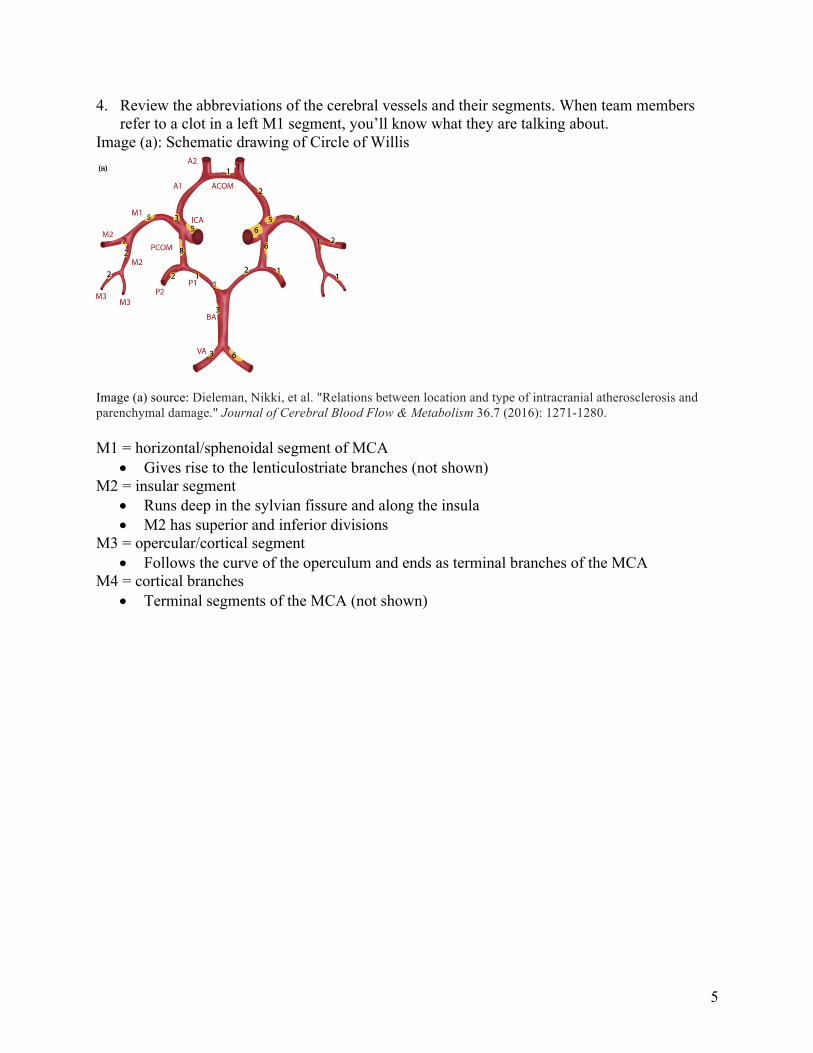
4. Review the abbreviations of the cerebral vessels and their segments. When team members refer to a clot in a left M1 segment, you’ll know what they are talking about.
Image (a): Schematic drawing of Circle of Willis Image (a) source: Dieleman, Nikki, et al. "Relations between location and type of intracranial atherosclerosis and parenchymal damage." Journal of Cerebral Blood Flow & Metabolism 36.7 (2016): 1271-1280. M1 = horizontal/sphenoidal segment of MCA
• Gives rise to the lenticulostriate branches (not shown) M2 = insular segment
• Runs deep in the sylvian fissure and along the insula • M2 has superior and inferior divisions
M3 = opercular/cortical segment • Follows the curve of the operculum and ends as terminal branches of the MCA
M4 = cortical branches • Terminal segments of the MCA (not shown)
6
Imaging Tips • CT = use to rule out hemorrhage, calculate ASPECT Score • CTA = use to assess patency of cerebral blood vessels • MRI is a powerful tool to image the brain and assess what parts of the brain might have been
affected in a stroke. An MRI sequence is a number of radiofrequency pulses and gradients that result in a set of images with particular appearance. The stroke unit utilizes a number of different MRI sequences in order to diagnose the location and extent of a patient’s stroke. Common sequences used include: Diffusion weight imaging (DWI), Apparent diffusion coefficient (ADC), FLASH, FLAIR, and rapid-sequence. • DWI assesses the ease with which water molecules move around within a tissue. Patient
has a stroke à decreased blood supply to brain parenchyma à decreased O2 supply to neurons à ATP pumps stop working à Na/K pumps stop working à cells swell à decreased movement of water from inside to outside of cells à restricted diffusion see in DWI à ischemic brain appears bright on DWI
o In order to confirm it’s a stroke and not artifact of the imaging, we compare the image to the ADC sequence
• ADC also assesses the ease with which water molecules move around within a tissue. However, on ADC, the acute ischemic pathology, such as a stroke, appears dark. Therefore a true ischemic stroke will be bright on DWI AND dark on ADC.
• FLASH (Fast low angle shot) = used to assess hemorrhage, and cerebral amyloid angiopathy
• FLAIR (Fluid-attenuate inversion recovery) = used to assess microvascular strokes • Rapid-sequence: This MRI sequence incorporates DWI and FLAIR sequences to quickly
(in less than 10 min) assess perfusion mismatch. We use this sequence to rule out stroke mimics, such as metabolic, traumatic, migranous, neoplastic, convulsive, and psychiatric disorders which account for 3-30% of patients presenting with acute neurologic deficits.
7
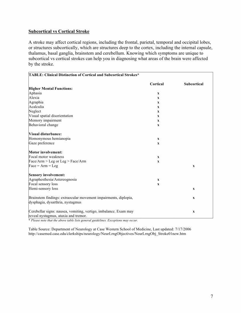
Subcortical vs Cortical Stroke A stroke may affect cortical regions, including the frontal, parietal, temporal and occipital lobes, or structures subcortically, which are structures deep to the cortex, including the internal capsule, thalamus, basal ganglia, brainstem and cerebellum. Knowing which symptoms are unique to subcortical vs cortical strokes can help you in diagnosing what areas of the brain were affected by the stroke. TABLE: Clinical Distinction of Cortical and Subcortical Strokes* Higher Mental Functions: Aphasia Alexia Agraphia Acalculia Neglect Visual spatial disorientation Memory impairment Behavioral change Visual disturbance: Homonymous hemianopia Gaze preference Motor involvement: Focal motor weakness Face/Arm > Leg or Leg > Face/Arm Face = Arm = Leg Sensory involvement: Agraphesthesia/Astereognosia Focal sensory loss Hemi-sensory loss Brainstem findings: extraocular movement impairments, diplopia, dysphagia, dysarthria, nystagmus Cerebellar signs: nausea, vomiting, vertigo, imbalance. Exam may reveal nystagmus, ataxia and tremor.
Cortical
x x x x x x x x
x x
x x
x x
Subcortical
x x
x
x
* Please note that the above table lists general guidelines. Exceptions may occur. Table Source: Department of Neurology at Case Western School of Medicine, Last updated: 7/17/2006 http://casemed.case.edu/clerkships/neurology/NeurLrngObjectives/NeurLrngObj_Stroke01new.htm
8
Creating a differential A stroke can be ischemic or hemorrhagic in origin. There are 4 main etiologies of ischemic stroke: Large Vessel Disease, Small vessel disease, Cardioembolic, and Cryptogenic. 1. Large vessel = Pathology in the Internal Carotids, Vertebral arteries, or Large arteries
originating from the Circle of Willis • Mechanisms of stroke:
o Intrinsic thrombosis à ischemia à symptoms often fluctuate (stuttering progression with periods of improvement)
§ Look for atherosclerotic risk factors (uncontrolled HTN, DM) o Hypoperfusion across a stenotic vessel o Emboli from an ulcerated atherosclerotic plaque (Artery to Artery embolism) à
symptoms are abrupt and usually maximal at the start 2. Small Vessel (Lacunar) = Pathology in the Small penetrating artery in the subcortex • Mechanism of stroke:
o Intrinsic lipohyalinosis or micro-atheroma in the small penetrating arteryà blocks blood flow à ischemic stroke
§ Deficits may wax and wane, and have repetitive stereotyped symptoms § Neurological deficits may fit with a lacunar syndrome such as pure motor
hemiparesis, pure hemi-sensory loss, or clumsy-hand dysarthria
3. Cardioembolic = A thrombus forms in the heart, and then embolizes to the brain • Risk Factors for thrombus formation in the atrium = having a sick heart, which can be
evidenced by having Afib, left atrial enlargement, an EKG significant for prior MIs • Endocarditis can lead to the formation of vegetations on the mitral valve, which can embolize
to the brain • Symptoms are abrupt and usually maximal at the start • Can see multiple infarcts within different vascular territories
Embolism), and more. There are 4 main hemorrhagic stroke subtypes: Epidural Hemorrhage, Subdural Hemorrhage, Subarachnoid hemorrhage, and intracerebral hemorrhage 1. Epidural Hemorrhage = Due to rupture of the middle meningeal artery, often caused by
traumatic brain injury • Lucid interval injury • Can lead to transtentorial herniation
2. Subdural Hemorrhage = Due to rupture of bridging veins, can be caused by acute trauma
(high-energy impact) or chronic trauma (cerebral atrophy, elderly, alcoholism)
9
3. Subarachnoid hemorrhage = Most commonly caused by rupture of a saccular aneurysm or an AVM • Severe HA at onset of neurologic symptoms • Meningeal irritation (eg. Neck stiffness) seen • Focal neurologic deficits uncommon
4. Intracerebral hemorrhage = Most commonly caused by uncontrolled HTN, coagulopathy,
illicit drug use (amphetamines, cocaine) • Symptoms progress over minutes to hours • Focal neurologic symptoms appear early (depending on site of bleed-in the basal ganglia,
cerebellum, thalamus, cerebral lobe, or brainstem), followed by features of increased ICP (vomiting and headache, bradycardia, reduced alertness)
Sometimes… it’s not a stroke. Remember to keep your differential wide, and to employ all the knowledge you’ve gained in your medical education. Don’t just zero in on stroke. Pathologies that can look like a stroke: Migraines, seizures, hypoglycemia, BPPV, Bell’s palsy
10
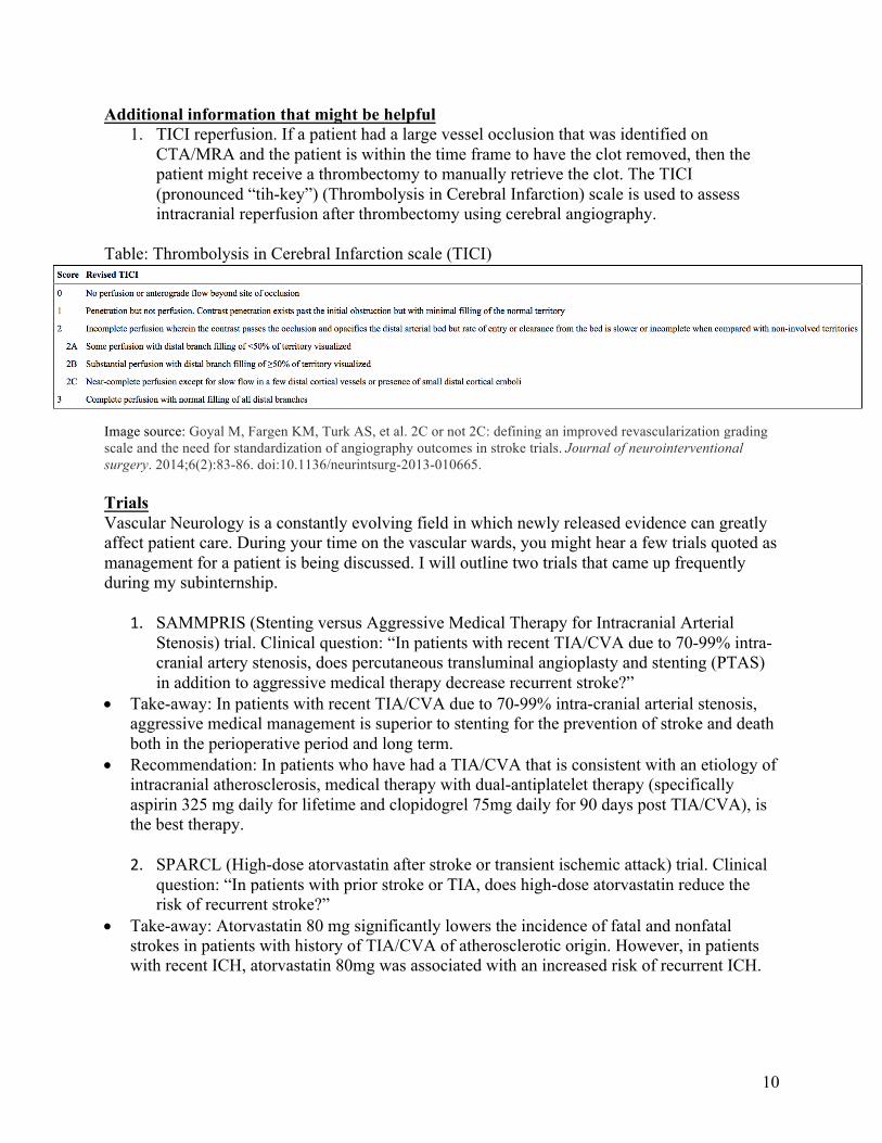
Additional information that might be helpful 1. TICI reperfusion. If a patient had a large vessel occlusion that was identified on
CTA/MRA and the patient is within the time frame to have the clot removed, then the patient might receive a thrombectomy to manually retrieve the clot. The TICI (pronounced “tih-key”) (Thrombolysis in Cerebral Infarction) scale is used to assess intracranial reperfusion after thrombectomy using cerebral angiography.
Table: Thrombolysis in Cerebral Infarction scale (TICI)
Image source: Goyal M, Fargen KM, Turk AS, et al. 2C or not 2C: defining an improved revascularization grading scale and the need for standardization of angiography outcomes in stroke trials. Journal of neurointerventional surgery. 2014;6(2):83-86. doi:10.1136/neurintsurg-2013-010665. Trials Vascular Neurology is a constantly evolving field in which newly released evidence can greatly affect patient care. During your time on the vascular wards, you might hear a few trials quoted as management for a patient is being discussed. I will outline two trials that came up frequently during my subinternship.
1. SAMMPRIS (Stenting versus Aggressive Medical Therapy for Intracranial Arterial Stenosis) trial. Clinical question: “In patients with recent TIA/CVA due to 70-99% intra-cranial artery stenosis, does percutaneous transluminal angioplasty and stenting (PTAS) in addition to aggressive medical therapy decrease recurrent stroke?”
• Take-away: In patients with recent TIA/CVA due to 70-99% intra-cranial arterial stenosis, aggressive medical management is superior to stenting for the prevention of stroke and death both in the perioperative period and long term.
• Recommendation: In patients who have had a TIA/CVA that is consistent with an etiology of intracranial atherosclerosis, medical therapy with dual-antiplatelet therapy (specifically aspirin 325 mg daily for lifetime and clopidogrel 75mg daily for 90 days post TIA/CVA), is the best therapy. 2. SPARCL (High-dose atorvastatin after stroke or transient ischemic attack) trial. Clinical
question: “In patients with prior stroke or TIA, does high-dose atorvastatin reduce the risk of recurrent stroke?”
• Take-away: Atorvastatin 80 mg significantly lowers the incidence of fatal and nonfatal strokes in patients with history of TIA/CVA of atherosclerotic origin. However, in patients with recent ICH, atorvastatin 80mg was associated with an increased risk of recurrent ICH.
11
Helpful websites/Resources: To practice neuroanatomy: http://headneckbrainspine.com/Neuroanatomy-modules.php To review material specific to cerebrovascular disorders: http://casemed.case.edu/clerkships/neurology/LearningObjectives.htm#Cerebrovascular_Disorders Dr. Ali Saad has a free smart phone app called “Stroke Trials” with summaries of the landmark stroke trials Useful Reference Books: Blumenfeld Clinical Neuroanatomy For the most up to date information on the rotation, consult the Alpert Medical School Brown Neurology Residency Handbook: https://goo.gl/1RQWwt Questions or Concerns about this document? Contact: Neesha Nama: [email protected] Kwame Adjepong: [email protected]