The Mitogen-Activated Protein Kinase (MAPK)-Activated ProteinKinases MK2 and MK3 Cooperate in Stimulation of Tumor
Necrosis Factor Biosynthesis and Stabilization of p38 MAPK�
N. Ronkina,1 A. Kotlyarov,1 O. Dittrich-Breiholz,2 M. Kracht,2 E. Hitti,1 K. Milarski,3R. Askew,3 S. Marusic,4 L.-L. Lin,4 M. Gaestel,1* and J.-B. Telliez4
Institute of Biochemistry1 and Institute of Pharmacology,2 Medical School Hannover, Carl-Neuberg-Strasse 1,30625 Hannover, Germany, and Biological Technology3 and Inflammation Department,4
Wyeth Research, 200 Cambridge Park Drive, Cambridge, Massachusetts 02140
Received 7 August 2006/Returned for modification 15 September 2006/Accepted 2 October 2006
MK2 and MK3 represent protein kinases downstream of p38 mitogen-activated protein kinase (MAPK).Deletion of the MK2 gene in mice resulted in an impaired inflammatory response although MK3, whichdisplays extensive structural similarities and identical functional properties in vitro, is still present. Here, weanalyze tumor necrosis factor (TNF) production and expression of p38 MAPK and tristetraprolin (TTP) inMK3-deficient mice and demonstrate that there are no significant differences with wild-type animals. We showthat in vivo MK2 and MK3 are expressed and activated in parallel. However, the level of activity of MK2 isalways significantly higher than that of MK3. Accordingly, we hypothesized that MK3 could have significanteffects only in an MK2-free background and generated MK2/MK3 double-knockout mice. Unexpectedly, thesemice are viable and show no obvious defects due to loss of compensation between MK2 and MK3. However,there is a further reduction of TNF production and expression of p38 and TTP in double-knockout micecompared to MK2-deficient mice. This finding, together with the observation that ectopically expressed MK3can rescue MK2 deficiency similarly to MK2, indicates that both kinases share the same physiological functionin vivo but are expressed to different levels.
Downstream of mitogen-activated protein kinases (MAPKs)different groups of MAPK-activated protein kinases (MAPKAPKs) have been defined (reviewed in reference 28). Theseenzymes transduce signals to target proteins that are not directsubstrates of the MAPKs and, therefore, serve to relay phos-phorylation-dependent signaling within MAPK cascades to di-verse cellular functions. One of these groups is formed by thethree MAPKAPKs, MK2, MK3 (also known as 3pK), and MK5(also designated PRAK) (reviewed in reference 12). WhileMK5 is mainly activated by the atypical MAPK ERK3 (29, 30),the remaining two kinases, MK2 and MK3, are directly down-stream of the MAPK p38�/� (7, 10, 24, 27, 31). Phosphoryla-tion of MK2 and MK3 by p38�/� at two or three major regu-latory sites leads to activation and coupled nuclear export ofboth enzymes, which are localized in the nucleus of restingcells (4, 8, 26, 36, 41).
A wide variety of substrates has been described for MK2including proteins interacting with the cytoskeleton, such assmall heat shock protein Hsp25 (33); mRNA-binding proteins,such as tristetraprolin (TTP) (6, 32); transcription factors, suchas heat shock factor 1 (38); and regulators of the cell cycle andapoptosis, such as Cdc25B/C (23). The phosphorylation siterecognition motifs of MK2 and MK3 are similar (20) or evenidentical (7). Despite the similar recognition motif, not allMK2 substrates have been described as MK3 substrates so far,
probably because in most cells MK2 activity dominates andmakes analysis of the minor MK3 activity dependent on anti-bodies which discriminate between both enzymes (7).
MK2-deficient mice are more resistant than wild type toendotoxic shock due to impaired production of cytokinessuch as tumor necrosis factor (TNF) (16). By genetic evi-dence it has been demonstrated that the mechanism bywhich MK2 stimulates lipopolysaccharide (LPS)-dependentTNF biosynthesis exists at the posttranscriptional level andis dependent on the adenine/uridine-rich element (ARE) inthe 3� untranslated region of TNF mRNA (25) and on theexistence of the TNF mRNA-destabilizing protein TTP (15).Probably, phosphorylation of TTP by MK2 inactivates itsmRNA-destabilizing activity and, in parallel, leads to stabi-lization and storage of phospho-TTP in complex with 14-3-3proteins until dephosphorylation reactivates TTP and down-regulates the inflammatory response (5, 6, 15, 32). Hence,phosphorylation of TTP by MK2 stabilizes TNF mRNA andstimulates its translation to TNF protein. The strong effectof deletion of MK2 on cytokine biosynthesis raised ques-tions about the role of MK3 in this process and producedserious doubts about the functional congruence of both en-zymes. Alternatively, MK3 could be responsible for residualcytokine biosynthesis or compensate an as yet undefinedfunction of MK2 in an unrelated process such as contribut-ing to polycomb-regulated development (37).
Here, we analyzed the role of MK3 by gene targeting strat-egy. By using an MK2-free genetic background, we were ableto assess MK3-specific effects in vivo and, hence, to define thephysiological role of this enzyme.
Generation of MK3 knockout and conditional knockout mice. MK3 knockoutmice were developed by gene-targeted mutagenesis in 129SvEvBrd embryonicstem (ES) cells and with Cre-mediated recombination. A conditional knockout(CKO) allele of MK3 was designed by Wyeth, and mice carrying this CKO allelewere created by Lexicon Genetics, Inc. Gene-targeted ES cell clones were iden-tified by Southern blotting and restriction fragment length polymorphism anal-ysis of HindIII-digested ES cell DNA using a 3� probe external to the targetingconstruct in which the wild-type band is 8.6 kb and the targeted band is 10.6 kb.The CKO allele contains LoxP sites flanking exon 1 and exon 2 including thetranslational start site. The MK3 KO allele was converted from the CKO allelevia germ line Cre-mediated deletion by crossing protamine-Cre transgenic mice(129SvEvBrd) with MK3-CKO mice. MK2 knockout mice were generated asdescribed previously (16). MK2�/� mice on the C57BL/6 genetic backgroundwere bred to MK3�/� mice on the mixed (129Sv � C57BL/6) genetic backgroundto obtain MK2�/� MK3�/� mice. F2 littermates were genotyped by PCR andused in experiments. All mice used in this study were maintained under specific-pathogen-free conditions and treated in accordance with local ethical guidelines.
Genotyping of MK3 conditional allele and MK3/MK2 knockout mice. Micewere genotyped by PCR analysis of proteinase K lysate of tail biopsies. Wild-type(WT) and MK3-CKO alleles were distinguished using PCR of an ampliconspanning the 5� LoxP insertion site with a forward primer of the sequence5�-ATTGATTGAGCCGGGCGTGGTG-3� and a reverse primer of the se-quence 5�-CCTGTAATTGCAGCGCGAGGAA-3�, yielding 416-bp WT prod-uct and a 514-bp CKO product. The MK3 knockout (deletion) allele was distin-guished from the WT allele by PCR of an amplicon spanning the deletionjunction using the forward primer 5�-ATTGATTGAGCCGGGCGTGGTG–3�and a reverse primer of the sequence 5�-CACAAGGTAGAGATTACGGCCAC-3�, yielding a 581-bp, MK3-KO-specific product. MK2 was genotyped as de-scribed previously (16).
Cell culture. Resident peritoneal macrophages were collected after intraper-itoneal injection of 5 ml of Dulbecco’s modified Eagle’s medium (DMEM) with10% fetal bovine serum, washed once with phosphate-buffered saline (PBS),resuspended in complete medium, and plated at 5 � 103 cells on chamber slides(Nunc, Naperville, Ill.). After 2 h at 37°C in a 95% air–5% CO2 incubator, themacrophages were washed twice with DMEM to remove nonadherent cells andcultivated for a further 16 h prior to stimulation.
To generate bone marrow-derived macrophages (BMDMs), bone marrowcells were flushed from the femurs of mice. Cells were cultured on 10-cm dishesin DMEM supplemented with 10% heat-inactivated fetal bovine serum, 2 mML-glutamine, 100 U/ml penicillin G, 100 �g/ml streptomycin, and 50 ng/ml re-combinant macrophage colony-stimulating factor (CSF) (Wyeth, Boston, MA)for 7 days.
Primary mouse embryonic fibroblasts (MEFs) were isolated from day 13.5mouse embryos. The heads and internal organs were removed from the embryos.The remaining tissues were then cut into small pieces, and single cells wereobtained by incubation in trypsin. Cells were then cultured in DMEM containing10% serum, 2 mM L-glutamine, 100 U of penicillin G/ml, and 100 �g of strep-tomycin/ml and used at passages 3 to 5. To immortalize primary MEFs, cells werecotransfected with pSV40Tag encoding simian virus 40 large T antigen andpREP8 plasmid (Invitrogen) in a 10:1 mixture; colonies were selected with 3 mMhistidinol (Sigma).
Cloning and site-directed mutagenesis. The full-length murine MK3 openreading frame was amplified from cDNA clone MGC:25617 distributed byBioCat (Heidelberg/Germany) using the forward primer MK3_EcoRI (5�-CATGGG AAT TCA TGG ATG GCG AGA CAG CAG GG-3�) and reverse primerMK3_SalI (5�-CAT GGG TCG ACG TTA CTG GTTGTT GCA TCC TTG-3�).The PCR product was digested by EcoRI/SalI and cloned into of pGEX-5x-1(Amersham).
For cloning into pENTR/D-TOPO (Invitrogen), mouse p38 full-length cDNAwas amplified by PCR using the primer pair 5�-CAC CTC GCA GGA GAGGCC CAC GTT CTA C-3� (forward) and 5�-GGA CTC CAT TTC TTC TTGGTC AAG-3� (reverse). The recombination reaction between the entry cloneand the pDEST15 vector for glutathione S-transferase (GST)-tagged alpha-p38bacterial expression was achieved with an LR Clonase Kit (Invitrogen). Site-directed mutagenesis was performed in pDEST15-alpha-p38 using a Quick-Change XL site-directed mutagenesis kit (Stratagene).
GST-p38 pull-down and kinase assay. Cells cultivated overnight in serum-freemedium were stimulated with either anisomycin, arsenite, or LPS (Sigma) usingthe concentrations and times indicated in the figures and figure legends. Forkinase assays and immunoblotting, cells were lysed in 50 mM Tris-HCl (pH 7.5),1 mM EGTA, 1 mM EDTA, 1 mM sodium orthovanadate, 50 mM sodium
fluoride, 1 mM sodium pyrophosphate, 0.27 M sucrose, 1% (vol/vol) TritonX-100, 0.1% (vol/vol) 2-mercaptoethanol, and complete proteinase inhibitorcocktail (Roche, East Sussex, United Kingdom). The lysates were centrifuged at13,000 rpm for 5 min at 4°C, and the supernatants were removed and stored at�80°C until use.
For GST pull-down 1 mg of lysate protein was incubated with 0.1 nmol ofrecombinant catalytically dead mutant GST-p38 (TGY/AFG) bound to glutathi-one-Sepharose 4B (Amersham Pharmacia Biotech). After five washes with im-munoprecipitation buffer (1� Tris-buffered saline, 50 mM NaF, 1% TritonX-100, 1 mM Na3VO4), the beads were used for kinase assay (16) or applied foran in-gel kinase assay (9). In both cases recombinant Hsp25 was used as asubstrate. Radioactivity incorporated into Hsp25 was visualized by phosphor-imaging using a Fuji Bas-1500, and the signal was quantified by the use of TINA2.09 software.
Western blotting. Soluble protein extract was run on sodium dodecyl sulfate–10% polyacrylamide gel electrophoresis (SDS–10% PAGE) gels and transferredto Hybond ECL membranes (Amersham Pharmacia Biotech). Blots were incu-bated for 2 h in PBS–1% Tween 20 containing 5% powdered skim milk. Afterthree washes with PBS–1% Tween 20, membranes were incubated for 16 h withthe primary antibody at 4°C and for 1 h with horseradish peroxidase-conjugatedsecondary antibodies (diluted 2,000-fold) at room temperature. Blots were de-veloped with an ECL detection kit (Santa Cruz Biotechnology), and the digitalchemiluminescence images were taken by a Luminescent Image Analyzer LAS-3000 (Fujifilm).
Antibodies. Anti-MK3 antibodies were raised against bacterially expressedMK3 protein. The full-length murine MK3 open reading frame was cloned inpGEX-5x-1 vector. Recombinant protein was affinity purified on glutathione-Sepharose and, the GST part was removed by Xa factor cleavage, injected intorabbits at Eurogentec (Seraing, Belgium). Antibodies that recognize p38 MAPK,phospho-p38 MAPK, MAPKAPK-2, and phospho-MAPKAPK-2 (Thr222) werefrom New England Biolabs. Antibody against Hsp25 was from Stressgene, andanti-phospho-Hsp25 (S86) was from Biosource. Anti-GAPDH (glyceraldehyde-3-phosphate dehydrogenase) antibody was from Chemicon. Antibodies against greenfluorescent protein (GFP), actin, and horseradish peroxidase-conjugated secondaryantibodies were from Santa Cruz. Anti-TTP antibody was previously described (22).
Retroviral gene transfer. Full-length murine MK2, MK3, and MK2 catalyti-cally dead mutant (K76R) were subcloned into the pMMP-IRES-GFP (kind giftof C. Klein, MHH, Hanover) bicistronic retroviral vector upstream of the inter-nal ribosome entry site (IRES). To obtain MEF cell lines stably expressing thekinases, retroviral supernatants were generated by transient transfection of theBD EcoPack 2-293 packaging cell line with bicistronic vectors encoding the geneof interest and GFP as marker and were used for infection. Cell lines with morethan 90% GFP-positive cells were used for experiments.
Measurement of cytokines. BMDMs were scraped from 10-cm dishes, and 104
cells per well were transferred to a 98-well plate and incubated in 100 �l ofmedium for 2 h before the addition of 1 �g/ml LPS for 2 and 6 h. Murine TNF,interleukin-6 (IL-6), and CXCL1 were measured using specific enzyme-linkedimmunosorbent assay (ELISA) kits (R&D) according to the manufacturer’sinstructions. In selected experiments cytokine profiling was performed usingMeso Scale Discovery (MSD) Multi-Spot plates and an MSD Sector Imager 6000reader (Gaithersburg, MD). A total of 10 cytokines were measured simulta-neously in each well of 96-well plates using an MSD 10-Plex Mouse CytokinePanel (IL-12p40, IL-6, KC [IL-8], IL-10, IL-1, RANTES, granulocyte macro-phage-CSF [GM-CSF], gamma interferon, monocyte chemoattractant protein 1,and TNF) according to the manufacturer’s instructions.
Oligonucleotide DNA microarray experiments. The inflammation array(Inflmus; MWG Biotech, now sold by Ocimum as Inflammation OciChip) usedin this study contains 155 validated oligonucleotide probes for 136 murine in-flammatory and 19 housekeeping genes. Total RNA from MK-2-deficient andreconstituted cell lines treated as indicated in the legend of Fig. 6 was purifiedwith a QIAGEN RNeasy kit followed by on-column DNase I digestion (QIAGEN).RNA was used to prepare Cy3-labeled cRNA by oligo(dT)-T7-primed double-stranded cDNA synthesis (cDNA synthesis system; Roche), followed by in vitrotranscription with T7 polymerase (MEGAscript T7 kit; Ambion) as directed bythe manufacturers. cRNA yield was determined photometrically. Equal amountsof cRNAs derived from approximately 1.5 �g of total RNA were hybridizedindividually to microarrays in preprepared hybridization solution (MWG Biotech) at42°C overnight and then washed sequentially in 2� SSC (1� SSC is 0.15 M NaClplus 0.015 M sodium citrate), 0.1% SDS, 1� SSC, and 0.5� SSC. Hybridized arrayswere scanned at maximal resolution on an Affymetrix 428 scanner at variable pho-tomultiplier tube voltage settings. Fluorescence intensity values from Cy3 channelswere processed using Imagene 4.2 software (Biodiscovery). Normalized values wereobtained by MAVI software (version Pro 2.5.1; MWG Biotech).
VOL. 27, 2007 ROLE OF MAPKAP KINASES MK2 AND MK3 171
Heat map visualization was performed by importing log 2-transformed ratiodata into the Mayday program (http://www.zbit.uni-tuebingen.de/pas/mayday/mayday.html).
Real-time PCR. cDNA prepared in parallel to the microarray experiments wasused to validate selected results by real-time PCR. Assays on demand (AppliedBiosystems; detailed information is in parentheses) for the following transcriptswere used: �-actin (Actb; Mm00607939_s1), I�B-� (Nfkbia; Mm00477798_m1),Cxcl1 (Mm00433859_m1), c-Jun (Jun; Mm00495062_s1), and Csf2(Mm00438328_m1). PCR was performed on an ABI7500 real-time PCR instru-
ment. The threshold cycle (CT) for each individual PCR product was calculatedby the instrument software, and CT values obtained for Nfkbia, Cxcl1, Jun, andCsf2 were normalized by subtracting the CT values obtained for Actb to obtainCT values. Mean values were calculated from two to four CT measurements; thestandard error of the mean for all corresponding CT values was less than 0.2.Relative expression of each mRNA was calculated by the CT method assum-ing a PCR efficiency of two.
LPS treatment of mice. LPS from Escherichia coli serotype 026:B6 (Sigma) wasdiluted in pyrogen-free saline, and 5 mg per kg of body weight was injected
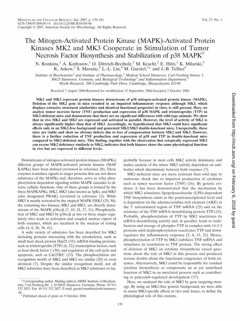
FIG. 1. Gene targeting strategy of MK3 deletion and absence of MK3 protein in MK3�/� tissues. (A) The MK3-CKO construct is composedof a 7-kb fragment of the MK3 gene with a 3-kb short arm and a 4-kb long arm flanking exons 1 and 2 and the neomycin resistance selectablemarker gene (Neor). Exons 1 and 2 and Neor are flanked with LoxP sites as indicated by solid triangles. Positions of the Hind III restriction sitesare indicated. Also indicated are positions of PCR primers, amplicons, and product sizes used for genotyping. (B) Southern blot analysis showingwild-type (�/�) and targeted (�/C) ES cell clones. PCR genotyping results from different sets of tail biopsies distinguishing WT (�) andconditional (C/C and C/�) alleles. (D) PCR results identifying KO (�) alleles after the MK3 KO allele was converted via germ line Cre-mediateddeletion by crossing protamine-Cre transgenic mice (129SvEvBrd) with MK3-CKO mice. (E) Endogenous MK3 and MK2 were precipitated from1 mg of spleen lysates by means of recombinant GST-p38 bound to glutathione-Sepharose 4B. Beads and 0.2 mg of whole lysates were subjectedto SDS-PAGE and Western blotting using anti-MK3 antiserum (upper blot) and anti-MK2 antibodies (lower blot). There is a specific 42-kDa MK3band precipitated from WT tissues which completely disappears in MK3�/� tissues. Endogenous MK3 is not detectable in whole cell lysates.ns,nonspecific band.
intraperitoneally into five mice of each genotype. Ninety minutes after injectionmice were sacrificed. Spleens were isolated and immediately frozen in liquidnitrogen. Spleen lysates were prepared using kinase assay lysis buffer and ana-lyzed by Western blotting as described above. Serum cytokines were quantifiedby ELISA or by a Multi-Spot cytokine assay as described above.
RESULTS
Generation and characterization of MK3 knockout mice.The MK3 gene was targeted in mice by homologous recombi-nation in ES cells, resulting in insertion of a floxed generegion spanning exons 1 and 2 and containing a neomycincassette (Fig. 1A and B) and generation of MK3 CKO mice(Fig. 1C). The MK3 KO allele was converted via germ lineCre-mediated deletion by crossing protamine-Cre trans-genic mice (129SvEvBrd) with MK3-CKO mice (Fig. 1D).MK3�/� KO mice are viable and fertile and do not displayabnormalities in tissue morphology or behavior. To prove thatdeletion of the gene resulted in deletion of the encoded pro-tein, we analyzed expression of MK3 in spleen cell lysates ofWT and MK3�/� mice by Western blotting. No MK3 bandcould be detected in wild-type lysates, although MK2 detectionwas clearly possible (Fig. 1E, lysate). Since the sensitivities ofthe anti-MK3 and anti-MK2 antisera used are comparable (notshown), this indicates that in wild-type tissues expression ofMK3 is much less than that of MK2. The MK3 mRNA expres-sion level detected by reverse transcription-PCR is also muchless than that of MK2 (data not shown). To overcome thesetechnical problems and to specifically enrich p38-binding pro-teins, we used GST-p38 pull-down before MK3 Western blot-ting. For wild-type spleen cells (Fig. 1E) and other tissues (notshown), a specific band for MK3 can be detected in the pull-down, which is completely missing in MK3�/� cells. This indi-cates that the deleted gene results in complete loss of MK3protein expression.
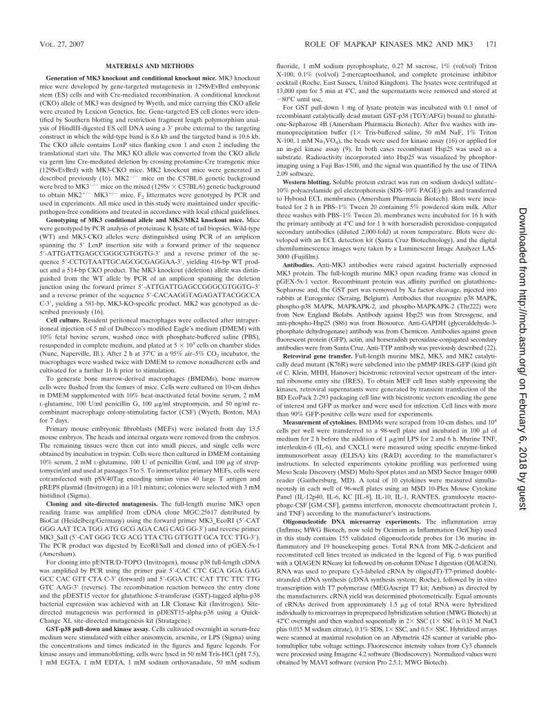
No significant changes in cytokine production, p38, andTTP levels in MK3�/� mice. The phenotype of MK2 deletionis very prominent, displaying defects in LPS-induced endotoxicreactions and cytokine production (16), reduction of p38 (17)and TTP levels (15), defects in chemotaxic cell migration (13),and increased susceptibility to infection (19). Since MK3 ex-hibits evolutionary, structural, and apparent extensive func-tional similarities to MK2 (12), we analyzed the phenotype ofMK3 deficiency in regard to cytokine production, p38, andTTP level. In contrast to MK2 knockout mice and cells,MK3�/� mice did not show a decrease either in LPS-inducedproduction of the cytokines IL-12p40, IL-6, KC (IL-8), IL-10,IL-1, RANTES, GM-CSF, gamma interferon, monocyte che-moattractant protein 1, and TNF or in IL-6 mRNA stability(not shown). Furthermore, in MK3-deficient macrophages(Fig. 2) and in different MK3�/� tissues (not shown), theprotein levels of p38 and TTP were not significantly changedcompared to the WT, and expression and activation of MK2were not significantly altered (Fig. 2). Hence, MK3 deletionresulted in a phenotype clearly different from the complexinflammation-impaired phenotype of mice deficient in MK2 inspite of the extensive structural and regulatory similarities ofMK2 and MK3. This discrepancy could be explained by differ-ent spatial-temporal expression of both enzymes.
MK3 and MK2 are ubiquitously expressed and phosphory-lated in parallel upon LPS stimulation. We analyzed protein
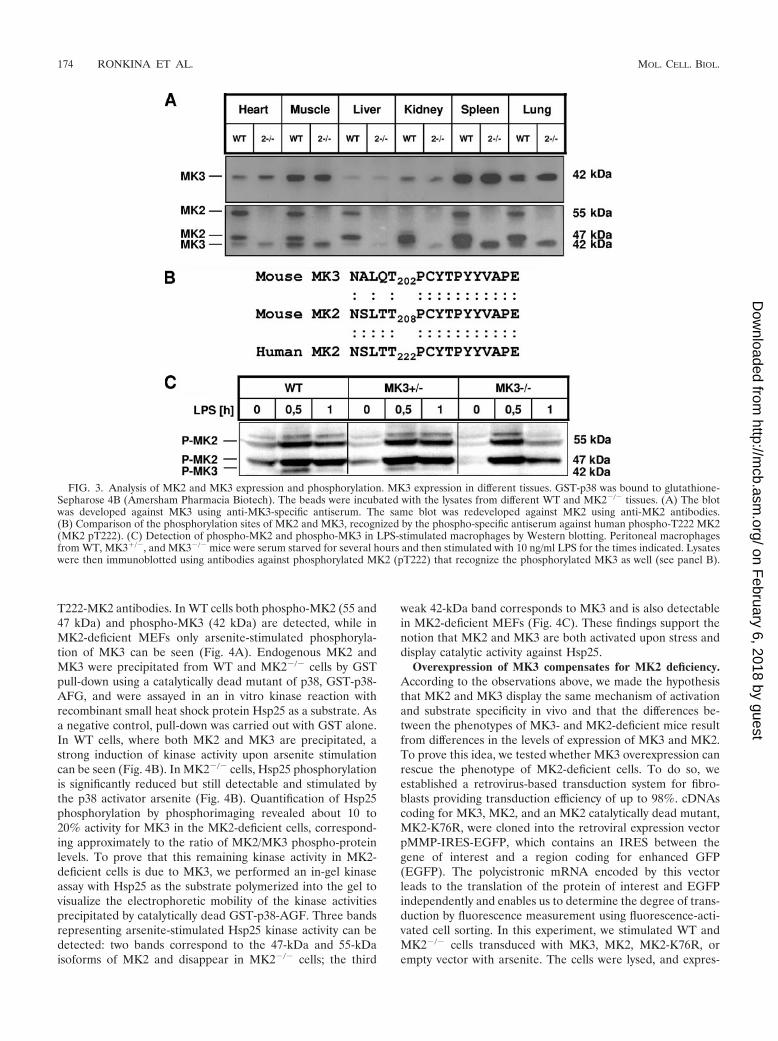
expression of MK3 and MK2 in different mouse tissues fromwild-type and MK2-deficient mice by using GST-p38 pull-downto enrich endogenous MK3 and MK2 and to avoid nonspecificbands in subsequent Western blot detection. MK3 proteinmigrating with an apparent molecular mass of 42 kDa is ubiq-uitously expressed at different levels in the tissues analyzed(Fig. 3A, upper blot). High levels of MK3 could be detected inthe spleen, lung, and skeletal muscle. The pattern of MK3expression is similar to MK2, which is detected on the sameblot as additional bands with apparent molecular masses of 47and 55 kDa (Fig. 3A, lower blot). There is no difference inMK3 expression between MK2�/� and WT tissues.
Next we analyzed the phosphorylation of MK3 at one of itsregulatory phosphorylation sites located in the activation loopof the kinase, threonine T203 in mouse, using an antiserumagainst phospho-T222 of human MK2 (Fig. 3 B). This anti-serum is well suited to detect both mouse phospho-T208-MK2and phospho-T203-MK3. A 42-kDa band specific for MK3 isphosphorylated in response to LPS, is reduced in macrophagesfrom MK3�/� animals, and disappears in MK3�/� macro-phages (Fig. 3C). It can be seen that MK3 phosphorylationupon LPS stimulation of primary macrophages has dynamicssimilar to p38 activation (Fig. 2) and MK2 phosphorylation,with the maximum reached after about 30 min and a subse-quent decrease.
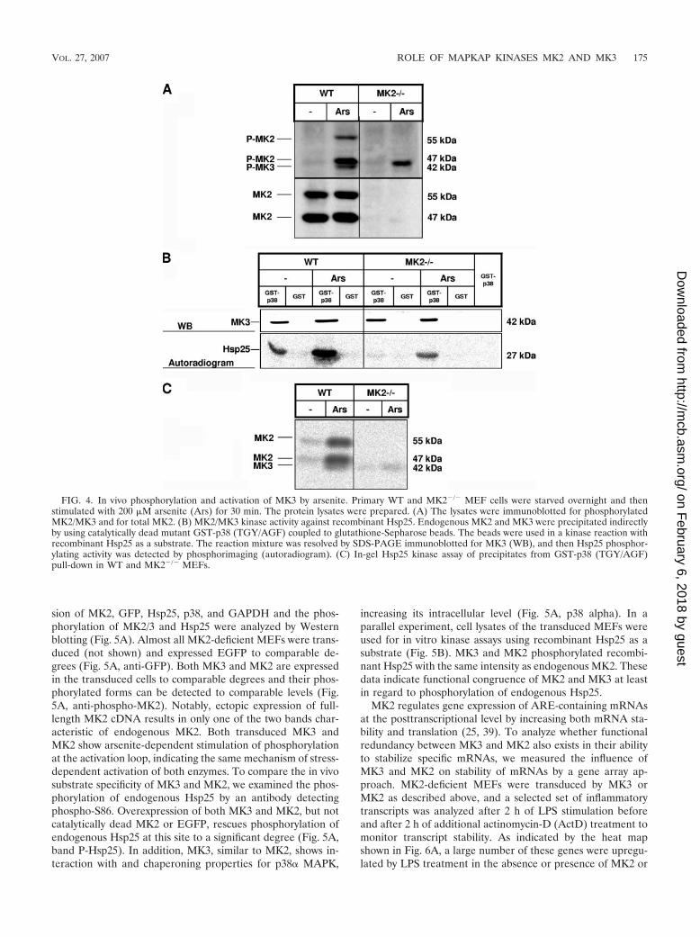
Detection of stress-stimulated catalytic activity of MK3 inMK2-deficient cells. To find out whether MK3 is activated inresponse to stress stimuli other than LPS, we stimulated pri-mary WT and MK2-deficient MEFs with arsenite. Phosphor-ylation of MK2 as well as MK3 upon arsenite treatment can bedemonstrated by Western blotting using the anti-phospho-
FIG. 2. Analysis of p38/MK2 signaling in MK3-deficient macro-phages. There were no significant changes in p38 MAPK, MK2, and TTPexpression and phosphorylation levels in MK3-deficient cells. Peritonealmacrophages from WT, MK3�/�, and MK3�/� mice were serum starvedfor several hours and then stimulated with 10 ng/ml LPS for the timesindicated. Lysates were then immunoblotted for total and phosphorylatedp38 MAPK and MK2 and with an antiserum that recognizes both thephosphorylated and nonphosphorylated forms of TTP.
VOL. 27, 2007 ROLE OF MAPKAP KINASES MK2 AND MK3 173
T222-MK2 antibodies. In WT cells both phospho-MK2 (55 and47 kDa) and phospho-MK3 (42 kDa) are detected, while inMK2-deficient MEFs only arsenite-stimulated phosphoryla-tion of MK3 can be seen (Fig. 4A). Endogenous MK2 andMK3 were precipitated from WT and MK2�/� cells by GSTpull-down using a catalytically dead mutant of p38, GST-p38-AFG, and were assayed in an in vitro kinase reaction withrecombinant small heat shock protein Hsp25 as a substrate. Asa negative control, pull-down was carried out with GST alone.In WT cells, where both MK2 and MK3 are precipitated, astrong induction of kinase activity upon arsenite stimulationcan be seen (Fig. 4B). In MK2�/� cells, Hsp25 phosphorylationis significantly reduced but still detectable and stimulated bythe p38 activator arsenite (Fig. 4B). Quantification of Hsp25phosphorylation by phosphorimaging revealed about 10 to20% activity for MK3 in the MK2-deficient cells, correspond-ing approximately to the ratio of MK2/MK3 phospho-proteinlevels. To prove that this remaining kinase activity in MK2-deficient cells is due to MK3, we performed an in-gel kinaseassay with Hsp25 as the substrate polymerized into the gel tovisualize the electrophoretic mobility of the kinase activitiesprecipitated by catalytically dead GST-p38-AGF. Three bandsrepresenting arsenite-stimulated Hsp25 kinase activity can bedetected: two bands correspond to the 47-kDa and 55-kDaisoforms of MK2 and disappear in MK2�/� cells; the third
weak 42-kDa band corresponds to MK3 and is also detectablein MK2-deficient MEFs (Fig. 4C). These findings support thenotion that MK2 and MK3 are both activated upon stress anddisplay catalytic activity against Hsp25.
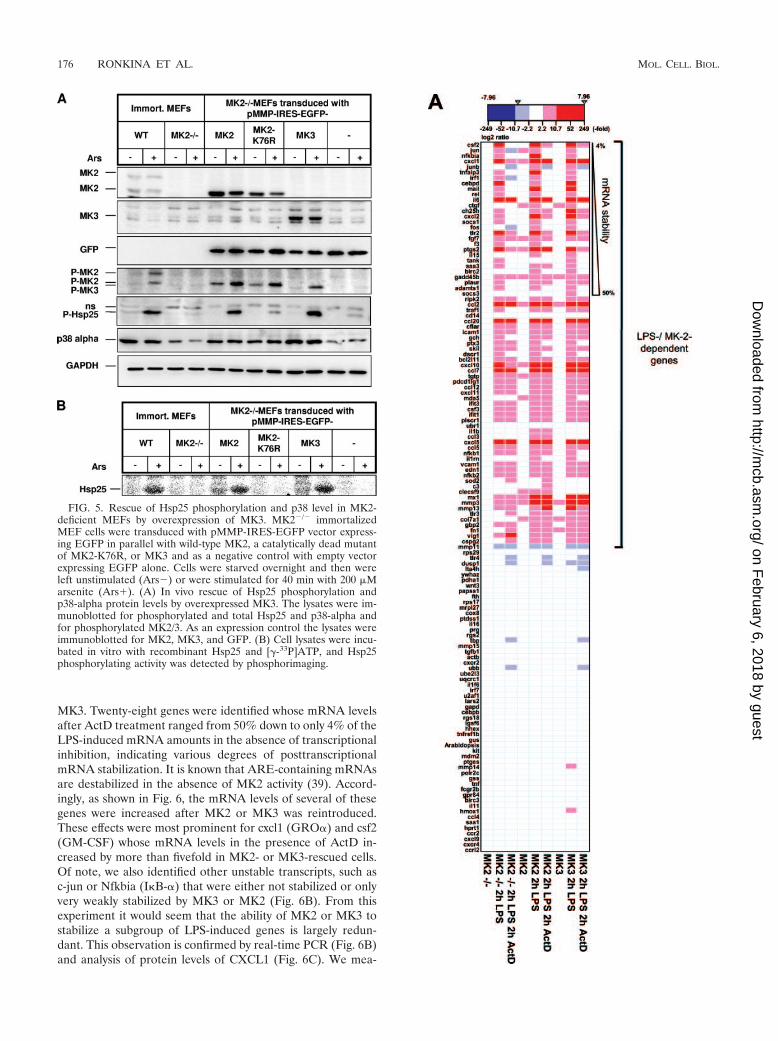
Overexpression of MK3 compensates for MK2 deficiency.According to the observations above, we made the hypothesisthat MK2 and MK3 display the same mechanism of activationand substrate specificity in vivo and that the differences be-tween the phenotypes of MK3- and MK2-deficient mice resultfrom differences in the levels of expression of MK3 and MK2.To prove this idea, we tested whether MK3 overexpression canrescue the phenotype of MK2-deficient cells. To do so, weestablished a retrovirus-based transduction system for fibro-blasts providing transduction efficiency of up to 98%. cDNAscoding for MK3, MK2, and an MK2 catalytically dead mutant,MK2-K76R, were cloned into the retroviral expression vectorpMMP-IRES-EGFP, which contains an IRES between thegene of interest and a region coding for enhanced GFP(EGFP). The polycistronic mRNA encoded by this vectorleads to the translation of the protein of interest and EGFPindependently and enables us to determine the degree of trans-duction by fluorescence measurement using fluorescence-acti-vated cell sorting. In this experiment, we stimulated WT andMK2�/� cells transduced with MK3, MK2, MK2-K76R, orempty vector with arsenite. The cells were lysed, and expres-
FIG. 3. Analysis of MK2 and MK3 expression and phosphorylation. MK3 expression in different tissues. GST-p38 was bound to glutathione-Sepharose 4B (Amersham Pharmacia Biotech). The beads were incubated with the lysates from different WT and MK2�/� tissues. (A) The blotwas developed against MK3 using anti-MK3-specific antiserum. The same blot was redeveloped against MK2 using anti-MK2 antibodies.(B) Comparison of the phosphorylation sites of MK2 and MK3, recognized by the phospho-specific antiserum against human phospho-T222 MK2(MK2 pT222). (C) Detection of phospho-MK2 and phospho-MK3 in LPS-stimulated macrophages by Western blotting. Peritoneal macrophagesfrom WT, MK3�/�, and MK3�/� mice were serum starved for several hours and then stimulated with 10 ng/ml LPS for the times indicated. Lysateswere then immunoblotted using antibodies against phosphorylated MK2 (pT222) that recognize the phosphorylated MK3 as well (see panel B).
sion of MK2, GFP, Hsp25, p38, and GAPDH and the phos-phorylation of MK2/3 and Hsp25 were analyzed by Westernblotting (Fig. 5A). Almost all MK2-deficient MEFs were trans-duced (not shown) and expressed EGFP to comparable de-grees (Fig. 5A, anti-GFP). Both MK3 and MK2 are expressedin the transduced cells to comparable degrees and their phos-phorylated forms can be detected to comparable levels (Fig.5A, anti-phospho-MK2). Notably, ectopic expression of full-length MK2 cDNA results in only one of the two bands char-acteristic of endogenous MK2. Both transduced MK3 andMK2 show arsenite-dependent stimulation of phosphorylationat the activation loop, indicating the same mechanism of stress-dependent activation of both enzymes. To compare the in vivosubstrate specificity of MK3 and MK2, we examined the phos-phorylation of endogenous Hsp25 by an antibody detectingphospho-S86. Overexpression of both MK3 and MK2, but notcatalytically dead MK2 or EGFP, rescues phosphorylation ofendogenous Hsp25 at this site to a significant degree (Fig. 5A,band P-Hsp25). In addition, MK3, similar to MK2, shows in-teraction with and chaperoning properties for p38� MAPK,
increasing its intracellular level (Fig. 5A, p38 alpha). In aparallel experiment, cell lysates of the transduced MEFs wereused for in vitro kinase assays using recombinant Hsp25 as asubstrate (Fig. 5B). MK3 and MK2 phosphorylated recombi-nant Hsp25 with the same intensity as endogenous MK2. Thesedata indicate functional congruence of MK2 and MK3 at leastin regard to phosphorylation of endogenous Hsp25.
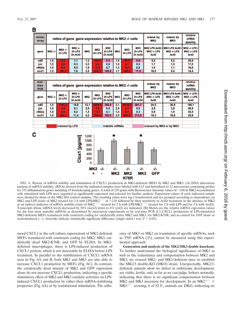
MK2 regulates gene expression of ARE-containing mRNAsat the posttranscriptional level by increasing both mRNA sta-bility and translation (25, 39). To analyze whether functionalredundancy between MK3 and MK2 also exists in their abilityto stabilize specific mRNAs, we measured the influence ofMK3 and MK2 on stability of mRNAs by a gene array ap-proach. MK2-deficient MEFs were transduced by MK3 orMK2 as described above, and a selected set of inflammatorytranscripts was analyzed after 2 h of LPS stimulation beforeand after 2 h of additional actinomycin-D (ActD) treatment tomonitor transcript stability. As indicated by the heat mapshown in Fig. 6A, a large number of these genes were upregu-lated by LPS treatment in the absence or presence of MK2 or
FIG. 4. In vivo phosphorylation and activation of MK3 by arsenite. Primary WT and MK2�/� MEF cells were starved overnight and thenstimulated with 200 �M arsenite (Ars) for 30 min. The protein lysates were prepared. (A) The lysates were immunoblotted for phosphorylatedMK2/MK3 and for total MK2. (B) MK2/MK3 kinase activity against recombinant Hsp25. Endogenous MK2 and MK3 were precipitated indirectlyby using catalytically dead mutant GST-p38 (TGY/AGF) coupled to glutathione-Sepharose beads. The beads were used in a kinase reaction withrecombinant Hsp25 as a substrate. The reaction mixture was resolved by SDS-PAGE immunoblotted for MK3 (WB), and then Hsp25 phosphor-ylating activity was detected by phosphorimaging (autoradiogram). (C) In-gel Hsp25 kinase assay of precipitates from GST-p38 (TGY/AGF)pull-down in WT and MK2�/� MEFs.
VOL. 27, 2007 ROLE OF MAPKAP KINASES MK2 AND MK3 175
MK3. Twenty-eight genes were identified whose mRNA levelsafter ActD treatment ranged from 50% down to only 4% of theLPS-induced mRNA amounts in the absence of transcriptionalinhibition, indicating various degrees of posttranscriptionalmRNA stabilization. It is known that ARE-containing mRNAsare destabilized in the absence of MK2 activity (39). Accord-ingly, as shown in Fig. 6, the mRNA levels of several of thesegenes were increased after MK2 or MK3 was reintroduced.These effects were most prominent for cxcl1 (GRO�) and csf2(GM-CSF) whose mRNA levels in the presence of ActD in-creased by more than fivefold in MK2- or MK3-rescued cells.Of note, we also identified other unstable transcripts, such asc-jun or Nfkbia (I�B-�) that were either not stabilized or onlyvery weakly stabilized by MK3 or MK2 (Fig. 6B). From thisexperiment it would seem that the ability of MK2 or MK3 tostabilize a subgroup of LPS-induced genes is largely redun-dant. This observation is confirmed by real-time PCR (Fig. 6B)and analysis of protein levels of CXCL1 (Fig. 6C). We mea-
FIG. 5. Rescue of Hsp25 phosphorylation and p38 level in MK2-deficient MEFs by overexpression of MK3. MK2�/� immortalizedMEF cells were transduced with pMMP-IRES-EGFP vector express-ing EGFP in parallel with wild-type MK2, a catalytically dead mutantof MK2-K76R, or MK3 and as a negative control with empty vectorexpressing EGFP alone. Cells were starved overnight and then wereleft unstimulated (Ars�) or were stimulated for 40 min with 200 �Marsenite (Ars�). (A) In vivo rescue of Hsp25 phosphorylation andp38-alpha protein levels by overexpressed MK3. The lysates were im-munoblotted for phosphorylated and total Hsp25 and p38-alpha andfor phosphorylated MK2/3. As an expression control the lysates wereimmunoblotted for MK2, MK3, and GFP. (B) Cell lysates were incu-bated in vitro with recombinant Hsp25 and [-33P]ATP, and Hsp25phosphorylating activity was detected by phosphorimaging.
sured CXCL1 in the cell culture supernatant of MK2-deficientMEFs transduced with constructs coding for MK2, MK3, cat-alytically dead MK2-K76R, and GFP by ELISA. In MK2-deficient macrophages, there is LPS-induced production ofCXCL1 protein, which is not detectable by ELISA before LPStreatment. In parallel to the stabilization of CXCL1 mRNAseen in Fig. 6A and B, both MK2 and MK3 are also able toincrease CXCL1 production by MEFs (Fig. 6C). In contrast,the catalytically dead mutant of MK2 and GFP expressionalone do not increase CXCL1 production, indicating a specificstimulatory effect of MK2 and MK3 catalytic activities on LPS-induced CXCL1 production by either their mRNA-stabilizingproperties (Fig. 6A) or by translational stimulation. The influ-
ence of MK3 or MK2 on translation of specific mRNAs, suchas TNF mRNA (25), cannot be measured using this experi-mental approach.
Generation and analysis of the MK2/MK3 double knockout.To further understand the biological significance of MK3 aswell as the redundancy and compensation between MK2 andMK3, we crossed MK2- and MK3-deficient mice to establishthe MK2/3 double-KO (DKO) strain. Unexpectedly, MK2/3-deficient animals show no defect in embryonic development,are viable, fertile, and, as far as we can judge, behave normally,indicating that there is no significant compensation betweenMK2 and MK3 necessary for development. In an MK2�/� �MK3�/� crossing, 6 of 82 F1 animals are DKO, indicating no
FIG. 6. Rescue of mRNA stability and stimulation of CXCL1 production in MK2-deficient MEFs by MK2 and MK3. (A) DNA microarrayanalysis of mRNA stability. cRNAs derived from the indicated samples were labeled with Cy3 and hybridized to 12 microarrays containing probesfor 155 inflammatory genes including 19 housekeeping genes. A total of 129 genes with fluorescence intensity values of �100 in MK2-reconstitutedcells stimulated with LPS were regarded as significantly expressed and selected for further analysis. Expression values of each indicated samplewere divided by those of the MK2 KO control samples. The resulting ratios were log 2 transformed and are grouped according to dependence onMK2 and LPS (ratio of MK2 treated for 2 h with LPS/MK2�/� of �2.0) followed by their sensitivity to ActD treatment in the absence of MK2as an indirect indicator of mRNA stability (ratio of MK2 �/� treated for 2 h with LPS/MK2 �/� treated for 2 h with LPS and for 2 h with ActD).Transcripts whose mRNA levels decreased by 50% (socs3) down to 4% (csf2) are indicated. (B) Shown are the relative mRNA expression valuesfor the four most unstable mRNAs as determined by microarray experiments or by real-time PCR (C) CXCL1 production of LPS-stimulatedMK2-deficient MEFs transduced with constructs coding for catalytically active MK2 and MK3, for MK2-K76R, and as control for GFP alone ornontransduced (�). Asterisks indicate statistically significant difference (single-sided t test, P � 0.05).
VOL. 27, 2007 ROLE OF MAPKAP KINASES MK2 AND MK3 177
significant deviation from Mendelian ratio (1 out of 16 ex-pected). Here we further analyzed MK2/3-deficient animals inregard to stabilization of p38 as well as TTP stabilization andTNF production in response to LPS.
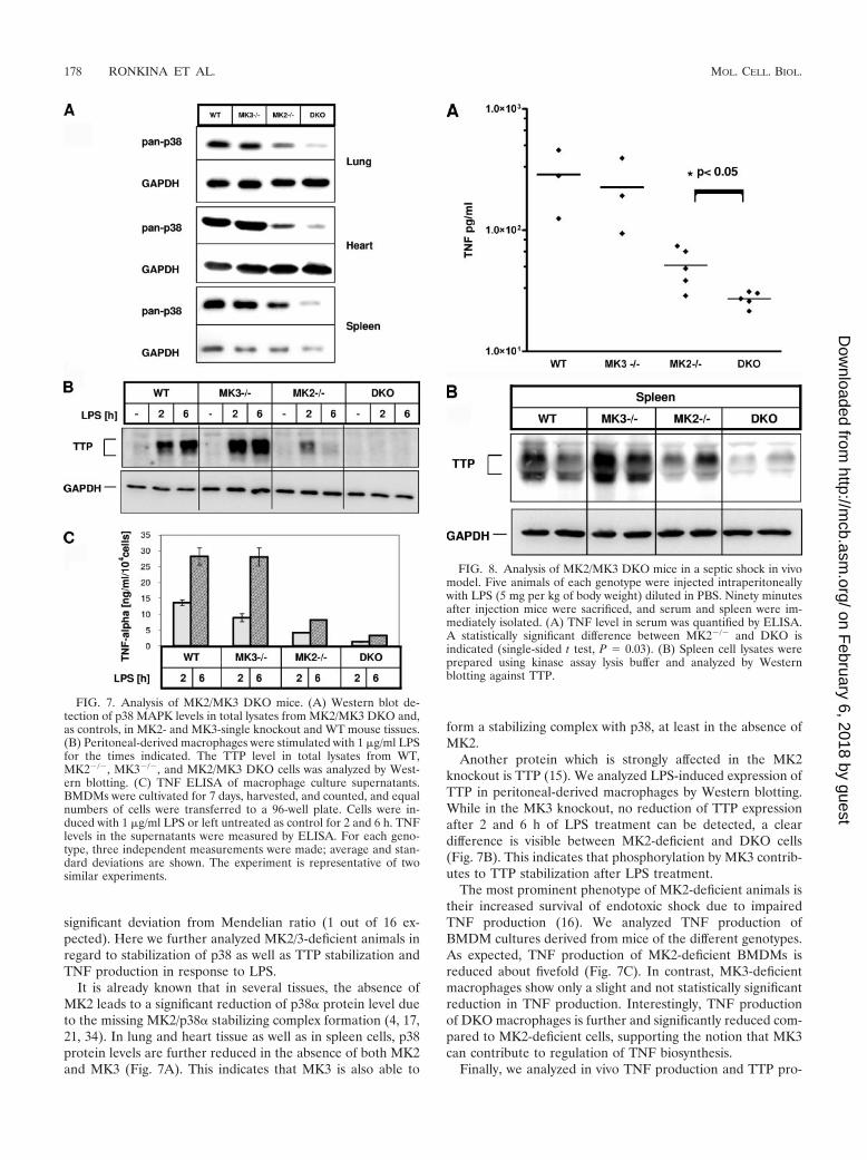
It is already known that in several tissues, the absence ofMK2 leads to a significant reduction of p38� protein level dueto the missing MK2/p38� stabilizing complex formation (4, 17,21, 34). In lung and heart tissue as well as in spleen cells, p38protein levels are further reduced in the absence of both MK2and MK3 (Fig. 7A). This indicates that MK3 is also able to
form a stabilizing complex with p38, at least in the absence ofMK2.
Another protein which is strongly affected in the MK2knockout is TTP (15). We analyzed LPS-induced expression ofTTP in peritoneal-derived macrophages by Western blotting.While in the MK3 knockout, no reduction of TTP expressionafter 2 and 6 h of LPS treatment can be detected, a cleardifference is visible between MK2-deficient and DKO cells(Fig. 7B). This indicates that phosphorylation by MK3 contrib-utes to TTP stabilization after LPS treatment.
The most prominent phenotype of MK2-deficient animals istheir increased survival of endotoxic shock due to impairedTNF production (16). We analyzed TNF production ofBMDM cultures derived from mice of the different genotypes.As expected, TNF production of MK2-deficient BMDMs isreduced about fivefold (Fig. 7C). In contrast, MK3-deficientmacrophages show only a slight and not statistically significantreduction in TNF production. Interestingly, TNF productionof DKO macrophages is further and significantly reduced com-pared to MK2-deficient cells, supporting the notion that MK3can contribute to regulation of TNF biosynthesis.
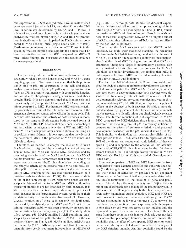
Finally, we analyzed in vivo TNF production and TTP pro-
FIG. 7. Analysis of MK2/MK3 DKO mice. (A) Western blot de-tection of p38 MAPK levels in total lysates from MK2/MK3 DKO and,as controls, in MK2- and MK3-single knockout and WT mouse tissues.(B) Peritoneal-derived macrophages were stimulated with 1 �g/ml LPSfor the times indicated. The TTP level in total lysates from WT,MK2�/�, MK3�/�, and MK2/MK3 DKO cells was analyzed by West-ern blotting. (C) TNF ELISA of macrophage culture supernatants.BMDMs were cultivated for 7 days, harvested, and counted, and equalnumbers of cells were transferred to a 96-well plate. Cells were in-duced with 1 �g/ml LPS or left untreated as control for 2 and 6 h. TNFlevels in the supernatants were measured by ELISA. For each geno-type, three independent measurements were made; average and stan-dard deviations are shown. The experiment is representative of twosimilar experiments.
FIG. 8. Analysis of MK2/MK3 DKO mice in a septic shock in vivomodel. Five animals of each genotype were injected intraperitoneallywith LPS (5 mg per kg of body weight) diluted in PBS. Ninety minutesafter injection mice were sacrificed, and serum and spleen were im-mediately isolated. (A) TNF level in serum was quantified by ELISA.A statistically significant difference between MK2�/� and DKO isindicated (single-sided t test, P 0.03). (B) Spleen cell lysates wereprepared using kinase assay lysis buffer and analyzed by Westernblotting against TTP.
tein expression in LPS-challenged mice. Five animals of eachgenotype were injected with LPS, and after 90 min the TNFlevel in serum was determined by ELISA, and TTP level inspleen of two randomly chosen animals of each genotype wasanalyzed by Western blotting (Fig. 8 A and B). TNF produc-tion is significantly further impaired in DKO animals com-pared to MK2-deficient mice (single-sided t test, P 0.03).Furthermore, semiquantitative detection of TTP protein in thespleen by Western blotting also supports the notion that TTPlevels are further reduced in DKO compared to MK2-KOmice. These findings are consistent with the results obtainedfor macrophages in vitro.
DISCUSSION
Here, we analyzed the functional overlap between the twostructurally related protein kinases MK2 and MK3 by a genetargeting approach. We provide evidence that both proteinstightly bind to p38, are coexpressed in the cells and tissuesanalyzed, are activated by the p38 pathway in response to stress(such as LPS or arsenite treatment) with comparable kinetics,and are able to phosphorylate endogenous small heat shockprotein Hsp25. It is also demonstrated that in most cells andtissues analyzed (except skeletal muscle), MK3 expression isminor compared to MK2. Furthermore, MK3 enzymatic activ-ity, probably as a result of the relatively low expression of theenzyme, is also minor compared to MK2 in the same cells. Thisbecomes obvious when the activity of both enzymes is moni-tored by the same antibody against both activated forms ofMK2 and MK3 after LPS treatment of peritoneal macrophagesand also when the enzymatic activities of WT and MK2-defi-cient MEFs are compared after arsenite stimulation using anin-gel kinase assay. Hence, it is not surprising that the effects ofthe deletion of MK3 in the presence of MK2 are minor anddifficult to detect.
Therefore, we decided to analyze the role of MK3 in anMK2-deficient background by analyzing how ectopic expres-sion of MK2 and MK3 can rescue MK2 deficiency and bycomparing the effects of the MK2 knockout and MK2/MK3double knockout. We demonstrate that both MK2 and MK3expression can rescue Hsp25 phosphorylation depending onthe catalytic activity of the enzyme, while MK2 and MK3 sta-bilize p38 to a similar degree as the catalytically inactive mu-tant of MK2, confirming the idea that binding between bothproteins leads to stabilization (17, 34). Furthermore, stabili-zation of the same group of LPS-responsive fibroblast tran-scripts is obtained with ectopic MK2 and MK3, while othertranscript stabilities are not changed by both enzymes. It isstill open whether the transcript-stabilizing properties ofboth enzymes in this experimental system result from theircatalytic activity or from their stabilization of p38. However,CXCL1 production of these cells can only be significantlyincreased by catalytically active MK2 and MK3. ARE-con-taining transcript stability has been previously assessed in ahuman system by microarray analysis (11). The study iden-tified several p38 MAPK-stabilized ARE-containing tran-scripts by means of the p38 inhibitor SB203580. In the ex-periment shown in Fig. 6, p38 MAP-stabilized mRNAs canbe rescued by MK2 or MK3 (e.g., cxcl1 and Gro�) or remainunstable after ActD treatment independent of MK2/MK3
(e.g., JUN B). Although both studies use different experi-mental designs and cell systems, i.e., pharmacological inhi-bition of p38 MAPK in a monocytic cell line (THP-1) versusreconstituted MK2-deficient embryonic fibroblasts as shownhere, these results suggest that MK2 or MK3 targets a subsetof ARE-containing inflammatory mRNAs that are stabilizedby the p38 MAPK pathway.
Comparing the MK2 knockout with the MK2/3 doubleknockout, we could show that MK3 stabilizes the remainingp38 level in the MK2-deficient background and that the role ofMK3 in TTP and TNF regulation is qualitatively indistinguish-able from the role of MK2. Taking into account that MK2 is anestablished therapeutic target of inflammatory diseases, suchas rheumatoid arthritis (14), and that small-molecule MK2inhibitors are under investigation, the finding that MK3 isindistinguishable from MK2 in its inflammatory functionwould favor MK2/3 dual inhibitors.
The fact that MK2/3-deficient DKO mice are viable andshow no obvious defects in embryonic development was unex-pected. We anticipated that MK2 and MK3 mutually compen-sate each other in development, since both enzymes were de-scribed to functionally interact with components of thedevelopmentally relevant polycomb complex involved in chro-matin remodeling (26, 37, 40); thus, we expected significantdefects in the absence of both enzymes. Possibly a more de-tailed analysis of, e.g., stem cell development in these animalsmay be necessary to identify minor but physiologically relevanteffects. The further reduction of p38 expression in MK2/3DKO compared to MK2-deficient tissue is also remarkable.However, the remaining level of p38 is obviously sufficient tocompensate the effects of embryonic lethality and placentaldevelopment described for the p38 knockout mice (1, 2, 35).This is similar to the finding that hypomorphic alleles of an-other protein kinase, PDK1, which lead to expression of only10% of the kinase, can rescue embryonic lethality of that en-zyme (18) and is supported by the observation that arsenite-stimulated ATF1/CREB phosphorylation by the p38 down-stream kinases MSK1/2 is not significantly reduced in MK2/3DKO cells (N. Ronkina, A. Kotlyarov, and M. Gaestel, unpub-lished data).
From our comparison of MK2 and MK3 here as well as fromcomparison of their catalytic activities and substrate specifici-ties (7), their regulation of subcellular localization (8, 26, 36),and their mode of activation by p38�/� (7), no significantdifference in the functions of both enzymes can be detected sofar. This is reminiscent of the situation of p38� and p38�,where p38� displays the major activity and p38� activity isminor and dispensable for signaling of the p38 pathway (3). Inboth cases, it is still enigmatic why both related enzymes havebeen stably maintained during evolution. MK2 and MK3 areboth present in birds and mammals, while only one MK2/3molecule is found in the lower vertebrates (12). It may well bethat there is an exemption from coexpression of both enzymesin one tissue or cell type not tested so far, making only theexpressed enzyme essential in these cells. Deletion of the en-zyme from these potential cells in mice obviously does not leadto a noticeable phenotype; however, we cannot exclude thepossibility that the effect of such specific MK3 function couldbe detected during a detailed and comprehensive analysis ofthe MK3-deficient animals. Another possibility could be the
VOL. 27, 2007 ROLE OF MAPKAP KINASES MK2 AND MK3 179
activation of one of the two enzymes by an alternative mech-anism, activator, and pathway, which is distinct from the p38pathway and responds to other signals (e.g., of specific Toll-like receptors) and stimuli. However, this pathway and its phys-iological function are still to be identified.
ACKNOWLEDGMENTS
We thank Stefanie Schumacher, Jessica Schwermann, and CorinnaMeier for plasmids pDEST15-alpha-p38, pMMP-IRES-GFP-MK2and -MK2-K76R, and pGEX-5x-1-MK3, respectively; Jeffrey Pelkerfor the ELISA; and Heike Schneider for gene array support. Geno-typing and mouse colony maintenance were done by Tatiana Iakovl-eva, Gretchen Ireland, Lauren Rusnak, and Brenda Lager.
This work was supported by Deutsche Forschungsgemeinschaftgrant GA453/10-1 and by the European Community grant RTN-HPRN-CT-2002-00255.
REFERENCES
1. Adams, R. H., A. Porras, G. Alonso, M. Jones, K. Vintersten, S. Panelli, A.Valladares, L. Perez, R. Klein, and A. R. Nebreda. 2000. Essential role ofp38� MAP kinase in placental but not embryonic cardiovascular develop-ment. Mol. Cell 6:109–116.
2. Allen, M., L. Svensson, M. Roach, J. Hambor, J. McNeish, and C. A. Gabel.2000. Deficiency of the stress kinase p38� results in embryonic lethality:characterization of the kinase dependence of stress responses of enzyme-deficient embryonic stem cells. J. Exp. Med. 191:859–870.
3. Beardmore, V. A., H. J. Hinton, C. Eftychi, M. Apostolaki, M. Armaka, J.Darragh, J. McIlrath, J. M. Carr, L. J. Armit, C. Clacher, L. Malone, G.Kollias, and J. S. Arthur. 2005. Generation and characterization of p38�(MAPK11) gene-targeted mice. Mol. Cell. Biol. 25:10454–10464.
4. Ben-Levy, R., S. Hooper, R. Wilson, H. F. Paterson, and C. J. Marshall.1998. Nuclear export of the stress-activated protein kinase p38 mediated byits substrate MAPKAP kinase-2. Curr. Biol. 8:1049–1057.
5. Brook, M., C. R. Tchen, T. Santalucia, J. McIlrath, J. S. Arthur, J.Saklatvala, and A. R. Clark. 2006. Posttranslational regulation of tristetra-prolin subcellular localization and protein stability by p38 mitogen-activatedprotein kinase and extracellular signal-regulated kinase pathways. Mol. Cell.Biol. 26:2408–2418.
6. Chrestensen, C. A., M. J. Schroeder, J. Shabanowitz, D. F. Hunt, J. W. Pelo,M. T. Worthington, and T. W. Sturgill. 2004. MAPKAP kinase 2 phosphor-ylates tristetraprolin on in vivo sites including Ser178, a site required for14-3-3 binding. J. Biol. Chem. 279:10176–10184.
7. Clifton, A. D., P. R. Young, and P. Cohen. 1996. A comparison of thesubstrate specificity of MAPKAP kinase-2 and MAPKAP kinase-3 and theiractivation by cytokines and cellular stress. FEBS Lett. 392:209–214.
8. Engel, K., A. Kotlyarov, and M. Gaestel. 1998. Leptomycin B-sensitive nu-clear export of MAPKAP kinase 2 is regulated by phosphorylation. EMBOJ. 17:3363–3371.
9. Engel, K., H. Schultz, F. Martin, A. Kotlyarov, K. Plath, M. Hahn, U.Heinemann, and M. Gaestel. 1995. Constitutive activation of mitogen-acti-vated protein kinase-activated protein kinase 2 by mutation of phosphory-lation sites and an A-helix motif. J. Biol. Chem. 270:27213–27221.
10. Freshney, N. W., L. Rawlinson, F. Guesdon, E. Jones, S. Cowley, J. Hsuan,and J. Saklatvala. 1994. Interleukin-1 activates a novel protein kinase cas-cade that results in the phosphorylation of Hsp27. Cell 78:1039–1049.
11. Frevel, M. A., T. Bakheet, A. M. Silva, J. G. Hissong, K. S. Khabar, and B. R.Williams. 2003. p38 mitogen-activated protein kinase-dependent and -inde-pendent signaling of mRNA stability of AU-rich element-containing tran-scripts. Mol. Cell. Biol. 23:425–436.
13. Hannigan, M. O., L. Zhan, Y. Ai, A. Kotlyarov, M. Gaestel, and C. K. Huang.2001. Abnormal migration phenotype of mitogen-activated protein kinase-activated protein kinase 2�/� neutrophils in zigmond chambers containingformyl-methionyl-leucyl-phenylalanine gradients. J. Immunol. 167:3953–3961.
14. Hegen, M., M. Gaestel, C. L. Nickerson-Nutter, L.-L. Lin, and J.-B. Telliez.2006. MAPKAP kinase 2-deficient mice are resistant to collagen-inducedarthritis. J. Immunol. 177:1913–1917.
15. Hitti, E., T. Iakovleva, M. Brook, S. Deppenmeier, A. D. Gruber, D. Radzioch,A. R. Clark, P. J. Blackshear, A. Kotlyarov, and M. Gaestel. 2006. Mitogen-activated protein kinase-activated protein kinase 2 regulates tumor necrosisfactor mRNA stability and translation mainly by altering tristetraprolin ex-pression, stability, and binding to adenine/uridine-rich element. Mol. Cell.Biol. 26:2399–2407.
16. Kotlyarov, A., A. Neininger, C. Schubert, R. Eckert, C. Birchmeier, H. D.Volk, and M. Gaestel. 1999. MAPKAP kinase 2 is essential for LPS-inducedTNF-alpha biosynthesis. Nat. Cell Biol. 1:94–97.
17. Kotlyarov, A., Y. Yannoni, S. Fritz, K. Laass, J. B. Telliez, D. Pitman, L. L.Lin, and M. Gaestel. 2002. Distinct cellular functions of MK2. Mol. Cell.Biol. 22:4827–4835.
18. Lawlor, M. A., A. Mora, P. R. Ashby, M. R. Williams, V. Murray-Tait, L.Malone, A. R. Prescott, J. M. Lucocq, and D. R. Alessi. 2002. Essential roleof PDK1 in regulating cell size and development in mice. EMBO J. 21:3728–3738.
19. Lehner, M. D., F. Schwoebel, A. Kotlyarov, M. Leist, M. Gaestel, and T.Hartung. 2002. Mitogen-activated protein kinase-activated protein kinase2-deficient mice show increased susceptibility to Listeria monocytogenes in-fection. J. Immunol. 168:4667–4673.
20. Ludwig, S., K. Engel, A. Hoffmeyer, G. Sithanandam, B. Neufeld, D. Palm,M. Gaestel, and U. R. Rapp. 1996. 3pK, a novel mitogen-activated protein(MAP) kinase-activated protein kinase, is targeted by three MAP kinasepathways. Mol. Cell. Biol. 16:6687–6697.
21. Lukas, S. M., R. R. Kroe, J. Wildeson, G. W. Peet, L. Frego, W. Davidson,R. H. Ingraham, C. A. Pargellis, M. E. Labadia, and B. G. Werneburg. 2004.Catalysis and function of the p38 alpha. MK2a signaling complex. Biochem-istry 43:9950–9960.
22. Mahtani, K. R., M. Brook, J. L. Dean, G. Sully, J. Saklatvala, and A. R.Clark. 2001. Mitogen-activated protein kinase p38 controls the expressionand posttranslational modification of tristetraprolin, a regulator of tumornecrosis factor alpha mRNA stability. Mol. Cell. Biol. 21:6461–6469.
23. Manke, I. A., A. Nguyen, D. Lim, M. Q. Stewart, A. E. Elia, and M. B. Yaffe.2005. MAPKAP kinase-2 is a cell cycle checkpoint kinase that regulates theG2/M transition and S phase progression in response to UV irradiation. Mol.Cell 17:37–48.
24. McLaughlin, M. M., S. Kumar, P. C. McDonnell, S. Van Horn, J. C. Lee,G. P. Livi, and P. R. Young. 1996. Identification of mitogen-activated protein(MAP) kinase-activated protein kinase-3, a novel substrate of CSBP p38MAP kinase. J. Biol. Chem. 271:8488–8492.
25. Neininger, A., D. Kontoyiannis, A. Kotlyarov, R. Winzen, R. Eckert, H. D.Volk, H. Holtmann, G. Kollias, and M. Gaestel. 2002. MK2 targets AU-richelements and regulates biosynthesis of tumor necrosis factor and interleu-kin-6 independently at different post-transcriptional levels. J. Biol. Chem.277:3065–3068.
26. Neufeld, B., A. Grosse-Wilde, A. Hoffmeyer, B. W. Jordan, P. Chen, D. Dinev,S. Ludwig, and U. R. Rapp. 2000. Serine/threonine kinases 3pK and MAPK-activated protein kinase 2 interact with the basic helix-loop-helix transcrip-tion factor E47 and repress its transcriptional activity. J. Biol. Chem. 275:20239–20242.
27. Rouse, J., P. Cohen, S. Trigon, M. Morange, A. Alonso-Llamazares, D.Zamanillo, T. Hunt, and A. R. Nebreda. 1994. A novel kinase cascadetriggered by stress and heat shock that stimulates MAPKAP kinase-2 andphosphorylation of the small heat shock proteins. Cell 78:1027–1037.
28. Roux, P. P., and J. Blenis. 2004. ERK and p38 MAPK-activated proteinkinases: a family of protein kinases with diverse biological functions. Micro-biol. Mol. Biol. Rev. 68:320–344.
29. Schumacher, S., K. Laass, S. Kant, Y. Shi, A. Visel, A. D. Gruber, A.Kotlyarov, and M. Gaestel. 11 November 2004, posting date. Scaffolding byERK3 regulates MK5 in development. EMBO J. 23:4770–4779.
30. Seternes, O. M., T. Mikalsen, B. Johansen, E. Michaelsen, C. G. Armstrong,N. A. Morrice, B. Turgeon, S. Meloche, U. Moens, and S. M. Keyse. 2December 2004, posting date. Activation of MK5/PRAK by the atypicalMAP kinase ERK3 defines a novel signal transduction pathway. EMBO J.23:4780–4791.
31. Sithanandam, G., F. Latif, F. M. Duh, R. Bernal, U. Smola, H. Li, I. Kuzmin,V. Wixler, L. Geil, and S. Shrestha. 1996. 3pK, a new mitogen-activatedprotein kinase-activated protein kinase located in the small cell lung cancertumor suppressor gene region. Mol. Cell. Biol. 16:868–876.
32. Stoecklin, G., T. Stubbs, N. Kedersha, S. Wax, W. F. Rigby, T. K. Blackwell,and P. Anderson. 11 March 2004, posting date. MK2-induced tristetraprolin:14-3-3 complexes prevent stress granule association and ARE-mRNA decay.EMBO J. 23:1313–1324.
33. Stokoe, D., D. G. Campbell, S. Nakielny, H. Hidaka, S. J. Leevers, C.Marshall, and P. Cohen. 1992. MAPKAP kinase-2: a novel protein kinaseactivated by mitogen-activated protein kinase. EMBO J. 11:3985–3994.
34. Sudo, T., K. Kawai, H. Matsuzaki, and H. Osada. 20 September 2005,posting date. p38 mitogen-activated protein kinase plays a key role in regu-lating MAPKAPK2 expression. Biochem. Biophys. Res. Commun. 337:415–421.
35. Tamura, K., T. Sudo, U. Senftleben, A. M. Dadak, R. Johnson, and M.Karin. 2000. Requirement for p38alpha in erythropoietin expression: a rolefor stress kinases in erythropoiesis. Cell 102:221–231.
36. Tanoue, T., R. Maeda, M. Adachi, and E. Nishida. 2001. Identification of adocking groove on ERK and p38 MAP kinases that regulates the specificityof docking interactions. EMBO J. 20:466–479.
37. Voncken, J. W., H. Niessen, B. Neufeld, U. Rennefahrt, V. Dahlmans, N.Kubben, B. Holzer, S. Ludwig, and U. R. Rapp. 24 November 2004, postingdate. MAPKAP kinase 3pK phosphorylates and regulates chromatin associ-ation of the polycomb group protein Bmi1. J. Biol. Chem. 280:5178–5187.
38. Wang, X., M. A. Khaleque, M. J. Zhao, R. Zhong, M. Gaestel, and S. K.
Calderwood. 8 November 2005, posting date. Phosphorylation of HSF1 byMAPK-activated protein kinase 2 on serine 121, inhibits transcriptionalactivity and promotes HSP90 binding. J. Biol. Chem. 281:782–791.
39. Winzen, R., M. Kracht, B. Ritter, A. Wilhelm, C. Y. Chen, A. B. Shyu, M.Muller, M. Gaestel, K. Resch, and H. Holtmann. 1999. The p38 MAP kinasepathway signals for cytokine-induced mRNA stabilization via MAP kinase-
activated protein kinase 2 and an AU-rich region-targeted mechanism.EMBO J. 18:4969–4980.
40. Yannoni, Y. M., M. Gaestel, and L. L. Lin. 2004. P66(ShcA) interacts withMAPKAP kinase 2 and regulates its activity. FEBS Lett. 564:205–211.
41. Zakowski, V., G. Keramas, K. Kilian, U. R. Rapp, and S. Ludwig. 2004. Mito-gen-activated 3p kinase is active in the nucleus. Exp. Cell Res. 299:101–109.
VOL. 27, 2007 ROLE OF MAPKAP KINASES MK2 AND MK3 181


![Human Mitogen-activated Protein Kinase Kinase 4 as a ......(CANCERRESEARCH57. 4177—4182,October 1, 1997] Advances in Brief Human Mitogen-activated Protein Kinase Kinase 4 as](https://static.documents.pub/doc/80x56/6082557b7810d746a5071f39/human-mitogen-activated-protein-kinase-kinase-4-as-a-cancerresearch57.jpg)


