41
THE MOLECULAR BASIS OF INHERITANCE Chapter 16
| Date post: | 02-Jan-2016 |
| Category: |
Documents |
| Upload: | phoebe-mills |
| View: | 216 times |
| Download: | 0 times |

THE MOLECULAR BASIS OF INHERITANCE
Chapter 16
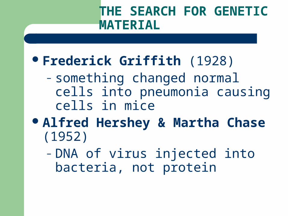
THE SEARCH FOR GENETIC MATERIAL
Frederick Griffith (1928) – something changed normal cells into
pneumonia causing cells in miceAlfred Hershey & Martha Chase (1952)
– DNA of virus injected into bacteria, not protein

Figure 16.1 Transformation of bacteria
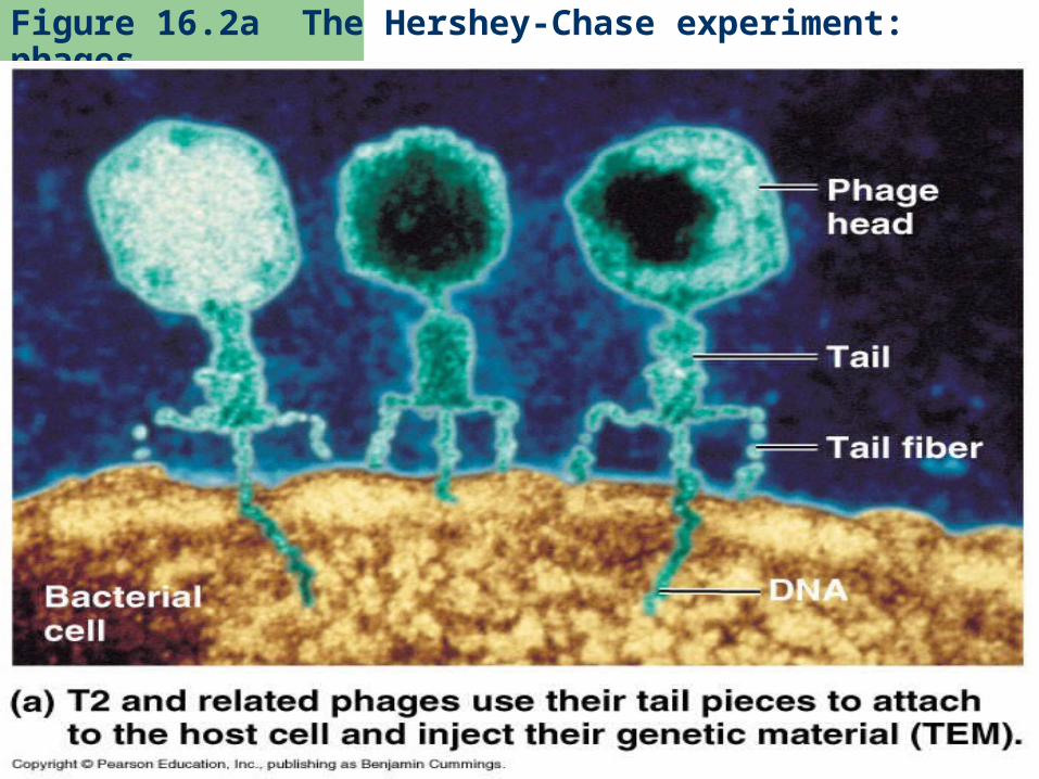
Figure 16.2a The Hershey-Chase experiment: phages

Figure 16.2ax Phages
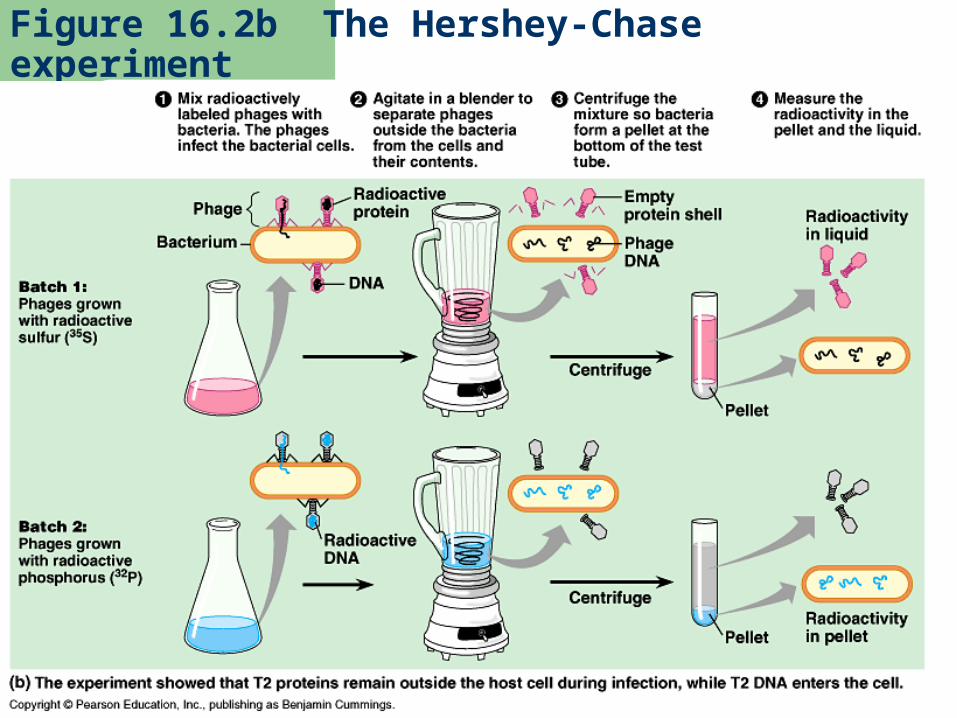
Figure 16.2b The Hershey-Chase experiment

Erwin Chargaff (1947)– Amounts of G = C and A = T in DNA
Rosalind Franklin (1950’s)– Photographed DNA using X-ray
crystallographyJames Watson and Francis Crick (1953)
– DNA is a double helix with base pairs as the rungs

Figure 16.4 Rosalind Franklin and her X-ray diffraction photo of DNA

Figure 16.3 The structure of a DNA stand

Figure 16.0 Watson and Crick

DNA STRUCTURE
4 Bases– Purines: adenine and guanine– Pyrimidine: cytosine and thymine– A – T and C – G
Hydrogen bonds connect the base pairs to make the rungs
Deoxyribose and phosphate make up the uprights
Nucleotide – a base, a sugar, and a phosphate

Purine and pyridimine

Figure 16.6 Base pairing in DNA

Figure 16.5 The double helix

SEMICONSERVATIVE MODEL OF REPLICATION
When DNA copies itself, the resulting “copy” is one of the original strands with a new strand

Figure 16.7 A model for DNA replication: the basic concept

Figure 16.8 Three alternative models of DNA replication

Figure 16.9 The Meselson-Stahl experiment tested three models of DNA replication (Layer 4)
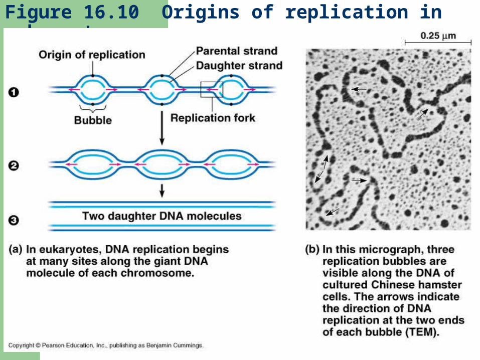
Figure 16.10 Origins of replication in eukaryotes
DNA REPLICATION
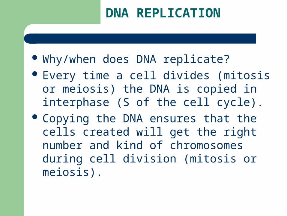
Why/when does DNA replicate? Every time a cell divides (mitosis or meiosis)
the DNA is copied in interphase (S of the cell cycle).
Copying the DNA ensures that the cells created will get the right number and kind of chromosomes during cell division (mitosis or meiosis).

DNA REPLICATION
Origins of replication – specific sequences of nucleotides that initiate replication
Replication fork – a Y shaped region where new nucleotides are added
DNA polymerases – enzymes that break H bonds (unwind helix) and add complementary nucleotides
Reaction driven by nucleoside triphosphates (like ATP)
Loose 2 phosphate groups to be added as a nucleotide

Figure 16.11 Incorporation of a nucleotide into a DNA strand

The 2 strands of a double helix are antiparallel
The sugar-phosphate backbones run in opposite directions
One is 5’ to 3’ while other is 3’ to 5’#1 is the C attached to a base and #5 is
the C attached to phosphate

Figure 16.12 The two strands of DNA are antiparallel

DNA polymerases can only attach nucleotides to the free 3’ end of existing an polynucleotide (already paired to complementary strand).
DNA polymerases add nucleotides in only the 5’ to 3’ direction
Leading strand – made by DNA polymerase following replication fork

Figure 16.13 Synthesis of leading and lagging strands during DNA replication

The other strand of DNA that is made is called the lagging strand
Polymerase makes a short strandAs bubble grows, more short strands are
made called Okazaki fragments

Figure 16.14 Priming DNA synthesis with RNA

Okazaki fragments are joined by DNA ligase
Priming: polymerase must attach nucleotides to an existing polynucleotide at a 3’ end
Primase (an enzyme) adds RNA nucleotides to make a primer ~10 bases long


Polymerase can then add its complementary nucleotides
Polymerase also replaces certain RNA nucleotides of primer
For lagging strand each fragment must be primed, but only one primer is needed for entire leading strand

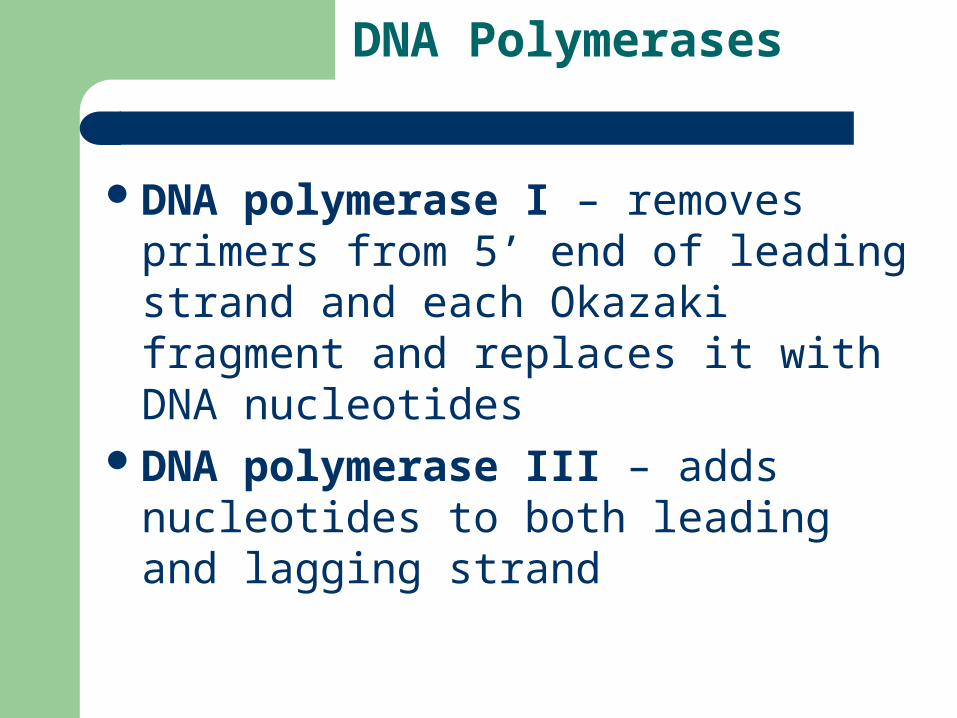
DNA Polymerases
DNA polymerase I – removes primers from 5’ end of leading strand and each Okazaki fragment and replaces it with DNA nucleotides
DNA polymerase III – adds nucleotides to both leading and lagging strand

OTHER ENZYMES…
Helicase – untwists helix at replication fork
Topoisomerase – relieves the strain that untwisting causes ahead of the replication fork
Single-strand binding proteins – hold strands apart while replication occurs

Figure 16.15 The main proteins of DNA replication and their functions

Figure 16.16 A summary of DNA replication

PROOFREADING
Mismatch repairing – polymerase checks each addition
Excision pair – nuclease cuts out bad section of strand
Replication Animation 1Replication Animation 2 (can see multiple
replication origins)
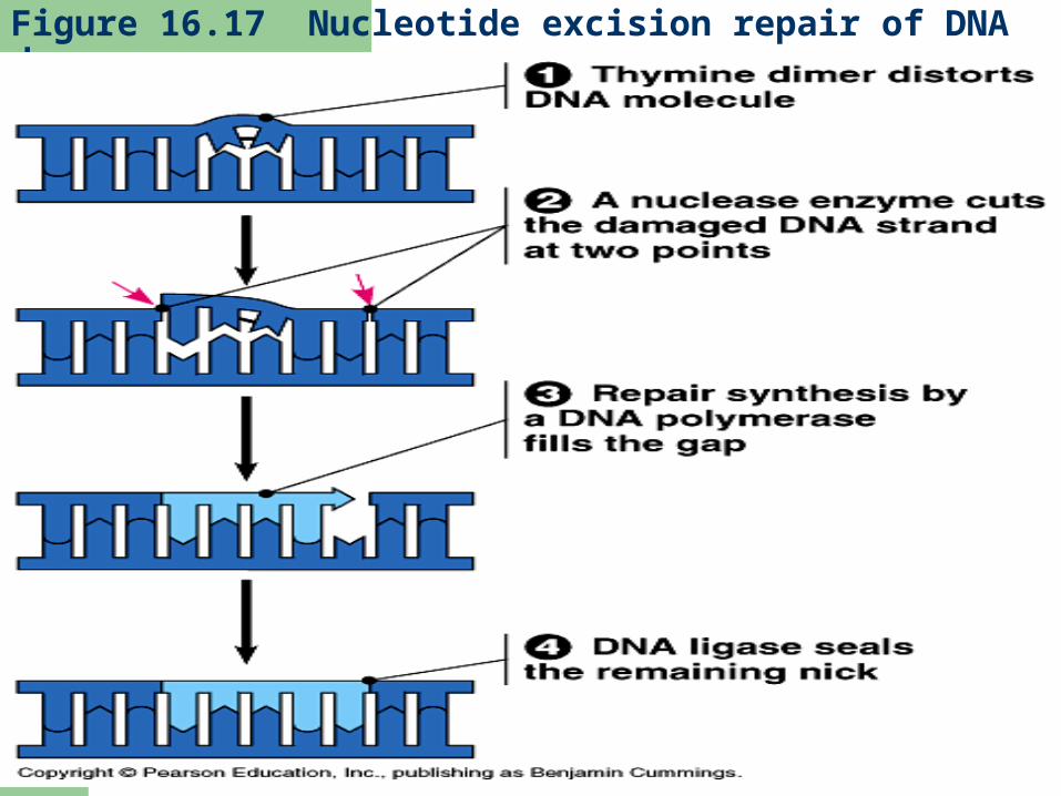
Figure 16.17 Nucleotide excision repair of DNA damage

Figure 16.18 The end-replication problem

TELOMERES
Impossible on lagging strand to copy the end of 5’ strand
This leaves a gap that would shorten DNA every time it replicates
Telomeres (TTAGGG) repeated many times protects genes by postponing erosion of genes from this shortening effect
Telomerase – lengthens telomeres– Contains an RNA sequence that is the template
for a telomere– Present in germ cells (for future gametes)– Increased activity in cancer cells (allows for more
cell division)

Figure 16.19a Telomeres and telomerase: Telomeres of mouse chromosomes


