The molecular characterisation of Escherichia coliK1 isolated from neonatal nasogastric feedingtubesAldukali Alkeskas1, Pauline Ogrodzki1, Mohamed Saad1, Naqash Masood1, Nasreddin R. Rhoma1, Karen Moore2,Audrey Farbos2, Konrad Paszkiewicz2 and Stephen Forsythe1*
Abstract
Background: The most common cause of Gram-negative bacterial neonatal meningitis is E. coli K1. It has a mortalityrate of 10–15 %, and neurological sequelae in 30–50 % of cases. Infections can be attributable to nosocomial sources,however the pre-colonisation of enteral feeding tubes has not been considered as a specific risk factor.
Methods: Thirty E. coli strains, which had been isolated in an earlier study, from the residual lumen liquid and biofilmsof neonatal nasogastric feeding tubes were genotyped using pulsed-field gel electrophoresis, and 7-loci multilocussequence typing. Potential pathogenicity and biofilm associated traits were determined using specific PCR probes,genome analysis, and in vitro tissue culture assays.
Results: The E. coli strains clustered into five pulsotypes, which were genotyped as sequence types (ST) 95, 73, 127, 394and 2076 (Achman scheme). The extra-intestinal pathogenic E. coli (ExPEC) phylogenetic group B2 ST95 serotypeO1:K1:NM strains had been isolated over a 2 week period from 11 neonates who were on different feeding regimes.The E. coli K1 ST95 strains encoded for various virulence traits associated with neonatal meningitis and extracellularmatrix formation. These strains attached and invaded intestinal, and both human and rat brain cell lines, and persistedfor 48 h in U937 macrophages. E. coli STs 73, 394 and 2076 also persisted in macrophages and invaded Caco-2 andhuman brain cells, but only ST394 invaded rat brain cells. E. coli ST127 was notable as it did not invade any cell lines.
Conclusions: Routes by which E. coli K1 can be disseminated within a neonatal intensive care unit are uncertain,however the colonisation of neonatal enteral feeding tubes may be one reservoir source which could constitute aserious health risk to neonates following ingestion.
BackgroundLiu et al. [1] reported that worldwide the estimatedmortality in children younger than 5 years in 2010 was7,600,000. Neonates, in particular those with very lowbirth-weight, are of particular concern due to theirweak immune system [2]. The risk of infection in neo-nates increases with low birth weight and additionallyprolonged hospitalization [3, 4]. Mortality among neo-nates is attributed to infectious causes, preterm birthcomplications, intrapartum-related complications, sep-sis and meningitis.
The most common cause of Gram-negative bacterialneonatal meningitis is E. coli K1. It has a mortality rateof 10–15 %, and neurological sequelae in 30–50 % ofcases [5–8]. Other bacteria responsible for neonataland infant morbidity and mortality include Group Bstreptococci (GBS), Enterobacter spp., Citrobacter koseri,Neisseria meningitidis, Serratia spp., and Cronobacter spp.[9, 10].E. coli serotypes associated with neonatal meningitis
are primarily O18:K1:H7, O1:K1, O7:K1, O83:K1 andthe more recently reported O45:K1:H7 [11–13]. Theseare in the E. coli extraintestinal pathogenic (ExPEC)subgroup B2, and are sequence type (ST) 95 (Achtmanscheme) [14]. Bacterial invasion across the blood–brainbarrier is multifactorial, requiring several genes forbinding, invasion and intracellular survival. Proposed
* Correspondence: [email protected] of Science and Technology, Nottingham Trent University, CliftonLane, Nottingham NG11 8NS, UKFull list of author information is available at the end of the article
virulence genetic determinants include ibeA, sfaS, cnf1,gimA, and ompA [15, 16]. However these are not alwayssupported in experimental assays and are not demon-strable in all E. coli strains isolated from cerebral spinalfluid [12, 13, 17].Although there have been some concerns regarding
bacterial biofilm formation inside neonatal nasogastricfeeding tubes by opportunistic pathogens, few system-atic studies have been undertaken [18–20]. Mehall et al.[18] reported both feeding intolerance and a link tonecrotizing enterocolitis in neonates following the bac-terial colonised of their feeding tubes. Previous studiesby Hurrell et al. [19] have revealed that in situ the in-side of such tubes can be colonised by a variety of fungiand various opportunistic bacterial pathogens produ-cing a biofilm of mixed microbial composition; Can-dida spp., E. coli, Enterobacter hormaechei, Klebsiellapneumoniae, Cronobacter sakazakii, Yersinia enterocoli-tica, and Pseudomonas fluorescens. The aim of this studywas to investigate the diversity of the E. coli strains previ-ously isolated by Hurrell et al. [19] from the residual liquidin the lumen and biofilm from 30 neonatal nasogastricfeeding tubes, which had been collected from among 129neonates on two intensive care units.
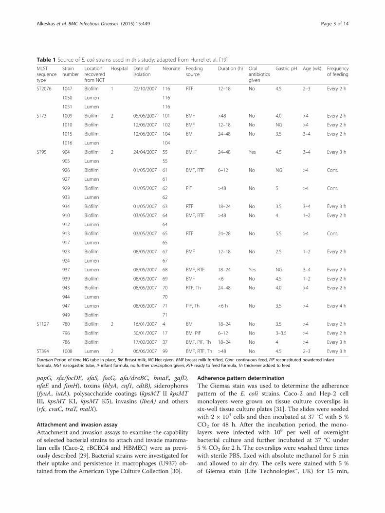
MethodsBacterial strains usedThirty isolates of E. coli were included in this study;Table 1. All isolates had previously been isolated byHurrell et al. [19] from the residual lumen liquid andbiofilms of nasogastric enteral feeding tubes on neonatalintensive care units at Hospital 1 (n = 3) and 2 (n = 27).
Pulsed-field gel electrophoresisPulsotypes were determined using pulsed-field gel electro-phoresis (PFGE) with XbaI and SpeI restriction enzymesas described by PulseNet [21]. Salmonella enterica ser-ovar Typhimurium H9812 was used as the referencestrain. Dendrogram construction and band assignmentwas achieved using BioNumerics software version 3.5.Dice coefficient, unweighted pair group method witharithmetic mean (UPGMA) were used for cluster ana-lysis. Less than 95 % of band similarity value was usedto consider the isolates to be non-clonal [22]. The tol-erance and optimization of the bands was 1.5 %.
Multilocus sequence typing (MLST)Sequence type (ST) of the E. coli isolates used the 7-lociMLST Achtman scheme (http://mlst.warwick.ac.uk/mlst/mlst/dbs/Ecoli). Seven housekeeping genes were ampli-fied by PCR using the primers for adk (adenylate kinase),fumC (fumarate hydratase), gyrB (DNA gyrase), icd (iso-citrate/isopropylmalate dehydrogenase), mdh (malatedehydrogenase), purA (adenylosuccinate dehydrogenase),
and recA (ATP/GTP binding motif ). The sequenceswere aligned using CLC Sequence Viewer 6.6 (http://www.clcbio.com). The trimmed allele sequences werecompared against the E. coli MLST database (http://mlst.warwick.ac.uk/mlst/mlst/dbs/Ecoli) and the sequencetypes were subsequently determined.
SerotypingThe O-antigen serotype was determined using compara-tive genomic analysis, and confirmed by laboratory ana-lysis (Statens Serum Institut) [23].
Motility determinationMotility was determined by measuring the zones ofgrowth in semi-solid agar [24]. A single colony fromeach strain was used to inoculate 3 ml of TSB whichwas then incubated at 37 °C with shaking incubator at200 rpm. The culture was diluted to 104 CFU/ml and3 μl of the suspension used to stab inoculate TSB sup-plemented with 0.4 % agar. The inoculated plates wereincubated overnight at 37 °C. Strains were analysedtwice, each time in triplicate.
Haemolysis reactionHaemolysis was examined by streaking on TSA-bloodagar plates containing 5 % sheep blood (Oxoid ThermoFischer Scientific, UK), and then incubating for 24 h forat 37 °C. The resultant colony morphology was re-corded after 24 h to determine the formation of eitherα- or β-haemolysis.
Antibiotic resistance determinationAntibiograms were determined using the disc diffusionmethod. Antibiotic discs were obtained from MASTGroup (UK). For each antibiotic the diameter of thezone was measured and then compared with standardmeasurements to determine if the strains were resistantor sensitive to the antibiotic [25]. The control strainswere E. coli NCTC 13351, E. coli NCTC 13352, E. coliNCTC 13353 and E. coli NCTC 10418. The presence ofthe β-lactamase resistance genes SHV, TEM, CTX-M,and OXA were screened for by a multiplex PCR assay[26]. Strains with known β-lactamase types were in-cluded as reference strains. These were E. coli NCTC13351 (TEM-3), E. coli NCTC 13353 (CTX-M-15, TEM,OXA), K. pneumoniae NCTC 13368 SHV-18. Genomeswere analysed using the Comprehensive AntibioticDatabase (CARD; http://arpcard.mcmaster.ca) for genesencoding antibiotic resistance [27].
PCR detection of virulence factor genesThe presence of 30 virulence factor genes was deter-mined using 5 multiplex PCR–based assays [28]. Thegene classes included adhesins (papAH, papC, papEF,
Alkeskas et al. BMC Infectious Diseases (2015) 15:449 Page 2 of 14
Attachment and invasion assayAttachment and invasion assays to examine the capabilityof selected bacterial strains to attach and invade mamma-lian cells (Caco-2, rBCEC4 and HBMEC) were as previ-ously described [29]. Bacterial strains were investigated fortheir uptake and persistence in macrophages (U937) ob-tained from the American Type Culture Collection [30].
Adherence pattern determinationThe Giemsa stain was used to determine the adherencepattern of the E. coli strains. Caco-2 and Hep-2 cellmonolayers were grown on tissue culture coverslips insix-well tissue culture plates [31]. The slides were seededwith 2 × 10⁴ cells and then incubated at 37 °C with 5 %CO2 for 48 h. After the incubation period, the mono-layers were infected with 108 per well of overnightbacterial culture and further incubated at 37 °C under5 % CO2 for 2 h. The coverslips were washed three timeswith sterile PBS, fixed with absolute methanol for 5 minand allowed to air dry. The cells were stained with 5 %of Giemsa stain (Life Technologies™, UK) for 15 min,
Table 1 Source of E. coli strains used in this study; adapted from Hurrel et al. [19]
MLSTsequencetype
Strainnumber
Locationrecoveredfrom NGT
Hospital Date ofisolation
Neonate Feedingsource
Duration (h) Oralantibioticsgiven
Gastric pH Age (wk) Frequencyof feeding
ST2076 1047 Biofilm 1 22/10/2007 116 RTF 12–18 No 4.5 2–3 Every 2 h
1050 Lumen 116
1051 Lumen 116
ST73 1009 Biofilm 2 05/06/2007 101 BMF >48 No 4.0 >4 Every 2 h
1010 Biofilm 12/06/2007 102 BMF 12–18 No NG >4 Every 2 h
1015 Biofilm 12/06/2007 104 BM 24–48 No 3.5 3–4 Every 2 h
1016 Lumen 104
ST95 904 Biofilm 2 24/04/2007 55 BM,IF 24–48 Yes 4.5 3–4 Every 3 h
905 Lumen 55
926 Biofilm 01/05/2007 61 BMF, RTF 6–12 No NG >4 Cont.
927 Lumen 61
929 Biofilm 01/05/2007 62 PIF >48 No 5 >4 Cont.
933 Lumen 62
934 Biofilm 01/05/2007 63 RTF 18–24 No 3.5 3–4 Every 3 h
910 Biofilm 03/05/2007 64 BMF, RTF >48 No 4 1–2 Every 2 h
912 Lumen 64
913 Biofilm 03/05/2007 65 RTF 24–28 No 5.5 >4 Cont.
917 Lumen 65
923 Biofilm 08/05/2007 67 BMF 12–18 No 2.5 1–2 Every 2 h
924 Lumen 67
937 Lumen 08/05/2007 68 BMF, RTF 18–24 Yes NG 3–4 Every 2 h
939 Biofilm 08/05/2007 69 BMF <6 No 4.5 1–2 Every 2 h
943 Biofilm 08/05/2007 70 RTF, Th 24–48 No 4.0 >4 Every 2 h
944 Lumen 70
947 Lumen 08/05/2007 71 PIF, Th <6 h No 3.5 >4 Every 4 h
949 Biofilm 71
ST127 780 Biofilm 2 16/01/2007 4 BM 18–24 No 3.5 >4 Every 2 h
796 Biofilm 30/01/2007 17 BM, PIF 6–12 No 3–3.5 >4 Every 2 h
786 Biofilm 17/02/2007 37 BMF, PIF, Th 18–24 No 4 >4 Every 3 h
ST394 1008 Lumen 2 06/06/2007 99 BMF, RTF, Th >48 No 4.5 2–3 Every 3 h
Duration Period of time NG tube in place, BM Breast milk, NG Not given, BMF breast milk fortified, Cont. continuous feed, PIF reconstituted powdered infantformula, NGT nasogastric tube, IF infant formula, no further description given, RTF ready to feed formula, Th thickener added to feed
Alkeskas et al. BMC Infectious Diseases (2015) 15:449 Page 3 of 14
washed with sterile PBS and allowed to air dry. The slidewas examined using light microscopy.
Genomic analysisBacterial DNA was extracted from 1-day old culturesof selected strains using GenElute™ bacterial genomekit (Sigma Aldrich®, USA). The genome sequenceswere generated on an Illumina MiSeq using v3 chem-istry and 300 bp paired end reads using dual indexedNextera XT libraries. The de novo assembly was per-formed using SPAdes assembly program and Quast[32]. Genome annotation used the SEED-based auto-mated annotation system provided by the RAST ser-ver (http://rast.nmpdr.org) and prokaryotic genomeannotation system PROKKA [33].The genome sequences obtained were compared to
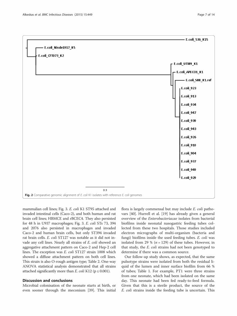
published chromosomal, plasmid and O-antigen se-quences for E. coli APEC O1 (Accession number CP000468), E. coli CE10 O7:K1 (Accession number GCA_000227625), E. coli S88 O45:K1:H7 (Accessionnumbers: chromosome CU928161, plasmid CU928146), O-antigens O1 and O2 (Accession numbers GU299791 and GU299792, respectively), and plasmids E.coli O1 pAPEC-O1-CelBM (Accession number DQ381420) and E. coli O18:K1 pRS218 (Accession numberCP007150) [1, 13, 17, 34–37]. The genomes were alsosearched for various biofilm formation associatedtraits. Whole genome alignment used Parsnp from theHarvest Tools software v1.1.2 with a reference genomeselected at random. Tree visualisation used FigTree v1.4.2,to construct a midpoint rooted tree.
Nucleotide sequence accession numbersThe Whole Genome Shotgun projects have been depos-ited at DDBJ/EMBL/GenBank under accession JQFB00000000, JQFC00000000, JQFD00000000, JQFQ00000000,JQFR00000000, JQFE00000000, JQFF00000000, JQFS00000000, JQFG00000000, JQFH00000000, JQFI00000000and JQFT00000000 for isolates 904, 910, 913, 923, 926,929, 934, 937, 939, 943, 947 and 949 respectively.
ResultsPulsed-field analysis of E. coli strainsAs shown in Fig. 1, the PFGE analysis of thirty of E. coliisolates from neonatal enteral feeding tubes from hos-pital 1 (n = 3) and hospital 2 (n = 27) showed the strainsclustered into four pulsotypes (PT1-4) and one unique(U) strain. These strains were isolated from the residualliquid in the tube and from biofilms on the inner wall of30/129 feeding tubes (Table 1). Three strains (1047,1050, 1051) belonging to PT1 had been isolated fromthe same neonate on the same day (22 October 2007).These were from both the lumen contents and biofilmwithin the tubes. This neonate had been fed ‘ready to
feed’ formula. Four strains (1009, 1010, 1015, 1016),previously isolated from both the residual liquid andbiofilm of feeding tubes, formed PT2. These had beenisolated on the same day (12 June 2007) from 3 neo-nates fed either breast milk or fortified breast milk.Three strains (786, 780, 796) from feeding tube biofilmsbelonged to PT4 and were isolated over a one monthperiod (16 January to 16 February 2007) from 2 differ-ent neonates fed breast milk, fortified breast milk, andreconstituted infant formula. There was one unique (U)strain (1008) from a neonate who had been fed bothfortified breast milk and reconstituted infant formula.Of particular interest were the nineteen strains belong-ing to PT3. These had isolated over a two week period(24 April to 8 May 2007) from 11 different neonates;Table 1. These neonates had been fed during the sam-pling period breast milk, fortified breast milk, reconsti-tuted infant formula, and ready to feed formula. Again,indistinguishable strains were isolated from both tubelumen contents and biofilms.
Multilocus sequence typingThe 7-loci MLST sequence types (ST) obtained for eightselected strains are given in Table 2. Five STs were iden-tified across the pulsotype groups, and were internallyconsistent within the clonal group. Pulsotypes 1 to 4 andthe unique isolate corresponded with sequence typesST2076, ST73, ST95, ST127 and ST394, respectively. Se-quence types 394 and 2076 differ by one nucleotide inthe parA allele. These strains had been isolated fromtwo hospitals at different times; 1 October 2007, PT4January and February 2007. All the sequence types be-long to the extra-intestinal (ExPEC) pathogenic E. coligroup B2; [38].
AntibiogramsAntimicrobial susceptibility of the E. coli strains is givenin Table 3. The two E. coli ST2076 strains 1047 and1050, which belong to PT1, showed resistance to thepenicillin antibiotics and were susceptible to all otherantibiotics. The PT2 strain 1009, belonging to ST73, wassusceptible to all antibiotics. The ST95 (PT3) E. colistrains 904, 923 and 939 were susceptible to all antibi-otics, except ampicillin. E. coli strain 780 (ST394, PT4)was resistance to ampicillin, and augmentin. E. colistrain 1008 (ST127, U) was susceptible to all antibiotics.The two closely related STs 394 and 2076 differed intheir susceptibility to piperacillin and meropenem;Table 3. The blatem gene, conferring resistance toampicillin, was found in E. coli ST95, ST394 andST2076 isolates and none harboured blaSHV, blaCTX-Mor blaOXA. E. coli strains 1008 (ST127, U) and 1009(ST73, PT2) did not encode any of these genes.
Alkeskas et al. BMC Infectious Diseases (2015) 15:449 Page 4 of 14
Haemolysis reaction on sheep blood agar α α α α α α α β
Motility NM NM NM NM NM NM NM Motile
MLST loci adk 21 21 36 37 37 37 21 13
fumC 35 35 24 38 38 38 35 14
gyrB 61 61 9 19 19 19 61 19
icd 52 52 13 37 37 37 52 36
mdh 5 5 17 17 17 17 5 23
purA 77 77 11 11 11 11 5 11
recA 4 4 25 26 26 26 4 10
O-antigenb O44 - O25:K5 O1:K1 - - O77 O-rough
Sequence type 2076 2076 73 95 95 95 394 127aSequence type given in parenthesis, bLaboratory determination by Statens Serum Institut of pulsotype representatives
Alkeskas et al. BMC Infectious Diseases (2015) 15:449 Page 5 of 14
Physiological traitsEight strains were selected as representatives of theinitial thirty E. coli strains for further detailed study;1047 (ST2076), 1050 (ST2076), 1009 (ST73), 904 (ST95),923 (ST95), 939 (ST95), 780 (ST127), and 1008 (ST394).The E. coli strains were non-motile and showed α-haemolysis on sheep blood agar, except for strain 1008ST394 which was motile and β-haemolytic; Table 2. Theserotype of pulsotype representatives were determinedby laboratory analysis (Statens Serum Institute). Thisrevealed a range of O-types including O1:K1 (ST95)and O25:K5 (ST73); Table 2.
Genome analysis of E. coli ST95 isolatesThe E. coli K1 phylogenetic group B2 ST95 strains withindistinguishable pulsotypes (PT3) had been isolatedfrom the feeding tubes of 11 neonates in an intensivecare unit over a two week period (Fig. 1). As givenabove, the representative PT3 strain (904) was laboratorydetermined to be O1:K1; Table 2. Given the significantassociation of E. coli K1 with neonatal meningitis, all thePT3 strains were genome sequenced. Their genome sizewas in the order of 4997507 bp, average G + C contentwas 50.7 % (ranging from 49.2 to 51.3 %). The genomeannotation indicated that all the E. coli ST95 strainswere serotype O1 and capsular type K1. Whole genomealignment was performed for conformational purposeswith a variety of publically available genomes of E. colistrains expressing different K antigens; Fig. 2. The ge-nomes also revealed the presence of genes encoding forcurli fimbriae and colanic acid which are associated withextracellular matrix production and biofilm formation.The range of antibiotic resistance encoding genes or
ORFs was predicted using the Comprehensive AntibioticDatabase (CARD; http://arpcard.mcmaster.ca) and has
been summarised in Table 4. This analysis revealed theE. coli ST95 strains had two streptomycin resistanceassociated genes (strA and strB) in the aminoglycosideresistance class. The number of predicted antibioticefflux genes in the 12 E. coli ST95 strains varied slightly.The majority (10/12) of strains had 52 genes, whereasone had 51 and the remainder had 53. This was the onlytrait which distinguished between the ST95 strains.Further studies will provide a more in-depth analysis ofthis small variation. Also the E. coli ST95, ST2076, andST127 had an additional β–lactamase gene comparedwith ST73 and ST394. Further detailed analysis is givenin the Additional file 1: Figure S1.
Virulence traitsThe presence of 30 virulence related traits were screenedusing PCR. The 30 virulence genes investigated, includeda range of traits including genes encoding for adhesins,invasins, capsule, toxins, siderophores and others. Thepresence of these genes differed depending on the strainsequence type; Table 5. For example, E. coli K1 ST95strains encoded adhesin genes fimH, papACEFG1,siderophores fyuA, traT and UPEC PAI. In contrast, theST127 E. coli K5 strain 1008 encoded sfaS, haemolysinhlyA and cnf. The UPEC PAI marker, malX from arche-typal ExPEC strain CFT073 (serotype O6:K2:H1), wasonly present in STs 73, 95, and 127. The aerobactinreceptor gene (iutA) was only in ST73 (PT2). The twoclosely related STs 394 and 2076 differed in the posses-sion of fimH and fvtA; Table 5.
Adhesion, invasion and persistence in mammalian celltypesIn vitro tissue culture assays showed that E. coli se-quence types varied in their ability to attach and invade
Table 3 Antibiograms of selected nasogastric tube E. coli isolates based on pulsotype
Antibiotic group Antibiotic ST2076 ST73PT2 ST95 ST394 ST127
(PT1)a (PT2) (PT3) (PT4) (U)
1047 1050 1009 904 923 939 780 1008
Cephalosporins Cefpodoxime S S S S S S S S
Cefotaxime S S S S S S S S
Ceftazidime S S S S S S S S
Penicillins Ampicillin R R S R R R R S
Augmentin R R S S S S R S
Piperacillin/Tazobactam R R S S S S S S
Fluoroquinolones Ciprofloxacin S S S S S S S S
Aminoglycosides Gentamicin S S S S S S S S
Carbapenems Imipenem S S S S S S S S
Meropenem S S S S S S R S
Miscellaneous Chloramphenicol S S S S S S S SaPulsotype is given in parenthesis
Alkeskas et al. BMC Infectious Diseases (2015) 15:449 Page 6 of 14
mammalian cell lines; Fig. 3. E. coli K1 ST95 attached andinvaded intestinal cells (Caco-2), and both human and ratbrain cell lines; HBMCE and rBCEC4. They also persistedfor 48 h in U937 macrophages; Fig. 3. E. coli STs 73, 394and 2076 also persisted in macrophages and invadedCaco-2 and human brain cells, but only ST394 invadedrat brain cells. E. coli ST127 was notable as it did not in-vade any cell lines. Nearly all strains of E. coli showed anaggregative attachment pattern on Caco-2 and Hep-2 celllines. The exception was E. coli ST127 strain 1008 whichshowed a diffuse attachment pattern on both cell lines.This strain is also O-rough antigen type; Table 2. One-wayANOVA statistical analysis demonstrated that all strainsattached significantly more than E. coli K12 (p ≤ 0.001).
Discussion and conclusionsMicrobial colonisation of the neonate starts at birth, oreven sooner through the meconium [39]. This initial
flora is largely commensal but may include E. coli patho-vars [40]. Hurrell et al. [19] has already given a generaloverview of the Enterobacteriacae isolates from bacterialbiofilms inside neonatal nasogastric feeding tubes col-lected from these two hospitals. Those studies includedelectron micrographs of multi-organism (bacteria andfungi) biofilms inside the used feeding tubes. E. coli wasisolated from 29 % (n = 129) of these tubes. However, inthat study, the E. coli strains had not been genotyped todetermine if there was a common source.Our follow-up study shows, as expected, that the same
pulsotype strains were isolated from both the residual li-quid of the lumen and inner surface biofilm from 66 %of tubes; Table 1. For example, PT1 were three strainsfrom one neonate, which had been isolated on the sameday. This neonate had been fed ready-to-feed formula.Given that this is a sterile product, the source of theE. coli strains inside the feeding tube is uncertain. This
Fig. 2 Comparative genomic alignment of E. coli K1 isolates with reference E. coli genomes
Alkeskas et al. BMC Infectious Diseases (2015) 15:449 Page 7 of 14
Table 4 Number of antibiotic resistance genes or open reading frames according to antibiotic classes as predicted using Comprehensive Antibiotic resistance Database (CARD;http://arpcard.mcmaster.ca)
E. coli isolate Sequence type Aminoglycoside β-lactamase Sulfonamide Polymyxin Peptide/Bacitracin Lincosamide Isoniazid/Miscellaneous Mac/lin/phe/str/lina Streptothricin Antibioticeffluxb
1047 2076 1 3 1 7 1 1 1 1 0 51
1009 73 0 2 0 8 1 1 1 1 0 51
904 95 2 3 1 7 1 1 1 1 0 52
910 95 2 3 1 7 1 1 1 1 0 52
913 95 2 3 1 7 1 1 1 1 0 52
923 95 2 3 1 7 1 1 1 1 0 52
926 95 2 3 1 7 1 1 1 1 0 52
929 95 2 3 1 7 1 1 1 1 0 51
934 95 2 3 1 7 1 1 1 1 0 52
937 95 2 3 1 7 1 1 1 1 0 52
939 95 2 3 1 7 1 1 1 1 0 53
943 95 2 3 1 7 1 1 1 1 0 52
947 95 2 3 1 7 1 1 1 1 0 52
949 95 2 3 1 7 1 1 1 1 0 52
780 127 1 3 0 7 1 2 1 1 1 52
1008 394 0 2 0 7 1 2 1 1 0 52aMacrolide, linezolid, phenicol, streptogramin, lincosamidebPredicted genes linked to antibiotic transport system or modulation of efflux systems
E. coli strain papG allele I ibeA kpsMT III kpsMT II k1 k5 hlyA cnf+ cdtB fyuA iutA malX rfc cvaC traT
1047 - - + + - + - - - - - - - - +
1050 - - + + - + - - - - - - - - +
1009 - - - + - + + + - + + + - - -
904 - - - + + - - - - + - + - - +
923 - - - + + - - - - + - + - - +
939 - - - + + - - - - + - + - - +
780 - - + + - + - - - + - - - - +
1008 - - + + - + + + - + - + - - +
PT pulsotype, U unique, ST sequence type
Table 5 Distribution virulence factors across selected E. coli isolates from nasogastric tubes based on pulsotype (Continued)
Alkeskas
etal.BM
CInfectious
Diseases
(2015) 15:449 Page
10of
14
Human intestinal cell (Caco-2) Macrophage (U937) survival
Rat brain (rBCEC4) Human brain (HBMEC)
a b
c d
Fig. 3 Attachment, invasion, and persistence of E. coli isolates in a intestinal b macrophage, c rat brain cells d human brain cells
Alkeskas et al. BMC Infectious Diseases (2015) 15:449 Page 11 of 14
issue has already been considered by Hurrell et al. [19]who proposed that a possible secondary source of theenteral tube flora was the throat due to gastroesophagealreflux. In preterm neonates this occurs 3–5 times perhour when the lower oesophageal sphincter relaxes. Thiswould increase the exposure of the feeding tube to thethroat flora.However, Fig. 1 also shows that the 30 E. coli isolates
only formed five pulsotypes, and therefore multiple in-distinguishable strains had been isolated from differentneonates over a one year period. PT4 strains had beenisolated from three neonates over a 4 week period.These neonates had all received breast milk, and twohad also received reconstituted infant formula. Thisdemonstrates the possible dissemination of strains in theneonatal intensive care unit. Of particular significancewas pulsotype 3 which was composed of 19 indistin-guishable E. coli strains from 11 neonates on differentfeeding regimes, over a two week period; Table 1. Thisreinforces the probability that strains were acquired dueto dispersion within the intensive care unit by carers andthe environment and not a specific feed source such ascontaminated infant formula.MLST revealed that each pulsotype corresponded
with a unique sequence type. It is noted that althoughST394 and ST2076 only differ in one nucleotide in theparA allele, that the strains differed in their antibioticsusceptibilities and virulence; Table 2. The E. coli ST127strain 1008 was the only strain which was motile, and alsoshowed β-haemolysis on sheep blood agar; Table 2.Since the clinical representation of the neonates in the
study was not available, the potential pathogenicity ofthe strains was assessed using both genetic analysis forvirulence traits (PCR-probes, and genome sequence ana-lysis) as well as in vitro tissue culture. E. coli K1 translo-cates from the neonatal intestines to the bloodstream,where they multiply and cross the blood–brain barrierby invading the brain microvascular endothelial cells.These steps were investigated using attachment and in-vasion studies of human colonic carcinoma epithelialcells (Caco-2), rat blood brain barrier cells (rBECE4) andhuman brain microvascular endothelial cells (HBMEC)tissue culture cells; Fig. 3. Macrophage survival wasstudied using the (U937) cell line of human monocytecells. These assays revealed there was considerable vari-ation in the presence of virulence traits and in vitropathogenicity according to the E. coli sequence type.The three E. coli K1 ST95 strains were notable for
their ability to attach and invade intestinal and both hu-man and rat brain cells at levels comparable to Salmon-ella enterica and C. koseri, respectively; Fig. 3a, c, d.Macrophage uptake and persistence was comparable toC. koseri; Fig. 3b. The three other sequence types(ST394, ST73, ST2076) also attached and invaded
human intestinal and brain cells, and ST394 was alsoable to invade rat brain cells.These five sequence types are in the ExPEC biogroup
B2, and combining the results of the motility assay withserotyping showed that the ST95 strains were E. coliO1:K1:NM. This group is of high significance due totheir strong association with neonatal meningitis, and onthis occasion 19 indistinguishable strains had been iso-lated from the tubes of 11 neonates. E. coli phylogroupB2 ExPEC strains of serotypes O1, O2, O18, and O45are most frequently in ST95, and have been a focus ofconsiderable research in recent years [12–14, 41,42]. Inorder to assess the virulence potential of the strains, atotal of 30 virulence genes were screened for. These in-cluded adhesins, invasins, capsule, toxins, siderophoresand others commonly associated with neonatal menin-gitic E. coli (NMEC), avian pathogenic E. coli (APEC)and uropathogenic E. coli (UPEC) [28].The presence of the virulence related genes differed
depending on the sequence type; Table 5. For example,E. coli O1:K1:NM ST95 strains encoded adhesin genesfimH, papACEFGI, papG allele II, siderophores fyuA(yersiniabactin receptor), and UPEC pathogenicity asso-ciated island (PAI) marker (malX) as well as the serumresistance associated gene traT. However despite the at-tachment and invasion of human and rat brain cells(Fig. 3), the ST95 strain however did not encode for ibeAor sfaS. Similarly Johnson et al. [17] reported their occur-rence in only 33 % and 59 % of NMEC strains (n = 70),respectively. In contrast, the β–haemolytic ST127 E. coliK5 strain 1008 which did not attach or invade any cellline encoded for the sfaS adhesin as well as the haemo-lysin encoded by hlyA and the cytotoxic necrotizingfactor (cnf ). MalX, a marker for a UPEC PAI from thearchetypal ExPEC strain CFT073 (serotype O6:K2:H1)[43] was present in sequence types 73, 95, and 127. Afuller description of the E. coli genomes derived fromthis study will be given in a separate publication.Mora et al. [38] reviewed the source and virulence
profiles of 59 ExPEC O1:K1:H7/NM ST95 strains ofanimal and human origin, recovered from different datesand geographic sources. They reported that some APECisolates may act as potential pathogens for humans frompoultry, suggesting no host specificity for this type ofisolate. In contrast, the strains in this study had beenisolated from preterm neonates in isolation units whohad been fed breast milk and infant formula. Microbio-logical analysis of the feeds and microbial carriage bystaff was not assessed at the time by Hurrell et al. [19].Therefore the source of these E. coli K1 strains is cur-rently uncertain.E. coli K1 are the second most common cause of
severe neonatal infections after Group B streptococcal(GBS) meningitis [44]. Although 85 % of infected neonates
Alkeskas et al. BMC Infectious Diseases (2015) 15:449 Page 12 of 14
recover, this is often not as full as would occur with olderinfants and children. Sources and dissemination of suchpathogenic organisms needs further investigation, espe-cially since E. coli K1 causes 80 % of neonatal meningitiscases. Neonates acquire their initial flora at birth from themother, environment and other carers. Maternal to childtransmission of E. coli has been reported, and has beenlinked to late-onset neonatal infection [45–47]. There isalso the possible transmission of E. coli K1 by nurses’hands [40]. In addition, recent microbiome studies haveindicated the possible dispersion of bacteria in the neo-natal intensive care units [48]. It should also be noted thatE. coli ST73, ST394 and ST2076 strains demonstrated theability to invade human cells lines and therefore theiroccurrence in nasogastric feeding tubes may additionallypose a pathogenic risk towards neonates.Due to confidentiality reasons, the clinical condition of
the specific neonates from whom the E. coli strains in thisstudy were isolated is not available. However during thiscollection period there were four cases of E. coli infection.It should be noted that the genomic analysis showed theE. coli K1 strains (ST95) had two streptomycin resistancegenes belonging to the aminoglycoside class antibiotics(Table 4). This could be of clinical significance since amino-glycoside antibiotics such as gentamicin are regularly usedas 1st and 2nd line combinations on NICUs. Since thepatients’ details and isolates were not available for analysis,no direct causal infection route from the nasogastric tubecan be made. Attribution of the source of the E. coli K1ST95 is not feasible as there no environmental sampling orscreening of carriage by staff or mothers. Neverthelessgiven the indistinguishable strains were obtained from neo-nates on different feeding regimes it seems probable thatstrains were disseminated in the NICU by carers and theenvironment, and not directly from a single feeding source.
Ethics statementIsolates from this study were obtained by culturing stockisolates. All clinical data are taken from a previous publi-cation [19].
Additional file
Additional file 1: Figure S1. Genomic analysis of E. coli sequencetypes using the Comprehensive Antibiotic Database (CARD: http://arpcard.mcmaster.ca). (DOC 1740 kb)
AbbreviationsCaco-2: Human colonic carcinoma epithelial cells; CFU/ml: Colony formingunits per millilitre; ExPEC: Extra-intestinal pathogenic E. coli; GBS: Group Bstreptococci; HBMEC: Human brain microvascular endothelial cells;MLST: Multilocus sequence typing; NICU: Neonatal intensive care unit;NM: Non-motile; NMEC: Neonatal meningitic E. coli; PAI: Pathogenicityassociated island; PCR: Polymerase chain reactions; PFGE: Pulsed-field gelelectrophoresis; PT: Pulsotype; rBECE4: Rat blood brain barrier cells;
ST: Sequence type; UPEC: Urinary pathogenic E. coli;UPGMA: Unweighted pair group method with arithmetic mean.
Competing interestsThe authors have no conflict of interests to declare.
Authors’ contributionsAA (PCR & tissue culture), PO & NM (bioinformatics), MS (PFGE), NR(phenotyping), KM, AF, KP (genome sequencing). SF wrote the first draftof the manuscript and managed the project. PO contributed to thewriting of the final version of the manuscript. All authors read andapproved the final manuscript.
AcknowledgementsThe authors thank the Libyan Ministry of Higher Education and NottinghamTrent University for their financial support. We also acknowledge thefollowing support: Wellcome Trust Institutional Strategic Support Fund(WT097835MF), Wellcome Trust Multi User Equipment Award (WT101650MA)and BBSRC LOLA award (BB/K003240/1).
FundingThis study was funded by the Libyan Ministry of Higher Education (AA, NR,MS), and Nottingham Trent University (PO, NM), Wellcome Trust InstitutionalStrategic Support Fund, Wellcome Trust Multi User Equipment Award andBBSRC (KM, AF, KP).
Author details1School of Science and Technology, Nottingham Trent University, CliftonLane, Nottingham NG11 8NS, UK. 2Wellcome Trust Biomedical InformaticsHub, Biosciences, Stocker Road, University of Exeter, Exeter EX4 4QD, UK.
Received: 29 June 2015 Accepted: 13 October 2015
References1. Li D, Liu B, Chen M, Guo D, Guo X, Liu F, et al. A multiplex PCR method to
2. Levy O, Zarember KA, Roy RM, Cywes C, Godowski PJ, Wessels MR. Selectiveimpairment of TLR-mediated innate immunity in human newborns:neonatal blood plasma reduces monocyte TNF-{alpha} induction bybacterial lipopeptides, lipopolysaccharide, and imiquimod, but preserves theresponse to R-848. J Immunol. 2004;173:4627–34.
3. Stoll BJ, Hansen NI, Higgins RD, Fanaroff AA, Duara S, Goldberg R, et al. Verylow birth weight preterm infants with early onset neonatal sepsis: thepredominance of gram-negative infections continues in the NationalInstitute of Child Health and Human Development Neonatal ResearchNetwork, 2002–2003. Ped Infect Dis J. 2005;24:635–9.
4. Klinger G, Levy I, Sirota L, Boyko V, Lerner-Geva L, Reichman B. Outcome ofearly-onset sepsis in a national cohort of very low birth weight infants.Pediatrics. 2010;125:e736–40.
5. de Louvois J, Halket S, Harvey D. Neonatal meningitis in England and Wales:sequelae at 5 years of age. Euro J Pediatr. 2005;164:730–4.
6. Kim KJ, Chung JW, Kim KS. 67-kDa laminin receptor promotes internalizationof cytotoxic necrotizing factor 1-expressing Escherichia coli K1 into humanbrain microvascular endothelial cells. J Biol Chem. 2005;280:1360–8.
7. Zhu L, Pearce D, Kim KS. Prevention of Escherichia coli K1 penetration of theblood–brain barrier by counteracting the host cell receptor and signalingmolecule involved in E. coli invasion of human brain microvascularendothelial cells. Infect Immun. 2010;78:3554–9.
8. Logue CM, Doetkott C, Mangiamele P, Wannemuehler YM, Johnson TJ,Tivendale KA, et al. Genotypic and phenotypic traits that distinguishneonatal meningitis-associated Escherichia coli from fecal E. coli isolates ofhealthy human hosts. Appl Environ Microbiol. 2012;78:5824–30.
9. Kaufman D, Fairchild KD. Clinical microbiology of bacterial and fungal sepsisin very-low-birth-weight infants. Clin Microbiol Rev. 2004;17:638–80.
10. Forsythe SJ, Dickins B, Jolley KA. Cronobacter, the emergent bacterialpathogen Enterobacter sakazakii comes of age; MLST and whole genomesequence analysis. BMC Genomics. 2014;15:1121.
11. Bonacorsi S, Bingen E. Molecular epidemiology of Escherichia coli causingneonatal meningitis. Intl J Med Microbiol. 2005;295:373–81.
Alkeskas et al. BMC Infectious Diseases (2015) 15:449 Page 13 of 14
12. Bonacorsi S, Clermont O, Houdouin V, Cordevant C, Brahimi N, Marecat A,et al. Molecular analysis and experimental virulence of French and NorthAmerican Escherichia coli neonatal meningitis isolates; identification ofnew virulent clone. J Infect Dis. 2003;187:1895–906.
13. Peigne C, Bidet P, Mahjoub-Messai F, Plainvert C, Barbe V, Médigue C.The plasmid of Escherichia coli strain S88 (O45: K1: H7) that causes neonatalmeningitis is closely related to avian pathogenic E. coli plasmids and isassociated with high-level bacteremia in a neonatal rat meningitis model.Infect Immun. 2009;77:2272–84.
14. Wirth T, Falush D, Lan R, Colles F, Mensa P, Wieler LH, et al. Sex andvirulence in Escherichia coli: an evolutionary perspective. Mol Microbiol.2006;60:1136–51.
15. Huang S-H, Jong AY. Cellular mechanisms of microbial proteins contributingto invasion of the blood–brain barrier. Cell Microbiol. 2001;3:277–87.
16. Wang Y, Kim KS. Role of OmpA and IbeB in Escherichia coli K1 invasion ofbrain microvascular endothelial cells in vitro and in vivo. Ped Res.2002;51:559–63.
17. Johnson JR, Oswald E, O’Bryan TT, Kuskowski MA, Spanjaard L. Phylogeneticdistribution of virulence-associated genes among Escherichia coli isolatesassociated with neonatal bacterial meningitis in The Netherlands. J InfectDis. 2002;185:774–84.
18. Mehall JR, Kite CA, Saltzman DA, Wallett T, Jackson RJ, Smith SD. Prospectivestudy of the incidence and complications of bacterial contamination ofenteral feeding in neonates. J Ped Surg. 2002;37:1177–82.
19. Hurrell E, Kucerova E, Loughlin M, Caubilla-Barron J, Hilton A, Armstrong R,et al. Neonatal enteral feeding tubes as loci for colonisation by members ofthe Enterobacteriaceae. BMC Infect Dis. 2009;9:46.
20. Hurrell E, Kucerova E, Loughlin M, Caubilla-Barron J, Forsythe SJ. Biofilmformation on enteral feeding tubes by Cronobacter sakazakii, Salmonellaserovars and other Enterobacteriaceae. Intl J Food Microbiol. 2009;136:227–31.
21. Ribot EM, Fair MA, Gautom R, Cameron DN, Hunter SB, Swaminathan B,et al. Standardization of pulsed-field gel electrophoresis protocols for thesubtyping of Escherichia coli O157:H7, Salmonella, and Shigella for PulseNet.Foodborne Pathog Dis. 2006;3:59–67.
22. Tenover FC, Arbeit RD, Goering RV, Mickelsen PA, Murray BE, Persing DH,et al. Interpreting chromosomal DNA restriction patterns produced bypulsed-field gel electrophoresis: criteria for bacterial strain typing.J Clin Microbiol. 1995;33:2233–9.
23. Ørskov F, Ørskov I. Serotyping of Escherichia coli. Methods Microbiol.1984;14:43–112.
24. Reller LB, Mirrett S. Motility-indole-lysine medium for presumptiveidentification of enteric pathogens of Enterobacteriaceae. J Clin Microbiol.1975;2:247–52.
25. British Society for Antimicrobial Chemotherapy. BSAC methods forantimicrobial susceptibility testing, version 14. 2015. http://bsac.org.uk/susceptibility/methodologylatestversion/.
26. Fang H, Ataker F, Hedin G, Dornbusch K. Molecular epidemiology ofextended-spectrum β-lactamases among Escherichia coli isolates collected ina Swedish hospital and its associated health care facilities from 2001 to2006. J Clin Microbiol. 2008;46:707–12.
27. McArthur AG, Waglechner N, Nizam F, Yan A, Azad MA, Baylay AJ, et al.The comprehensive antibiotic resistance database. Antimicrob AgentsChemother. 2013;57:3348–57.
28. Johnson JR, Stell AL. Extended virulence genotypes of Escherichia coli strainsfrom patients with urosepsis in relation to phylogeny and hostcompromise. J Infect Dis. 2000;181:261–72.
29. Townsend SM, Hurrell E, Gonzalez-Gomez I, Lowe L, Frye JG, Forsythe S,et al. Enterobacter sakazakii invades brain capillary endothelial cells, persistsin human macrophages influencing cytokine secretion and induces severebrain pathology in the neonatal rat. Microbiology. 2007;153:3538–47.
30. Townsend SM, Pollack HA, Gonzalez-Gomez I, Shimada H, Badger JL.Citrobacter koseri brain abscess in the neonatal rat: survival and replicationwithin human and rat macrophages. Infect Immun. 2003;71:5871–80.
31. Ruttler ME, Yanzon CS, Cuitino MJ, Renna NF, Pizarro MA, Ortiz AM.Evaluation of a multiplex PCR method to detect enteroaggregativeEscherichia coli. Biocell. 2006;30:301–8.
32. Prjibelski AD, Vasilinetc I, Bankevich A, Gurevich A, Krivosheeva T, Nurk S,et al. ExSPAnder: a universal repeat resolver for DNA fragment assembly.Bioinformatics. 2014;30:i293–301.
33. Seemann T. Prokka: rapid prokaryotic genome annotation. Bioinformatics.2014;30:2068–9.
34. Johnson TJ, Johnson SJ, Nolan LK. Complete DNA sequence of a ColBMplasmid from avian pathogenic Escherichia coli suggests that it evolvedfrom closely related ColV virulence plasmids. J Bacteriol. 2006;188:5975–83.
35. Johnson TJ, Kariyawasam S, Wannemuehler Y, Mangiamele P, Johnson SJ,Doetkott C, et al. The genome sequence of avian pathogenic Escherichiacoli strain O1:K1:H7 shares strong similarities with human extraintestinalpathogenic E. coli genomes. J Bacteriol. 2007;189:3228–36.
36. Lu S, Zhang X, Zhu Y, Kim KS, Yang J, Jin Q. Complete genome sequence ofthe neonatal-meningitis-associated Escherichia coli strain CE10. J Bacteriol.2011;193:7005.
37. Wijetunge DS, Karunathilake KH, Chaudhari A, Katani R, Dudley EG, Kapur V,et al. Complete nucleotide sequence of pRS218, a large virulence plasmid,that augments pathogenic potential of meningitis-associated Escherichia colistrain RS218. BMC Microbiol. 2014;14:203.
38. Mora A, López C, Dabhi G, Blanco M, Blanco JE, Alonso MP, et al.Extraintestinal pathogenic Escherichia coli O1:K1:H7/NM from human andavian origin: detection of clonal groups B2 ST95 and D ST59 with differenthost distribution. BMC Microbiol. 2009;9:132.
39. Moles L, Gómez M, Heilig H, Bustos G, Fuentes S, de Vos W, et al. Bacterialdiversity in meconium of preterm neonates and evolution of their fecalmicrobiota during the first month of life. PLoS One. 2013;8, e66986.
40. Alos JI, Lambert T, Courvalin P. Comparison of two molecular methods fortracing nosocomial transmission of Escherichia coli K1 in a neonatal unit.J Clin Microbiol. 1993;31:1704–9.
41. Bidet P, Metais A, Mahjoub-Messai F, Durand L, Dehem M, Aujard Y, et al.Detection and identification by PCR of a highly virulent phylogeneticsubgroup among extraintestinal pathogenic Escherichia coli B2 strains.Appl Environ Microbiol. 2007;73:2373–7.
42. Wang S, Meng Q, Dai J, Han X, Han Y, Ding C, et al. Development of anallele-specific PCR assay for simultaneous sero-typing of avian pathogenicEscherichia coli predominant O1, O2, O18 and O78 strains. PLoS One.2014;9, e96904.
43. Guyer DM, Kao JS, Mobley HLT. Genomic analysis of a pathogenicity islandin uropathogenic Escherichia coli CFT073: distribution of homologoussequences among isolates from patients with pyelonephritis, cystitis, andcatheter-associated bacteriuria and from fecal samples. Infect Immun.1998;66:4411–7.
45. Bingen E, Denamur E, Brahimi N, Elion J. Genotyping may provide rapididentification of Escherichia coli K1 organisms that cause neonatalmeningitis. Clin Infect Dis. 1996;22:152–6.
46. Raymond J, Lopez E, Bonacorsi S, Poyart C, Moriette G, Jarreau PH,et al. Evidence for transmission of Escherichia coli from mother to child inlate-onset neonatal infection. Pediatr Infect Dis J. 2008;27:186–8.
47. de Muinck EJ, Oien T, Storrø O, Johnsen R, Stenseth NC, Rønningen KS, et al.Diversity, transmission and persistence of Escherichia coli in a cohort ofmothers and their infants. Environ Microbiol Rep. 2011;3:352–9.
48. Hewitt KM, Mannino FL, Gonzalez A, Chase JH, Caporaso JG, Knight R, et al.Bacterial diversity in two neonatal intensive care units (NICUs). PLoS One.2013;8;e54703.
Submit your next manuscript to BioMed Centraland take full advantage of:
• Convenient online submission
• Thorough peer review
• No space constraints or color figure charges
• Immediate publication on acceptance
• Inclusion in PubMed, CAS, Scopus and Google Scholar
• Research which is freely available for redistribution
Submit your manuscript at www.biomedcentral.com/submit
Alkeskas et al. BMC Infectious Diseases (2015) 15:449 Page 14 of 14