The molecular mechanisms of the complement system 1 Complement activation, regulation and molecular basis for complement‐related diseases Goran Bajic a , Søren E. Degn b,c , Steffen Thiel b , Gregers R. Andersen a a Department of Molecular Biology and Genetics, Aarhus University b Department of Biomedicine, Aarhus University c Program in Cellular and Molecular Medicine, Children’s Hospital, Boston, MA, USA Correspondence should be addressed to S.T. or G.R.A. Email: [email protected]. Office Phone: +45 87167851. Mobile Phone: +45 29270890. Department of Biomedicine. Bartholins Allé 6, building 1242, room 563. 8000 Aarhus C. Denmark. Email: [email protected]. Office Phone: +45 51446530. Department of Molecular Biology and Genetics, Gustav Wiedsvej 10C, 8000 Aarhus C. Denmark. Abstract The complement system is an essential element of the innate immune response that becomes activated upon recognition of molecular patterns associated with microorganisms, abnormal host cells, and modified molecules in the extracellular environment. The resulting proteolytic cascade tags the complement activator for elimination and elicits a pro‐inflammatory response leading to recruitment and activation of immune cells from both the innate and adaptive branches of the immune system. Through these activities complement functions in the first line of defense against
Transcript
The molecular mechanisms of the complement system
1
Complement activation, regulation and molecular basis for complement‐related diseases Goran Bajica, Søren E. Degnb,c, Steffen Thielb, Gregers R. Andersena
a Department of Molecular Biology and Genetics, Aarhus University
b Department of Biomedicine, Aarhus University
c Program in Cellular and Molecular Medicine, Children’s Hospital, Boston, MA, USA
Correspondence should be addressed to S.T. or G.R.A.
Email: [email protected]. Office Phone: +45 87167851. Mobile Phone: +45 29270890. Department
of Biomedicine. Bartholins Allé 6, building 1242, room 563. 8000 Aarhus C. Denmark.
Email: [email protected]. Office Phone: +45 51446530. Department of Molecular Biology and
Genetics, Gustav Wiedsvej 10C, 8000 Aarhus C. Denmark.
Abstract
The complement system is an essential element of the innate immune response that becomes
activated upon recognition of molecular patterns associated with microorganisms, abnormal host
cells, and modified molecules in the extracellular environment. The resulting proteolytic cascade
tags the complement activator for elimination and elicits a pro‐inflammatory response leading to
recruitment and activation of immune cells from both the innate and adaptive branches of the
immune system. Through these activities complement functions in the first line of defense against
The molecular mechanisms of the complement system
2
pathogens but also contributes significantly to the maintenance of homeostasis and prevention of
autoimmunity. Activation of complement and the subsequent biological responses occur primarily
in the extracellular environment. However, recent studies have demonstrated autocrine signaling
by complement activation in intracellular vesicles, while the presence of a cytoplasmic receptor
serves to detect complement‐opsonized intracellular pathogens. Furthermore, breakthroughs in
both functional and structural studies now make it possible to describe many of the intricate
molecular mechanisms underlying complement activation and the subsequent downstream
events, as well as its cross‐talk with, e.g. signaling pathways, the coagulation system and adaptive
immunity. We present an integrated and updated view of complement based on structural and
functional data, and describe the new roles attributed to complement. Finally we discuss how the
structural and mechanistic understanding of the complement system rationalizes the genetic
defects conferring uncontrolled activation or other undesirable effects of complement.
The molecular mechanisms of the complement system
3
Glosssary
3MC Malpuech, Michels and Mingarelli‐Carnevale
7TM Seven transmembrane
aHUS Atypical hemolytic uremic syndrome
AMD Age‐related macular degeneration
AP Alternative pathway of complement
Bb Activated factor B
C3aR C3a anaphylatoxin chemotactic receptor
C5aR1 C5a anaphylatoxin chemotactic receptor 1
C5aR2 C5a anaphylatoxin chemotactic receptor 2
CCP Complement control protein
CFHR Complement factor H related protein
CL Collectin
CP Classical pathway of complement
CR Complement receptor
CRD Carbohydrate‐recognition domain
CUB Complement C1r/C1s, Uegf, Bmp1
CVF Cobra venom factor
DAF Decay‐accelerating factor
DAMP Danger‐associated molecular pattern
EGF Epidermal growth factor
The molecular mechanisms of the complement system
4
EM Electron microscopy
FB Factor B
FBG Fibrinogen
FD Factor D
FH Factor H
FHL‐1 Factor H‐like protein 1
FI Factor I
FP Properdin
GAG Glycosaminoglycan
GPCR G‐protein coupled receptor
IR Ischemia‐reperfusion
LP Lectin pathway of complement
MAC Membrane attack complex
MAp19 MBL‐associated protein of 19 kDa
MAp44 MBL‐associated protein of 44 kDa
MASP MBL‐associated serine protease
MBL Mannan‐binding lectin
MCP Membrane cofactor protein
MG Macroglobulin
MPGNII Membranoproliferative glomerulonephritis
type II
PAMP Pathogen‐associated molecular pattern
The molecular mechanisms of the complement system
5
PNH Paroxysmal nocturnal hemoglobinuria
PRM Pattern‐recognition molecule
SAXS Small‐angle X‐ray scattering
SCR Short consensus repeat
SLE Systemic lupus erythematosus
SP Serine protease
TP The terminal pathway of complement
vWA Von Willebrand factor A
The molecular mechanisms of the complement system
6
Introduction
The complement system is canonically regarded as a major effector within innate immunity. As a
universally distributed defense mechanism in blood and interstitial fluids, complement is one of
the first lines of defense against pathogenic microorganisms that breach the mechanical and
chemical barriers of the body. It is a germline‐encoded system of more than 50 circulating and
membrane‐bound proteins. The majority of the circulating proteins are produced in the liver,
although extrahepatic complement biosynthesis does occur in many other cell types including
fibroblasts, T‐ and B‐cells, adipocytes, endothelial cells etc. (Morgan & Gasque, 1997). The local
production of complement proteins appears to be sufficient for the generation of humoral
immune responses and is the main source of complement in immune‐privileged sites such as the
brain and the eye (Barnum, 1995; Gadjeva et al, 2002).
Complement was first identified as the heat‐sensitive fraction of human plasma that
‘’complemented’’ antibodies in their ability to kill bacteria. Although complement is usually
considered pro‐inflammatory it has also proven important in the homeostatic processes leading to
the removal of dying cells presenting danger‐associated molecular patterns (DAMPs) where
complement triggers a sterile inflammatory response leading to essentially the same vascular and
cellular inflammatory state (Rock et al, 2010). More recent research additionally associates
complement with transport of immune complexes, and regulation of humoral immunity (Carroll &
Isenman, 2012). Furthermore, recent findings that implicate complement in angiogenesis and
synaptic pruning highlight the role of complement during development in mice (Haynes et al,
2013; Schafer et al, 2012; Stephan et al, 2012). Complement receptors, effectors and regulators
intertwine into a complex network interacting with other crucial pathways such as the coagulation
The molecular mechanisms of the complement system
7
pathway and Toll‐like receptor sensing and signaling (Amara et al, 2008; Hawlisch & Kohl, 2006).
Clearly, this intricate network and its interplay with other systems need to be carefully controlled.
If this fails complement can target host tissues and cause organ damage leading to autoimmune
and chronic inflammatory diseases.
A longstanding observation in the clinic that complement deficiencies lead to autoimmune
diseases is now being explained by the genetic, functional and structural studies of complement
regulators. One of the complement pattern recognition molecules (C1q) has been identified as an
important player in the clearance of autoantigens offering an explanation as to why deficiency of
C1q presents the strongest known genetic predisposition for development of the autoimmune
disease systemic lupus erythematosus (SLE) with near complete penetrance (Pickering et al, 2000).
Acquired complement deficiencies are also frequently observed in SLE and thought to contribute
to pathogenesis. Examples of such acquired deficiencies are lowered C1q levels caused by
autoantibodies against C1q, or a lowered concentration of two other two pivotal complement
proteins (C3 and C4). Thus a well‐established tool to monitor SLE activity used worldwide is
measurements of C3 and C4 levels (also as part of disease scoring systems, e.g. SLEDAI)
(Bombardier et al, 1992; Gladman et al, 2002).
After the emergence of genome sequencing it likewise became clear that polymorphisms of
complement genes were quite frequent (>5%). Recent elaborate reviews on the genetics
underlying complement deficiencies are found in references (de Cordoba, 2015; de Cordoba et al,
2012; Liszewski & Atkinson, 2015; Rodriguez et al, 2014). The individual‐specific ensemble of
polymorphisms in genes encoding complement proteins and regulators can significantly influence
the balance between complement activation and regulation, and the set of polymorphisms which
The molecular mechanisms of the complement system
8
determines the intrinsic complement activity is referred to as the complotype of an individual (for
review see (Harris et al, 2012)). In the following we provide an overview of the molecular
mechanisms of complement activation and regulation and couple this to the rapidly growing
information concerning the structure of complement proteins and their complexes with particular
emphasis on understanding the role of complement proteins in health and disease.
Complement activation
Upon complement activation, structural rearrangements, proteolytic cleavages and the assembly
of proteolytic and lytic complexes occur. In this way, complement can be ubiquitously present in
an inactive form but become activated locally. Many of the molecules and processes we describe
in this Review are illustrated in Figure 1. Complement is activated through the classical pathway
(CP), the lectin pathway (LP) and the alternative pathway (AP). The recognition of invading
microorganisms by the complement system can occur directly via recognition of pathogen‐
associated molecular patterns (PAMPs) by soluble pattern recognition molecules (PRMs). In
humans these are complement protein C1q, mannan‐binding lectin (MBL), collectin‐LK (CL‐LK) or
the three ficolins L/M/H (also denoted ficolin‐1, ‐2 and ‐3) (Degn & Thiel, 2013). In the classical and
lectin pathways binding of PRMs to a PAMP or a DAMP (the activator) confers activation of
zymogen proteases in complex with the PRMs. Within the CP, the C1 complex consists of the PRM
C1q associated with the serine proteases C1r and C1s organized as a calcium‐dependent C1r2s2
tetramer (Arlaud et al, 2001). In antibody‐dependent CP activation the globular heads of C1q bind
to the Fc moieties of multivalent IgG‐antigen complexes or to antigen‐bound IgM (Nayak et al,
2012) (Fig 1A). In addition to antibody‐antigen complexes, a variety of other ligands have been
suggested for C1q (Fig 1A). This includes molecular patterns on certain bacteria, viruses, parasites
The molecular mechanisms of the complement system
9
and mycoplasma, indicating a role as an antibody‐independent PRM. C1q has also been reported
to bind to C‐reactive protein (CRP) in complex with exposed phosphocholine residues on bacteria
(Szalai et al, 1999), providing a further means of host defense (Fig 1A). Other C1q ligands are
pentraxin‐3 (PTX‐3), serum amyloid P component, β‐amyloid fibrils, as well as tissue‐damage
elements such as DNA and mitochondrial membranes (Kang et al, 2009) (Fig 1A). C1q likewise
recognizes a variety of DAMPs exposed by apoptotic cells explaining its linkage to SLE (see below).
C1q has been reported to directly bind phosphatidylserine exposed on apoptotic cells (Paidassi et
al, 2008), although more recent data suggest that the binding targets are rather DNA, histones,
and Annexins A2 and A5 on the apoptotic cell surface (Martin et al, 2012). Recently, the proteins
SCARF1 and LAIR‐1 were invoked as immunomodulatory receptors for C1q‐opsonized apoptotic
cells, potentially explaining the role of C1q in SLE (Ramirez‐Ortiz et al, 2013; Son et al, 2012).
Following C1q‐ligand binding, C1r autoactivates and subsequently cleaves C1s, which may then
cleave C4 into the fragments C4a and C4b (Fig 1A). The nascent C4b can be covalently bound to
the activator via an exposed internal thioester leading to irreversible tagging of the activator. C2
binds activator‐bound C4b and is cleaved by C1s to generate the active serine protease C2a bound
to C4b resulting in the CP C3 convertase C4b2a (Muller‐Eberhard et al, 1967). The C3 convertase
cleaves C3 into the anaphylatoxin C3a and the major opsonin of the complement system, C3b,
which like C4b, becomes covalently coupled to the activator through its exposed thioester (Law &
Dodds, 1997).
Activation of the lectin pathway (LP) is initiated by the collectins MBL and CL‐LK or one of the
three ficolins (Fig 1A). MBL and CL‐LK harbor Ca2+‐dependent carbohydrate‐recognition domains
(CRDs) and collagen‐like regions through which they trimerize. Such trimers oligomerize in larger
The molecular mechanisms of the complement system
10
complexes (Fig 1A and 2A), allowing high‐avidity binding (KD ≈ 10‐9 M) based on multiple low‐
affinity interactions of their CRDs (KD ≈ 10‐3 M) (Degn & Thiel, 2013; Kawasaki et al, 1983). Ficolins
are structurally similar to collectins, but instead of C‐type lectin domains they possess fibrinogen
lysophosphatidylcholine (LPC)) either directly or antibody‐bound. This recognition induces
autoactivation of C1r, which subsequently activates C1s. This is followed by cleavage of C4 and C2
by C1s and the subsequent formation of the CP C3 convertase C4b2a. Cleavage of C4 exposes an
internal thioester, which causes C4b to become covalently attached to the activator surface, in
The molecular mechanisms of the complement system
38
turn tethering the convertase activity to the activator. In the lectin pathway patterns of glycans
are detected via MBL, CL‐LK or ficolins leading to activation of MASPs and formation of the same
C3 convertase, C4b2a. C3 convertases cleave C3 into C3b, which also becomes covalently attached
to the activator surface. Surface‐associated C3b recruits FB, which leads to FB activation and the
formation of C3bBb, the AP C3 convertase, which cleaves more C3 and amplifies complement
activation. In addition to the surface‐bound C3 convertase, a fluid‐phase convertase can be
formed by association of water‐reacted C3, termed C3(H20), to FB thus constantly maintaining a
low level of complement activation in solution (tick‐over). Both of the surface‐bound C3
convertases can bind a C3b molecule whereby the C5 convertases are formed. These cleave C5
into C5a and C5b and thus initiating the terminal pathway and leading to formation of the
membrane attack complex (MAC). Complement opsonins and PRMs are shown in purple, whereas
the proteolytically active complexes are shown in light pink. B) Complement activation and
amplification are attenuated on host surfaces. The healthy cells express membrane‐bound or
attract soluble regulators that irreversibly dissociate convertases (DAF, CR1 and FH, C4BP) and act
as cofactors for FI‐mediated degradation of C3b and C4b (MCP, FH, CR1, C4BP) or prevent MAC
assembly (CD59). Soluble regulators also prevent formation of the MAC (clusterin, vitronectin).
Recently, it was discovered that complement mediates a potent intracellular immune response to
non‐enveloped viruses. Deposition and covalent attachment of C3 onto pathogens in the
extracellular environment serves as a marker of cellular invasion because C3 products in the
cytosol are detected by an as yet unidentified receptor. This receptor signals through MAVS and
induces an antiviral state by triggering the transcription of pro‐inflammatory cytokines.
Intracellular complement immunity is independent of professional immune cells and is conserved
in mammals.
The molecular mechanisms of the complement system
39
Figure 2. Large macromolecular complexes of complement proteins assembled upon
complement activation. The order of panels A‐F reflects the order of appearance starting from
activation in the LP and ending with MAC assembly in the TP. A) SAXS model of the MBL:MASP‐1
complex with MBL (green) associated with a MASP‐1 homodimer with its serine protease domains
(orange) protruding away from the MBL collagen stems in agreement with an intercomplex
activation mechanism. B) Crystal structure of the C4:MASP‐2 complex (RCSB ID 4FXG) with the
substrate (C4, blue with the anaphylatoxin domain in yellow) making contacts at two distinct sites;
the CCP domains (grey) and the SP domain (orange). C) Crystal structure of C4b (RSCB ID 4XAM)
with the TE domain colored in grey and the reactive thioester covalently bound to the membrane
shown as a red sphere. D) Crystal structure of the ternary C3bB:D complex (RSCB ID 2XWB). FB
binds C3b (green, with the TE domain in grey) via its vWA and 3 CCP domains (gray). The SP
domain (orange) is in the closed state. FD (magenta) is recruited to FB. E) Structural model of the
AP C3 convertase in complex with a C3 substrate (blue) generated by superimposing C3bBb
stabilized with SCIN (RCSB ID 2WIN) and the C5:CVF complex (RCSB ID 3PVM). The anaphylatoxin
moiety (yellow) is released upon cleavage. (F) Crystal structure of the C5bC6 complex (RCSB ID
4A5W) revealing conformational rearrangements occurring upon C5 cleavage to C5b (blue),
reminiscent of those observed in the C3/C4 to C3b/C4b conversion. G) Structural model of FH
binding to C3b (green) generated by superimposing C3b bound to FH CCP1‐4 (light yellow, RCSB ID
2WII) and TE domain bound to FH CCP19‐20 (dark yellow, RCSB ID 4ONT), CCP5‐18 are not
illustrated. FH also interacts with host glycans. The binding of FH prepares C3b for FI binding and
cleavage. In all panels the red surface approximates the activator such as the surface of an LPS
layer on a pathogenic bacterium. Importantly, this is separated from the cell membrane, thus
panel F does not imply that C6 extends into the membrane.
The molecular mechanisms of the complement system
40
Figure 3. Complement factor H family regulators. A) Domain organization of complement factor H
(FH). C3‐binding domains are highlighted in yellow and glycosaminoglycan (GAG)‐contacting
domains in blue. Complement factor H‐like 1 (FHL1) and complement factor H related (CFHR)
proteins are represented below according to their sequence similarity to FH. CFHRs share high
sequence similarity with each other and with FH. All CFHRs contain domains homologous to the FH
GAG‐ and TED‐binding domains but lack the domains homologous to the FH N‐terminal CCP1‐4. B)
Models of FH recruited to non‐activating surfaces. FH may be recruited to the self‐surface‐bound
C3b and establish bivalent contacts with one C3b molecule. Owing to its flexible structure, FH may
bind two surface‐bound C3b molecules (or iC3b and C3d). C) CFHRs form homo‐ and heterodimers.
The N‐terminal CCP1‐2 domains of CFHR1 crystallize as head‐to‐tail dimers (RSCB ID 3ZD2). Due to
the high sequence identity between the CCP1‐2 of CFHR1, 2 and 5, the three proteins are able to
form homo‐ and heterodimers in the serum and thus modulate complement activation in a more
complex manner.
Figure 4. The structural characterization of anaphylatoxins and the role of CR3 and CR2 in
antigen trafficking in the lymph node. A) Structure of C3a and C5a anaphylatoxins. The overall
structure of human C3a (RCSB ID 4HW5) indicates a four‐helix bundle fold (alpha helices are
indicated) stabilized by three disulfide bridges (yellow sticks, cysteine numbering is indicated).
Underneath is shown a superimposition of the C5a moiety from the intact C5 (light blue, RSCB ID
3CU7) with C5asdesArg (teal, RSCB ID 3HQB), and the C5aA8 antagonist (dark blue, RSCB ID 4P39).
The swing‐out motion of the α1‐helix is indicated with an arrow. C5a residues involved in C5aR1
binding are indicated in red sticks. B) Atomic model of the CR3 (yellow) and CR2 (light blue) co‐
ligation to C3d (purple) generated by superimposing the CR3 I domain:C3d (RCSB ID 4M76) and
The molecular mechanisms of the complement system
41
CR2 CCP1‐2:C3d (RSCB ID 3OED) complexes. A ternary complex between a C3 opsonized antigen,
CR2 and CR3 could be of physiological relevance, see the next panel. C) Complement dependent
antigen transport, uptake, recycling and presentation occurs in the lymph node. 1) Immune
complexes (IC) containing complement‐opsonized antigens drain with the afferent lymphatics into
the subcapsular sinus (SCS). 2) Complement‐opsonized antigen is taken up by subcapsular sinus
macrophages (SSM) via complement receptor 3 (CR3) and shunted across the subcapsular sinus
floor. 3) The antigen is handed off to non‐cognate B cells via complement receptor 2 (CR2), which
transport it into the follicle. 4a) Antigen is delivered to follicular dendritic cells (FDCs) via CR1 and
CR2. FDCs are subsequently able to retain antigen for long periods of time in a recycling
compartment. 4b) Low molecular weight antigen is delivered directly into the follicle through
conduits. 5) Cognate B cells can probe antigen arrayed on the surface of FDCs and BCR signaling is
enhanced by co‐ligation of CR2 by antigen‐IC‐associated complement fragments. The color coding
is consistent with Fig. 4B.
Figure 5. The molecular basis of complement associated disease. A) Crystal structure of the
zymogen mutant G666E (red sticks) of the MASP‐3 CCP1‐CCP2‐SP fragment (RCSB ID 4KKD). The SP
domain polymorphisms associated with the 3MC syndrome (H497Y, C630R, G666E and G687R) are
indicated. B) Zoom‐in on the active site of the zymogen mutant G666E of MASP‐3. E666 is making
multiple polar contacts (dashed black lines) with the α turn of the active site restraining the
peptide chain in a locked position that keeps the catalytic residues S664 and H497 too far apart. C)
Zoom‐in on the catalytic pocket of the MASP‐1 SP domain (RCSB ID 4IGD). The equivalent to E666
in MASP‐3 is G648 making only a single hydrogen bond, which is insufficient to restrain the
catalytic S646 away from the catalytic H490. D) Crystal structure of FH CCP6‐8 in complex with
The molecular mechanisms of the complement system
42
sulfated glycans (RSCB ID 2UWN). All 3 CCP domains interact with host glycans. The carbohydrate‐
binding residues are colored in blue. The H402 risk variant is highlighted in green. E) FH CCP19‐20
in complex with host‐specific glycans (RSCB ID 4ONT). The FH residues in contact with the TE
domain of C3b, iC3b or C3d is shown in pink; glycan interacting residues are shown in blue; aHUS‐
associated mutations are shown in yellow; aHUS‐associated mutations overlapping with glycan‐
binding residues are highlighted in green. F) Mapping of the aHUS‐associated C3 polymorphisms
on C3b responsible for decreased cofactor activity of FH (green dot), MCP (pink dot) and both FH
and MCP (yellow dot). Mapped is also the FH interaction area (yellow) explaining why aHUS is
triggered when the polymorphisms occur on this C3b interface. G) The C3b interaction area (cyan)
is mapped on the surface of Bb (orange). aHUS‐associated polymorphisms of FB are highlighted
with yellow dots. H) Crystal structure of the CVF:C5 complex (RCSB ID 3PVM). The CVF interaction
area (mapped in green) on C5 (blue) overlaps with that of the C5 convertase. Eculizumab prevents
C5 recognition by the C5 convertases and is used in treatment of PNH. The red dot indicates the
position of C5 Arg885, a polymorphism associated with the lack of response to Eculizumab
treatment of certain PNH patients. The Eculizumab epitope comprises the Arg885 and thus
overlaps with the putative convertase binding area.
Acknowledgements. This work was supported by the LUNA nanomedicine center, The Lundbeck
Foundation (GRA and ST), the Danish Science Research Council (GRA) and the Danish council for
Independent Research, Medical Sciences (ST). GRA received a Hallas‐Møller stipend from the
Novo‐Nordisk Foundation. SD was supported by a Marie Curie International Outgoing Fellowship
within the 7th European Community Framework Programme.
Conflict of interest. GRA declares collaboration with Alexion Pharmaceuticals
The molecular mechanisms of the complement system
43
References
Agarwal S, Ferreira VP, Cortes C, Pangburn MK, Rice PA, Ram S (2010) An evaluation of the role of properdin in alternative pathway activation on Neisseria meningitidis and Neisseria gonorrhoeae. Journal of immunology 185: 507‐516
Alcorlo M, Tortajada A, Rodriguez de Cordoba S, Llorca O (2013) Structural basis for the stabilization of the complement alternative pathway C3 convertase by properdin. Proceedings of the National Academy of Sciences of the United States of America 110: 13504‐13509
Aleshin AE, DiScipio RG, Stec B, Liddington RC (2012) Crystal structure of C5b‐6 suggests structural basis for priming assembly of the membrane attack complex. The Journal of biological chemistry 287: 19642‐19652
Alper CA, Boenisch T, Watson L (1972) Genetic polymorphism in human glycine‐rich beta‐glycoprotein. The Journal of experimental medicine 135: 68‐80
Amara U, Flierl MA, Rittirsch D, Klos A, Chen H, Acker B, Bruckner UB, Nilsson B, Gebhard F, Lambris JD, Huber‐Lang M (2010) Molecular intercommunication between the complement and coagulation systems. Journal of immunology 185: 5628‐5636
Amara U, Rittirsch D, Flierl M, Bruckner U, Klos A, Gebhard F, Lambris JD, Huber‐Lang M (2008) Interaction between the coagulation and complement system. Advances in experimental medicine and biology 632: 71‐79
Andrews PW, Knowles BB, Parkar M, Pym B, Stanley K, Goodfellow PN (1985) A human cell‐surface antigen defined by a monoclonal antibody and controlled by a gene on human chromosome 1. Annals of human genetics 49: 31‐39
Arlaud GJ, Gaboriaud C, Thielens NM, Rossi V, Bersch B, Hernandez JF, Fontecilla‐Camps JC (2001) Structural biology of C1: dissection of a complex molecular machinery. Immunological reviews 180: 136‐145
The molecular mechanisms of the complement system
44
Asgari E, Le Friec G, Yamamoto H, Perucha E, Sacks SS, Kohl J, Cook HT, Kemper C (2013) C3a modulates IL‐1beta secretion in human monocytes by regulating ATP efflux and subsequent NLRP3 inflammasome activation. Blood 122: 3473‐3481
Axelgaard E, Jensen L, Dyrlund TF, Nielsen HJ, Enghild JJ, Thiel S, Jensenius JC (2013) Investigations on collectin liver 1. The Journal of biological chemistry 288: 23407‐23420
Bajic G, Yatime L, Klos A, Andersen GR (2013a) Human C3a and C3a desArg anaphylatoxins have conserved structures, in contrast to C5a and C5a desArg. Protein science : a publication of the Protein Society 22: 204‐212
Bajic G, Yatime L, Sim RB, Vorup‐Jensen T, Andersen GR (2013b) Structural insight on the recognition of surface‐bound opsonins by the integrin I domain of complement receptor 3. Proceedings of the National Academy of Sciences of the United States of America 110: 16426‐16431
Bally I, Ancelet S, Moriscot C, Gonnet F, Mantovani A, Daniel R, Schoehn G, Arlaud GJ, Thielens NM (2013) Expression of recombinant human complement C1q allows identification of the C1r/C1s‐binding sites. Proceedings of the National Academy of Sciences of the United States of America 110: 8650‐8655
Bally I, Rossi V, Lunardi T, Thielens NM, Gaboriaud C, Arlaud GJ (2009) Identification of the C1q‐binding Sites of Human C1r and C1s: a refined three‐dimensional model of the C1 complex of complement. The Journal of biological chemistry 284: 19340‐19348
Bamberg CE, Mackay CR, Lee H, Zahra D, Jackson J, Lim YS, Whitfeld PL, Craig S, Corsini E, Lu B, Gerard C, Gerard NP (2010) The C5a receptor (C5aR) C5L2 is a modulator of C5aR‐mediated signal transduction. The Journal of biological chemistry 285: 7633‐7644
Barlow PN, Hageman GS, Lea SM (2008) Complement factor H: using atomic resolution structure to illuminate disease mechanisms. Advances in experimental medicine and biology 632: 117‐142
Barnum SR (1995) Complement biosynthesis in the central nervous system. Critical reviews in oral biology and medicine : an official publication of the American Association of Oral Biologists 6: 132‐146
The molecular mechanisms of the complement system
45
Berends ET, Kuipers A, Ravesloot MM, Urbanus RT, Rooijakkers SH (2014) Bacteria under stress by complement and coagulation. FEMS microbiology reviews 38: 1146‐1171
Blackmore TK, Sadlon TA, Ward HM, Lublin DM, Gordon DL (1996) Identification of a heparin binding domain in the seventh short consensus repeat of complement factor H. Journal of immunology 157: 5422‐5427
Blaum BS, Hannan JP, Herbert AP, Kavanagh D, Uhrin D, Stehle T (2014) Structural basis for sialic acid‐mediated self‐recognition by complement factor H. Nature chemical biology
Blom AM, Villoutreix BO, Dahlback B (2004) Complement inhibitor C4b‐binding protein‐friend or foe in the innate immune system? Molecular immunology 40: 1333‐1346
Bokisch VA, Muller‐Eberhard HJ (1970) Anaphylatoxin inactivator of human plasma: its isolation and characterization as a carboxypeptidase. The Journal of clinical investigation 49: 2427‐2436
Bombardier C, Gladman DD, Urowitz MB, Caron D, Chang CH (1992) Derivation of the SLEDAI. A disease activity index for lupus patients. The Committee on Prognosis Studies in SLE. Arthritis Rheum 35: 630‐640
Brier S, Pflieger D, Le Mignon M, Bally I, Gaboriaud C, Arlaud GJ, Daniel R (2010) Mapping surface accessibility of the C1r/C1s tetramer by chemical modification and mass spectrometry provides new insights into assembly of the human C1 complex. The Journal of biological chemistry 285: 32251‐32263
Brinkmann CR, Jensen L, Dagnaes‐Hansen F, Holm IE, Endo Y, Fujita T, Thiel S, Jensenius JC, Degn SE (2013) Mitochondria and the lectin pathway of complement. The Journal of biological chemistry 288: 8016‐8027
Brodsky RA (2014) Paroxysmal nocturnal hemoglobinuria. Blood 124: 2804‐2811
Carroll MC (2000) The role of complement in B cell activation and tolerance. Adv Immunol 74: 61‐88
Carroll MC, Isenman DE (2012) Regulation of humoral immunity by complement. Immunity 37: 199‐207
Chen CB, Wallis R (2004) Two mechanisms for mannose‐binding protein modulation of the activity of its associated serine proteases. The Journal of biological chemistry 279: 26058‐26065
Chen X, Yu Y, Mi LZ, Walz T, Springer TA (2012) Molecular basis for complement recognition by integrin alphaXbeta2. Proceedings of the National Academy of Sciences of the United States of America 109: 4586‐4591
Chenoweth DE, Goodman MG, Weigle WO (1982) Demonstration of a specific receptor for human C5a anaphylatoxin on murine macrophages. The Journal of experimental medicine 156: 68‐78
Cianflone K, Xia Z, Chen LY (2003) Critical review of acylation‐stimulating protein physiology in humans and rodents. Biochimica et biophysica acta 1609: 127‐143
Clark SJ, Higman VA, Mulloy B, Perkins SJ, Lea SM, Sim RB, Day AJ (2006) His‐384 allotypic variant of factor H associated with age‐related macular degeneration has different heparin binding properties from the non‐disease‐associated form. The Journal of biological chemistry 281: 24713‐24720
Cook WJ, Galakatos N, Boyar WC, Walter RL, Ealick SE (2010) Structure of human desArg‐C5a. Acta crystallographica Section D, Biological crystallography 66: 190‐197
Cortes C, Ohtola JA, Saggu G, Ferreira VP (2012) Local release of properdin in the cellular microenvironment: role in pattern recognition and amplification of the alternative pathway of complement. Frontiers in immunology 3: 412
The molecular mechanisms of the complement system
47
Cortesio CL, Jiang W (2006) Mannan‐binding lectin‐associated serine protease 3 cleaves synthetic peptides and insulin‐like growth factor‐binding protein 5. Arch Biochem Biophys 449: 164‐170
Coulthard LG, Woodruff TM (2015) Is the Complement Activation Product C3a a Proinflammatory Molecule? Re‐evaluating the Evidence and the Myth. Journal of immunology 194: 3542‐3548
Cui W, Lapointe M, Gauvreau D, Kalant D, Cianflone K (2009) Recombinant C3adesArg/acylation stimulating protein (ASP) is highly bioactive: a critical evaluation of C5L2 binding and 3T3‐L1 adipocyte activation. Molecular immunology 46: 3207‐3217
Dahl MR, Thiel S, Matsushita M, Fujita T, Willis AC, Christensen T, Vorup‐Jensen T, Jensenius JC (2001) MASP‐3 and its association with distinct complexes of the mannan‐binding lectin complement activation pathway. Immunity 15: 127‐135
Davis AE, 3rd (1988) C1 inhibitor and hereditary angioneurotic edema. Annual review of immunology 6: 595‐628
de Cordoba SR (2015) Complement genetics and susceptibility to inflammatory disease. Lessons from genotype‐phenotype correlations. Immunobiology
de Cordoba SR, Tortajada A, Harris CL, Morgan BP (2012) Complement dysregulation and disease: from genes and proteins to diagnostics and drugs. Immunobiology 217: 1034‐1046
Degn SE, Hansen AG, Steffensen R, Jacobsen C, Jensenius JC, Thiel S (2009) MAp44, a human protein associated with pattern recognition molecules of the complement system and regulating the lectin pathway of complement activation. Journal of immunology 183: 7371‐7378
Degn SE, Jensen L, Olszowski T, Jensenius JC, Thiel S (2013a) Co‐complexes of MASP‐1 and MASP‐2 associated with the soluble pattern‐recognition molecules drive lectin pathway activation in a manner inhibitable by MAp44. Journal of immunology 191: 1334‐1345
The molecular mechanisms of the complement system
48
Degn SE, Jensenius JC, Thiel S (2014a) The pro‐factor D cleaving activity of MASP‐1/‐3 is not required for alternative pathway function. Journal of immunology 192: 5447‐5448
Degn SE, Kjaer TR, Kidmose RT, Jensen L, Hansen AG, Tekin M, Jensenius JC, Andersen GR, Thiel S (2014b) Complement activation by ligand‐driven juxtaposition of discrete pattern recognition complexes. Proceedings of the National Academy of Sciences of the United States of America 111: 13445‐13450
Degn SE, Thiel S (2013) Humoral pattern recognition and the complement system. Scand J Immunol 78: 181‐193
Degn SE, Thiel S, Jensenius JC (2013b) Recombinant expression of the autocatalytic complement protease MASP‐1 is crucially dependent on co‐expression with its inhibitor, C1 inhibitor. Protein expression and purification 88: 173‐182
Degn SE, Thiel S, Nielsen O, Hansen AG, Steffensen R, Jensenius JC (2011) MAp19, the alternative splice product of the MASP2 gene. Journal of immunological methods 373: 89‐101
Diebolder CA, Beurskens FJ, de Jong RN, Koning RI, Strumane K, Lindorfer MA, Voorhorst M, Ugurlar D, Rosati S, Heck AJ, van de Winkel JG, Wilson IA, Koster AJ, Taylor RP, Saphire EO, Burton DR, Schuurman J, Gros P, Parren PW (2014) Complement is activated by IgG hexamers assembled at the cell surface. Science 343: 1260‐1263
Dragon‐Durey MA, Fremeaux‐Bacchi V, Loirat C, Blouin J, Niaudet P, Deschenes G, Coppo P, Fridman WH, Weiss L (2004) Heterozygous and homozygous factor H deficiencies associated with hemolytic uremic syndrome or membranoproliferative glomerulonephritis: Report and genetic analysis of 16 cases. J Am Soc Nephrol 15: 787‐795
Duncan RC, Bergstrom F, Coetzer TH, Blom AM, Wijeyewickrema LC, Pike RN (2012) Multiple domains of MASP‐2, an initiating complement protease, are required for interaction with its substrate C4. Molecular immunology 49: 593‐600
The molecular mechanisms of the complement system
49
Eberhardt HU, Buhlmann D, Hortschansky P, Chen Q, Bohm S, Kemper MJ, Wallich R, Hartmann A, Hallstrom T, Zipfel PF, Skerka C (2013) Human factor H‐related protein 2 (CFHR2) regulates complement activation. PloS one 8: e78617
Esparza‐Gordillo J, Soria JM, Buil A, Almasy L, Blangero J, Fontcuberta J, de Cordoba SR (2004) Genetic and environmental factors influencing the human factor H plasma levels. Immunogenetics 56: 77‐82
Fang Y, Xu C, Fu YX, Holers VM, Molina H (1998) Expression of complement receptors 1 and 2 on follicular dendritic cells is necessary for the generation of a strong antigen‐specific IgG response. Journal of immunology 160: 5273‐5279
Farzan M, Schnitzler CE, Vasilieva N, Leung D, Kuhn J, Gerard C, Gerard NP, Choe H (2001) Sulfated tyrosines contribute to the formation of the C5a docking site of the human C5a anaphylatoxin receptor. The Journal of experimental medicine 193: 1059‐1066
Fearon DT (1980) Identification of the membrane glycoprotein that is the C3b receptor of the human erythrocyte, polymorphonuclear leukocyte, B lymphocyte, and monocyte. The Journal of experimental medicine 152: 20‐30
Fearon DT, Austen KF (1975) Properdin: binding to C3b and stabilization of the C3b‐dependent C3 convertase. The Journal of experimental medicine 142: 856‐863
Fearon DT, Austen KF, Ruddy S (1973) Formation of a hemolytically active cellular intermediate by the interaction between properdin factors B and D and the activated third component of complement. The Journal of experimental medicine 138: 1305‐1313
Ferreira VP, Herbert AP, Hocking HG, Barlow PN, Pangburn MK (2006) Critical role of the C‐terminal domains of factor H in regulating complement activation at cell surfaces. Journal of immunology 177: 6308‐6316
Ferreira VP, Pangburn MK, Cortes C (2010) Complement control protein factor H: the good, the bad, and the inadequate. Molecular immunology 47: 2187‐2197
The molecular mechanisms of the complement system
50
Forneris F, Ricklin D, Wu J, Tzekou A, Wallace RS, Lambris JD, Gros P (2010) Structures of C3b in complex with factors B and D give insight into complement convertase formation. Science 330: 1816‐1820
Fredslund F, Laursen NS, Roversi P, Jenner L, Oliveira CL, Pedersen JS, Nunn MA, Lea SM, Discipio R, Sottrup‐Jensen L, Andersen GR (2008) Structure of and influence of a tick complement inhibitor on human complement component 5. Nature immunology 9: 753‐760
Frimat M, Tabarin F, Dimitrov JD, Poitou C, Halbwachs‐Mecarelli L, Fremeaux‐Bacchi V, Roumenina LT (2013) Complement activation by heme as a secondary hit for atypical hemolytic uremic syndrome. Blood 122: 282‐292
Fritsche LG, Lauer N, Hartmann A, Stippa S, Keilhauer CN, Oppermann M, Pandey MK, Kohl J, Zipfel PF, Weber BH, Skerka C (2010) An imbalance of human complement regulatory proteins CFHR1, CFHR3 and factor H influences risk for age‐related macular degeneration (AMD). Human molecular genetics 19: 4694‐4704
Gadjeva M, Verschoor A, Brockman MA, Jezak H, Shen LM, Knipe DM, Carroll MC (2002) Macrophage‐derived complement component C4 can restore humoral immunity in C4‐deficient mice. Journal of immunology 169: 5489‐5495
Ghannam A, Fauquert JL, Thomas C, Kemper C, Drouet C (2014) Human complement C3 deficiency: Th1 induction requires T cell‐derived complement C3a and CD46 activation. Molecular immunology 58: 98‐107
Gingras AR, Girija UV, Keeble AH, Panchal R, Mitchell DA, Moody PC, Wallis R (2011) Structural basis of mannan‐binding lectin recognition by its associated serine protease MASP‐1: implications for complement activation. Structure 19: 1635‐1643
Gladman DD, Ibanez D, Urowitz MB (2002) Systemic lupus erythematosus disease activity index 2000. The Journal of rheumatology 29: 288‐291
The molecular mechanisms of the complement system
51
Goicoechea de Jorge E, Caesar JJ, Malik TH, Patel M, Colledge M, Johnson S, Hakobyan S, Morgan BP, Harris CL, Pickering MC, Lea SM (2013) Dimerization of complement factor H‐related proteins modulates complement activation in vivo. Proceedings of the National Academy of Sciences of the United States of America 110: 4685‐4690
Gordon DL, Kaufman RM, Blackmore TK, Kwong J, Lublin DM (1995) Identification of complement regulatory domains in human factor H. Journal of immunology 155: 348‐356
Guo RF, Ward PA (2005) Role of C5A in inflammatory responses. Annual review of immunology 23: 821‐852
Hadders MA, Beringer DX, Gros P (2007) Structure of C8alpha‐MACPF reveals mechanism of membrane attack in complement immune defense. Science 317: 1552‐1554
Hadders MA, Bubeck D, Roversi P, Hakobyan S, Forneris F, Morgan BP, Pangburn MK, Llorca O, Lea SM, Gros P (2012) Assembly and regulation of the membrane attack complex based on structures of C5b6 and sC5b9. Cell reports 1: 200‐207
Harboe M, Garred P, Karlstrom E, Lindstad JK, Stahl GL, Mollnes TE (2009) The down‐stream effects of mannan‐induced lectin complement pathway activation depend quantitatively on alternative pathway amplification. Molecular immunology 47: 373‐380
Harboe M, Ulvund G, Vien L, Fung M, Mollnes TE (2004) The quantitative role of alternative pathway amplification in classical pathway induced terminal complement activation. Clinical and experimental immunology 138: 439‐446
Harris CL, Heurich M, Rodriguez de Cordoba S, Morgan BP (2012) The complotype: dictating risk for inflammation and infection. Trends in immunology 33: 513‐521
Haupt K, Kraiczy P, Wallich R, Brade V, Skerka C, Zipfel PF (2007) Binding of human factor H‐related protein 1 to serum‐resistant Borrelia burgdorferi is mediated by borrelial complement regulator‐acquiring surface proteins. The Journal of infectious diseases 196: 124‐133
The molecular mechanisms of the complement system
52
Hawlisch H, Kohl J (2006) Complement and Toll‐like receptors: key regulators of adaptive immune responses. Molecular immunology 43: 13‐21
Hawlisch H, Wills‐Karp M, Karp CL, Kohl J (2004) The anaphylatoxins bridge innate and adaptive immune responses in allergic asthma. Molecular immunology 41: 123‐131
Haynes T, Luz‐Madrigal A, Reis ES, Echeverri Ruiz NP, Grajales‐Esquivel E, Tzekou A, Tsonis PA, Lambris JD, Del Rio‐Tsonis K (2013) Complement anaphylatoxin C3a is a potent inducer of embryonic chick retina regeneration. Nature communications 4: 2312
He S, Atkinson C, Qiao F, Cianflone K, Chen X, Tomlinson S (2009) A complement‐dependent balance between hepatic ischemia/reperfusion injury and liver regeneration in mice. The Journal of clinical investigation 119: 2304‐2316
Hebecker M, Jozsi M (2012) Factor H‐related protein 4 activates complement by serving as a platform for the assembly of alternative pathway C3 convertase via its interaction with C3b protein. The Journal of biological chemistry 287: 19528‐19536
Heesters BA, Chatterjee P, Kim YA, Gonzalez SF, Kuligowski MP, Kirchhausen T, Carroll MC (2013) Endocytosis and recycling of immune complexes by follicular dendritic cells enhances B cell antigen binding and activation. Immunity 38: 1164‐1175
Heinen S, Hartmann A, Lauer N, Wiehl U, Dahse HM, Schirmer S, Gropp K, Enghardt T, Wallich R, Halbich S, Mihlan M, Schlotzer‐Schrehardt U, Zipfel PF, Skerka C (2009) Factor H‐related protein 1 (CFHR‐1) inhibits complement C5 convertase activity and terminal complex formation. Blood 114: 2439‐2447
The molecular mechanisms of the complement system
53
Hellwage J, Jokiranta TS, Koistinen V, Vaarala O, Meri S, Zipfel PF (1999) Functional properties of complement factor H‐related proteins FHR‐3 and FHR‐4: binding to the C3d region of C3b and differential regulation by heparin. FEBS letters 462: 345‐352
Helmy KY, Katschke KJ, Jr., Gorgani NN, Kljavin NM, Elliott JM, Diehl L, Scales SJ, Ghilardi N, van Lookeren Campagne M (2006) CRIg: a macrophage complement receptor required for phagocytosis of circulating pathogens. Cell 124: 915‐927
Henriksen ML, Brandt J, Andrieu JP, Nielsen C, Jensen PH, Holmskov U, Jorgensen TJ, Palarasah Y, Thielens NM, Hansen S (2013a) Heteromeric complexes of native collectin kidney 1 and collectin liver 1 are found in the circulation with MASPs and activate the complement system. Journal of immunology 191: 6117‐6127
Henriksen ML, Brandt J, Iyer SS, Thielens NM, Hansen S (2013b) Characterization of the interaction between collectin 11 (CL‐11, CL‐K1) and nucleic acids. Molecular immunology 56: 757‐767
Herbert AP, Deakin JA, Schmidt CQ, Blaum BS, Egan C, Ferreira VP, Pangburn MK, Lyon M, Uhrin D, Barlow PN (2007) Structure shows that a glycosaminoglycan and protein recognition site in factor H is perturbed by age‐related macular degeneration‐linked single nucleotide polymorphism. The Journal of biological chemistry 282: 18960‐18968
Hillebrandt S, Wasmuth HE, Weiskirchen R, Hellerbrand C, Keppeler H, Werth A, Schirin‐Sokhan R, Wilkens G, Geier A, Lorenzen J, Kohl J, Gressner AM, Matern S, Lammert F (2005) Complement factor 5 is a quantitative trait gene that modifies liver fibrogenesis in mice and humans. Nature genetics 37: 835‐843
Hofmeyer T, Schmelz S, Degiacomi MT, Dal Peraro M, Daneschdar M, Scrima A, van den Heuvel J, Heinz DW, Kolmar H (2013) Arranged sevenfold: structural insights into the C‐terminal oligomerization domain of human C4b‐binding protein. J Mol Biol 425: 1302‐1317
Honore C, Rorvig S, Hummelshoj T, Skjoedt MO, Borregaard N, Garred P (2010) Tethering of Ficolin‐1 to cell surfaces through recognition of sialic acid by the fibrinogen‐like domain. Journal of leukocyte biology 88: 145‐158
The molecular mechanisms of the complement system
54
Huber‐Lang M, Sarma JV, Zetoune FS, Rittirsch D, Neff TA, McGuire SR, Lambris JD, Warner RL, Flierl MA, Hoesel LM, Gebhard F, Younger JG, Drouin SM, Wetsel RA, Ward PA (2006) Generation of C5a in the absence of C3: a new complement activation pathway. Nature medicine 12: 682‐687
Huber‐Lang MS, Sarma JV, McGuire SR, Lu KT, Padgaonkar VA, Younkin EM, Guo RF, Weber CH, Zuiderweg ER, Zetoune FS, Ward PA (2003) Structure‐function relationships of human C5a and C5aR. Journal of immunology 170: 6115‐6124
Hughes‐Jones NC, Gardner B (1979) Reaction between the isolated globular sub‐units of the complement component C1q and IgG‐complexes. Molecular immunology 16: 697‐701
Iwaki D, Kanno K, Takahashi M, Endo Y, Lynch NJ, Schwaeble WJ, Matsushita M, Okabe M, Fujita T (2006) Small mannose‐binding lectin‐associated protein plays a regulatory role in the lectin complement pathway. Journal of immunology 177: 8626‐8632
Iwaki D, Kanno K, Takahashi M, Endo Y, Matsushita M, Fujita T (2011) The role of mannose‐binding lectin‐associated serine protease‐3 in activation of the alternative complement pathway. Journal of immunology 187: 3751‐3758
Janssen BJ, Christodoulidou A, McCarthy A, Lambris JD, Gros P (2006) Structure of C3b reveals conformational changes that underlie complement activity. Nature 444: 213‐216
Janssen BJ, Huizinga EG, Raaijmakers HC, Roos A, Daha MR, Nilsson‐Ekdahl K, Nilsson B, Gros P (2005) Structures of complement component C3 provide insights into the function and evolution of immunity. Nature 437: 505‐511
Jozsi M, Zipfel PF (2008) Factor H family proteins and human diseases. Trends in immunology 29: 380‐387
Kang YH, Tan LA, Carroll MV, Gentle ME, Sim RB (2009) Target pattern recognition by complement proteins of the classical and alternative pathways. Advances in experimental medicine and biology 653: 117‐128
The molecular mechanisms of the complement system
55
Kawasaki N, Kawasaki T, Yamashina I (1983) Isolation and characterization of a mannan‐binding protein from human serum. Journal of biochemistry 94: 937‐947
Kemper C, Mitchell LM, Zhang L, Hourcade DE (2008) The complement protein properdin binds apoptotic T cells and promotes complement activation and phagocytosis. Proceedings of the National Academy of Sciences of the United States of America 105: 9023‐9028
Khan S, Nan R, Gor J, Mulloy B, Perkins SJ (2012) Bivalent and co‐operative binding of complement factor H to heparan sulfate and heparin. The Biochemical journal 444: 417‐428
Kidmose RT, Laursen NS, Dobo J, Kjaer TR, Sirotkina S, Yatime L, Sottrup‐Jensen L, Thiel S, Gal P, Andersen GR (2012) Structural basis for activation of the complement system by component C4 cleavage. Proceedings of the National Academy of Sciences of the United States of America 109: 15425‐15430
Kim YU, Carroll MC, Isenman DE, Nonaka M, Pramoonjago P, Takeda J, Inoue K, Kinoshita T (1992) Covalent binding of C3b to C4b within the classical complement pathway C5 convertase. Determination of amino acid residues involved in ester linkage formation. The Journal of biological chemistry 267: 4171‐4176
Kjaer TR, Hansen AG, Sorensen UB, Nielsen O, Thiel S, Jensenius JC (2011) Investigations on the pattern recognition molecule M‐ficolin: quantitative aspects of bacterial binding and leukocyte association. Journal of leukocyte biology 90: 425‐437
Kjaer TR, Le LTM, Pedersen JS, Sander B, Golas M, Jensenius JC, Andersen GR, Thiel S (2014) Structural insights into the initiating complex of the lectin pathway of complement activation. Structure In press
Kjaer TR, Thiel S, Andersen GR (2013) Toward a structure‐based comprehension of the lectin pathway of complement. Molecular immunology 56: 413‐422
Klos A, Tenner AJ, Johswich KO, Ager RR, Reis ES, Kohl J (2009) The role of the anaphylatoxins in health and disease. Molecular immunology 46: 2753‐2766
The molecular mechanisms of the complement system
56
Klos A, Wende E, Wareham KJ, Monk PN (2013) International Union of Basic and Clinical Pharmacology. [corrected]. LXXXVII. Complement peptide C5a, C4a, and C3a receptors. Pharmacological reviews 65: 500‐543
Krisinger MJ, Goebeler V, Lu Z, Meixner SC, Myles T, Pryzdial EL, Conway EM (2012) Thrombin generates previously unidentified C5 products that support the terminal complement activation pathway. Blood 120: 1717‐1725
Kuhn S, Zipfel PF (1996) Mapping of the domains required for decay acceleration activity of the human factor H‐like protein 1 and factor H. European journal of immunology 26: 2383‐2387
Kunert A, Losse J, Gruszin C, Huhn M, Kaendler K, Mikkat S, Volke D, Hoffmann R, Jokiranta TS, Seeberger H, Moellmann U, Hellwage J, Zipfel PF (2007) Immune evasion of the human pathogen Pseudomonas aeruginosa: elongation factor Tuf is a factor H and plasminogen binding protein. Journal of immunology 179: 2979‐2988
La Bonte LR, Pavlov VI, Tan YS, Takahashi K, Takahashi M, Banda NK, Zou C, Fujita T, Stahl GL (2012) Mannose‐binding lectin‐associated serine protease‐1 is a significant contributor to coagulation in a murine model of occlusive thrombosis. Journal of immunology 188: 885‐891
Lacroix M, Dumestre‐Perard C, Schoehn G, Houen G, Cesbron JY, Arlaud GJ, Thielens NM (2009) Residue Lys57 in the collagen‐like region of human L‐ficolin and its counterpart Lys47 in H‐ficolin play a key role in the interaction with the mannan‐binding lectin‐associated serine proteases and the collectin receptor calreticulin. Journal of immunology 182: 456‐465
Laudisi F, Spreafico R, Evrard M, Hughes TR, Mandriani B, Kandasamy M, Morgan BP, Sivasankar B, Mortellaro A (2013) Cutting edge: the NLRP3 inflammasome links complement‐mediated inflammation and IL‐1beta release. Journal of immunology 191: 1006‐1010
Laursen NS, Andersen KR, Braren I, Spillner E, Sottrup‐Jensen L, Andersen GR (2011) Substrate recognition by complement convertases revealed in the C5‐cobra venom factor complex. EMBO J 30: 606‐616
The molecular mechanisms of the complement system
57
Laursen NS, Magnani F, Gottfredsen RH, Petersen SV, Andersen GR (2012) Structure, function and control of complement C5 and its proteolytic fragments. Current molecular medicine 12: 1083‐1097
Law SK, Dodds AW (1997) The internal thioester and the covalent binding properties of the complement proteins C3 and C4. Protein science : a publication of the Protein Society 6: 263‐274
Li M, Atmaca‐Sonmez P, Othman M, Branham KE, Khanna R, Wade MS, Li Y, Liang L, Zareparsi S, Swaroop A, Abecasis GR (2006) CFH haplotypes without the Y402H coding variant show strong association with susceptibility to age‐related macular degeneration. Nature genetics 38: 1049‐1054
Li R, Coulthard LG, Wu MC, Taylor SM, Woodruff TM (2013) C5L2: a controversial receptor of complement anaphylatoxin, C5a. FASEB J 27: 855‐864
Linton SM, Morgan BP (1999) Complement activation and inhibition in experimental models of arthritis. Molecular immunology 36: 905‐914
Liszewski MK, Atkinson JP (2015) Complement regulators in human disease: lessons from modern genetics. J Intern Med 277: 294‐305
Liszewski MK, Kolev M, Le Friec G, Leung M, Bertram PG, Fara AF, Subias M, Pickering MC, Drouet C, Meri S, Arstila TP, Pekkarinen PT, Ma M, Cope A, Reinheckel T, Rodriguez de Cordoba S, Afzali B, Atkinson JP, Kemper C (2013) Intracellular complement activation sustains T cell homeostasis and mediates effector differentiation. Immunity 39: 1143‐1157
Lo JC, Ljubicic S, Leibiger B, Kern M, Leibiger IB, Moede T, Kelly ME, Chatterjee Bhowmick D, Murano I, Cohen P, Banks AS, Khandekar MJ, Dietrich A, Flier JS, Cinti S, Bluher M, Danial NN, Berggren PO, Spiegelman BM (2014) Adipsin is an adipokine that improves beta cell function in diabetes. Cell 158: 41‐53
Makou E, Herbert AP, Barlow PN (2013) Functional anatomy of complement factor H. Biochemistry 52: 3949‐3962
The molecular mechanisms of the complement system
58
Manuelian T, Hellwage J, Meri S, Caprioli J, Noris M, Heinen S, Jozsi M, Neumann HPH, Remuzzi G, Zipfel PF (2003) Mutations in factor H reduce binding affinity to C3b and heparin and surface attachment to endothelial cells in hemolytic uremic syndrome. Journal of Clinical Investigation 111: 1181‐1190
Marinozzi MC, Vergoz L, Rybkine T, Ngo S, Bettoni S, Pashov A, Cayla M, Tabarin F, Jablonski M, Hue C, Smith RJ, Noris M, Halbwachs‐Mecarelli L, Donadelli R, Fremeaux‐Bacchi V, Roumenina LT (2014) Complement factor B mutations in atypical hemolytic uremic syndrome‐disease‐relevant or benign? J Am Soc Nephrol 25: 2053‐2065
Martin M, Leffler J, Blom AM (2012) Annexin A2 and A5 serve as new ligands for C1q on apoptotic cells. The Journal of biological chemistry 287: 33733‐33744
Mastellos D, Papadimitriou JC, Franchini S, Tsonis PA, Lambris JD (2001) A novel role of complement: mice deficient in the fifth component of complement (C5) exhibit impaired liver regeneration. Journal of immunology 166: 2479‐2486
Matsushita M (2013) Ficolins in complement activation. Molecular immunology 55: 22‐26
Matsushita M, Thiel S, Jensenius JC, Terai I, Fujita T (2000) Proteolytic activities of two types of mannose‐binding lectin‐associated serine protease. Journal of immunology 165: 2637‐2642
Matthews KW, Mueller‐Ortiz SL, Wetsel RA (2004) Carboxypeptidase N: a pleiotropic regulator of inflammation. Molecular immunology 40: 785‐793
McRae JL, Duthy TG, Griggs KM, Ormsby RJ, Cowan PJ, Cromer BA, McKinstry WJ, Parker MW, Murphy BF, Gordon DL (2005) Human factor H‐related protein 5 has cofactor activity, inhibits C3 convertase activity, binds heparin and C‐reactive protein, and associates with lipoprotein. Journal of immunology 174: 6250‐6256
Medicus RG, Gotze O, Muller‐Eberhard HJ (1976) Alternative pathway of complement: recruitment of precursor properdin by the labile C3/C5 convertase and the potentiation of the pathway. The Journal of experimental medicine 144: 1076‐1093
The molecular mechanisms of the complement system
59
Medof ME, Kinoshita T, Nussenzweig V (1984) Inhibition of complement activation on the surface of cells after incorporation of decay‐accelerating factor (DAF) into their membranes. The Journal of experimental medicine 160: 1558‐1578
Megyeri M, Mako V, Beinrohr L, Doleschall Z, Prohaszka Z, Cervenak L, Zavodszky P, Gal P (2009) Complement protease MASP‐1 activates human endothelial cells: PAR4 activation is a link between complement and endothelial function. Journal of immunology 183: 3409‐3416
Meri S, Pangburn MK (1990) A mechanism of activation of the alternative complement pathway by the classical pathway: protection of C3b from inactivation by covalent attachment to C4b. European journal of immunology 20: 2555‐2561
Meri S, Waldmann H, Lachmann PJ (1991) Distribution of protectin (CD59), a complement membrane attack inhibitor, in normal human tissues. Laboratory investigation; a journal of technical methods and pathology 65: 532‐537
Metlay JP, Witmer‐Pack MD, Agger R, Crowley MT, Lawless D, Steinman RM (1990) The distinct leukocyte integrins of mouse spleen dendritic cells as identified with new hamster monoclonal antibodies. The Journal of experimental medicine 171: 1753‐1771
Morgan BP, Berg CW, Harris CL (2005) ''Homologous restriction'' in complement lysis: roles of membrane complement regulators. Xenotransplantation 12: 258‐265
Morgan BP, Gasque P (1997) Extrahepatic complement biosynthesis: where, when and why? Clinical and experimental immunology 107: 1‐7
Morgan HP, Schmidt CQ, Guariento M, Blaum BS, Gillespie D, Herbert AP, Kavanagh D, Mertens HD, Svergun DI, Johansson CM, Uhrin D, Barlow PN, Hannan JP (2011) Structural basis for engagement by complement factor H of C3b on a self surface. Nature structural & molecular biology 18: 463‐470
The molecular mechanisms of the complement system
60
Mortensen S, Kidmose RT, Petersen SV, Szilagyi A, Prohaszka Z, Andersen GR (2015) Structural Basis for the Function of Complement Component C4 within the Classical and Lectin Pathways of Complement. Journal of immunology 194: 5488‐5496
Moskovich O, Fishelson Z (2007) Live cell imaging of outward and inward vesiculation induced by the complement c5b‐9 complex. The Journal of biological chemistry 282: 29977‐29986
Mukherjee P, Thomas S, Pasinetti GM (2008) Complement anaphylatoxin C5a neuroprotects through regulation of glutamate receptor subunit 2 in vitro and in vivo. Journal of neuroinflammation 5: 5
Muller‐Eberhard HJ, Polley MJ, Calcott MA (1967) Formation and functional significance of a molecular complex derived from the second and the fourth component of human complement. The Journal of experimental medicine 125: 359‐380
Murphy B, Georgiou T, Machet D, Hill P, McRae J (2002) Factor H‐related protein‐5: a novel component of human glomerular immune deposits. American journal of kidney diseases : the official journal of the National Kidney Foundation 39: 24‐27
Nayak A, Pednekar L, Reid KB, Kishore U (2012) Complement and non‐complement activating functions of C1q: a prototypical innate immune molecule. Innate immunity 18: 350‐363
Nishimura J, Yamamoto M, Hayashi S, Ohyashiki K, Ando K, Brodsky AL, Noji H, Kitamura K, Eto T, Takahashi T, Masuko M, Matsumoto T, Wano Y, Shichishima T, Shibayama H, Hase M, Li L, Johnson K, Lazarowski A, Tamburini P, Inazawa J, Kinoshita T, Kanakura Y (2014) Genetic variants in C5 and poor response to eculizumab. The New England journal of medicine 370: 632‐639
Noris M, Remuzzi G (2009) Atypical hemolytic‐uremic syndrome. The New England journal of medicine 361: 1676‐1687
Oppermann M, Manuelian T, Jozsi M, Brandt E, Jokiranta TS, Heinen S, Meri S, Skerka C, Gotze O, Zipfel PF (2006) The C‐terminus of complement regulator Factor H mediates target recognition: evidence for a compact conformation of the native protein. Clinical and experimental immunology 144: 342‐352
The molecular mechanisms of the complement system
61
Paidassi H, Tacnet‐Delorme P, Garlatti V, Darnault C, Ghebrehiwet B, Gaboriaud C, Arlaud GJ, Frachet P (2008) C1q binds phosphatidylserine and likely acts as a multiligand‐bridging molecule in apoptotic cell recognition. Journal of immunology 180: 2329‐2338
Pangburn MK, Schreiber RD, Müller‐Eberhard HJ (1981) Formation of the initial C3 convertase of the alternative complement pathway. Acquisition of C3b‐like activities by spontaneous hydrolysis of the putative thioester in native C3. The Journal of experimental medicine 154: 856‐867
Perkins SJ, Fung KW, Khan S (2014) Molecular Interactions between Complement Factor H and Its Heparin and Heparan Sulfate Ligands. Frontiers in immunology 5: 126
Phan TG, Green JA, Gray EE, Xu Y, Cyster JG (2009) Immune complex relay by subcapsular sinus macrophages and noncognate B cells drives antibody affinity maturation. Nature immunology 10: 786‐793
Phan TG, Grigorova I, Okada T, Cyster JG (2007) Subcapsular encounter and complement‐dependent transport of immune complexes by lymph node B cells. Nature immunology 8: 992‐1000
Phillips AE, Toth J, Dodds AW, Girija UV, Furze CM, Pala E, Sim RB, Reid KB, Schwaeble WJ, Schmid R, Keeble AH, Wallis R (2009) Analogous interactions in initiating complexes of the classical and lectin pathways of complement. Journal of immunology 182: 7708‐7717
Pickering MC, Botto M, Taylor PR, Lachmann PJ, Walport MJ (2000) Systemic lupus erythematosus, complement deficiency, and apoptosis. Adv Immunol 76: 227‐324
Podack ER, Kolb WP, Muller‐Eberhard HJ (1978) The C5b‐6 complex: formation, isolation, and inhibition of its activity by lipoprotein and the S‐protein of human serum. Journal of immunology 120: 1841‐1848
Poursharifi P, Lapointe M, Fisette A, Lu H, Roy C, Munkonda MN, Fairlie DP, Cianflone K (2014) C5aR and C5L2 act in concert to balance immunometabolism in adipose tissue. Molecular and cellular endocrinology 382: 325‐333
The molecular mechanisms of the complement system
62
Prosser BE, Johnson S, Roversi P, Herbert AP, Blaum BS, Tyrrell J, Jowitt TA, Clark SJ, Tarelli E, Uhrín D, Barlow PN, Sim RB, Day AJ, Lea SM (2007) Structural basis for complement factor H linked age‐related macular degeneration. The Journal of experimental medicine 204: 2277‐2283
Ramirez‐Ortiz ZG, Pendergraft WF, 3rd, Prasad A, Byrne MH, Iram T, Blanchette CJ, Luster AD, Hacohen N, El Khoury J, Means TK (2013) The scavenger receptor SCARF1 mediates the clearance of apoptotic cells and prevents autoimmunity. Nature immunology 14: 917‐926
Reynes M, Aubert JP, Cohen JH, Audouin J, Tricottet V, Diebold J, Kazatchkine MD (1985) Human follicular dendritic cells express CR1, CR2, and CR3 complement receptor antigens. Journal of immunology 135: 2687‐2694
Rittirsch D, Flierl MA, Nadeau BA, Day DE, Huber‐Lang M, Mackay CR, Zetoune FS, Gerard NP, Cianflone K, Kohl J, Gerard C, Sarma JV, Ward PA (2008) Functional roles for C5a receptors in sepsis. Nature medicine 14: 551‐557
Rock KL, Latz E, Ontiveros F, Kono H (2010) The sterile inflammatory response. Annual review of immunology 28: 321‐342
Rodriguez de Cordoba S, Rubinstein P (1984) Genetic polymorphism of human factor H (beta 1H). Journal of immunology 132: 1906‐1908
Rodriguez E, Rallapalli PM, Osborne AJ, Perkins SJ (2014) New functional and structural insights from updated mutational databases for complement factor H, Factor I, membrane cofactor protein and C3. Bioscience reports 34
Rooijakkers SH, Wu J, Ruyken M, van Domselaar R, Planken KL, Tzekou A, Ricklin D, Lambris JD, Janssen BJ, van Strijp JA, Gros P (2009) Structural and functional implications of the alternative complement pathway C3 convertase stabilized by a staphylococcal inhibitor. Nature immunology 10: 721‐727
The molecular mechanisms of the complement system
63
Rooryck C, Diaz‐Font A, Osborn DP, Chabchoub E, Hernandez‐Hernandez V, Shamseldin H, Kenny J, Waters A, Jenkins D, Kaissi AA, Leal GF, Dallapiccola B, Carnevale F, Bitner‐Glindzicz M, Lees M, Hennekam R, Stanier P, Burns AJ, Peeters H, Alkuraya FS, Beales PL (2011) Mutations in lectin complement pathway genes COLEC11 and MASP1 cause 3MC syndrome. Nature genetics 43: 197‐203
Ross GD (2000) Regulation of the adhesion versus cytotoxic functions of the Mac‐1/CR3/alphaMbeta2‐integrin glycoprotein. Critical reviews in immunology 20: 197‐222
Rossi V, Cseh S, Bally I, Thielens NM, Jensenius JC, Arlaud GJ (2001) Substrate specificities of recombinant mannan‐binding lectin‐associated serine proteases‐1 and ‐2. The Journal of biological chemistry 276: 40880‐40887
Rossi V, Teillet F, Thielens NM, Bally I, Arlaud GJ (2005) Functional characterization of complement proteases C1s/mannan‐binding lectin‐associated serine protease‐2 (MASP‐2) chimeras reveals the higher C4 recognition efficacy of the MASP‐2 complement control protein modules. The Journal of biological chemistry 280: 41811‐41818
Roversi P, Johnson S, Caesar JJ, McLean F, Leath KJ, Tsiftsoglou SA, Morgan BP, Harris CL, Sim RB, Lea SM (2011) Structural basis for complement factor I control and its disease‐associated sequence polymorphisms. Proceedings of the National Academy of Sciences of the United States of America 108: 12839‐12844
Ruseva MM, Takahashi M, Fujita T, Pickering MC (2014) C3 dysregulation due to factor H deficiency is mannan‐binding lectin‐associated serine proteases (MASP)‐1 and MASP‐3 independent in vivo. Clinical and experimental immunology 176: 84‐92
Saggu G, Cortes C, Emch HN, Ramirez G, Worth RG, Ferreira VP (2013) Identification of a novel mode of complement activation on stimulated platelets mediated by properdin and C3(H2O). Journal of immunology 190: 6457‐6467
Sayah S, Jauneau AC, Patte C, Tonon MC, Vaudry H, Fontaine M (2003) Two different transduction pathways are activated by C3a and C5a anaphylatoxins on astrocytes. Brain research Molecular brain research 112: 53‐60
The molecular mechanisms of the complement system
64
Schafer DP, Lehrman EK, Kautzman AG, Koyama R, Mardinly AR, Yamasaki R, Ransohoff RM, Greenberg ME, Barres BA, Stevens B (2012) Microglia sculpt postnatal neural circuits in an activity and complement‐dependent manner. Neuron 74: 691‐705
Schatz‐Jakobsen JA, Yatime L, Larsen C, Petersen SV, Klos A, Andersen GR (2014) Structural and functional characterization of human and murine C5a anaphylatoxins. Acta crystallographica Section D, Biological crystallography 70: 1704‐1717
Schmidt CQ, Herbert AP, Kavanagh D, Gandy C, Fenton CJ, Blaum BS, Lyon M, Uhrin D, Barlow PN (2008) A new map of glycosaminoglycan and C3b binding sites on factor H. Journal of immunology 181: 2610‐2619
Schramm EC, Roumenina LT, Rybkine T, Chauvet S, Vieira‐Martins P, Hue C, Maga T, Valoti E, Wilson V, Jokiranta S, Smith RJ, Noris M, Goodship T, Atkinson JP, Fremeaux‐Bacchi V (2015) Functional mapping of the interactions between complement C3 and regulatory proteins using atypical hemolytic uremic syndrome‐associated mutations. Blood
Seddon JM, Yu Y, Miller EC, Reynolds R, Tan PL, Gowrisankar S, Goldstein JI, Triebwasser M, Anderson HE, Zerbib J, Kavanagh D, Souied E, Katsanis N, Daly MJ, Atkinson JP, Raychaudhuri S (2013) Rare variants in CFI, C3 and C9 are associated with high risk of advanced age‐related macular degeneration. Nature genetics 45: 1366‐1370
Selman L, Hansen S (2012) Structure and function of collectin liver 1 (CL‐L1) and collectin 11 (CL‐11, CL‐K1). Immunobiology 217: 851‐863
Shinjyo N, Stahlberg A, Dragunow M, Pekny M, Pekna M (2009) Complement‐derived anaphylatoxin C3a regulates in vitro differentiation and migration of neural progenitor cells. Stem cells 27: 2824‐2832
Sirmaci A, Walsh T, Akay H, Spiliopoulos M, Sakalar YB, Hasanefendioglu‐Bayrak A, Duman D, Farooq A, King MC, Tekin M (2010) MASP1 mutations in patients with facial, umbilical, coccygeal, and auditory findings of Carnevale, Malpuech, OSA, and Michels syndromes. Am J Hum Genet 87: 679‐686
The molecular mechanisms of the complement system
65
Skjoedt MO, Hummelshoj T, Palarasah Y, Honore C, Koch C, Skjodt K, Garred P (2010) A novel mannose‐binding lectin/ficolin‐associated protein is highly expressed in heart and skeletal muscle tissues and inhibits complement activation. The Journal of biological chemistry 285: 8234‐8243
Son M, Santiago‐Schwarz F, Al‐Abed Y, Diamond B (2012) C1q limits dendritic cell differentiation and activation by engaging LAIR‐1. Proceedings of the National Academy of Sciences of the United States of America 109: E3160‐3167
Stephan AH, Barres BA, Stevens B (2012) The complement system: an unexpected role in synaptic pruning during development and disease. Annual review of neuroscience 35: 369‐389
Strey CW, Markiewski M, Mastellos D, Tudoran R, Spruce LA, Greenbaum LE, Lambris JD (2003) The proinflammatory mediators C3a and C5a are essential for liver regeneration. The Journal of experimental medicine 198: 913‐923
Szalai AJ, Agrawal A, Greenhough TJ, Volanakis JE (1999) C‐reactive protein: structural biology and host defense function. Clinical chemistry and laboratory medicine : CCLM / FESCC 37: 265‐270
Takahashi K, Chang WC, Takahashi M, Pavlov V, Ishida Y, La Bonte L, Shi L, Fujita T, Stahl GL, Van Cott EM (2011) Mannose‐binding lectin and its associated proteases (MASPs) mediate coagulation and its deficiency is a risk factor in developing complications from infection, including disseminated intravascular coagulation. Immunobiology 216: 96‐102
Takahashi M, Ishida Y, Iwaki D, Kanno K, Suzuki T, Endo Y, Homma Y, Fujita T (2010) Essential role of mannose‐binding lectin‐associated serine protease‐1 in activation of the complement factor D. The Journal of experimental medicine 207: 29‐37
Takata Y, Kinoshita T, Kozono H, Takeda J, Tanaka E, Hong K, Inoue K (1987) Covalent association of C3b with C4b within C5 convertase of the classical complement pathway. The Journal of experimental medicine 165: 1494‐1507
The molecular mechanisms of the complement system
66
Tam JC, Bidgood SR, McEwan WA, James LC (2014) Intracellular sensing of complement C3 activates cell autonomous immunity. Science 345: 1256070
Tedder TF, Fearon DT, Gartland GL, Cooper MD (1983) Expression of C3b receptors on human be cells and myelomonocytic cells but not natural killer cells. Journal of immunology 130: 1668‐1673
Tegla CA, Cudrici C, Patel S, Trippe R, 3rd, Rus V, Niculescu F, Rus H (2011) Membrane attack by complement: the assembly and biology of terminal complement complexes. Immunologic research 51: 45‐60
Teillet F, Dublet B, Andrieu JP, Gaboriaud C, Arlaud GJ, Thielens NM (2005) The two major oligomeric forms of human mannan‐binding lectin: chemical characterization, carbohydrate‐binding properties, and interaction with MBL‐associated serine proteases. Journal of immunology 174: 2870‐2877
Teillet F, Gaboriaud C, Lacroix M, Martin L, Arlaud GJ, Thielens NM (2008) Crystal structure of the CUB1‐EGF‐CUB2 domain of human MASP‐1/3 and identification of its interaction sites with mannan‐binding lectin and ficolins. The Journal of biological chemistry 283: 25715‐25724
Timmann C, Leippe M, Horstmann RD (1991) Two major serum components antigenically related to complement factor H are different glycosylation forms of a single protein with no factor H‐like complement regulatory functions. Journal of immunology 146: 1265‐1270
Torreira E, Tortajada A, Montes T, Rodriguez de Cordoba S, Llorca O (2009) 3D structure of the C3bB complex provides insights into the activation and regulation of the complement alternative pathway convertase. Proceedings of the National Academy of Sciences of the United States of America 106: 882‐887
Triantafilou K, Hughes TR, Triantafilou M, Morgan BP (2013) The complement membrane attack complex triggers intracellular Ca2+ fluxes leading to NLRP3 inflammasome activation. Journal of cell science 126: 2903‐2913
Tschopp J, French LE (1994) Clusterin: modulation of complement function. Clinical and experimental immunology 97 Suppl 2: 11‐14
The molecular mechanisms of the complement system
67
van den Elsen JM, Isenman DE (2011) A crystal structure of the complex between human complement receptor 2 and its ligand C3d. Science 332: 608‐611
Venkatraman Girija U, Gingras AR, Marshall JE, Panchal R, Sheikh MA, Gal P, Schwaeble WJ, Mitchell DA, Moody PC, Wallis R (2013) Structural basis of the C1q/C1s interaction and its central role in assembly of the C1 complex of complement activation. Proceedings of the National Academy of Sciences of the United States of America 110: 13916‐13920
Wallis R, Mitchell DA, Schmid R, Schwaeble WJ, Keeble AH (2010) Paths reunited: Initiation of the classical and lectin pathways of complement activation. Immunobiology 215: 1‐11
Weber M, Blair E, Simpson CV, O'Hara M, Blackburn PE, Rot A, Graham GJ, Nibbs RJ (2004) The chemokine receptor D6 constitutively traffics to and from the cell surface to internalize and degrade chemokines. Molecular biology of the cell 15: 2492‐2508
Weiler JM (1989) Regulation of C5 convertase activity by properdin, factors B and H. Immunologic research 8: 305‐315
Weismann D, Hartvigsen K, Lauer N, Bennett KL, Scholl HP, Charbel Issa P, Cano M, Brandstatter H, Tsimikas S, Skerka C, Superti‐Furga G, Handa JT, Zipfel PF, Witztum JL, Binder CJ (2011) Complement factor H binds malondialdehyde epitopes and protects from oxidative stress. Nature 478: 76‐81
Wieme RJ, Demeulenaere L (1967) Genetically determined electrophoretic variant of the human complement component C'3. Nature 214: 1042‐1043
Wiesmann C, Katschke KJ, Yin J, Helmy KY, Steffek M, Fairbrother WJ, McCallum SA, Embuscado L, DeForge L, Hass PE, van Lookeren Campagne M (2006) Structure of C3b in complex with CRIg gives insights into regulation of complement activation. Nature 444: 217‐220
The molecular mechanisms of the complement system
68
Wilken HC, Gotze O, Werfel T, Zwirner J (1999) C3a(desArg) does not bind to and signal through the human C3a receptor. Immunology letters 67: 141‐145
Woodruff TM, Arumugam TV, Shiels IA, Reid RC, Fairlie DP, Taylor SM (2003) A potent human C5a receptor antagonist protects against disease pathology in a rat model of inflammatory bowel disease. Journal of immunology 171: 5514‐5520
Wu J, Wu YQ, Ricklin D, Janssen BJ, Lambris JD, Gros P (2009) Structure of complement fragment C3b‐factor H and implications for host protection by complement regulators. Nature immunology 10: 728‐733
Yongqing T, Wilmann PG, Reeve SB, Coetzer TH, Smith AI, Whisstock JC, Pike RN, Wijeyewickrema LC (2013) The x‐ray crystal structure of mannose‐binding lectin‐associated serine proteinase‐3 reveals the structural basis for enzyme inactivity associated with the Carnevale, Mingarelli, Malpuech, and Michels (3MC) syndrome. The Journal of biological chemistry 288: 22399‐22407
Yoshizawa S, Nagasawa K, Yae Y, Niho Y, Okochi K (1997) A thermolabile beta 2‐macroglycoprotein (TMG) and the antibody against TMG in patients with systemic lupus erythematosus. Clinica chimica acta; international journal of clinical chemistry 264: 219‐225
Zaferani A, Vives RR, van der Pol P, Navis GJ, Daha MR, van Kooten C, Lortat‐Jacob H, Seelen MA, van den Born J (2012) Factor h and properdin recognize different epitopes on renal tubular epithelial heparan sulfate. The Journal of biological chemistry 287: 31471‐31481
Zarbin MA (2004) Current concepts in the pathogenesis of age‐related macular degeneration. Archives of ophthalmology 122: 598‐614
Zhan X, Larson DE, Wang C, Koboldt DC, Sergeev YV, Fulton RS, Fulton LL, Fronick CC, Branham KE, Bragg‐Gresham J, Jun G, Hu Y, Kang HM, Liu D, Othman M, Brooks M, Ratnapriya R, Boleda A, Grassmann F, von Strachwitz C, Olson LM, Buitendijk GH, Hofman A, van Duijn CM, Cipriani V, Moore AT, Shahid H, Jiang Y, Conley YP, Morgan DJ, Kim IK, Johnson MP, Cantsilieris S, Richardson AJ, Guymer RH, Luo H, Ouyang H, Licht C, Pluthero FG, Zhang MM, Zhang K, Baird PN, Blangero J, Klein ML, Farrer LA, DeAngelis MM, Weeks DE, Gorin MB, Yates JR, Klaver CC, Pericak‐Vance MA, Haines JL, Weber BH, Wilson RK, Heckenlively JR, Chew EY, Stambolian D, Mardis ER, Swaroop A, Abecasis GR (2013) Identification of a rare coding variant in complement 3 associated with age‐related macular degeneration. Nature genetics 45: 1375‐1379
The molecular mechanisms of the complement system
69
Zhang M, Takahashi K, Alicot EM, Vorup‐Jensen T, Kessler B, Thiel S, Jensenius JC, Ezekowitz RA, Moore FD, Carroll MC (2006) Activation of the lectin pathway by natural IgM in a model of ischemia/reperfusion injury. Journal of immunology 177: 4727‐4734
Zipfel PF, Lauer N, Skerka C (2010) The role of complement in AMD. Advances in experimental medicine and biology 703: 9‐24
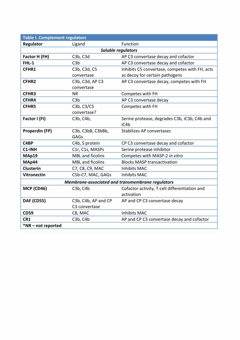
Table I. Complement regulators
Regulator Ligand Function
Soluble regulators
Factor H (FH) C3b, C3d AP C3 convertase decay and cofactor
FHL‐1 C3b AP C3 convertase decay and cofactor
CFHR1 C3b, C3d, C5 convertase
Inhibits C5 convertase, competes with FH, acts as decoy for certain pathogens
CFHR2 C3b, C3d, AP C3 convertase
AP C3 convertase decay, competes with FH
CFHR3 NR Competes with FH
CFHR4 C3b AP C3 convertase decay
CFHR5 C3b, C3/C5 convertase?
Competes with FH
Factor I (FI) C3b, C4b, Serine protease, degrades C3b, iC3b, C4b and iC4b
Properdin (FP) C3b, C3bB, C3bBb, GAGs
Stabilizes AP convertases
C4BP C4b, S protein CP C3 convertase decay and cofactor
C1‐INH C1r, C1s, MASPs Serine protease inhibitor
MAp19 MBL and ficolins Competes with MASP‐2 in vitro
MAp44 MBL and ficolins Blocks MASP transactivation
Clusterin C7, C8, C9, MAC Inhibits MAC
Vitronectin C5b‐C7, MAC, GAGs Inhibits MAC
Membrane‐associated and transmembrane regulators
MCP (CD46) C3b, C4b Cofactor activity, T‐cell differentiation and activation
DAF (CD55) C3b, C4b, AP and CP C3 convertase
AP and CP C3 convertase decay
CD59 C8, MAC Inhibits MAC
CR1 C3b, C4b AP and CP C3 convertase decay and cofactor