94
The Muscular System 1
| Date post: | 16-Dec-2015 |
| Category: |
Documents |
| Upload: | evangeline-bishop |
| View: | 215 times |
| Download: | 0 times |
The Muscular System
1

The Muscular SystemMuscles are responsible
for all types of body movement
Three basic muscle types are found in the body
–Skeletal muscle–Cardiac muscle–Smooth muscle
2

Characteristics of MusclesMuscle cells are elongated (muscle cell = muscle fiber)Contraction of muscles is due to the movement of microfilamentsAll muscles share some terminology– Prefix myo refers to muscle– Prefix mys refers to muscle– Prefix sarco refers to flesh
3
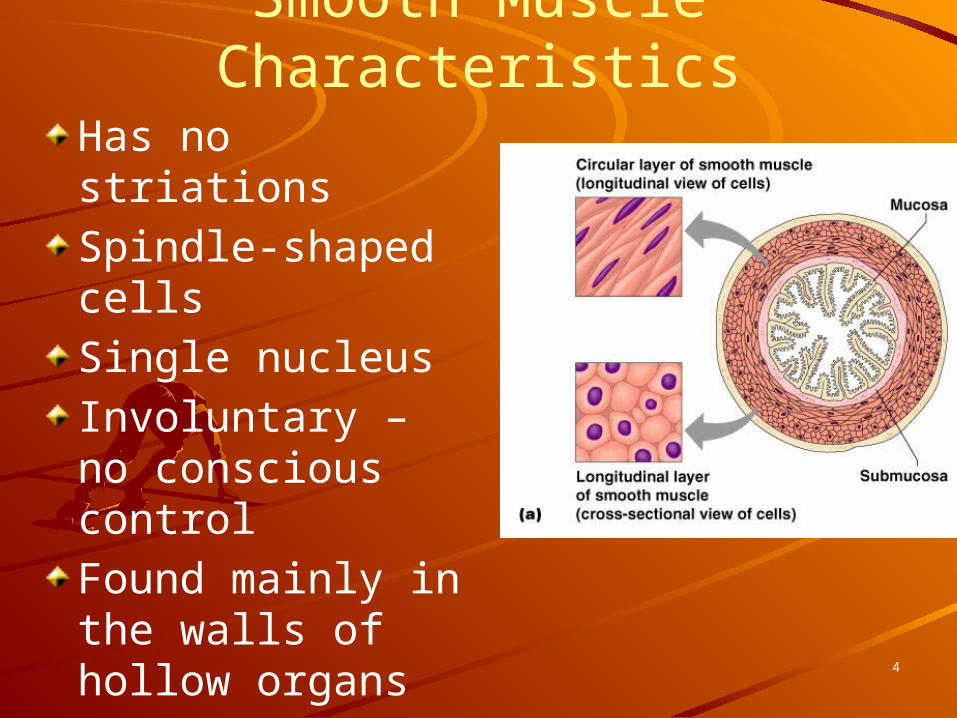
Smooth Muscle CharacteristicsHas no striationsSpindle-shaped cellsSingle nucleusInvoluntary – no conscious controlFound mainly in the walls of hollow organs
4

Cardiac Muscle CharacteristicsHas striationsUsually has a single nucleusJoined to another muscle cell at an intercalated discInvoluntaryFound only in the heart
5
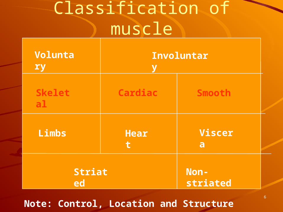
Classification of muscleVoluntary Involuntary
Limbs Heart Viscera
Striated Non-striated
Skeletal Cardiac Smooth
Note: Control, Location and Structure6
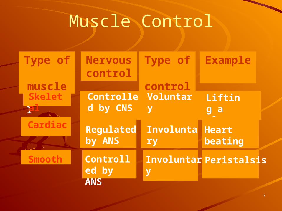
Muscle Control
Type of muscle
Nervouscontrol
Type of control
Example
SkeletalSkeletal Controlled by CNS
Voluntary Lifting a glass
Cardiac Regulated by ANS
Involuntary Heart beating
Smooth Controlled by ANS
Involuntary Peristalsis
7

There are about 650 muscles in the human body. They enable us to move, maintain posture and generate heat. In this section we will only study a sample of the major muscles.
Skeletal Muscle
8
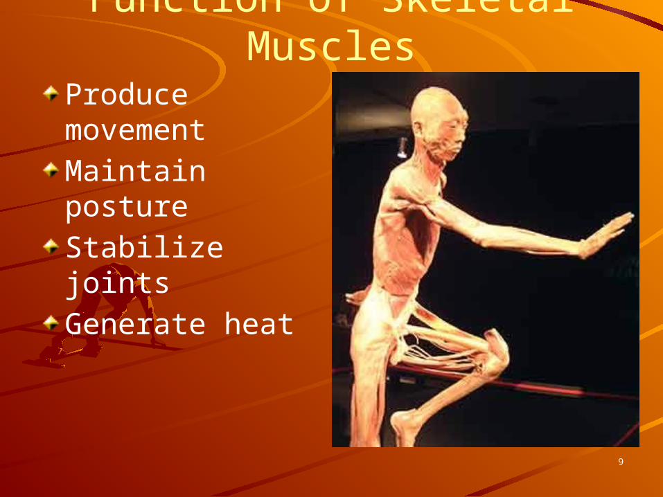
Function of Skeletal MusclesProduce movementMaintain postureStabilize jointsGenerate heat
9
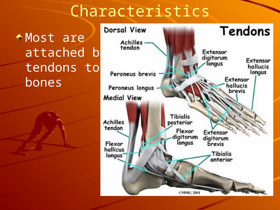
Skeletal Muscle Characteristics
Most are attached by tendons to bones
10

Skeletal Muscle CharacteristicsCells are multinucleateStriated – have visible bandingVoluntary – subject to conscious controlCells are surrounded and bundled by connective tissue
11

Connective Tissue Wrappings of Skeletal Muscle
Endomysium – around single muscle fiberPerimysium – around a fascicle (bundle) of fibers
12

Connective Tissue Wrappings of Skeletal Muscle
Epimysium – covers the entire skeletal muscleFascia – on the outside of the epimysium
13

Skeletal Muscle AttachmentsEpimysium blends into a connective tissue attachment– Tendon – cord-like structure– Aponeuroses – sheet-like
structure
Sites of muscle attachment– Bones– Cartilages– Connective tissue coverings
14

Microscopic Anatomy of Skeletal Muscle
Cells are multinucleateNuclei are just beneath the sarcolemma
15
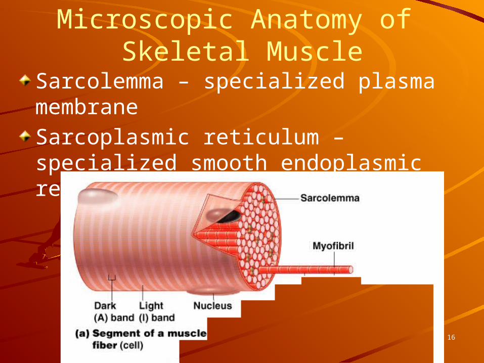
Microscopic Anatomy of Skeletal Muscle
Sarcolemma – specialized plasma membraneSarcoplasmic reticulum – specialized smooth endoplasmic reticulum
16

Microscopic Anatomy of Skeletal Muscle
Myofibril– Bundles of myofilaments– Myofibrils are aligned to give distinct
bandsI band =light bandA band = dark band
17

Microscopic Anatomy of Skeletal Muscle
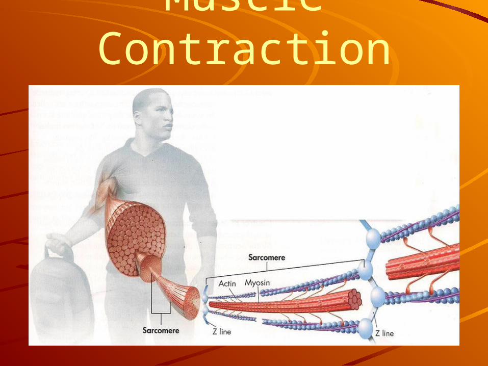
Sarcomere– Contractile unit of a muscle fiber
18

Microscopic Anatomy of Skeletal Muscle
Organization of the sarcomere– Thick filaments = myosin filaments
Composed of the protein myosinHas ATPase enzymes
19

Microscopic Anatomy of Skeletal Muscle
Organization of the sarcomere– Thin filaments = actin filaments
Composed of the protein actin
20

Microscopic Anatomy of Skeletal Muscle
Myosin filaments have heads (extensions, or cross bridges)Myosin and actin overlap somewhat
21

Microscopic Anatomy of Skeletal Muscle
At rest, there is a bare zone that lacks actin filamentsSarcoplasmic reticulum (SR) – for storage of calcium
22
To Summarize . . .
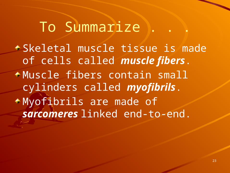
Skeletal muscle tissue is made of cells called muscle fibers.Muscle fibers contain small cylinders called myofibrils.Myofibrils are made of sarcomeres linked end-to-end.
23
Muscle Contraction
24
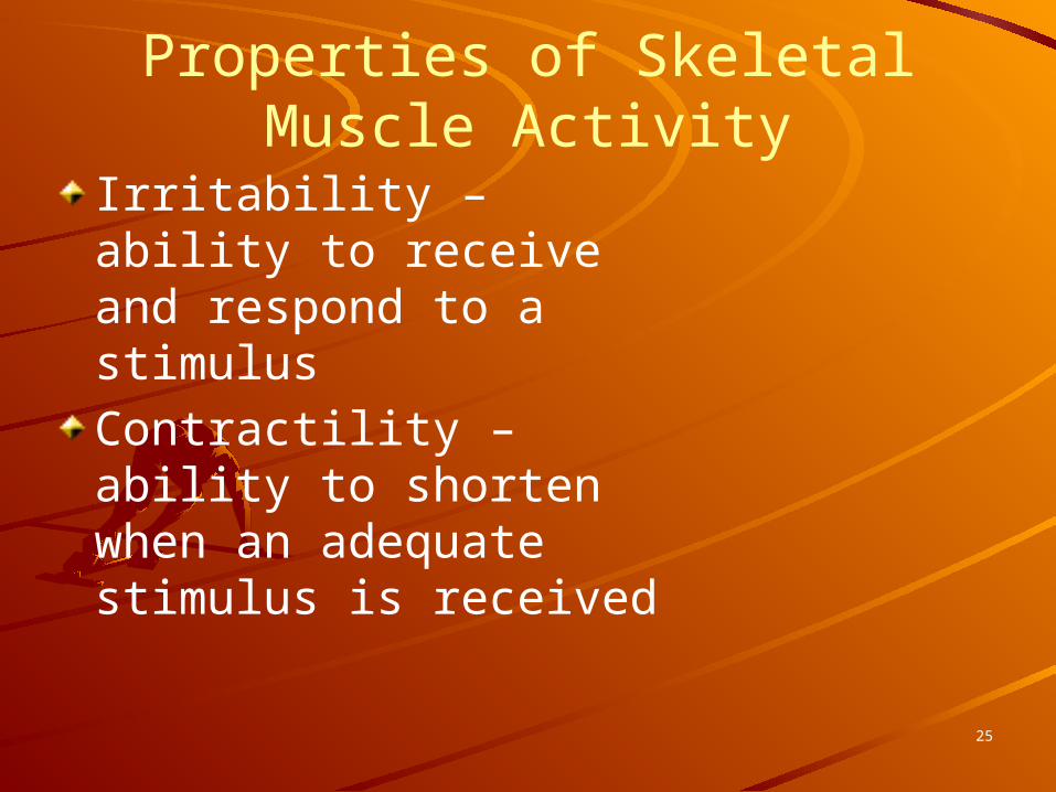
Properties of Skeletal Muscle Activity
Irritability – ability to receive and respond to a stimulusContractility – ability to shorten when an adequate stimulus is received
25

Nerve Stimulus to MusclesSkeletal muscles must be stimulated by a nerve to contractMotor unit– One neuron– Muscle cells
stimulated by that neuron
26

Nerve Stimulus to MusclesNeuromuscular junctions – association site of nerve and muscle
27
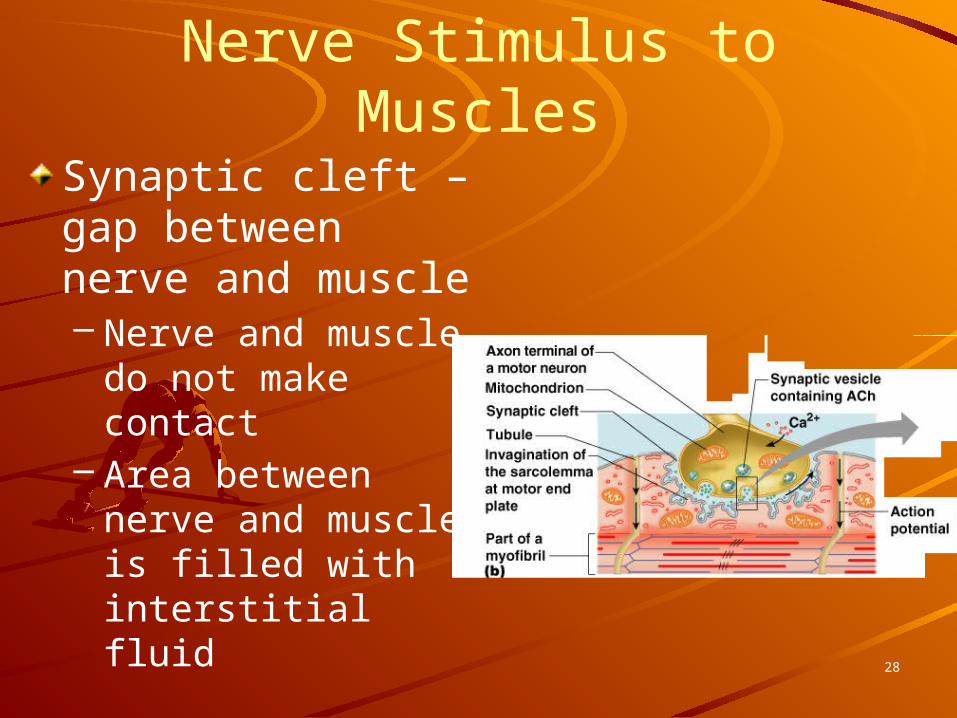
Nerve Stimulus to MusclesSynaptic cleft – gap between nerve and muscle– Nerve and muscle
do not make contact
– Area between nerve and muscle is filled with interstitial fluid
28
Transmission of Nerve Impulse to Muscle
Neurotransmitter – chemical released by nerve upon arrival of nerve impulse– The neurotransmitter for skeletal muscle
is acetylcholine
Neurotransmitter attaches to receptors on the sarcolemmaSarcolemma becomes permeable to sodium (Na+)
29

Transmission of Nerve Impulse to Muscle
Sodium rushing into the cell generates an action potentialOnce started, muscle contraction cannot be stopped
30
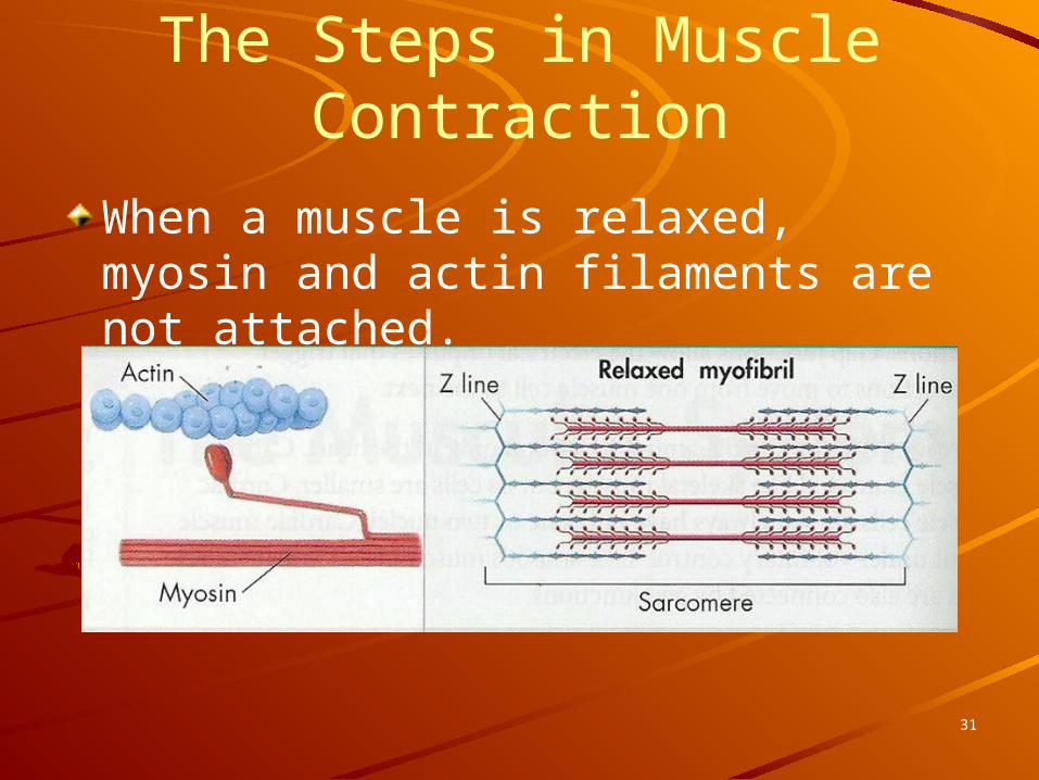
When a muscle is relaxed, myosin and actin filaments are not attached.
The Steps in Muscle Contraction
31

1. Myosin attaches to a binding site on an actin filament. Calcium is required to make a binding site available for myosin.
The Steps in Muscle Contraction
32

The Sliding Filament Theory
33

2. The myosin head rotates and causes the actin filament to slide along the myosin filament. The sliding causes the filaments to overlap more, and the sarcomere becomes shorter.
The Steps in Muscle Contraction
34
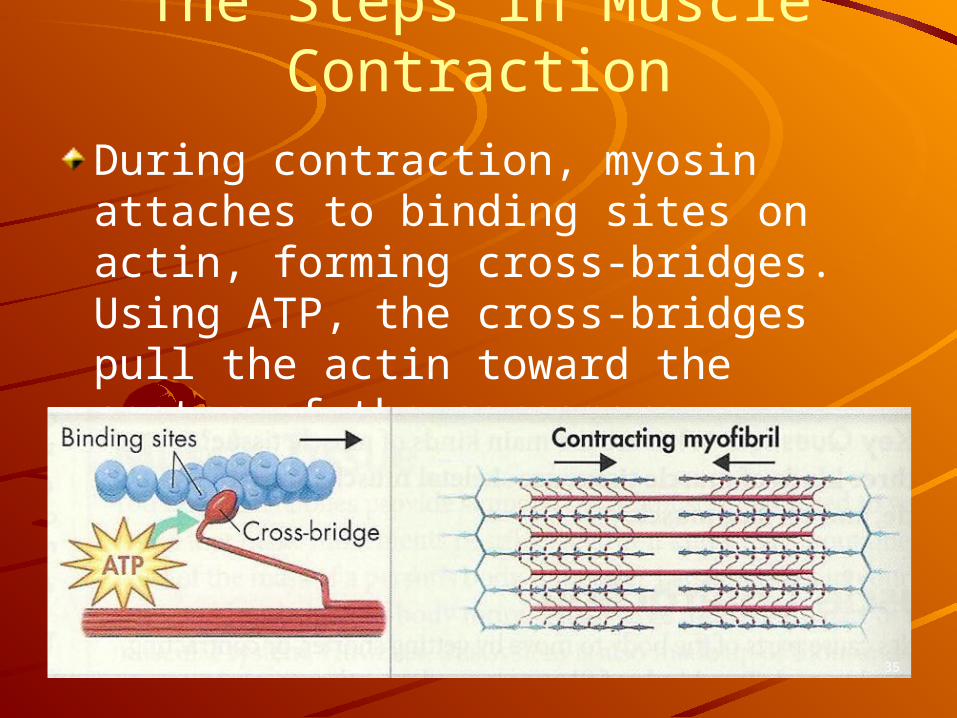
During contraction, myosin attaches to binding sites on actin, forming cross-bridges. Using ATP, the cross-bridges pull the actin toward the center of the sarcomere.
The Steps in Muscle Contraction
35

3. After the myosin head has rotated as far as it can, it must let go of the actin fiber. ATP is required for myosin to detach from actin. The myosin head snaps back into its original position, using the energy in the ATP. The ATP becomes ADP and releases a phosphate ion.
The Steps in Muscle Contraction
36
The Steps in Muscle Contraction
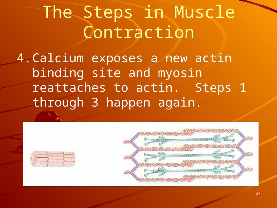
4. Calcium exposes a new actin binding site and myosin reattaches to actin. Steps 1 through 3 happen again.
37

The cross-bridges break, myosin binds to another site, and the cycle begins again until the muscle fiber is contracted.
The Steps in Muscle Contraction
38

To summarize . . .
Myosin filaments bind to actin filaments, actin filaments move inward, and sarcomeres shorten to cause muscle contraction.
39

Contraction of a Skeletal MuscleMuscle fiber contraction is “all or none”Within a skeletal muscle, not all fibers may be stimulated during the same intervalDifferent combinations of muscle fiber contractions may give differing responsesGraded responses – different degrees of skeletal muscle shortening
40
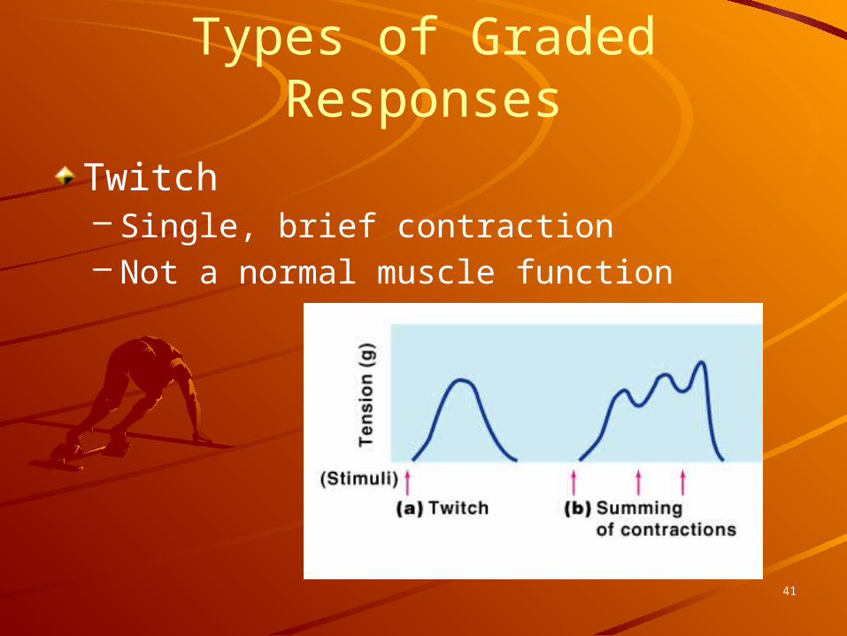
Types of Graded Responses
Twitch– Single, brief contraction– Not a normal muscle function
41
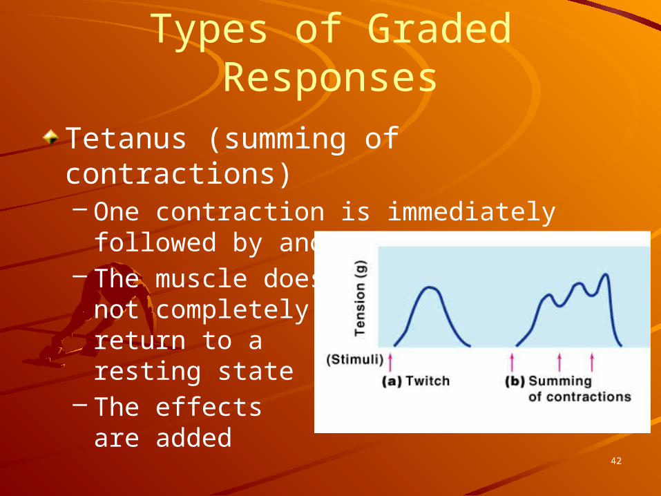
Types of Graded Responses
Tetanus (summing of contractions)– One contraction is immediately followed
by another– The muscle does
not completely return to a resting state
– The effects are added
42
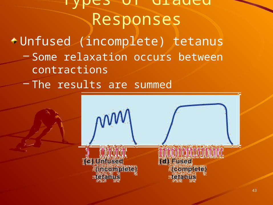
Types of Graded ResponsesUnfused (incomplete) tetanus– Some relaxation occurs between
contractions– The results are summed
43
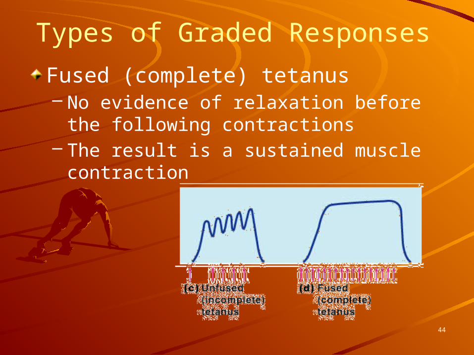
Types of Graded Responses
Fused (complete) tetanus– No evidence of relaxation before the
following contractions– The result is a sustained muscle
contraction
44
Muscle Response to Strong Stimuli
Muscle force depends upon the number of fibers stimulatedMore fibers contracting results in greater muscle tensionMuscles can continue to contract unless they run out of energy
45
Energy for Muscle Contraction
Initially, muscles used stored ATP for energy– Bonds of ATP are broken to release
energy– Only 4-6 seconds worth of ATP is stored
by muscles
After this initial time, other pathways must be utilized to produce ATP
46

Energy for Muscle ContractionDirect phosphorylation– Muscle cells contain
creatine phosphate (CP)CP is a high-energy molecule
– After ATP is depleted, ADP is left
– CP transfers energy to ADP, to regenerate ATP
– CP supplies are exhausted in about 20 seconds
47

Energy for Muscle ContractionAerobic Respiration– Series of metabolic
pathways that occur in the mitochondria
– Glucose is broken down to carbon dioxide and water, releasing energy
– This is a slower reaction that requires continuous oxygen
48
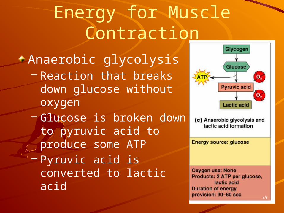
Energy for Muscle Contraction
Anaerobic glycolysis– Reaction that breaks
down glucose without oxygen
– Glucose is broken down to pyruvic acid to produce some ATP
– Pyruvic acid is converted to lactic acid
49

Energy for Muscle ContractionAnaerobic glycolysis (continued)– This reaction is
not as efficient, but is fast
Huge amounts of glucose are neededLactic acid produces muscle fatigue
50

Begin October 18, 2010
51
The Muscular System
52
Five Golden Rules of Gross Muscle Activity
1. all muscles cross at least one joint2. bulk of muscles lies proximal to the
joint crossed3. all muscles have at least 2
attachments: origin & insertion4. muscles only pull/never push5. during contraction the muscle
insertion moves toward the origin
53

Muscles and Body Movements
Movement is attained due to a muscle moving an attached bone
54
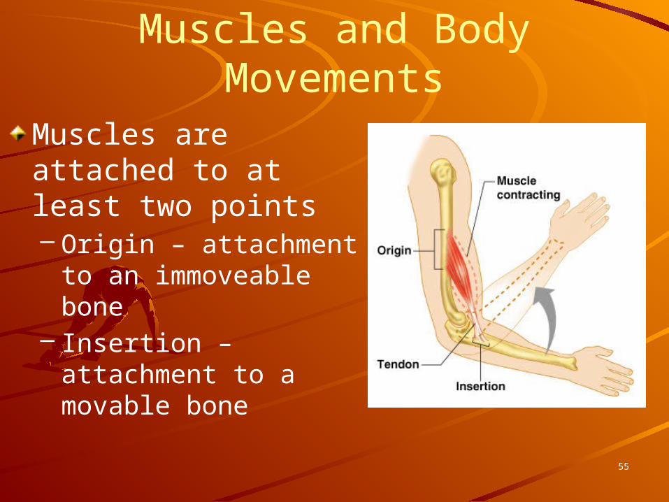
Muscles and Body MovementsMuscles are attached to at least two points– Origin – attachment to
an immoveable bone– Insertion – attachment
to a movable bone
55

The Muscular System
56

Types of Muscle ContractionsIsotonic contractions– Myofilaments are able to slide past each
other during contractions– The muscle shortens
Isometric contractions– Tension in the muscles increases– The muscle is unable to shorten
Myofilaments “skidding their wheels”
57

Muscle ToneSome fibers are contracted even in a relaxed muscleDifferent fibers contract at different times to provide muscle toneThe process of
stimulating various fibers is under involuntary control
58
Muscle Tone
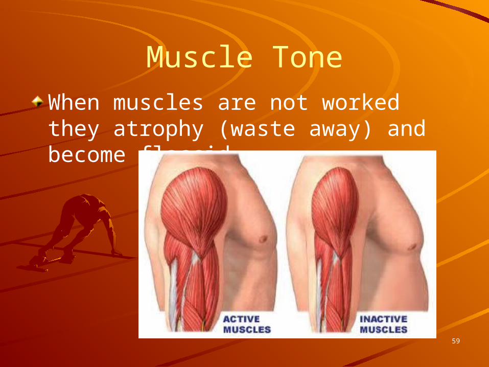
When muscles are not worked they atrophy (waste away) and become flaccid.
59

Effects of Exercise on MuscleAerobics result in stronger muscles due to increase blood supply Muscle fibers increase mitochondria and oxygen storage Muscle becomes more fatigue resistantHeart enlarges to pump
more blood to bodyDoes not increase skeletal
muscle size
60
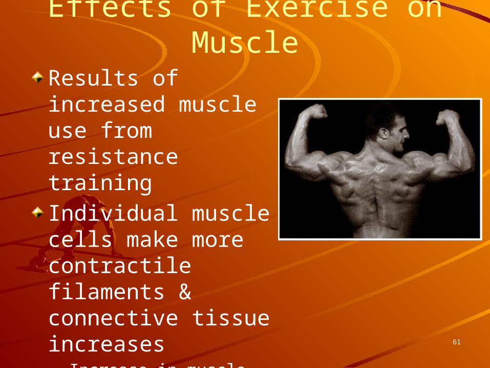
Results of increased muscle use from resistance trainingIndividual muscle cells make more contractile filaments & connective tissue increases– Increase in muscle size– Increase in muscle
strength
Effects of Exercise on Muscle
61

Begin October 19, 2010
62
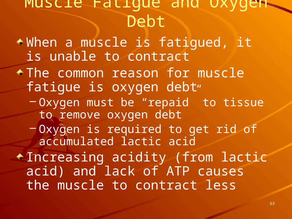
Muscle Fatigue and Oxygen DebtWhen a muscle is fatigued, it is unable to contractThe common reason for muscle fatigue is oxygen debt– Oxygen must be “repaid” to tissue to
remove oxygen debt– Oxygen is required to get rid of
accumulated lactic acidIncreasing acidity (from lactic acid) and lack of ATP causes the muscle to contract less
63

The Muscular System
64
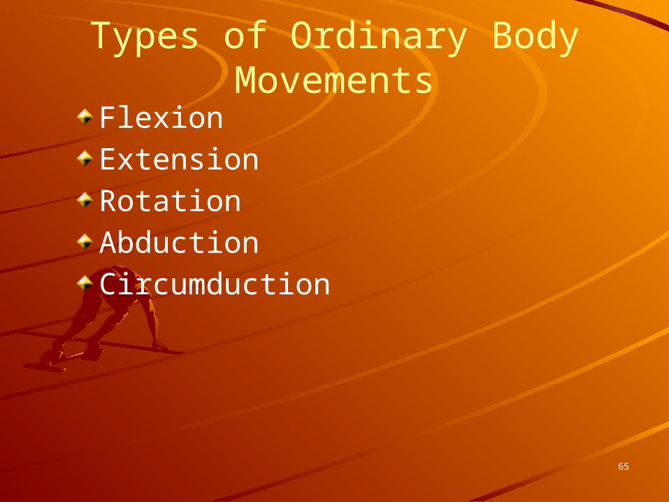
Types of Ordinary Body Movements
FlexionExtensionRotationAbductionCircumduction
65

Flexion & Extension
66
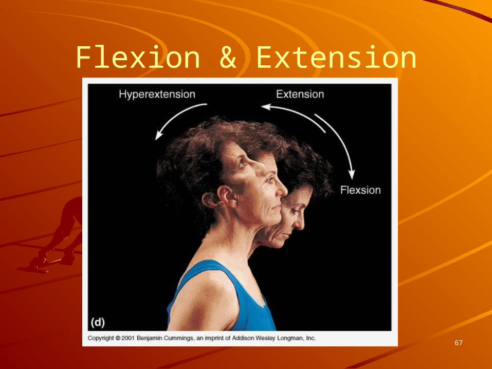
Flexion & Extension
67

Rotation
68

Abduction & Adduction
69

Special Movements
DorsiflexionPlantar flexionInversionEversionSupinationPronationOpposition
70

Dorsiflexion & Plantar Flexion
71

Inversion & Eversion
72
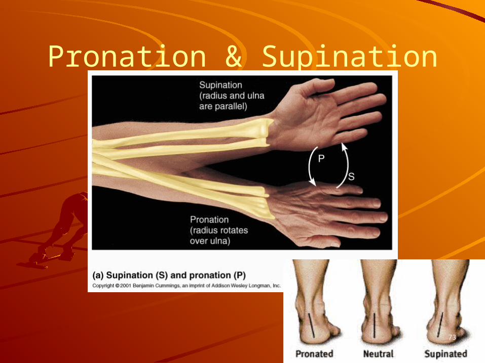
Pronation & Supination
73
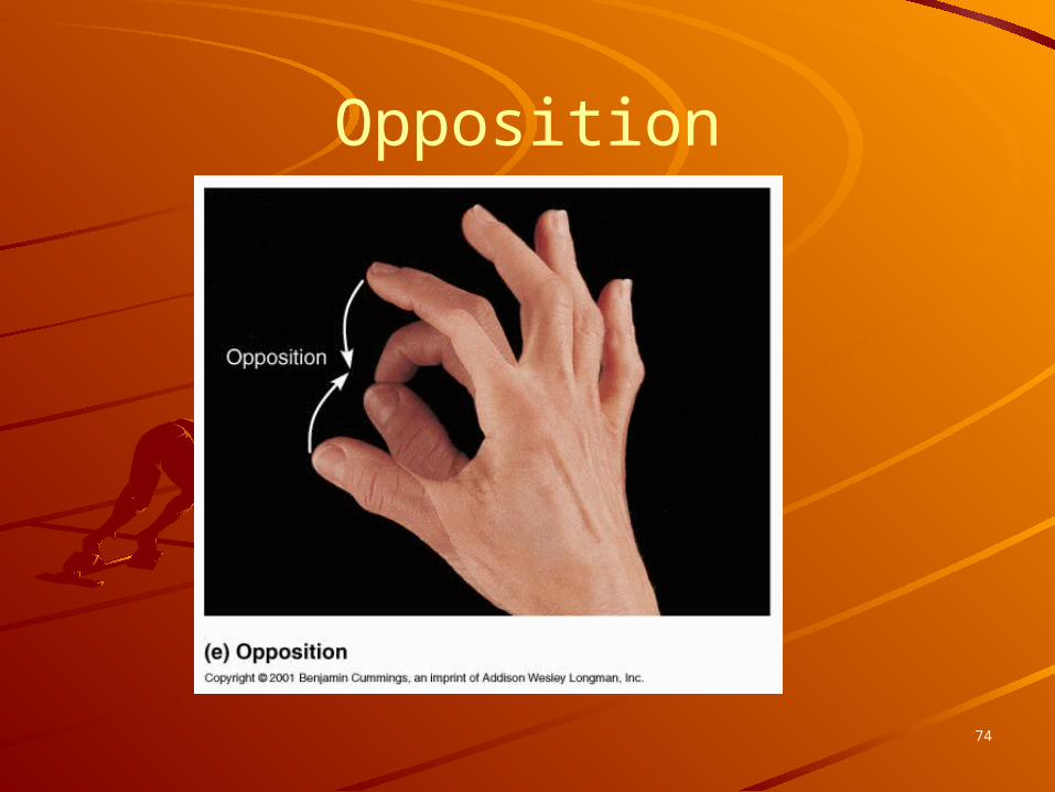
Opposition
74

75

76

77

Diseases and Disorders of the Muscular System
Myalgia: Muscle pain due to strain, tearing of muscle fibers. It also is a symptom of an immune response along with a fever.
Myositis: Inflammation of muscle tissue due to injury or disease.
Charley Horse (fibromyositis): Inflammation of muscle tissue and the tendons associated with that muscle due to injury (tear or severe bruising- contusion)
Cramps: Painful, involuntary muscle spasms78
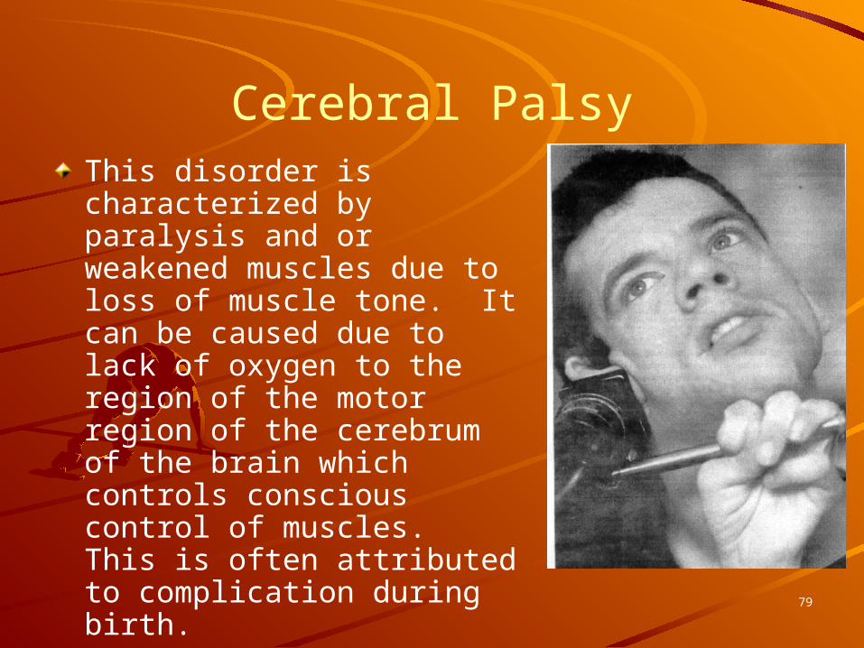
Cerebral PalsyThis disorder is characterized by paralysis and or weakened muscles due to loss of muscle tone. It can be caused due to lack of oxygen to the region of the motor region of the cerebrum of the brain which controls conscious control of muscles. This is often attributed to complication during birth.
79

PoliomyelitisPoliomyelitis: Polio is due to a viral infection which affects the motor neurons that control skeletal muscles. It often leads to paralysis and can result in death by paralysis of the diaphragm. Due to vaccine developed by Jonas Salk, the virus has been virtually eliminated in the US. However, it still poses a threat in developing countries. 80
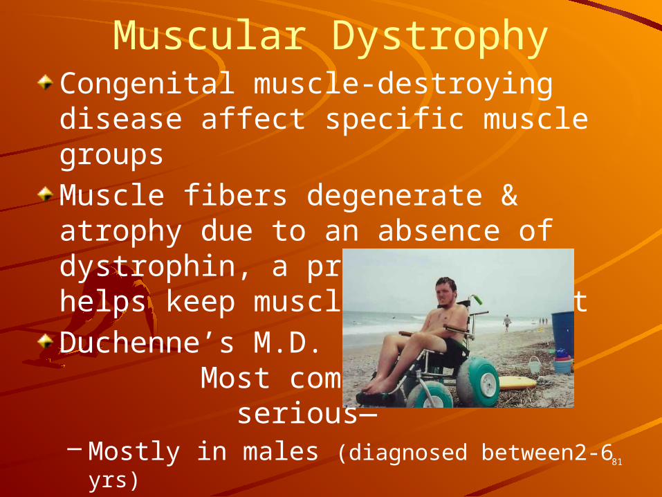
Muscular DystrophyCongenital muscle-destroying disease affect specific muscle groupsMuscle fibers degenerate & atrophy due to an absence of dystrophin, a protein that helps keep muscle cells intactDuchenne’s M.D. Most common & serious—– Mostly in males (diagnosed between2-6 yrs)– Survival is rare beyond early 30’s– X-linked recessive
81

Myasthenia gravisRare adult disease caused by antibodies to acetylcholine receptors at the neuromuscular junction which prevents the muscle contraction from occurring Drooping upper eyelids, difficulty swallowing & talking, muscle weakness & fatigueDeath occurs when respiratory muscles cease to function
82
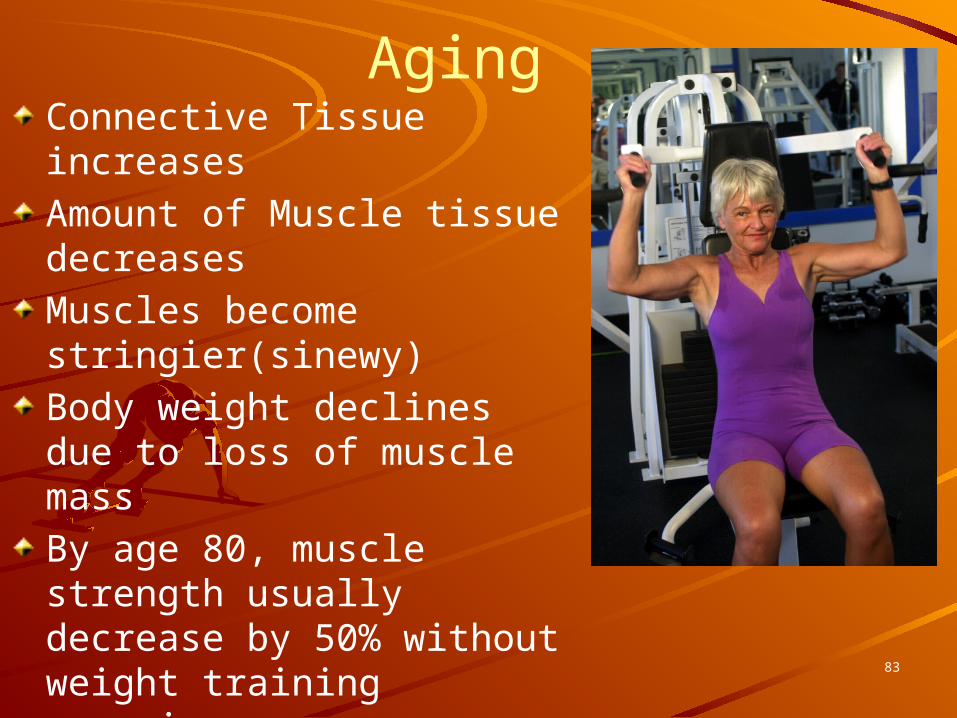
AgingConnective Tissue increasesAmount of Muscle tissue decreasesMuscles become stringier(sinewy)Body weight declines due to loss of muscle massBy age 80, muscle strength usually decrease by 50% without weight training exercises
83
Types of MusclesPrime mover – muscle with the major responsibility for a certain movementAntagonist – muscle that opposes or reverses a prime moverSynergist – muscle that aids a prime mover in a movement and helps prevent rotationFixator – stabilizes the origin of a prime mover
84

Naming of Skeletal Muscles
Direction of muscle fibers– Example: rectus (straight)
Relative size of the muscle– Example: maximus (largest)
85

Naming of Skeletal Muscles
Location of the muscle– Example: many muscles are named for
bones (e.g., temporalis)
Number of origins– Example: triceps (three heads)
86

Naming of Skeletal Muscles
Location of the muscle’s origin and insertion– Example: sterno (on the sternum)
Shape of the muscle– Example: deltoid (triangular)
Action of the muscle– Example: flexor and extensor (flexes or
extends a bone)
87

Head and Neck Muscles
88
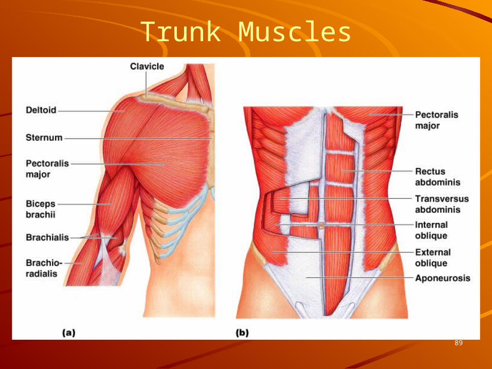
Trunk Muscles
89

Deep Trunk and Arm Muscles
90

Muscles of the Pelvis, Hip, and
Thigh
91

Muscles of the Lower
Leg
92

Superficial Muscles: Anterior
93
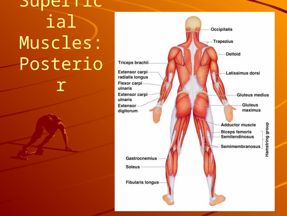
Superficial Muscles: Posterior
94


