65
The Muscular System Chapter 7
| Date post: | 17-Dec-2015 |
| Category: |
Documents |
| Upload: | gabriella-brown |
| View: | 257 times |
| Download: | 3 times |
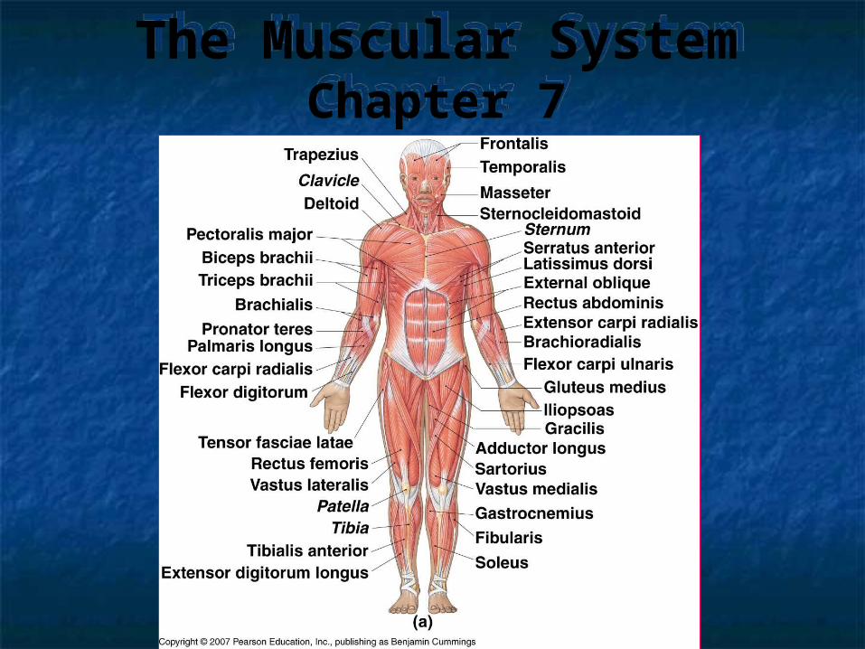
The Muscular SystemChapter 7
The Muscular SystemChapter 7
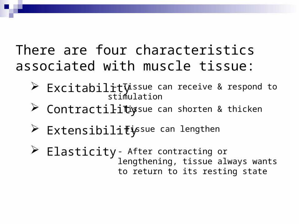
There are four characteristics associated with muscle tissue:
Excitability
Contractility
Extensibility
Elasticity
- Tissue can receive & respond to stimulation
- Tissue can shorten & thicken
- Tissue can lengthen
- After contracting or lengthening, tissue always wants to return to its resting state
The characteristics of muscle tissue enable it to perform some important functions, including:
Movement – both voluntary & involuntary
Maintaining posture
Supporting soft tissues within body cavities
Guarding entrances & exits of the body
Maintaining body temperature
Types of muscle tissue:
• Skeletal
• Cardiac
• Smooth (Visceral)
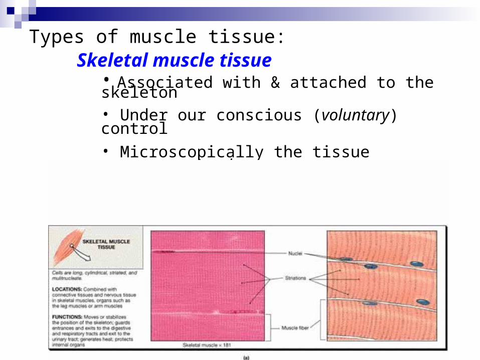
Types of muscle tissue:Skeletal muscle tissue
• Associated with & attached to the skeleton• Under our conscious (voluntary) control• Microscopically the tissue appears striated • Cells are long, cylindrical & multinucleate
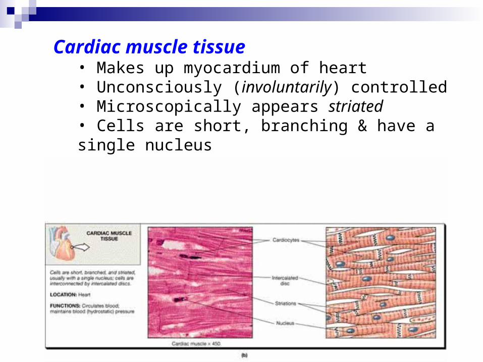
Cardiac muscle tissue• Makes up myocardium of heart• Unconsciously (involuntarily) controlled• Microscopically appears striated• Cells are short, branching & have a single nucleus• Cells connect to each other at intercalated discs
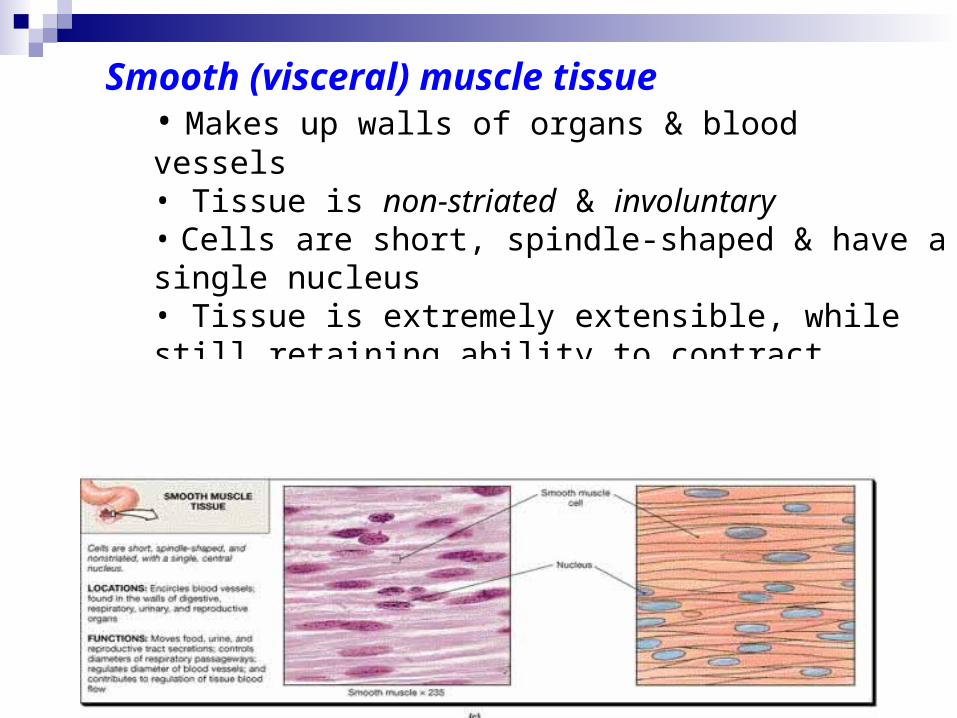
Smooth (visceral) muscle tissue• Makes up walls of organs & blood vessels• Tissue is non-striated & involuntary• Cells are short, spindle-shaped & have a single nucleus• Tissue is extremely extensible, while still retaining ability to contract
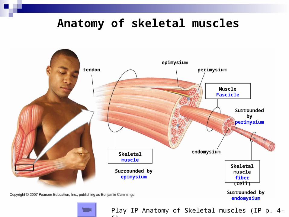
Anatomy of skeletal muscles
Skeletal muscle
fiber (cell)
Muscle Fascicle
Surrounded by perimysium
Surrounded by endomysium
endomysium
perimysium
Skeletal muscle
Surrounded by epimysium
epimysium
tendon
Play IP Anatomy of Skeletal muscles (IP p. 4-6)
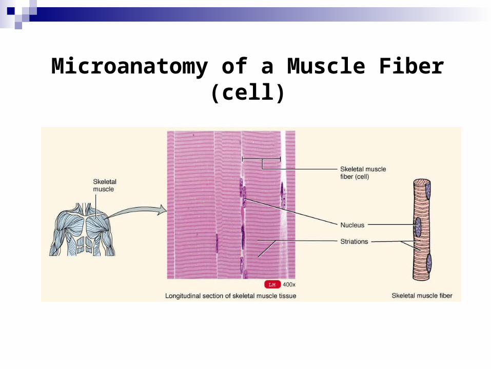
Microanatomy of a Muscle Fiber (cell)
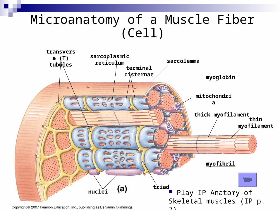
Microanatomy of a Muscle Fiber (Cell)
sarcolemma
transverse (T) tubules sarcoplasmic
reticulumterminal cisternae
myofibril
thin myofilament
thick myofilament
triad
mitochondria
Play IP Anatomy of Skeletal muscles (IP p. 7)
nuclei
myoglobin
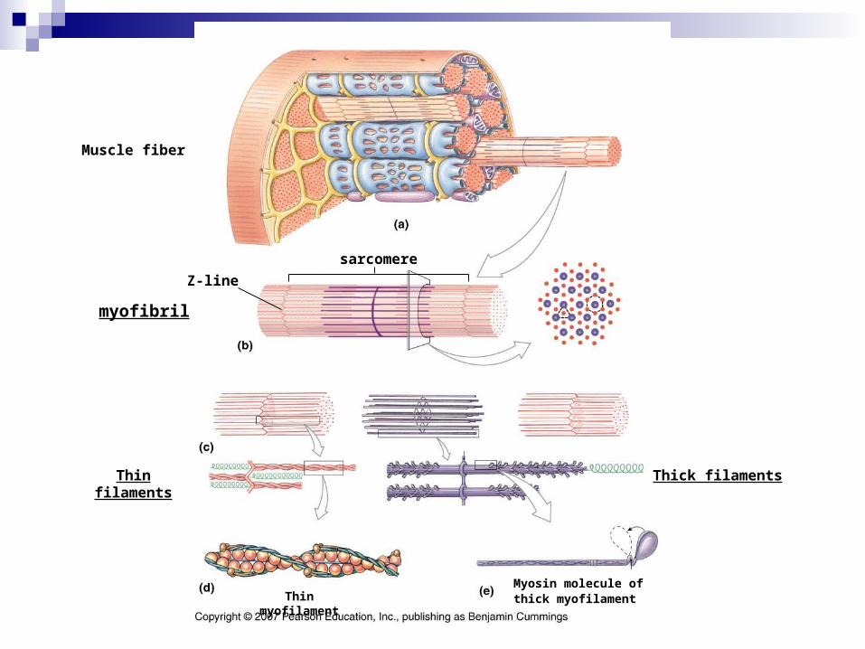
Muscle fiber
myofibril
Thin filaments Thick filaments
Thin myofilamentMyosin molecule ofthick myofilament
sarcomere
Z-line
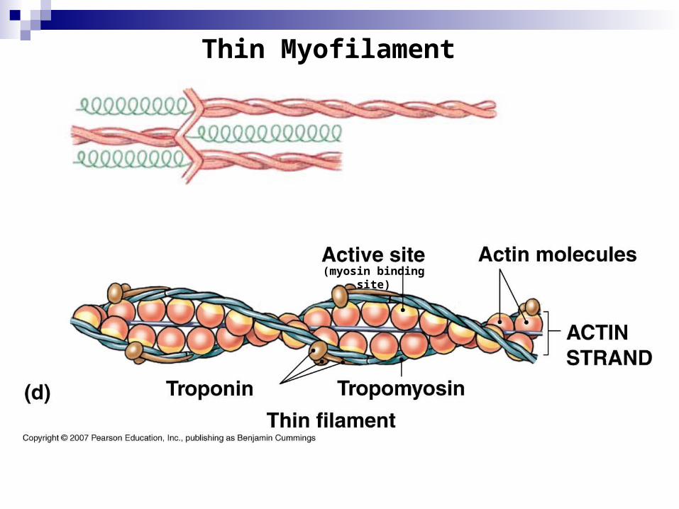
Thin Myofilament
(myosin binding site)
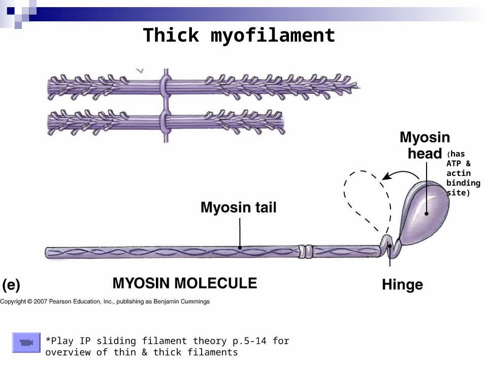
Thick myofilament
(has ATP & actin binding site)
*Play IP sliding filament theory p.5-14 for overview of thin & thick filaments
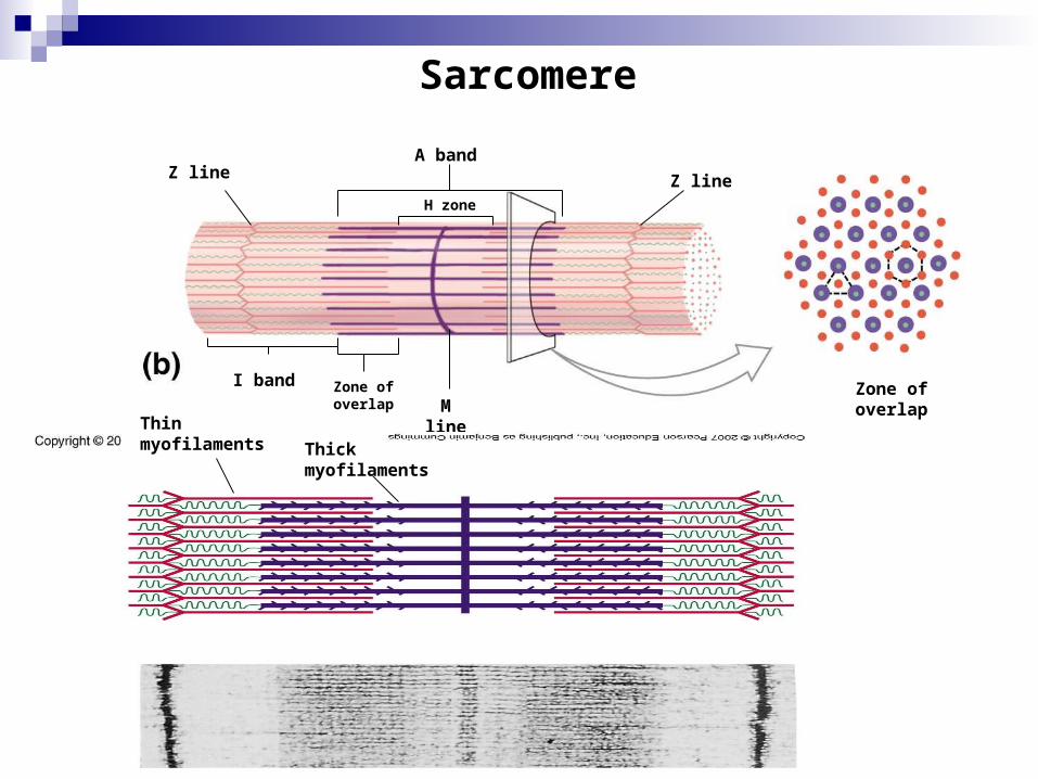
Sarcomere
Z line Z line
A band
H zone
I band Zone of overlap M line
Zone of overlap
Thin myofilaments Thick
myofilaments
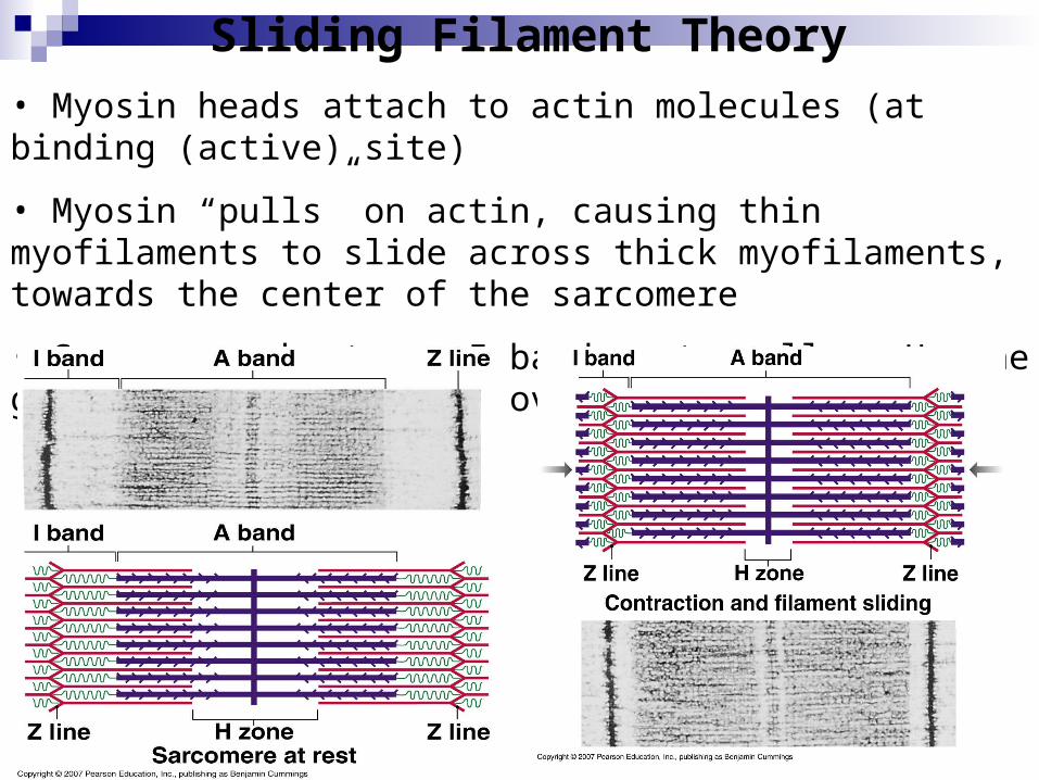
Sliding Filament Theory
• Myosin heads attach to actin molecules (at binding (active) site)
• Myosin “pulls” on actin, causing thin myofilaments to slide across thick myofilaments, towards the center of the sarcomere
• Sarcomere shortens, I bands get smaller, H zone gets smaller, & zone of overlap increases
• As sarcomeres shorten, myofibril shortens. As myofibrils shorten, so does muscle fiber
• Once a muscle fiber begins to contract, it will contract maximally
• This is known as the “all or none” principle
Play IP sliding filament theory p. 28
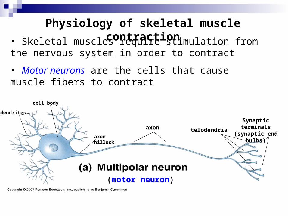
Physiology of skeletal muscle contraction
• Skeletal muscles require stimulation from the nervous system in order to contract
• Motor neurons are the cells that cause muscle fibers to contract
(motor neuron)
cell body
dendrites
axonSynaptic terminals
(synaptic end bulbs)telodendriaaxon hillock

telodendria
Synaptic terminal
(end bulb)
Neuromuscular junction
Synaptic vessicles
containing Ach
Motor end plateof sarcolemma
Synaptic cleftNeuromuscular
junction

Overview of Events at the neuromuscular junction
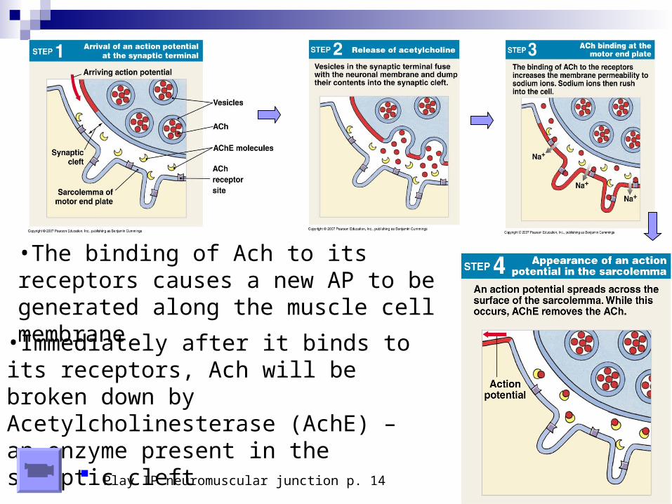
• An action potential (AP), an electrical impulse, travels down the axon of the motor neuron to the end bulbs (synaptic terminals)
• The AP causes the synaptic vesicles to fuse with the end bulb membrane, resulting in the release of Acetylcholine (Ach) into the synaptic cleft
• Ach diffuses across the synaptic cleft & binds to Ach receptors on the motor end plate
• The binding of Ach to its receptors causes a new AP to be generated along the muscle cell membrane
• Immediately after it binds to its receptors, Ach will be broken down by Acetylcholinesterase (AchE) – an enzyme present in the synaptic cleft
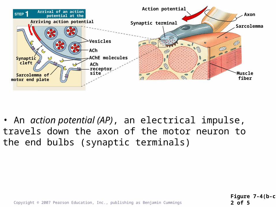
Figure 7-4(b-c)2 of 5Copyright © 2007 Pearson Education, Inc., publishing as Benjamin Cummings
Synapticcleft
Arrival of an action potential at the synaptic terminal
Sarcolemma ofmotor end plate
Arriving action potential
Vesicles
ACh
AChE molecules
AChreceptorsite
Action potential
Synaptic terminal
Axon
Sarcolemma
Musclefiber
• An action potential (AP), an electrical impulse, travels down the axon of the motor neuron to the end bulbs (synaptic terminals)
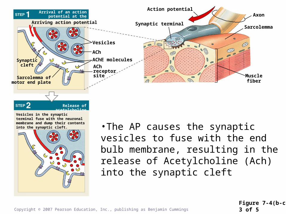
Figure 7-4(b-c)3 of 5Copyright © 2007 Pearson Education, Inc., publishing as Benjamin Cummings
Synapticcleft
Vesicles in the synaptic terminal fuse with the neuronal membrane and dump their contents into the synaptic cleft.
Release of acetylcholine
Arrival of an action potential at the synaptic terminal
Sarcolemma ofmotor end plate
Arriving action potential
Vesicles
ACh
AChE molecules
AChreceptorsite
Action potential
Synaptic terminal
Axon
Sarcolemma
Musclefiber
•The AP causes the synaptic vesicles to fuse with the end bulb membrane, resulting in the release of Acetylcholine (Ach) into the synaptic cleft
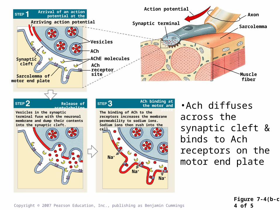
Figure 7-4(b-c)4 of 5Copyright © 2007 Pearson Education, Inc., publishing as Benjamin Cummings
Synapticcleft
Vesicles in the synaptic terminal fuse with the neuronal membrane and dump their contents into the synaptic cleft.
The binding of ACh to the receptors increases the membrane permeability to sodium ions. Sodium ions then rush into the cell.
ACh binding at the motor and plateRelease of acetylcholine
Arrival of an action potential at the synaptic terminal
Sarcolemma ofmotor end plate
Arriving action potential
Vesicles
ACh
AChE molecules
AChreceptorsite
Action potential
Synaptic terminal
Axon
Sarcolemma
Musclefiber
Na+
Na+
Na+
•Ach diffuses across the synaptic cleft & binds to Ach receptors on the motor end plate
•The binding of Ach to its receptors causes a new AP to be generated along the muscle cell membrane
•Immediately after it binds to its receptors, Ach will be broken down by Acetylcholinesterase (AchE) – an enzyme present in the synaptic cleft
Play IP neuromuscular junction p. 14
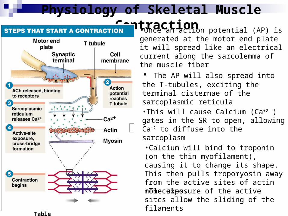
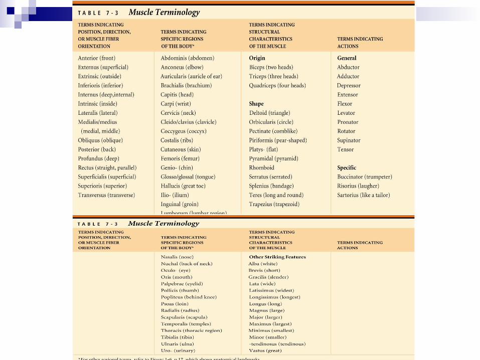
Table 7-1
Physiology of Skeletal Muscle Contraction
•Once an action potential (AP) is generated at the motor end plate it will spread like an electrical current along the sarcolemma of the muscle fiber
• The AP will also spread into the T-tubules, exciting the terminal cisternae of the sarcoplasmic reticula
•This will cause Calcium (Ca+2 ) gates in the SR to open, allowing Ca+2 to diffuse into the sarcoplasm
•Calcium will bind to troponin (on the thin myofilament), causing it to change its shape. This then pulls tropomyosin away from the active sites of actin molecules.
•The exposure of the active sites allow the sliding of the filaments
Copyright © 2007 Pearson Education, Inc., publishing as Benjamin Cummings
Figure 7-53 of 7
Resting sarcomere
Myosin head
Active-site exposure
Troponin
ActinTropomyosin
ADP
P+
ADP
P +
ADP
P+
Active site
Sarcoplasm
Ca2+
Ca2+
ADP
P +
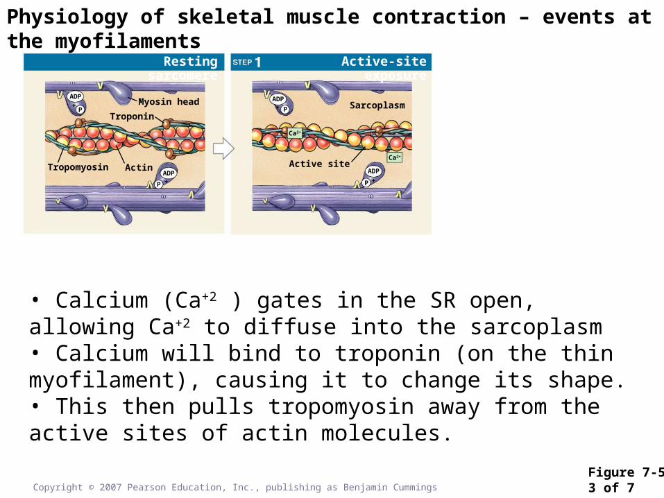
• Calcium (Ca+2 ) gates in the SR open, allowing Ca+2 to diffuse into the sarcoplasm• Calcium will bind to troponin (on the thin myofilament), causing it to change its shape. • This then pulls tropomyosin away from the active sites of actin molecules.
Physiology of skeletal muscle contraction – events at the myofilaments
Copyright © 2007 Pearson Education, Inc., publishing as Benjamin Cummings
Figure 7-54 of 7
Resting sarcomere
Myosin head
Active-site exposure Cross-bridge formation
Troponin
ActinTropomyosin
ADP
P+
ADP
P +
ADP
P+
Active site
Sarcoplasm
Ca2+
Ca2+
ADP
P +
ADP
+ P
Ca2+
ADP+P
Ca2+
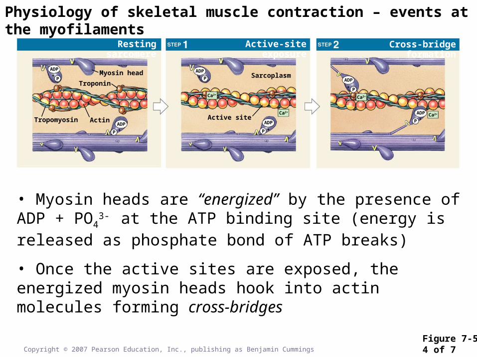
• Myosin heads are “energized” by the presence of ADP + PO43-
at the ATP binding site (energy is released as phosphate bond of ATP breaks)
• Once the active sites are exposed, the energized myosin heads hook into actin molecules forming cross-bridges
Physiology of skeletal muscle contraction – events at the myofilaments
Copyright © 2007 Pearson Education, Inc., publishing as Benjamin Cummings
Figure 7-55 of 7
Resting sarcomere
Myosin head
Active-site exposure Cross-bridge formation
Pivoting of myosin head
Troponin
ActinTropomyosin
ADP
P+
ADP
P +
ADP
P+
Active site
Sarcoplasm
Ca2+
Ca2+
ADP
P +
ADP
+ P
Ca2+
ADP+P
Ca2+
Ca2+
ADP + P
Ca2+
ADP + P
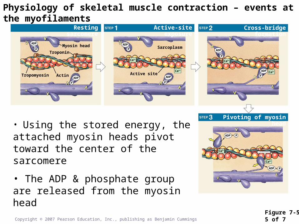
• Using the stored energy, the attached myosin heads pivot toward the center of the sarcomere
• The ADP & phosphate group are released from the myosin head
Physiology of skeletal muscle contraction – events at the myofilaments
Resting sarcomere
Myosin head
Active-site exposure
Cross bridge detachment
Cross-bridge formation
Pivoting of myosin head
Troponin
ActinTropomyosin
ADP
P+
ADP
P +
ADP
P+
Active site
Sarcoplasm
Ca2+
Ca2+
ADP
P +
ADP
+ P
Ca2+
ADP+P
Ca2+
Ca2+
ADP + P
Ca2+
ADP + P
Ca2+
ATP
ATP
Ca2+
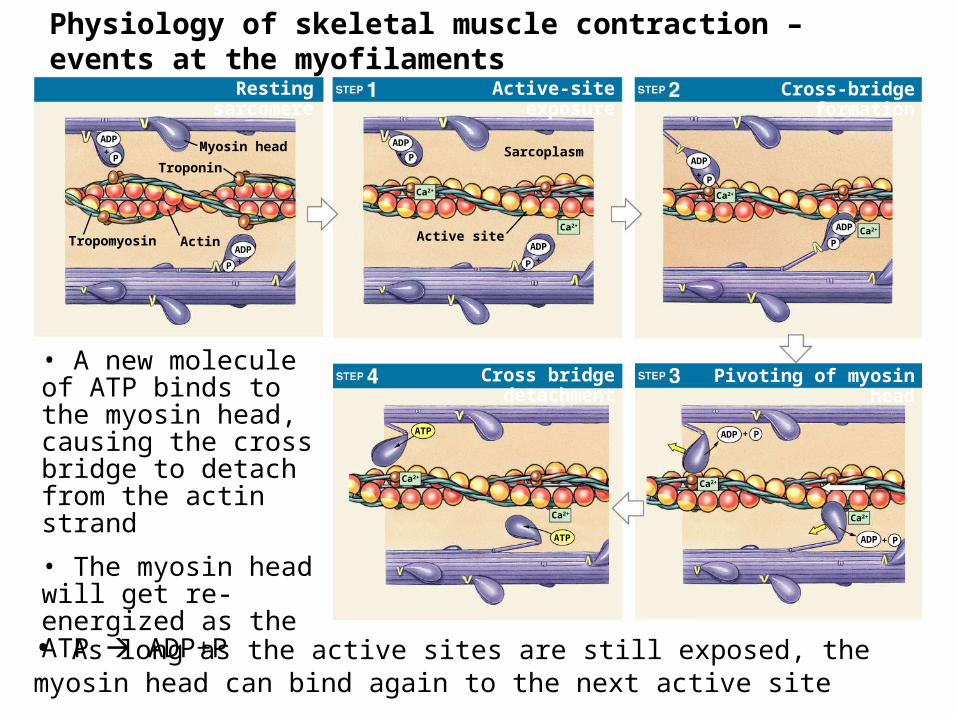
• A new molecule of ATP binds to the myosin head, causing the cross bridge to detach from the actin strand
• The myosin head will get re-energized as the ATP ADP+P
• As long as the active sites are still exposed, the myosin head can bind again to the next active site
Physiology of skeletal muscle contraction – events at the myofilaments
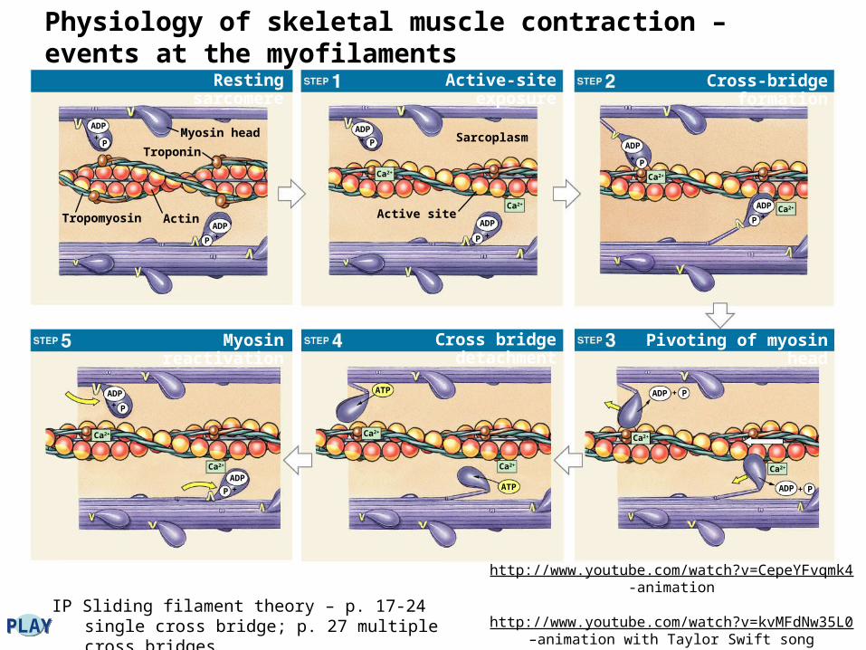
IP Sliding filament theory – p. 17-24 single cross bridge; p. 27 multiple cross bridgesPLAYPLAY
Resting sarcomere
Myosin head
Myosin reactivation
Active-site exposure
Cross bridge detachment
Cross-bridge formation
Pivoting of myosin head
Troponin
ActinTropomyosin
ADP
P+
ADP
P +
ADP
P+
Active site
Sarcoplasm
Ca2+
Ca2+
ADP
P +
ADP
+ P
Ca2+
ADP+P
Ca2+
Ca2+
ADP + P
Ca2+
ADP + P
Ca2+
ATP
ATP
Ca2+
Ca2+
Ca2+
ADP
P +
+ P
ADP
http://www.youtube.com/watch?v=CepeYFvqmk4 -animation
http://www.youtube.com/watch?v=kvMFdNw35L0 –
animation with Taylor Swift song
Physiology of skeletal muscle contraction – events at the myofilaments
Physiology of Skeletal Muscle Contraction
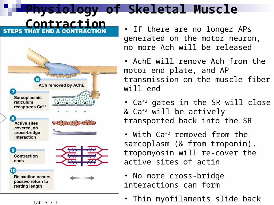
• If there are no longer APs generated on the motor neuron, no more Ach will be released
• AchE will remove Ach from the motor end plate, and AP transmission on the muscle fiber will end
• Ca+2 gates in the SR will close & Ca+2 will be actively transported back into the SR
• With Ca+2 removed from the sarcoplasm (& from troponin), tropomyosin will re-cover the active sites of actin
• No more cross-bridge interactions can form
• Thin myofilaments slide back to their resting state
Table 7-1
Skeletal muscle fibers shorten as thick filaments interact with thin filaments (“cross bridge”) and sliding occurs (“power stroke”). The trigger for contraction is the calcium ions released by the SR when the muscle fiber is stimulated by its motor neuron. Contraction is an active process; relaxation and the return to resting length is entirely passive.
These physiological processes describe what happen at the cellular level – how skeletal muscle fibers contract
But what about at the organ level? How do skeletal muscles (like your biceps brachii) contract to create useful movement?
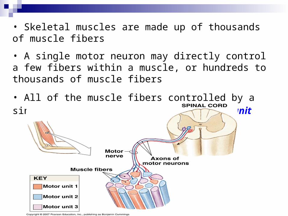
• Skeletal muscles are made up of thousands of muscle fibers
• A single motor neuron may directly control a few fibers within a muscle, or hundreds to thousands of muscle fibers
• All of the muscle fibers controlled by a single motor neuron constitute a motor unit
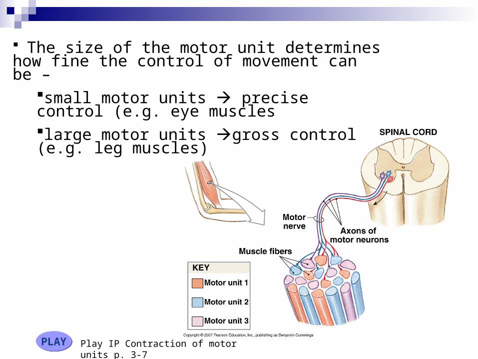
The size of the motor unit determines how fine the control of movement can be –
small motor units precise control (e.g. eye muscleslarge motor units gross control (e.g. leg muscles)
Play IP Contraction of motor units p. 3-7PLAYPLAY
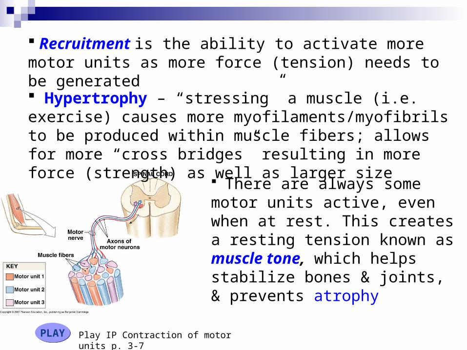
Recruitment is the ability to activate more motor units as more force (tension) needs to be generated
There are always some motor units active, even when at rest. This creates a resting tension known as muscle tone, which helps stabilize bones & joints, & prevents atrophy
Play IP Contraction of motor units p. 3-7PLAYPLAY
Hypertrophy – “stressing” a muscle (i.e. exercise) causes more myofilaments/myofibrils to be produced within muscle fibers; allows for more “cross bridges” resulting in more force (strength) as well as larger size
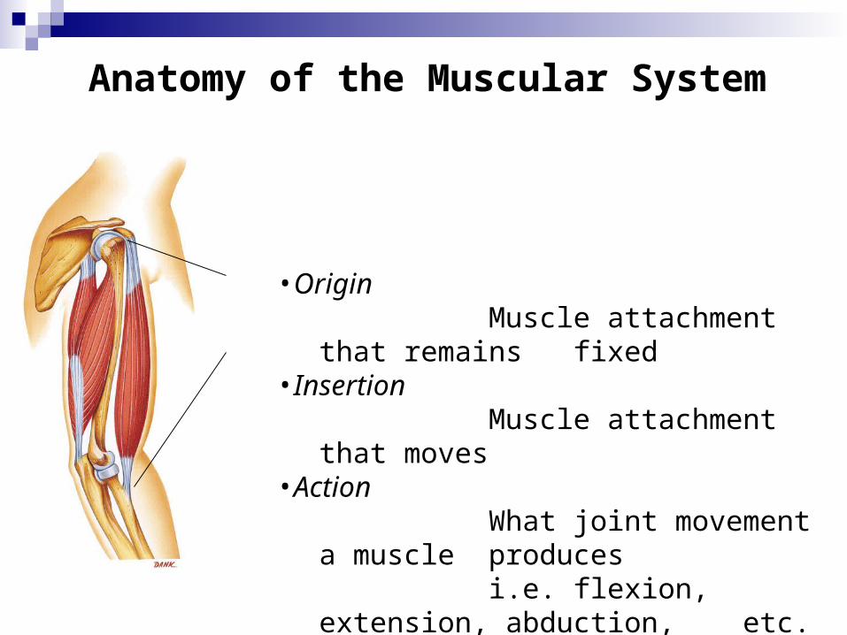
Anatomy of the Muscular System
•OriginMuscle attachment that
remains fixed•Insertion
Muscle attachment that moves
•ActionWhat joint movement a
muscle producesi.e. flexion, extension,
abduction, etc.

• For muscles to create a movement, they can only pull, not push
• Muscles in the body rarely work alone, & are usually arranged in groups surrounding a joint
• A muscle that contracts to create the desired action is known as an agonist or prime mover
• A muscle that helps the agonist is a synergist
• A muscle that opposes the action of the agonist, therefore undoing the desired action is an antagonist
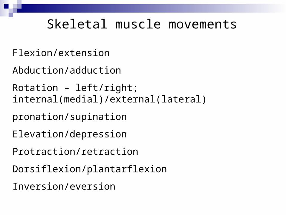
Skeletal muscle movements
Flexion/extension
Abduction/adduction
Rotation – left/right; internal(medial)/external(lateral)
pronation/supination
Elevation/depression
Protraction/retraction
Dorsiflexion/plantarflexion
Inversion/eversion
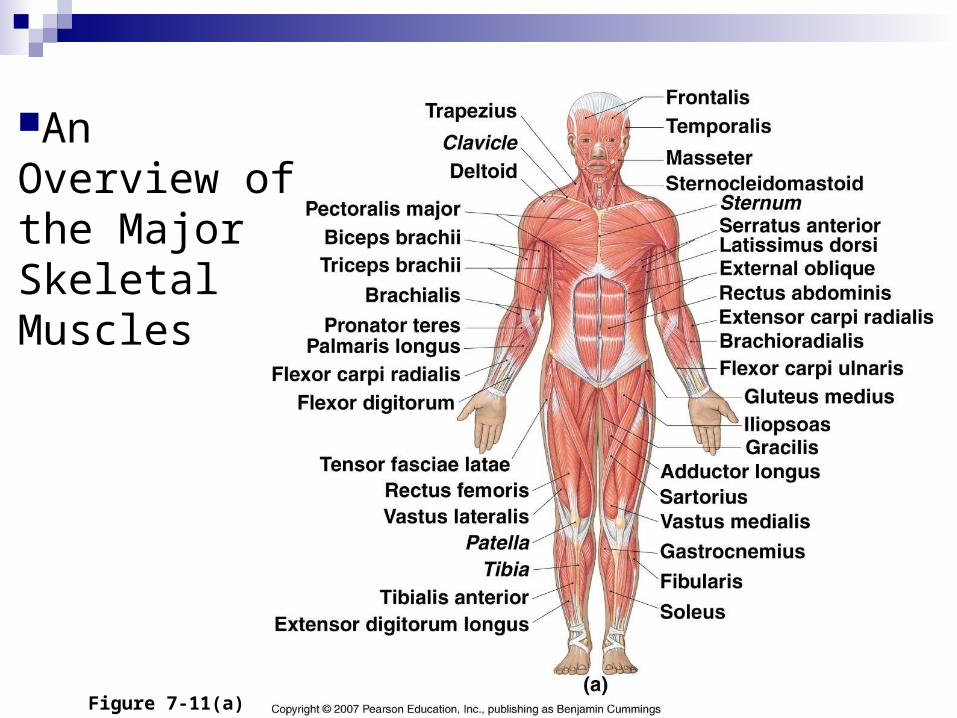
An Overview of the Major Skeletal Muscles
Figure 7-11(a)
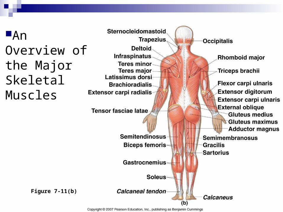
An Overview of the Major Skeletal Muscles
Figure 7-11(b)


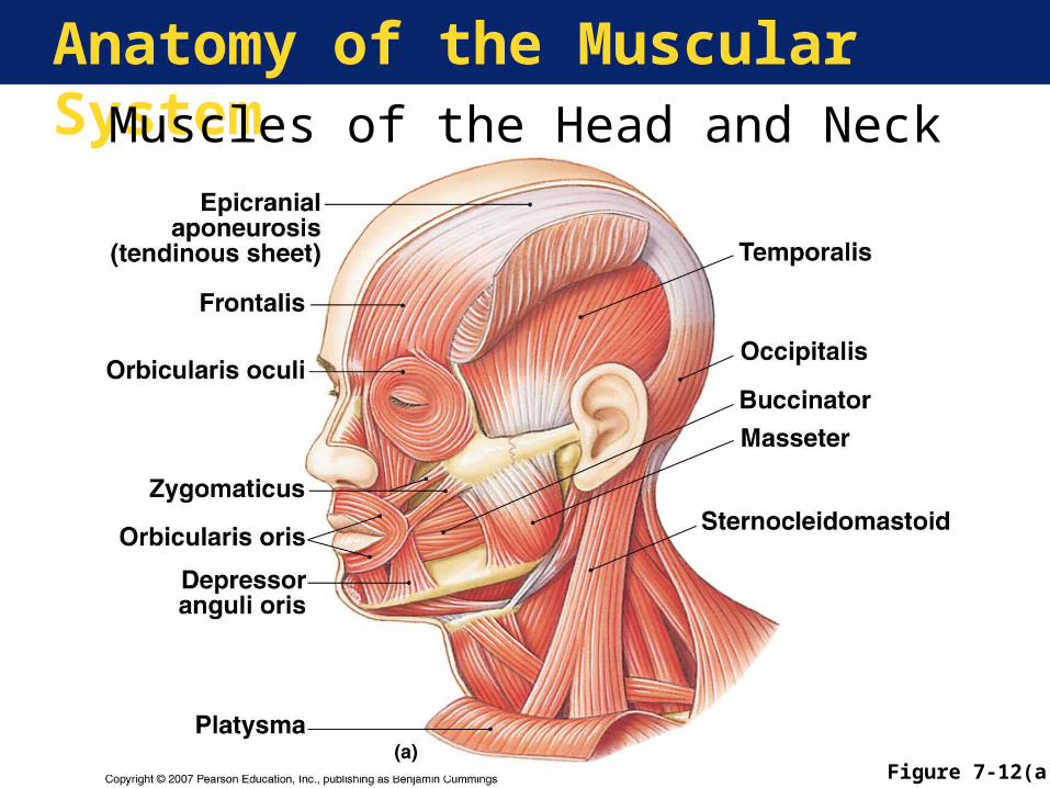
Anatomy of the Muscular SystemMuscles of the Head and Neck
Figure 7-12(a)
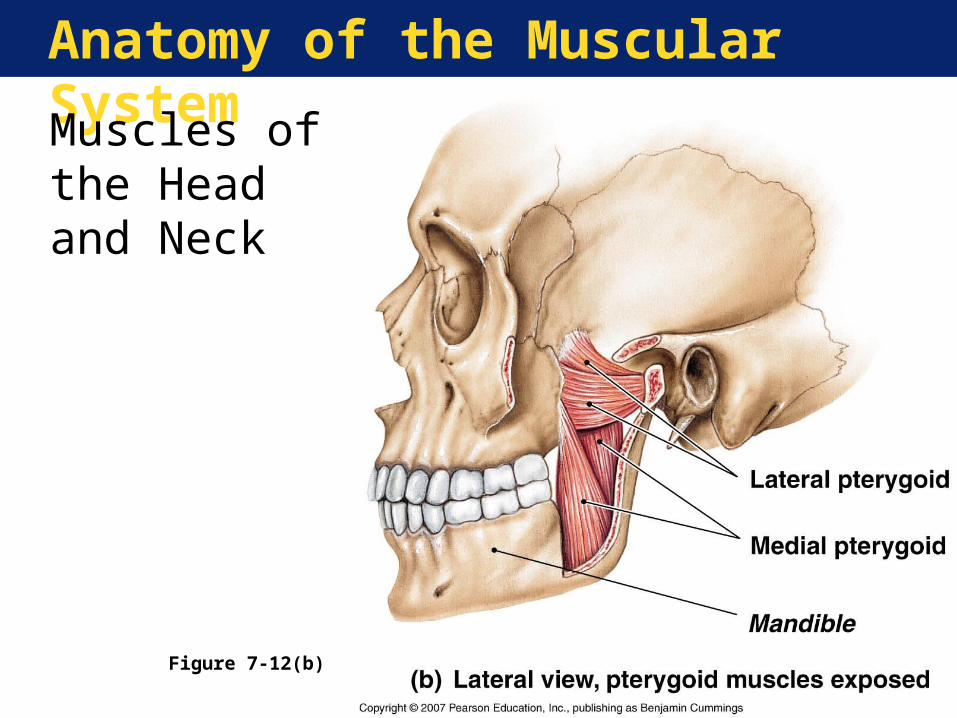
Anatomy of the Muscular System
Muscles of the Head and Neck
Figure 7-12(b)
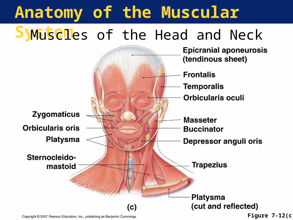
Anatomy of the Muscular SystemMuscles of the Head and Neck
Figure 7-12(c)
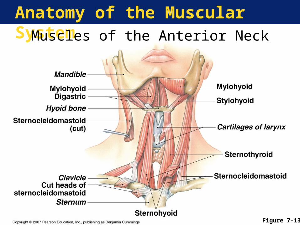
Anatomy of the Muscular SystemMuscles of the Anterior Neck
Figure 7-13
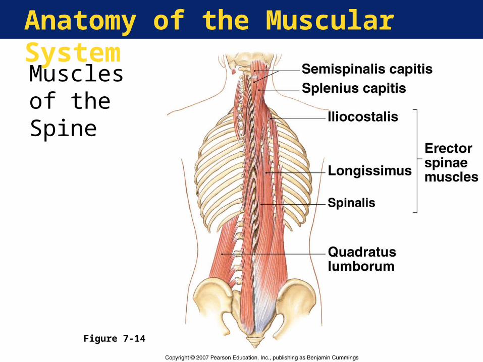
Anatomy of the Muscular System
Muscles of the Spine
Figure 7-14
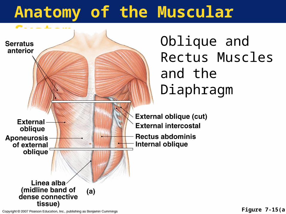
Anatomy of the Muscular System
Figure 7-15(a)
Oblique and Rectus Muscles and the Diaphragm
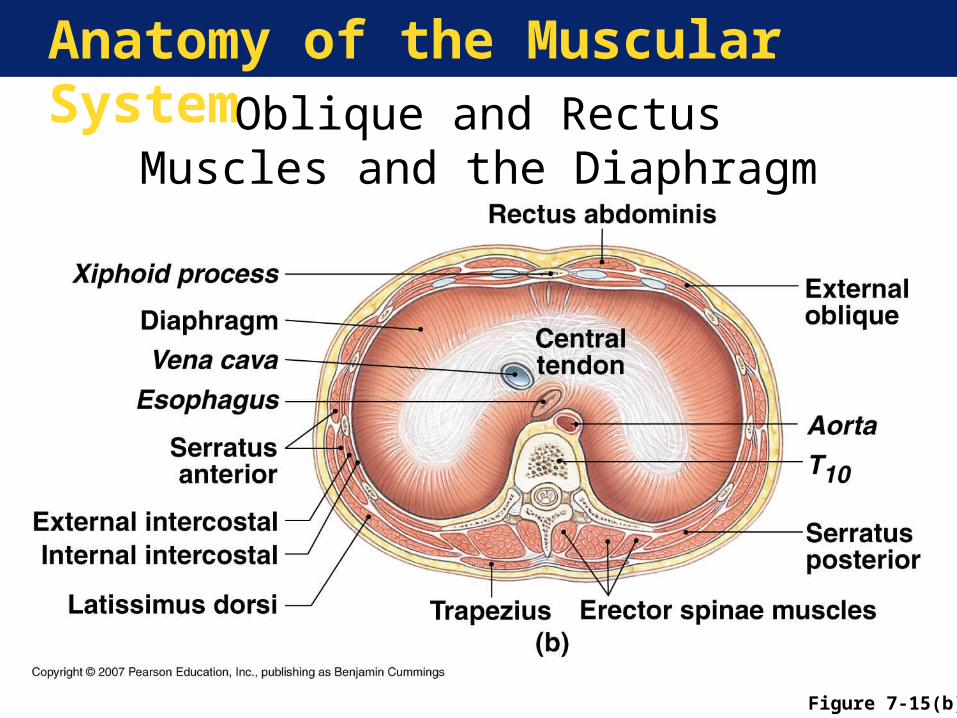
Anatomy of the Muscular SystemOblique and Rectus Muscles and
the Diaphragm
Figure 7-15(b)
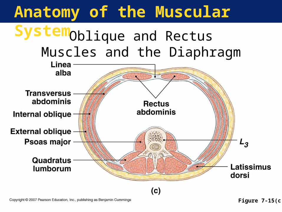
Anatomy of the Muscular System
Oblique and Rectus Muscles and the Diaphragm
Figure 7-15(c)
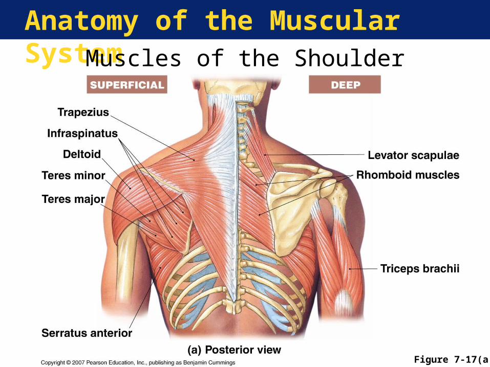
Anatomy of the Muscular SystemMuscles of the Shoulder
Figure 7-17(a)
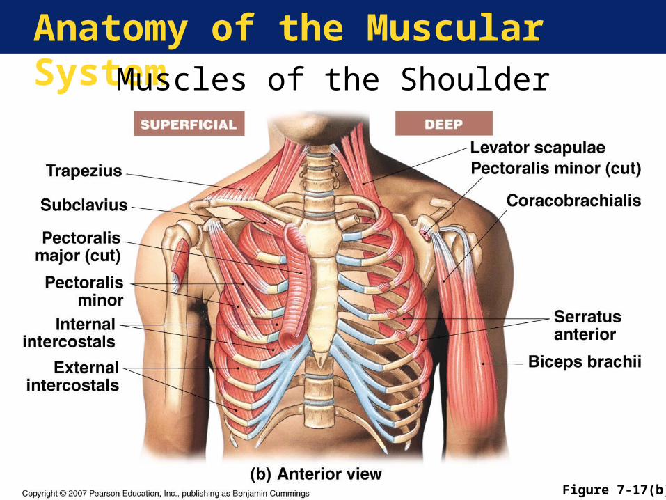
Anatomy of the Muscular SystemMuscles of the Shoulder
Figure 7-17(b)
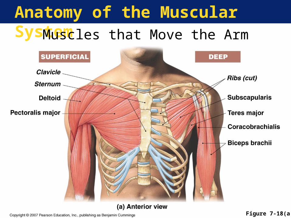
Anatomy of the Muscular SystemMuscles that Move the Arm
Figure 7-18(a)
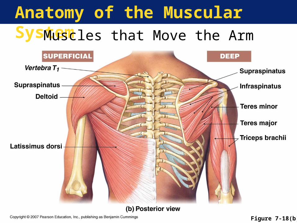
Anatomy of the Muscular SystemMuscles that Move the Arm
Figure 7-18(b)
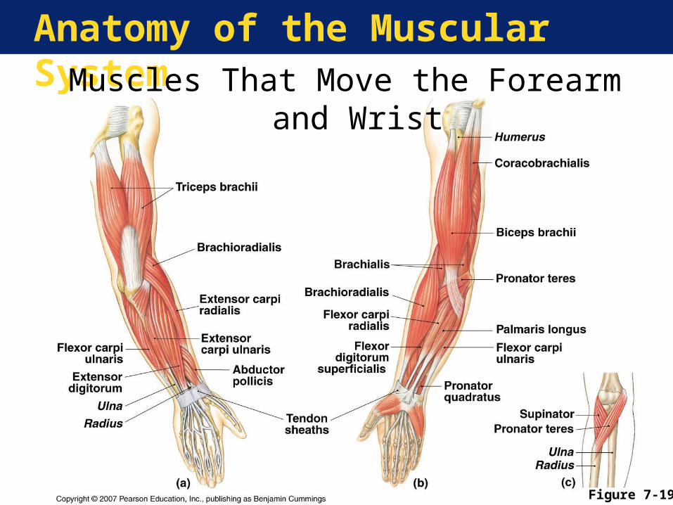
Anatomy of the Muscular SystemMuscles That Move the Forearm and Wrist
Figure 7-19
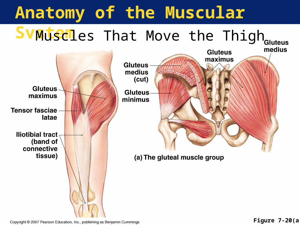
Anatomy of the Muscular SystemMuscles That Move the Thigh
Figure 7-20(a)
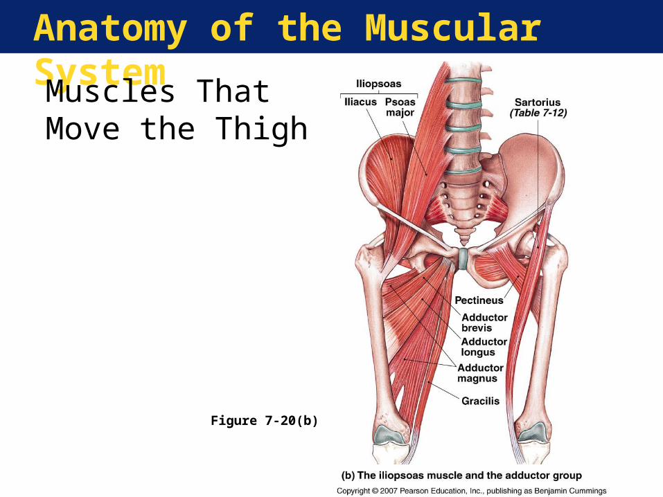
Anatomy of the Muscular System
Muscles That Move the Thigh
Figure 7-20(b)
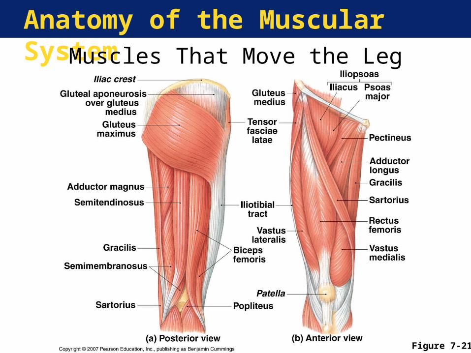
Anatomy of the Muscular System
Figure 7-21
Muscles That Move the Leg
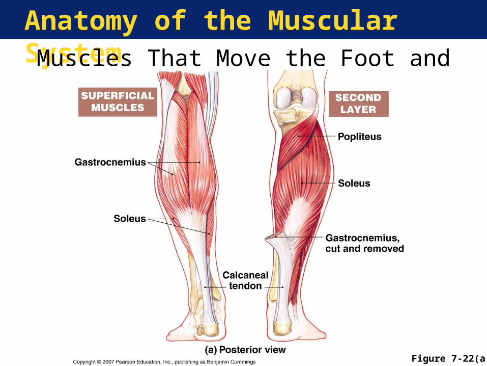
Anatomy of the Muscular SystemMuscles That Move the Foot and Toes
Figure 7-22(a)
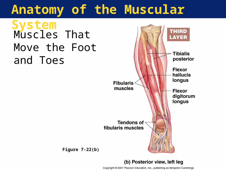
Anatomy of the Muscular System
Figure 7-22(b)
Muscles That Move the Foot and Toes
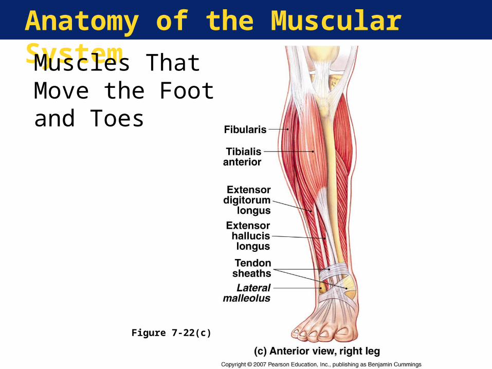
Anatomy of the Muscular System
Figure 7-22(c)
Muscles That Move the Foot and Toes
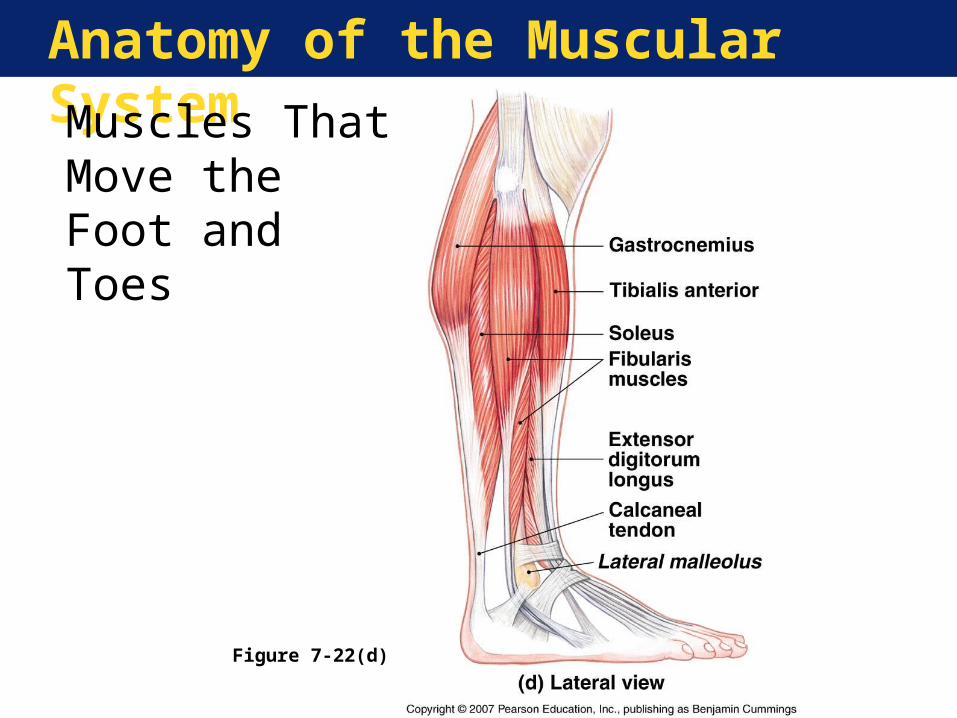
Anatomy of the Muscular System
Figure 7-22(d)
Muscles That Move the Foot and Toes


