Page 1
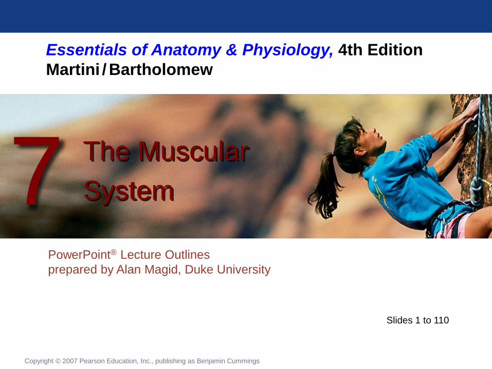
Essentials of Anatomy & Physiology, 4th Edition
Martini / Bartholomew
PowerPoint® Lecture Outlines
prepared by Alan Magid, Duke University
The Muscular
System 7
Copyright © 2007 Pearson Education, Inc., publishing as Benjamin Cummings
Slides 1 to 110
Page 2
Overview of Muscular System
Types of Muscle Tissue
• Under voluntary control
• Skeletal muscles
• The muscular system
• Under involuntary control
• Cardiac muscle
• Heart wall
• Smooth muscle
• Visceral organs
Copyright © 2007 Pearson Education, Inc., publishing as Benjamin Cummings
Page 3
Overview of Muscular System
• Skeletal muscles attach to bones
directly or indirectly
• Perform five functions
• Produce movement of skeleton
• Maintain posture and body position
• Support soft tissues
• Guard entrances and exits
• Maintain body temperature
Copyright © 2007 Pearson Education, Inc., publishing as Benjamin Cummings
Page 4
Anatomy of Skeletal Muscles
Gross Anatomy
• Connective tissue organization
• Epimysium
• Fibrous covering of whole muscle
• Perimysium
• Fibrous covering of fascicle
• Endomysium
• Fibrous covering of a single cell (a
muscle fiber)
• Tendons (or aponeurosis)
Copyright © 2007 Pearson Education, Inc., publishing as Benjamin Cummings
Page 5
Anatomy of Skeletal Muscles
The Organization of a Skeletal Muscle
Figure 7-1
Page 6
Anatomy of Skeletal Muscles
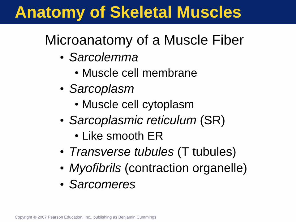
Microanatomy of a Muscle Fiber
• Sarcolemma
• Muscle cell membrane
• Sarcoplasm
• Muscle cell cytoplasm
• Sarcoplasmic reticulum (SR)
• Like smooth ER
• Transverse tubules (T tubules)
• Myofibrils (contraction organelle)
• Sarcomeres
Copyright © 2007 Pearson Education, Inc., publishing as Benjamin Cummings
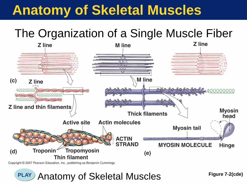
Page 7
Anatomy of Skeletal Muscles
Sarcomere—Repeating structural unit
of the myofibril
• Components of a sarcomere
• Myofilaments
• Thin filaments (mostly actin)
• Thick filaments (mostly myosin)
• Z lines at each end
• Anchor for thin filaments
Copyright © 2007 Pearson Education, Inc., publishing as Benjamin Cummings
Page 8
Anatomy of Skeletal Muscles
The Organization of a Single Muscle Fiber
Figure 7-2(a)
Page 9
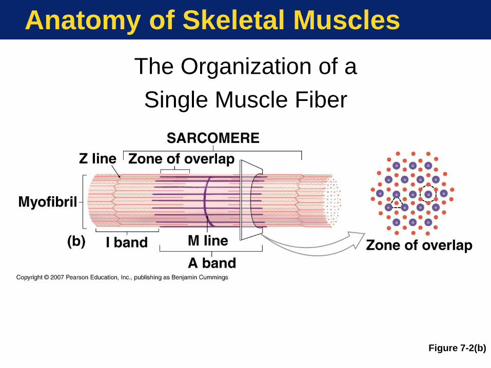
Anatomy of Skeletal Muscles
The Organization of a
Single Muscle Fiber
Figure 7-2(b)
Page 10
Anatomy of Skeletal Muscles
The Organization of a Single Muscle Fiber
Figure 7-2(cde) Anatomy of Skeletal Muscles PLAY
Page 11
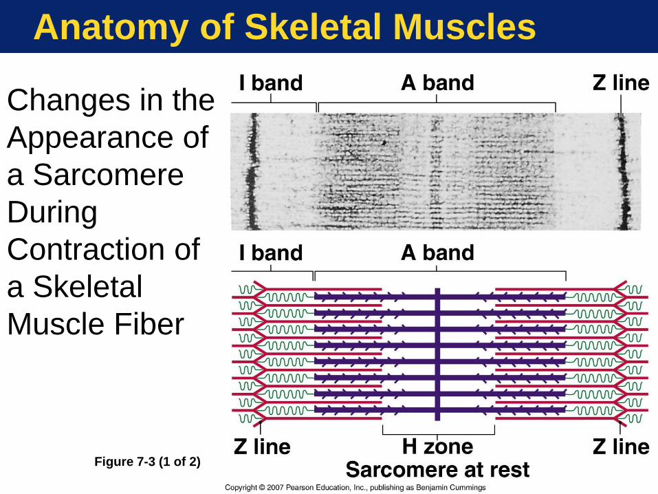
Anatomy of Skeletal Muscles
Changes in the
Appearance of
a Sarcomere
During
Contraction of
a Skeletal
Muscle Fiber
Figure 7-3 (1 of 2)
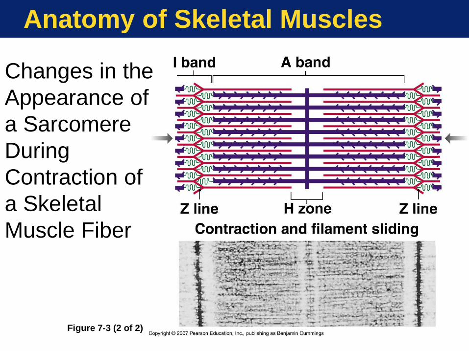
Page 12
Anatomy of Skeletal Muscles
Changes in the
Appearance of
a Sarcomere
During
Contraction of
a Skeletal
Muscle Fiber
Figure 7-3 (2 of 2)
Page 13
Control of Muscle Contraction
Steps in Neuromuscular Transmission
• Motor neuron action potential
• Acetylcholine (ACh) release and
binding
• Action potential in sarcolemma
• T tubule action potential
• Calcium release from SR
Copyright © 2007 Pearson Education, Inc., publishing as Benjamin Cummings
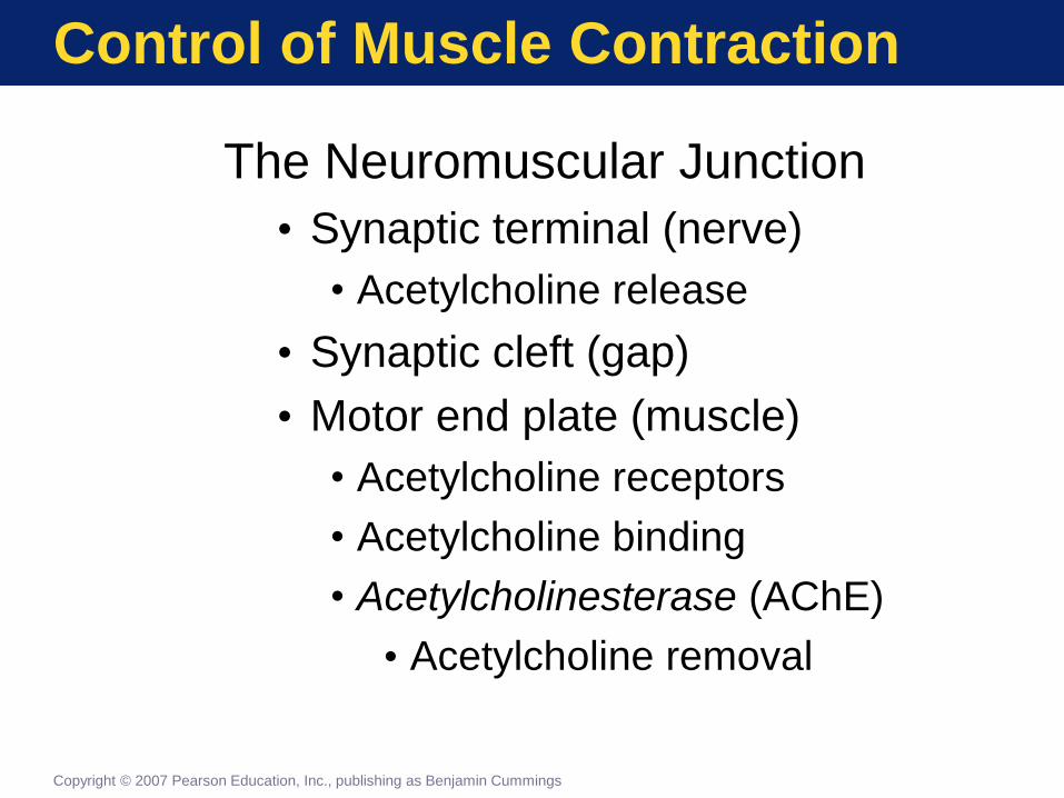
Page 14
Control of Muscle Contraction
The Neuromuscular Junction
• Synaptic terminal (nerve)
• Acetylcholine release
• Synaptic cleft (gap)
• Motor end plate (muscle)
• Acetylcholine receptors
• Acetylcholine binding
• Acetylcholinesterase (AChE)
• Acetylcholine removal
Copyright © 2007 Pearson Education, Inc., publishing as Benjamin Cummings
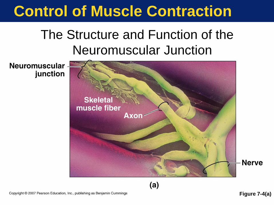
Page 15
Control of Muscle Contraction
The Structure and Function of the
Neuromuscular Junction
Figure 7-4(a)
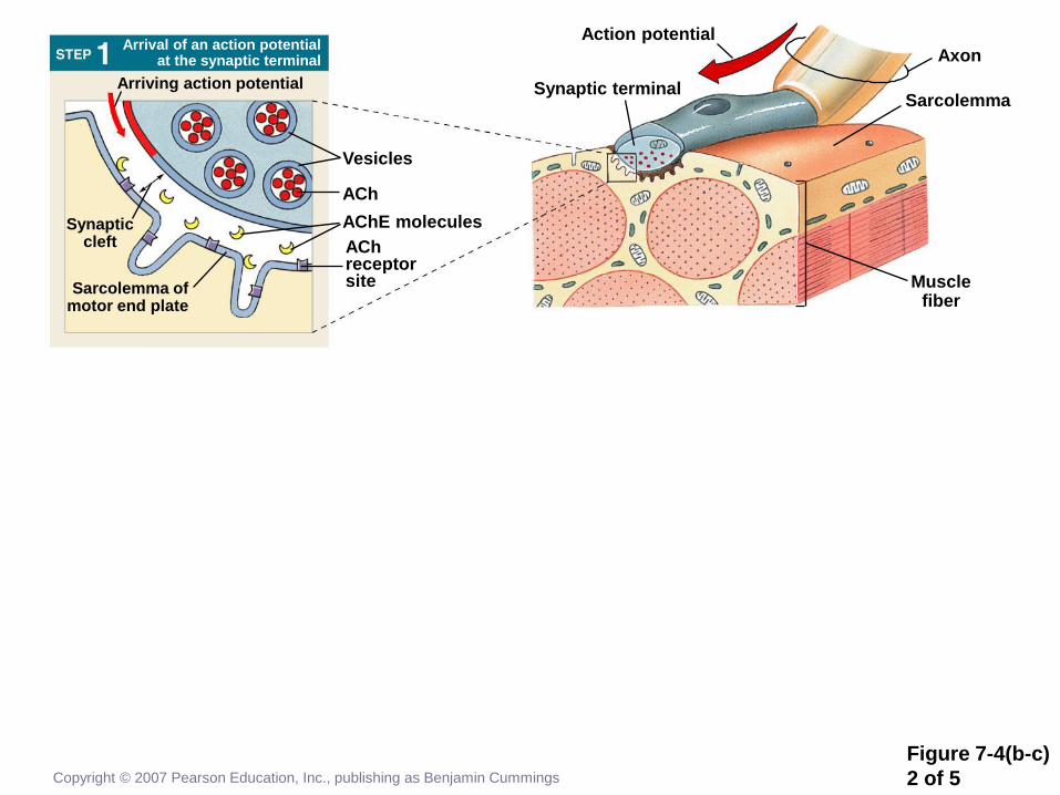
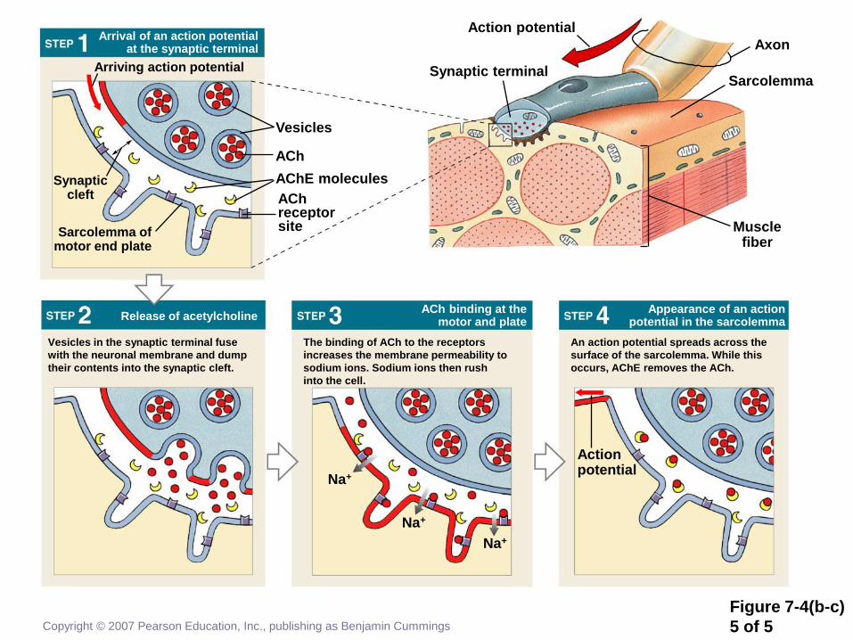
Page 16
Figure 7-4(b-c)
1 of 5 Copyright © 2007 Pearson Education, Inc., publishing as Benjamin Cummings
Synaptic cleft
Vesicles in the synaptic terminal fuse
with the neuronal membrane and dump
their contents into the synaptic cleft.
The binding of ACh to the receptors
increases the membrane permeability to
sodium ions. Sodium ions then rush
into the cell.
An action potential spreads across the
surface of the sarcolemma. While this
occurs, AChE removes the ACh.
Appearance of an action potential in the sarcolemma
ACh binding at the motor and plate
Release of acetylcholine
Arrival of an action potential at the synaptic terminal
Sarcolemma of motor end plate
Arriving action potential
Vesicles
ACh
AChE molecules
ACh receptor site
Action potential
Synaptic terminal
Axon
Sarcolemma
Muscle fiber
Action potential
Na+
Na+
Na+
Page 17
Figure 7-4(b-c)
2 of 5 Copyright © 2007 Pearson Education, Inc., publishing as Benjamin Cummings
Synaptic cleft
Arrival of an action potential at the synaptic terminal
Sarcolemma of motor end plate
Arriving action potential
Vesicles
ACh
AChE molecules
ACh receptor site
Action potential
Synaptic terminal
Axon
Sarcolemma
Muscle fiber
Page 18
Figure 7-4(b-c)
3 of 5 Copyright © 2007 Pearson Education, Inc., publishing as Benjamin Cummings
Synaptic cleft
Vesicles in the synaptic terminal fuse
with the neuronal membrane and dump
their contents into the synaptic cleft.
Release of acetylcholine
Arrival of an action potential at the synaptic terminal
Sarcolemma of motor end plate
Arriving action potential
Vesicles
ACh
AChE molecules
ACh receptor site
Action potential
Synaptic terminal
Axon
Sarcolemma
Muscle fiber
Page 19
Figure 7-4(b-c)
4 of 5 Copyright © 2007 Pearson Education, Inc., publishing as Benjamin Cummings
Synaptic cleft
Vesicles in the synaptic terminal fuse
with the neuronal membrane and dump
their contents into the synaptic cleft.
The binding of ACh to the receptors
increases the membrane permeability to
sodium ions. Sodium ions then rush
into the cell.
ACh binding at the motor and plate
Release of acetylcholine
Arrival of an action potential at the synaptic terminal
Sarcolemma of motor end plate
Arriving action potential
Vesicles
ACh
AChE molecules
ACh receptor site
Action potential
Synaptic terminal
Axon
Sarcolemma
Muscle fiber
Na+
Na+
Na+
Page 20
Figure 7-4(b-c)
5 of 5 Copyright © 2007 Pearson Education, Inc., publishing as Benjamin Cummings
Synaptic cleft
Vesicles in the synaptic terminal fuse
with the neuronal membrane and dump
their contents into the synaptic cleft.
The binding of ACh to the receptors
increases the membrane permeability to
sodium ions. Sodium ions then rush
into the cell.
An action potential spreads across the
surface of the sarcolemma. While this
occurs, AChE removes the ACh.
Appearance of an action potential in the sarcolemma
ACh binding at the motor and plate
Release of acetylcholine
Arrival of an action potential at the synaptic terminal
Sarcolemma of motor end plate
Arriving action potential
Vesicles
ACh
AChE molecules
ACh receptor site
Action potential
Synaptic terminal
Axon
Sarcolemma
Muscle fiber
Action potential
Na+
Na+
Na+
Page 21
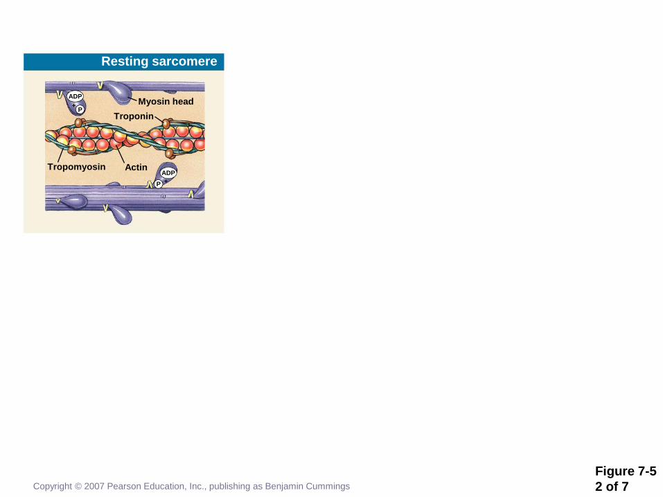
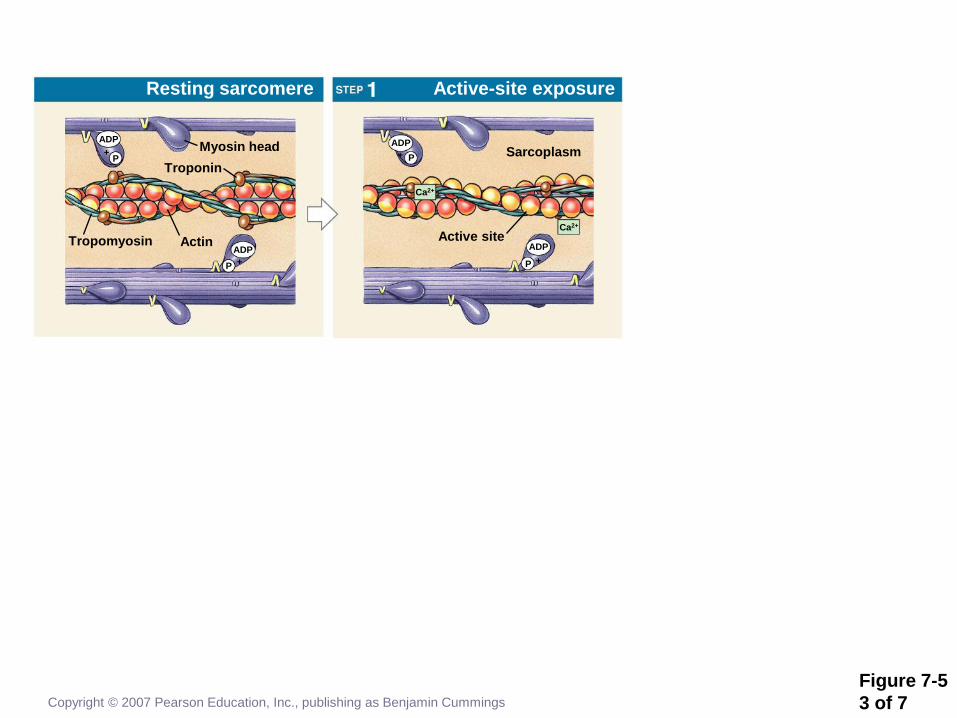
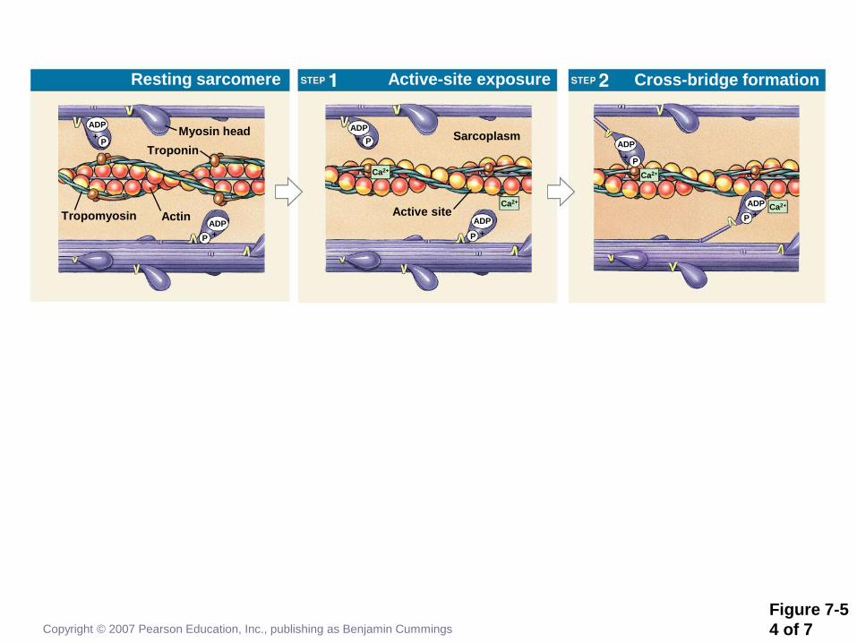
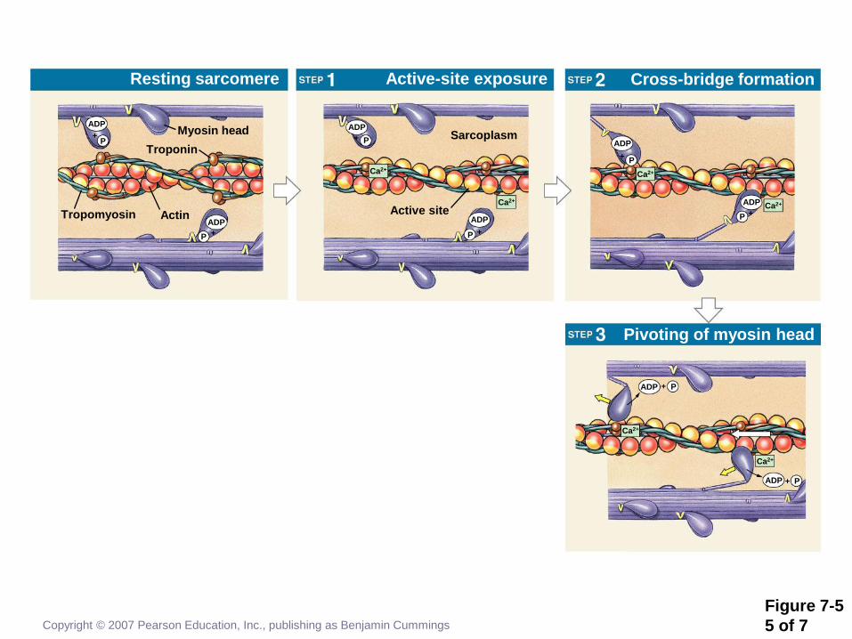
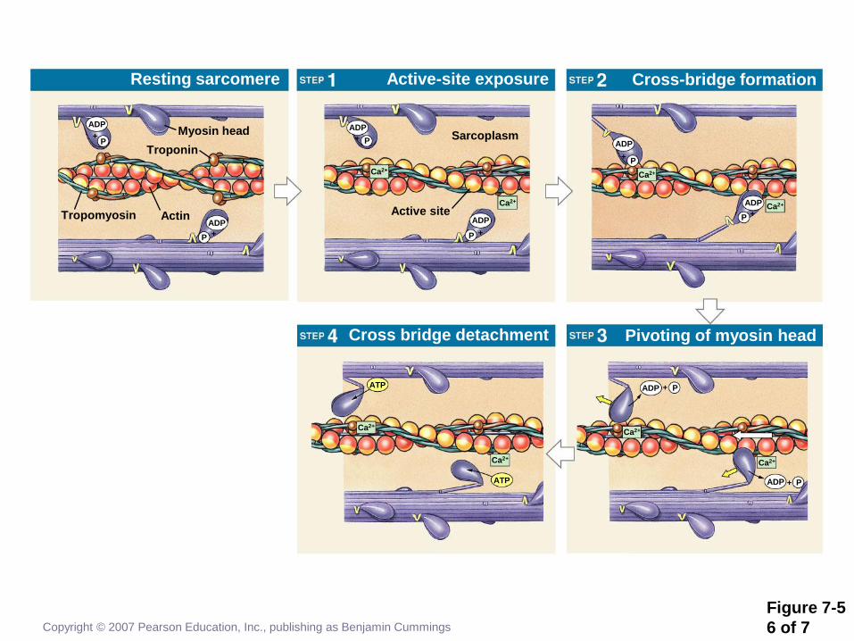
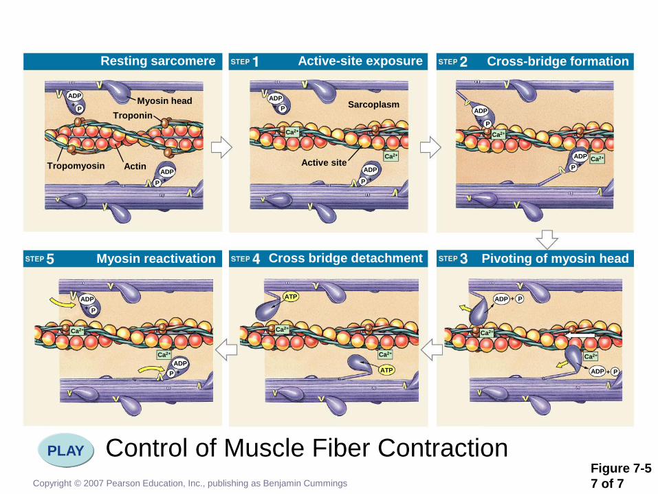
Anatomy of Skeletal Muscles
The Contraction Process
• Actin active sites and myosin cross-bridges
interact
• Thin filaments slide past thick filaments
• Cross-bridges undergo a cycle of movement
• Attach, pivot, detach, return
• Troponin-tropomyosin control interaction
• Prevent interaction at rest
Copyright © 2007 Pearson Education, Inc., publishing as Benjamin Cummings
Page 22
Copyright © 2007 Pearson Education, Inc., publishing as Benjamin Cummings
Figure 7-5
1 of 7
Resting sarcomere
Myosin head
Myosin reactivation
Active-site exposure
Cross bridge detachment
Cross-bridge formation
Pivoting of myosin head
Troponin
Actin Tropomyosin
ADP
P +
ADP
P +
ADP
P +
Active site
Sarcoplasm
Ca2+
Ca2+
ADP
P +
ADP
+ P
Ca2+
ADP
+ P
Ca2+
Ca2+
ADP + P
Ca2+
ADP + P
Ca2+
ATP
ATP
Ca2+
Ca2+
Ca2+
ADP
P +
+ P
ADP
Page 23
Copyright © 2007 Pearson Education, Inc., publishing as Benjamin Cummings
Figure 7-5
2 of 7
Resting sarcomere
Myosin head
Troponin
Actin Tropomyosin
ADP
P +
ADP
P +
Page 24
Copyright © 2007 Pearson Education, Inc., publishing as Benjamin Cummings
Figure 7-5
3 of 7
Resting sarcomere
Myosin head
Active-site exposure
Troponin
Actin Tropomyosin
ADP
P +
ADP
P +
ADP
P +
Active site
Sarcoplasm
Ca2+
Ca2+
ADP
P +
Page 25
Copyright © 2007 Pearson Education, Inc., publishing as Benjamin Cummings
Figure 7-5
4 of 7
Resting sarcomere
Myosin head
Active-site exposure Cross-bridge formation
Troponin
Actin Tropomyosin
ADP
P +
ADP
P +
ADP
P +
Active site
Sarcoplasm
Ca2+
Ca2+
ADP
P +
ADP
+ P
Ca2+
ADP
+ P
Ca2+
Page 26
Copyright © 2007 Pearson Education, Inc., publishing as Benjamin Cummings
Figure 7-5
5 of 7
Resting sarcomere
Myosin head
Active-site exposure Cross-bridge formation
Pivoting of myosin head
Troponin
Actin Tropomyosin
ADP
P +
ADP
P +
ADP
P +
Active site
Sarcoplasm
Ca2+
Ca2+
ADP
P +
ADP
+ P
Ca2+
ADP
+ P
Ca2+
Ca2+
ADP + P
Ca2+
ADP + P
Page 27
Copyright © 2007 Pearson Education, Inc., publishing as Benjamin Cummings
Figure 7-5
6 of 7
Resting sarcomere
Myosin head
Active-site exposure
Cross bridge detachment
Cross-bridge formation
Pivoting of myosin head
Troponin
Actin Tropomyosin
ADP
P +
ADP
P +
ADP
P +
Active site
Sarcoplasm
Ca2+
Ca2+
ADP
P +
ADP
+ P
Ca2+
ADP
+ P
Ca2+
Ca2+
ADP + P
Ca2+
ADP + P
Ca2+
ATP
ATP
Ca2+
Page 28
Control of Muscle Fiber Contraction PLAY
Copyright © 2007 Pearson Education, Inc., publishing as Benjamin Cummings
Figure 7-5
7 of 7
Resting sarcomere
Myosin head
Myosin reactivation
Active-site exposure
Cross bridge detachment
Cross-bridge formation
Pivoting of myosin head
Troponin
Actin Tropomyosin
ADP
P +
ADP
P +
ADP
P +
Active site
Sarcoplasm
Ca2+
Ca2+
ADP
P +
ADP
+ P
Ca2+
ADP
+ P
Ca2+
Ca2+
ADP + P
Ca2+
ADP + P
Ca2+
ATP
ATP
Ca2+
Ca2+
Ca2+
ADP
P +
+ P
ADP
Page 29
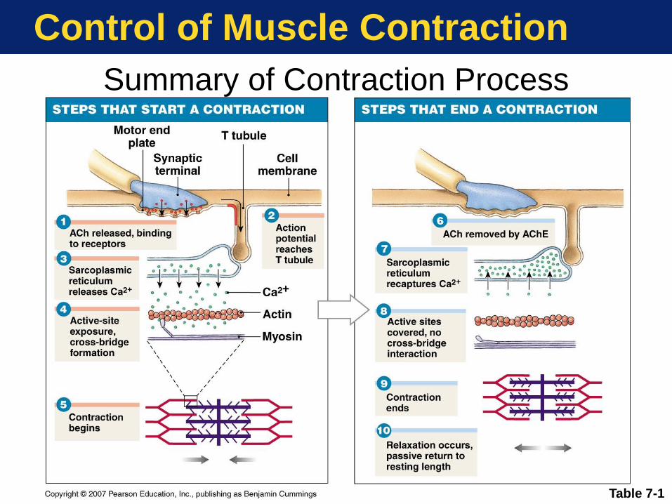
Control of Muscle Contraction
Table 7-1
Summary of Contraction Process
Page 30
Control of Muscle Contraction
Key Note
Skeletal muscle fibers shorten as thin
filaments interact with thick filaments and
sliding occurs. The trigger for contraction
is the calcium ions released by the SR
when the muscle fiber is stimulated by its
motor neuron. Contraction is an active
process; relaxation and the return to
resting length is entirely passive.
Copyright © 2007 Pearson Education, Inc., publishing as Benjamin Cummings
Page 31
Muscle Mechanics
Some Basic Muscle Definitions
• Muscle tension—The pulling force on the
tendons that muscle cells generate when
contracting
• Muscle twitch—A brief contraction-relaxation
response to a single action potential
Copyright © 2007 Pearson Education, Inc., publishing as Benjamin Cummings
Page 32
Muscle Mechanics
The Twitch and Development of Tension
Figure 7-6
Page 33
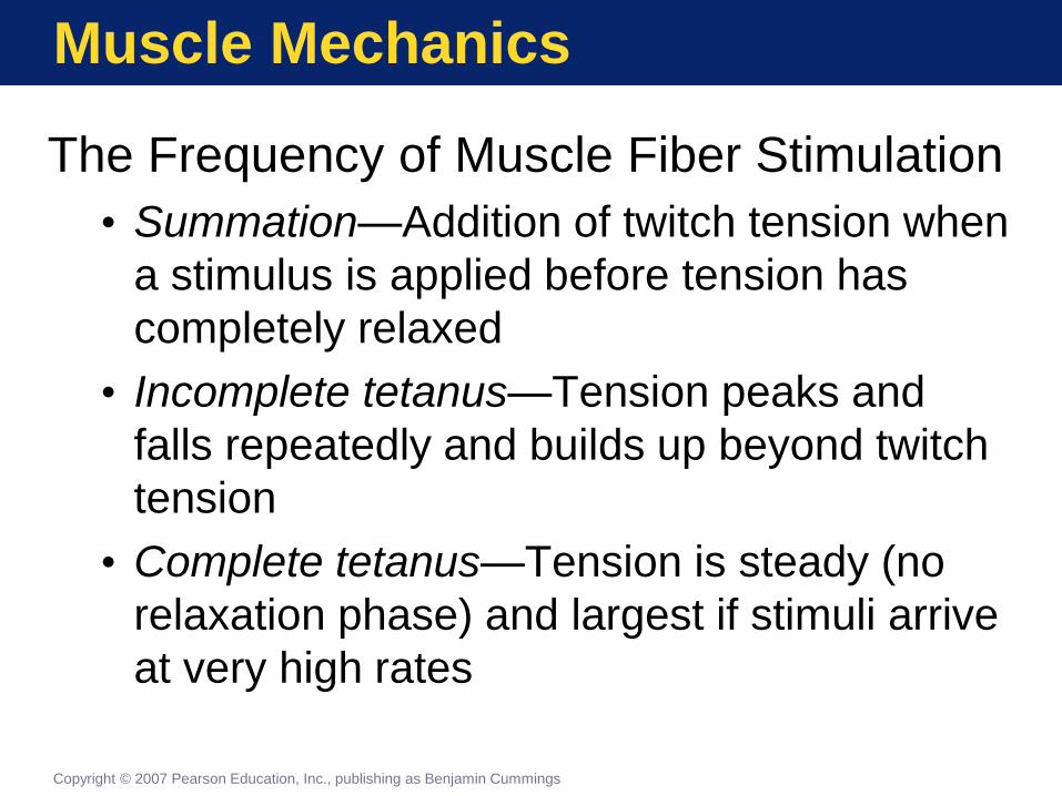
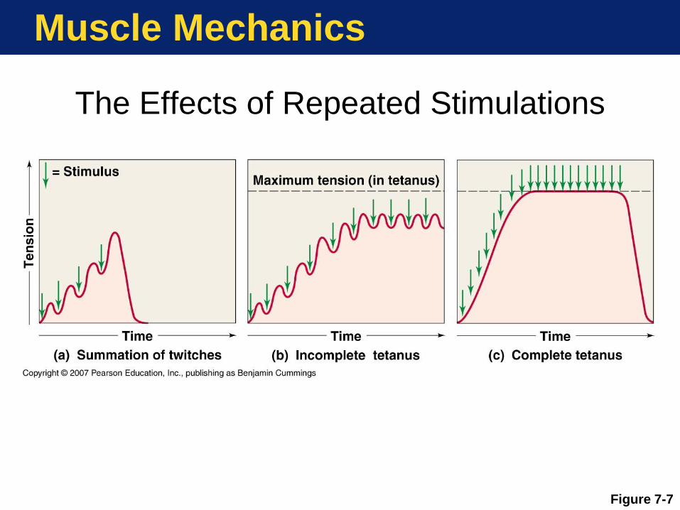
Muscle Mechanics
The Frequency of Muscle Fiber Stimulation
• Summation—Addition of twitch tension when
a stimulus is applied before tension has
completely relaxed
• Incomplete tetanus—Tension peaks and
falls repeatedly and builds up beyond twitch
tension
• Complete tetanus—Tension is steady (no
relaxation phase) and largest if stimuli arrive
at very high rates
Copyright © 2007 Pearson Education, Inc., publishing as Benjamin Cummings
Page 34
Muscle Mechanics
The Effects of Repeated Stimulations
Figure 7-7
Page 35
Muscle Mechanics
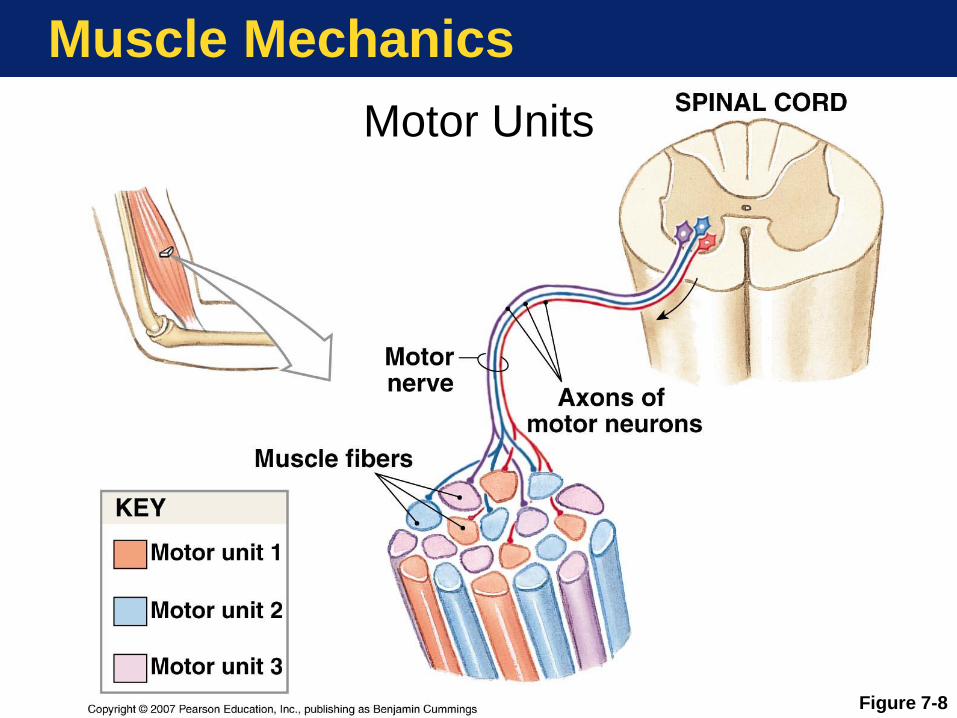
Motor Units
• Motor Unit —A motor neuron and all
the muscle cells it controls
• Recruitment—To increase muscle
tension by activating more motor units
• Small motor units provide finer control
• Motor units are intermixed in the
muscle to pull evenly on the tendon
Copyright © 2007 Pearson Education, Inc., publishing as Benjamin Cummings
Page 36
Muscle Mechanics
Motor Units
Figure 7-8
Page 37
Muscle Mechanics
Key Note
All voluntary (intentional) movements
involve the sustained, sub-tetanic
contractions of skeletal muscle fibers
organized into distinct motor units. The
force generated can be increased by
increasing the frequency of action
potentials or by recruiting additional
motor units.
Copyright © 2007 Pearson Education, Inc., publishing as Benjamin Cummings
Page 38
Muscle Mechanics
• Muscle tone—Tension in a “resting” muscle produced by a low level of spontaneous motor neuron activity. Distinct from resting tension produced by passive stretching.
• Function of muscle tone
• Stabilizes bones, joints
• Prevents atrophy (muscle wasting )
Copyright © 2007 Pearson Education, Inc., publishing as Benjamin Cummings
Page 39
Muscle Mechanics
Types of Contractions
• Isotonic contraction
The tension (load) on a muscle stays
constant (iso = same, tonic = tension)
during a movement. (Example: lifting a
baby)
• Isometric contraction
The length of a muscle stays constant
(iso = same, metric = length) during a
“contraction” (Example: holding a baby
at arms length)
Copyright © 2007 Pearson Education, Inc., publishing as Benjamin Cummings
Page 40
Muscle Mechanics
Muscle Elongation
• Muscle contracts actively
• Muscles can only pull
• Muscles never push
• Muscle elongates passively
• Elastic forces
• Contraction of opposing
muscles
• Effects of gravity
Copyright © 2007 Pearson Education, Inc., publishing as Benjamin Cummings
Page 41
Energetics of Muscle Contraction
ATP and Creatine Phosphate Reserves
• Muscle contraction consumes much ATP
• ATP transfers energy directly to cycling
cross-bridges and calcium pumping
• CP stores energy and regenerates ATP
• CP transfers its energy to ADP
• Creatine phosphokinase (CPK) catalyzes
• ADP (2 “P”s) becomes ATP(3 “P”s)
• CP levels greatly exceed ATP levels
Copyright © 2007 Pearson Education, Inc., publishing as Benjamin Cummings
Page 42
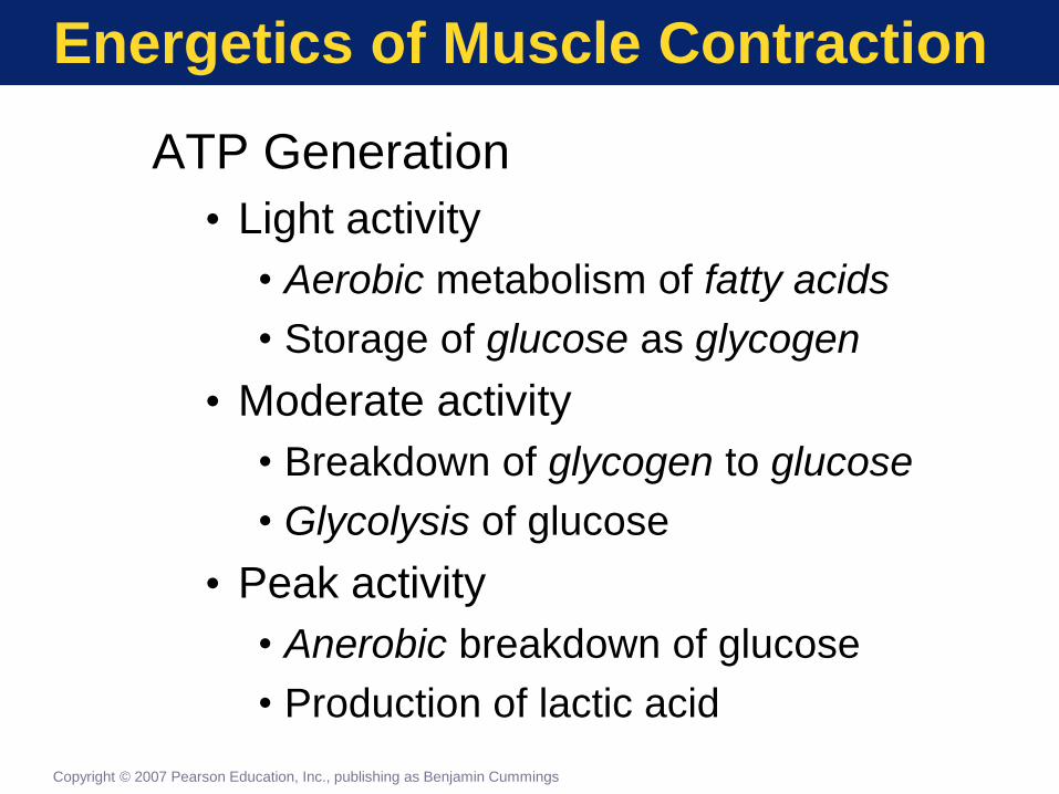
Energetics of Muscle Contraction
ATP Generation
• Light activity
• Aerobic metabolism of fatty acids
• Storage of glucose as glycogen
• Moderate activity
• Breakdown of glycogen to glucose
• Glycolysis of glucose
• Peak activity
• Anerobic breakdown of glucose
• Production of lactic acid
Copyright © 2007 Pearson Education, Inc., publishing as Benjamin Cummings
Page 43
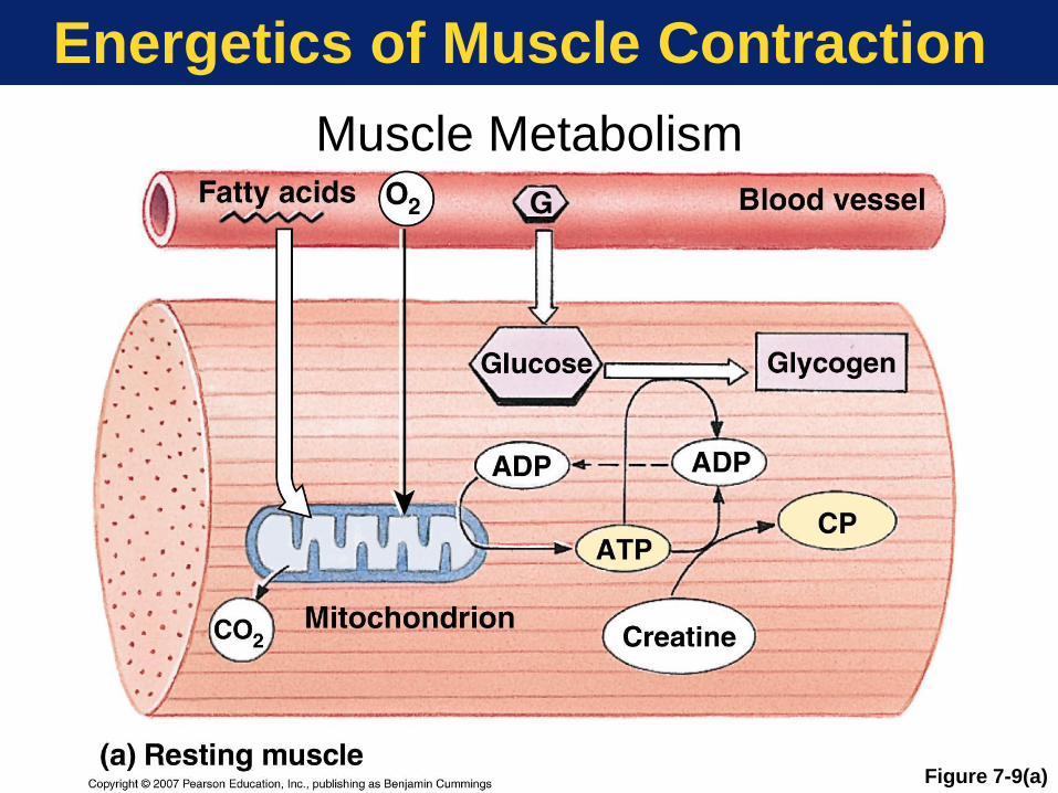
Energetics of Muscle Contraction
Muscle Metabolism
Figure 7-9(a)
Page 44
Energetics of Muscle Contraction
Muscle Metabolism
Figure 7-9(b)
Page 45
Energetics of Muscle Contraction
Muscle Metabolism
Figure 7-9(c)
Page 46
Energetics of Muscle Contraction
Muscle Fatigue—When a muscle
loses ability to contract due to a low
pH (lactic acid buildup), low ATP
levels, or other problems
Recovery Period—Time after muscle
activity that it takes to restore pre-
exertion conditions
Oxygen Debt—Amount of excess
oxygen used during the recovery
period Copyright © 2007 Pearson Education, Inc., publishing as Benjamin Cummings
Page 47
Energetics of Muscle Contraction
Key Note
Skeletal muscles at rest metabolize
fatty acids and store glycogen. During
light activity, muscles can generate
ATP through the aerobic breakdown of
carbohydrates, lipids, or amino acids.
At peak levels of activity, most of the
energy is provided by anaerobic
reactions that generate lactic acid.
Copyright © 2007 Pearson Education, Inc., publishing as Benjamin Cummings
Page 48
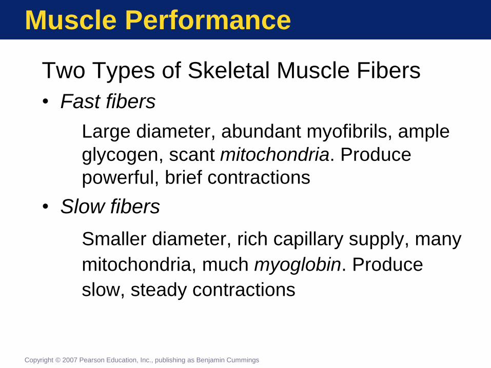
Muscle Performance
Two Types of Skeletal Muscle Fibers
• Fast fibers
Large diameter, abundant myofibrils, ample
glycogen, scant mitochondria. Produce
powerful, brief contractions
• Slow fibers
Smaller diameter, rich capillary supply, many
mitochondria, much myoglobin. Produce
slow, steady contractions
Copyright © 2007 Pearson Education, Inc., publishing as Benjamin Cummings
Page 49
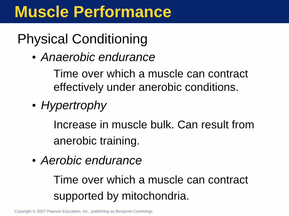
Muscle Performance
Physical Conditioning
• Anaerobic endurance
Time over which a muscle can contract
effectively under anerobic conditions.
• Hypertrophy
Increase in muscle bulk. Can result from
anerobic training.
• Aerobic endurance
Time over which a muscle can contract
supported by mitochondria. Copyright © 2007 Pearson Education, Inc., publishing as Benjamin Cummings
Page 50
Muscle Performance
Key Note
What you don’t use, you lose. When
motor units are inactive for days or
weeks, muscle fibers break down their
contractile proteins and grow smaller
and weaker. If inactive for long periods,
muscle fibers may be replaced by
fibrous tissue.
Copyright © 2007 Pearson Education, Inc., publishing as Benjamin Cummings
Page 51
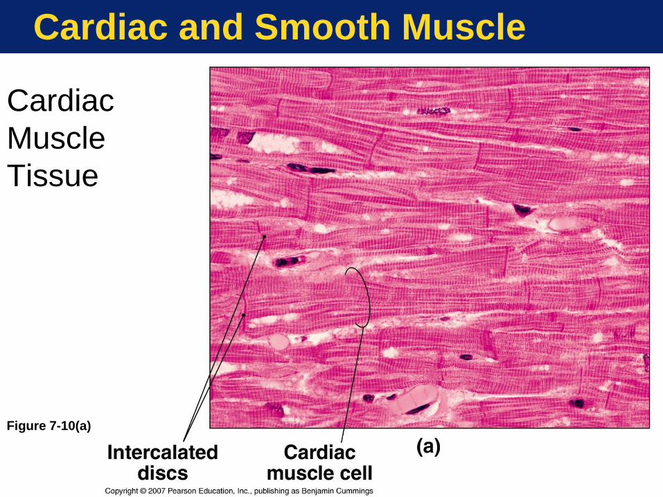
Cardiac and Smooth Muscle
Cardiac Muscle Tissue
• Small cells
• Single nucleus/cell
• Aerobic metabolism
• Intercalated discs
• Long contraction time
• Self-exciting (automaticity)
• No tetanic contraction
Copyright © 2007 Pearson Education, Inc., publishing as Benjamin Cummings
Page 52
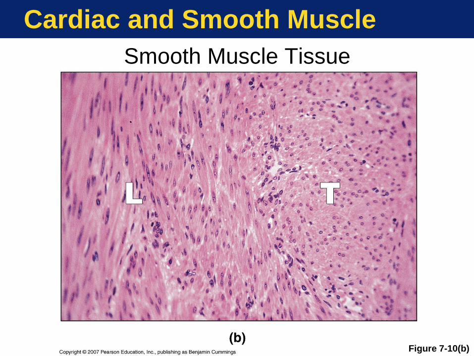
Cardiac and Smooth Muscle
Smooth Muscle Tissue
• Nonstriated cells (no sarcomeres)
• Calcium control of contraction
different from striated muscle
• Wide range of operating lengths
• Involuntary muscle
• Under hormonal or local control
• Pacesetter cells
• Motor neurons often unneeded
Copyright © 2007 Pearson Education, Inc., publishing as Benjamin Cummings
Page 53
Cardiac and Smooth Muscle
Figure 7-10(a)
Cardiac
Muscle
Tissue
Page 54
Cardiac and Smooth Muscle
Figure 7-10(b)
Smooth Muscle Tissue
Page 55
Cardiac and Smooth Muscle
Table 7-2
Page 56
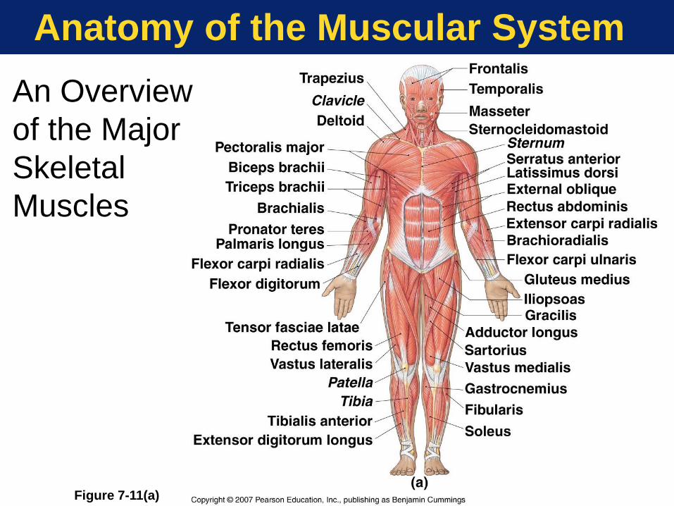
Anatomy of the Muscular System
An Overview
of the Major
Skeletal
Muscles
Figure 7-11(a)
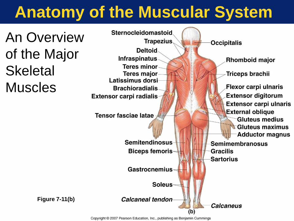
Page 57
Anatomy of the Muscular System
An Overview
of the Major
Skeletal
Muscles
Figure 7-11(b)
Page 58
Anatomy of the Muscular System
Origins, Insertions, and Actions
• Origin
Muscle attachment that remains fixed
• Insertion
Muscle attachment that moves
• Action
What joint movement a muscle produces
Copyright © 2007 Pearson Education, Inc., publishing as Benjamin Cummings
Page 59
Anatomy of the Muscular System
Primary Action Categories
• Prime mover (agonist)
• Main muscle in an action
• Synergist
• Helper muscle in an action
• Antagonist
• Opposed muscle to an action
Copyright © 2007 Pearson Education, Inc., publishing as Benjamin Cummings
Page 60
Anatomy of the Muscular System
Muscle Terminology
• Names of muscles provide clues to
location, orientation, or action
• Axial musculature—Muscles with origins
on the axial skeleton that position and
move head, spine, rib cage
• Appendicular musculature—Muscles that
stabilize or move appendicular
components
Copyright © 2007 Pearson Education, Inc., publishing as Benjamin Cummings
Page 61
Anatomy of the Muscular System
The Axial Muscles
• Four groups of axial muscles
• Head and neck
• Spine
• Trunk
• Pelvic floor
Copyright © 2007 Pearson Education, Inc., publishing as Benjamin Cummings
Page 62
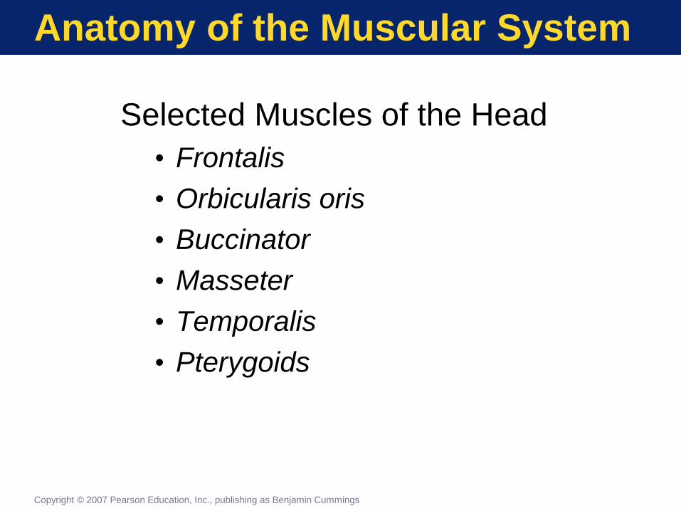
Anatomy of the Muscular System
Selected Muscles of the Head
• Frontalis
• Orbicularis oris
• Buccinator
• Masseter
• Temporalis
• Pterygoids
Copyright © 2007 Pearson Education, Inc., publishing as Benjamin Cummings
Page 63
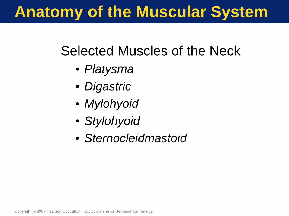
Anatomy of the Muscular System
Selected Muscles of the Neck
• Platysma
• Digastric
• Mylohyoid
• Stylohyoid
• Sternocleidmastoid
Copyright © 2007 Pearson Education, Inc., publishing as Benjamin Cummings
Page 64
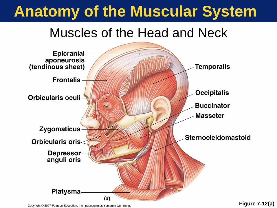
Anatomy of the Muscular System
Muscles of the Head and Neck
Figure 7-12(a)
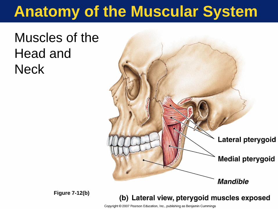
Page 65
Anatomy of the Muscular System
Muscles of the
Head and
Neck
Figure 7-12(b)
Page 66
Anatomy of the Muscular System
Muscles of the Head and Neck
Figure 7-12(c)
Page 67
Anatomy of the Muscular System
Muscles of the Anterior Neck
Figure 7-13
Page 68
Anatomy of the Muscular System
Selected Muscles of the Spine
• Splenius capitis
• Semispinalis capitis
• Erector spinae groups
• Spinalis
• Longissimus
• Iliocostalis
Copyright © 2007 Pearson Education, Inc., publishing as Benjamin Cummings
Page 69
Anatomy of the Muscular System
Muscles of
the Spine
Figure 7-14
Page 70
Anatomy of the Muscular System
Axial Muscles of the Trunk
• Thoracic region
• External intercostals
• Internal intercostals
• Diaphragm
• Abdominal region
• Rectus abdominis
• External oblique
• Internal oblique
• Transversus abdominis
Copyright © 2007 Pearson Education, Inc., publishing as Benjamin Cummings
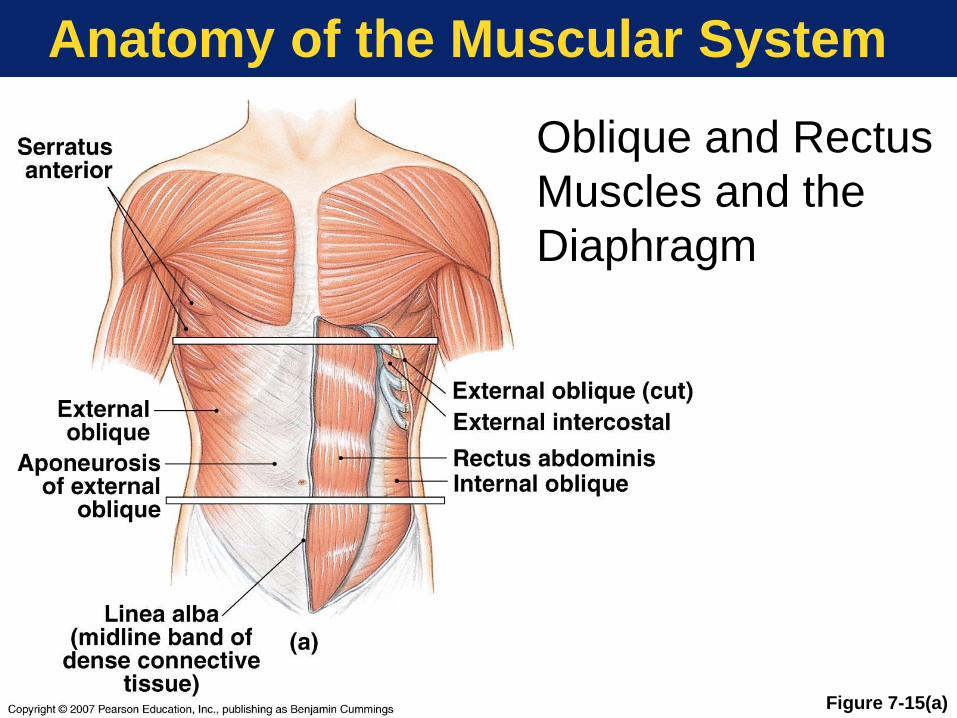
Page 71
Anatomy of the Muscular System
Figure 7-15(a)
Oblique and Rectus
Muscles and the
Diaphragm
Page 72
Anatomy of the Muscular System
Oblique and Rectus Muscles and
the Diaphragm
Figure 7-15(b)
Page 73
Anatomy of the Muscular System
Oblique and Rectus Muscles and
the Diaphragm
Figure 7-15(c)
Page 74
Anatomy of the Muscular System
Muscles of the Pelvic Floor (Perineum)
• Sheets of muscle
• From sacrum and coccyx
• To pubis and ischium
• Pelvic organ support
• Control of material passing through
urethra and anus
Copyright © 2007 Pearson Education, Inc., publishing as Benjamin Cummings
Page 75
Anatomy of the Muscular System
Muscles of the Perineum—Female
Figure 7-16(a)
Page 76
Anatomy of the Muscular System
Muscles of the Perineum—Male
Figure 7-16(b)
Page 77
Anatomy of the Muscular System
The Appendicular Muscles
• Two functionally distinct groups
• Muscles of the shoulder and
upper limbs
• Muscles of the pelvic girdle
and lower limbs
Copyright © 2007 Pearson Education, Inc., publishing as Benjamin Cummings
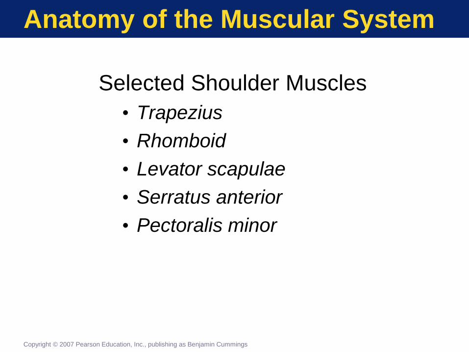
Page 78
Anatomy of the Muscular System
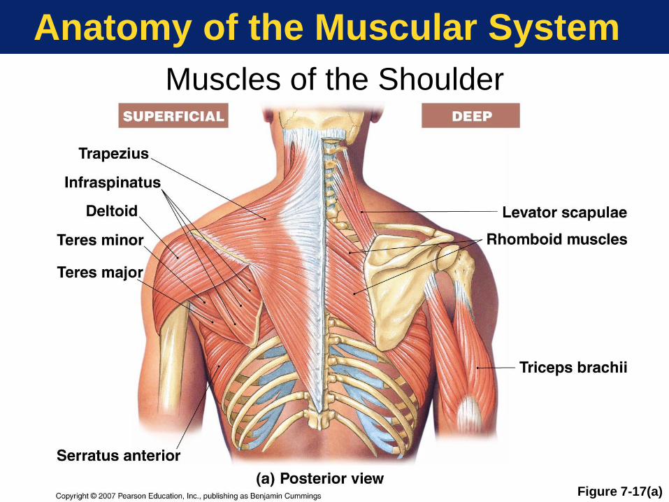
Selected Shoulder Muscles
• Trapezius
• Rhomboid
• Levator scapulae
• Serratus anterior
• Pectoralis minor
Copyright © 2007 Pearson Education, Inc., publishing as Benjamin Cummings
Page 79
Anatomy of the Muscular System
Muscles of the Shoulder
Figure 7-17(a)
Page 80
Anatomy of the Muscular System
Muscles of the Shoulder
Figure 7-17(b)
Page 81
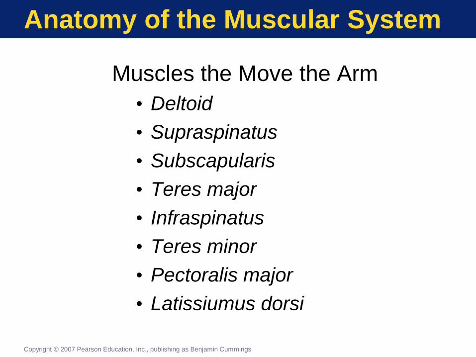
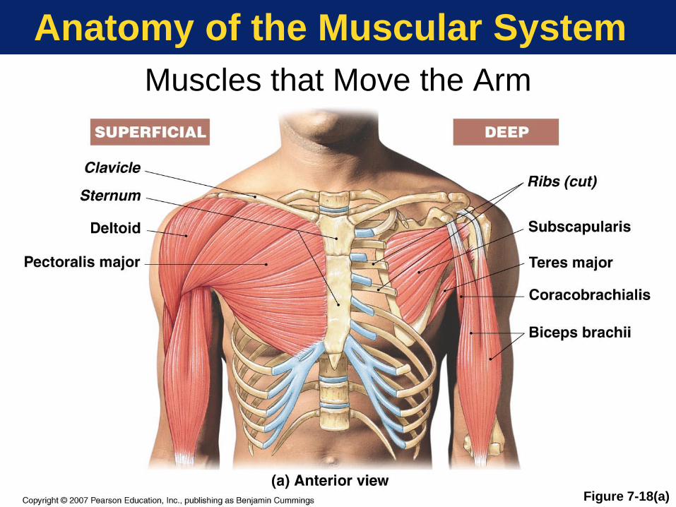
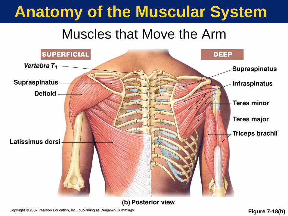
Anatomy of the Muscular System
Muscles the Move the Arm
• Deltoid
• Supraspinatus
• Subscapularis
• Teres major
• Infraspinatus
• Teres minor
• Pectoralis major
• Latissiumus dorsi
Copyright © 2007 Pearson Education, Inc., publishing as Benjamin Cummings
Page 82
Anatomy of the Muscular System
Muscles that Move the Arm
Figure 7-18(a)
Page 83
Anatomy of the Muscular System
Muscles that Move the Arm
Figure 7-18(b)
Page 84
Anatomy of the Muscular System
Muscles That Move the Forearm
• Biceps brachii
• Triceps brachii
• Brachialis
• Brachioradialis
• Pronators
• Supinator
Copyright © 2007 Pearson Education, Inc., publishing as Benjamin Cummings
Page 85
Anatomy of the Muscular System
Muscles That Move the Wrist
• Wrist flexors
• Flexor carpi ulnaris
• Flexor carpi radialis
• Palmaris longus
• Wrist extensors
• Extensor carpi radialis
• Extensor carpi ulnaris
Copyright © 2007 Pearson Education, Inc., publishing as Benjamin Cummings
Page 86
Anatomy of the Muscular System
Muscles That Move the Forearm and Wrist
Figure 7-19
Page 87
Anatomy of the Muscular System
Muscle of the Pelvis and Lower Limbs
• Three functional groups
• Thigh movement
• Leg movement
• Ankle, foot, and toe movement
Copyright © 2007 Pearson Education, Inc., publishing as Benjamin Cummings
Page 88
Anatomy of the Muscular System
Muscles That Move the Thigh
• Gluteal muscles
• Thigh adductors
• Adductor magnus
• Adductor brevis
• Adductor longus
• Pectineus
• Gracilis
• Thigh flexors
• Iliopsoas (psoas major + iliacus)
Copyright © 2007 Pearson Education, Inc., publishing as Benjamin Cummings
Page 89
Anatomy of the Muscular System
Muscles That Move the Thigh
Figure 7-20(a)
Page 90
Anatomy of the Muscular System
Muscles That Move
the Thigh
Figure 7-20(b)
Page 91
Anatomy of the Muscular System
Flexors of the Knee
• Biceps femoris
• Semimembranosus
• Semitendinosus
• Sartorius
• Popliteus
• Synergist muscle unlocks knee
Copyright © 2007 Pearson Education, Inc., publishing as Benjamin Cummings
Page 92
Anatomy of the Muscular System
Extensors of the Knee
• Quadriceps femoris group
• Rectus femoris
• Vastus lateralis
• Vastus intermedius
• Vastus medialis
Copyright © 2007 Pearson Education, Inc., publishing as Benjamin Cummings
Page 93
Anatomy of the Muscular System
Figure 7-21
Muscles That Move the Leg
Page 94
Anatomy of the Muscular System
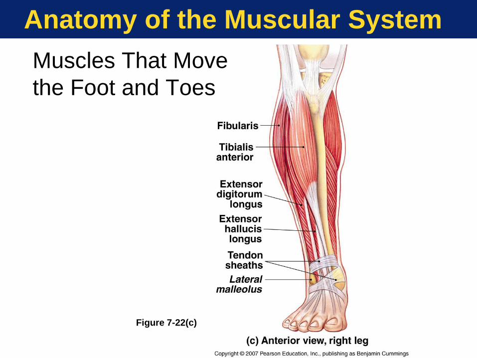
Muscles That Move the Foot
• Plantar flexion
• Gastrocnemius
• Soleus
• Eversion and plantar flexion
• Fibularis (peroneus)
• Dorsiflexion
• Tibialis anterior
Copyright © 2007 Pearson Education, Inc., publishing as Benjamin Cummings
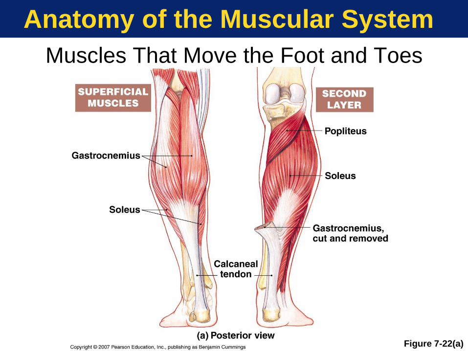
Page 95
Anatomy of the Muscular System
Muscles That Move the Foot and Toes
Figure 7-22(a)
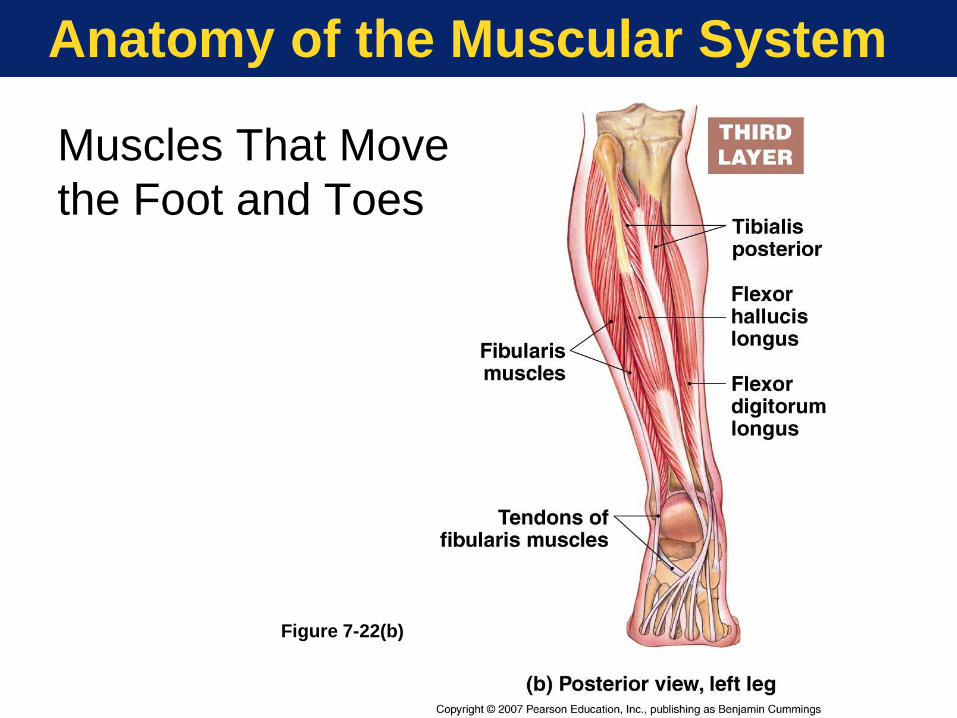
Page 96
Anatomy of the Muscular System
Figure 7-22(b)
Muscles That Move
the Foot and Toes
Page 97
Anatomy of the Muscular System
Figure 7-22(c)
Muscles That Move
the Foot and Toes
Page 98
Anatomy of the Muscular System
Figure 7-22(d)
Muscles That
Move the Foot
and Toes
Page 99
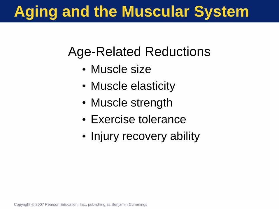
Aging and the Muscular System
Age-Related Reductions
• Muscle size
• Muscle elasticity
• Muscle strength
• Exercise tolerance
• Injury recovery ability
Copyright © 2007 Pearson Education, Inc., publishing as Benjamin Cummings
Page 100
The Muscular System
in Perspective
FIGURE 7-23
Functional Relationships Between
the Muscular System and Other Systems
Figure 7-23
1 of 11 Copyright © 2007 Pearson Education, Inc., publishing as Benjamin Cummings
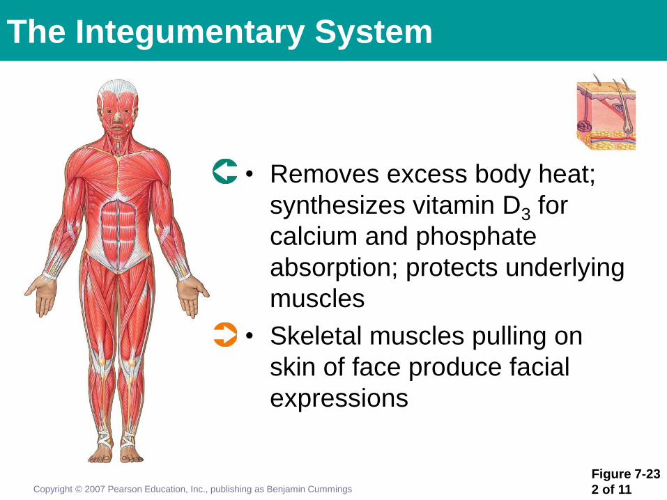
Page 101
Figure 7-23
2 of 11
• Removes excess body heat;
synthesizes vitamin D3 for
calcium and phosphate
absorption; protects underlying
muscles
• Skeletal muscles pulling on
skin of face produce facial
expressions
Copyright © 2007 Pearson Education, Inc., publishing as Benjamin Cummings
The Integumentary System
Page 102
Figure 7-23
3 of 11 Copyright © 2007 Pearson Education, Inc., publishing as Benjamin Cummings
The Skeletal System
• Maintains normal calcium and phosphate levels in body fluids; supports skeletal muscles; provides sites of attachment
• Provides movement and support; stresses exerted by tendons maintain bone mass; stabilizes bones and joints
Page 103
Figure 7-23
4 of 11
The Nervous System
• Controls skeletal muscle contractions; adjusts activities of respiratory and cardiovascular systems during periods of muscular activity
• Muscle spindles monitor body position; facial muscles express emotion; muscles of the larynx, tongue, lips and cheeks permit speech
Copyright © 2007 Pearson Education, Inc., publishing as Benjamin Cummings
Page 104
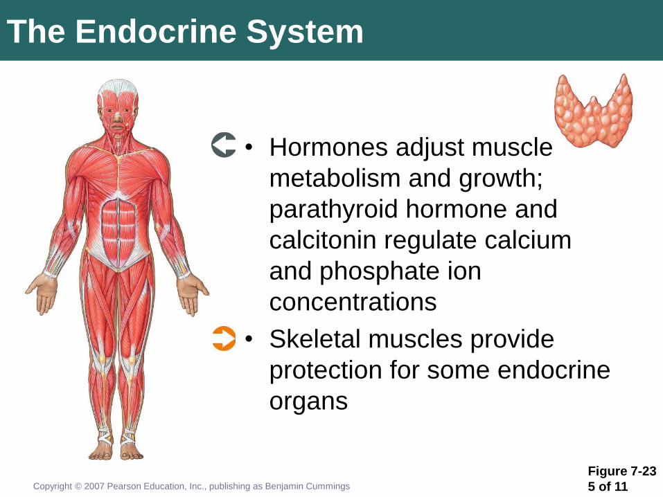
Figure 7-23
5 of 11
The Endocrine System
• Hormones adjust muscle
metabolism and growth;
parathyroid hormone and
calcitonin regulate calcium
and phosphate ion
concentrations
• Skeletal muscles provide
protection for some endocrine
organs
Copyright © 2007 Pearson Education, Inc., publishing as Benjamin Cummings
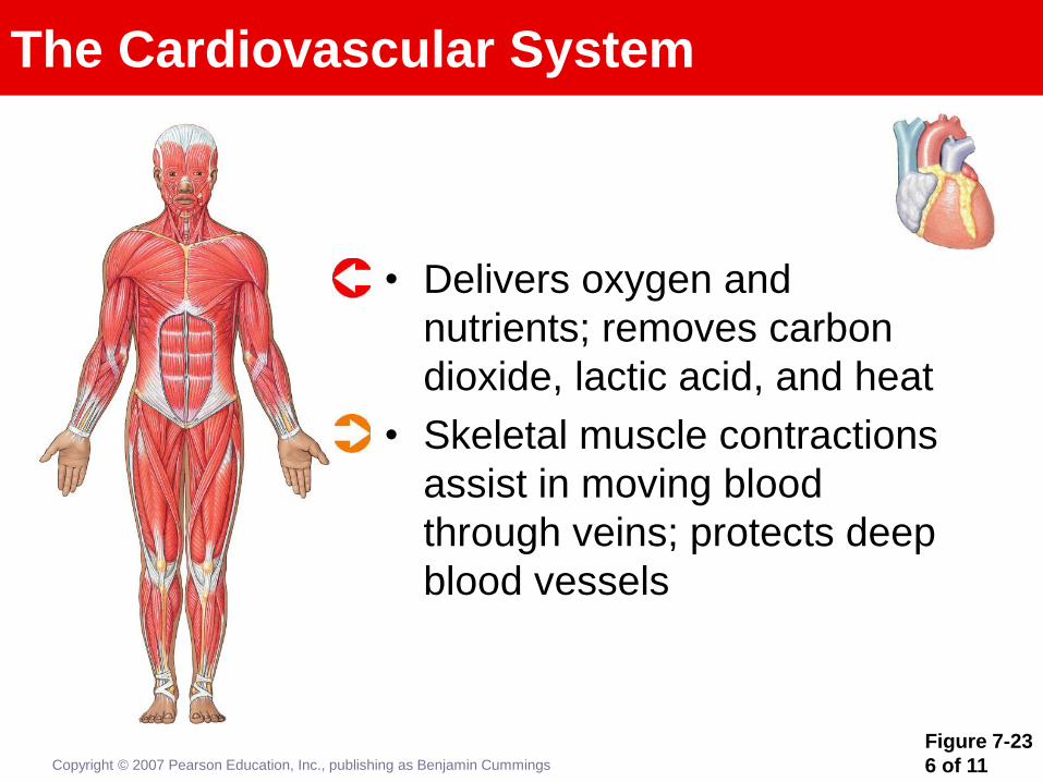
Page 105
Figure 7-23
6 of 11
The Cardiovascular System
• Delivers oxygen and
nutrients; removes carbon
dioxide, lactic acid, and heat
• Skeletal muscle contractions
assist in moving blood
through veins; protects deep
blood vessels
Copyright © 2007 Pearson Education, Inc., publishing as Benjamin Cummings
Page 106
Figure 7-23
7 of 11
The Lymphatic System
• Defends skeletal muscles
against infection and assists
in tissue repairs after injury
• Protects superficial lymph
nodes and the lymphatic
vessels in the
abdominopelvic cavity
Copyright © 2007 Pearson Education, Inc., publishing as Benjamin Cummings
Page 107
Figure 7-23
8 of 11
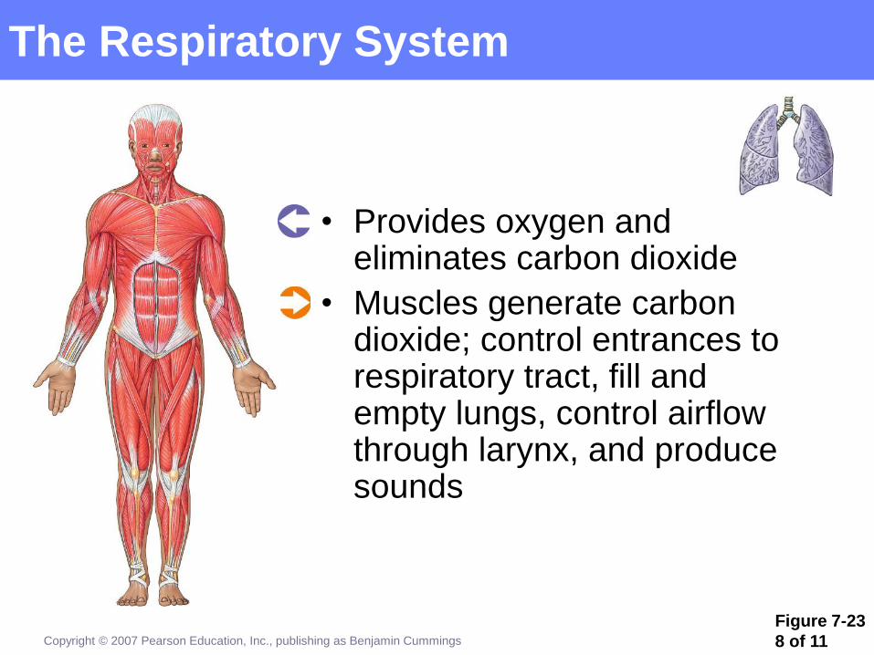
The Respiratory System
• Provides oxygen and eliminates carbon dioxide
• Muscles generate carbon dioxide; control entrances to respiratory tract, fill and empty lungs, control airflow through larynx, and produce sounds
Copyright © 2007 Pearson Education, Inc., publishing as Benjamin Cummings
Page 108
Figure 7-23
9 of 11
The Digestive System
Copyright © 2007 Pearson Education, Inc., publishing as Benjamin Cummings
• Provides nutrients; liver
regulates blood glucose and
fatty acid levels and removes
lactic acid from circulation
• Protects and supports soft
tissues in abdominal cavity;
controls entrances to and
exits from digestive tract
Page 109
Figure 7-23
10 of 11
The Urinary System
• Removes waste products of
protein metabolism; assists in
regulation of calcium and
phosphate concentrations
• External sphincter controls
urination by constricting
urethra
Copyright © 2007 Pearson Education, Inc., publishing as Benjamin Cummings
Page 110
Figure 7-23
11 of 11
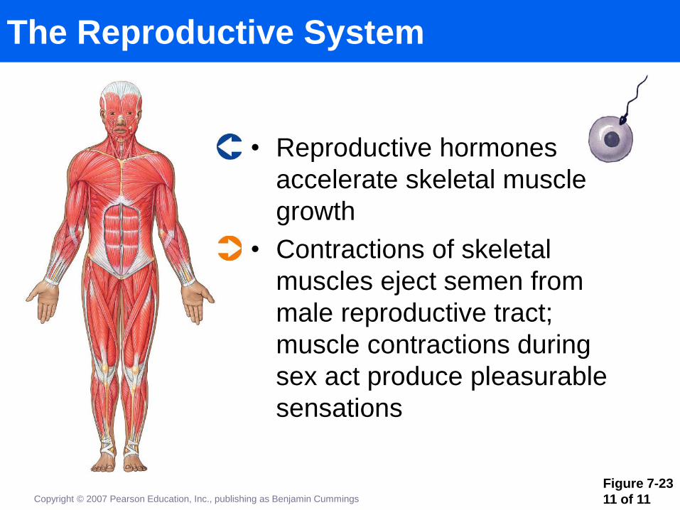
The Reproductive System
• Reproductive hormones
accelerate skeletal muscle
growth
• Contractions of skeletal
muscles eject semen from
male reproductive tract;
muscle contractions during
sex act produce pleasurable
sensations
Copyright © 2007 Pearson Education, Inc., publishing as Benjamin Cummings