The nephrogenic potential of the transcriptionfactors osr1, osr2, hnf1b, lhx1 and pax8 assessedin Xenopus animal capsChristiane Drews, Sabine Senkel, Gerhart U Ryffel*
Abstract
Background: The three distinct types of kidneys, pronephros, mesonephros and metanephros, developconsecutively in vertebrates. The earliest form of embryonic kidney, the pronephros, is derived from intermediatemesoderm and the first expressed genes localized in the pronephros anlage are the transcription factors osr1, osr2,hnf1b, lhx1 and pax8, here referred to as the early nephrogenic transcription factors. However, the pathwayinducing nephrogenesis and the network of theses factors are poorly understood. Treatment of theundifferentiated animal pole explant (animal cap) of Xenopus with activin A and retinoic acid induces pronephrosformation providing a powerful tool to analyze key molecular events in nephrogenesis.
Results: We have investigated the expression kinetics of the early nephrogenic transcription factors in activin Aand retinoic acid treated animal caps and their potential to induce pronephric differentiation. In treated animalcaps, expression of osr1, osr2, hnf1b and lhx1 are induced early, whereas pax8 expression occurs later implying anindirect activation. Activin A alone is able to induce osr2 and lhx1 after three hours treatment in animal caps whileretinoic acid fails to induce any of these nephrogenic transcription factors. The early expression of the fivetranscription factors and their interference with pronephros development when overexpressed in embryos suggestthat these factors potentially induce nephrogenesis upon expression in animal caps. But no pronephrosdevelopment is achieved by either overexpression of OSR1, by HNF1B injection with activin A treatment, or thecombined application of LHX1 and PAX8, although they influenced the expression of several early nephrogenictranscription factors in some cases. In an additional approach we could show that HNF1B induces several genesimportant in nephrogenesis and regulates lhx1 expression by an HNF1 binding site in the lhx1 promoter.
Conclusions: The early nephrogenic transcription factors play an important role in nephrogenesis, but have nopronephros induction potential upon overexpression in animal caps. They activate transcriptional cascades thatpartially reflect the gene activation initiated by activin A and retinoic acid. Significantly, HNF1B activates the lhx1promoter directly, thus extending the known activin A regulation of the lhx1 gene via an activin A responsiveelement.
BackgroundDuring vertebrate development three kidney types ofincreasing complexity (pronephros, mesonephros andmetanephros) form successively from the intermediatemesoderm, located between the paraxial mesoderm(developing somites) and the lateral plate [1]. The pro-nephros is the simplest, functional form of kidney in lar-val stages of fish and amphibians and consists of three
major components: glomus, tubules and duct. In adultsthe pronephros is replaced by the mesonephros. Inmammals the pronephros is not functional, but requiredfor mesonephros formation that is replaced by the meta-nephros, the kidney of the adult [2].All components of the pronephros arise from inter-
mediate mesoderm, but the signals that direct patteringof the presumptive pronephric mesoderm towards pro-nephric lineages are unknown. Experiments showed thatthe anterior somites are crucial for pronephros develop-ment and provide an essential first signal. If the anterior
somites are removed [3] or separated from the presump-tive pronephros [4], pronephroi do not form. Anteriorsomites can also induce pronephric tubules in unspeci-fied intermediate mesoderm [3]. Although the exact tim-ing and nature of the signal provided by the anteriorsomites are yet unknown, wnt11b expressed throughoutthe anterior somites has recently been shown as a cru-cial signal [5].Xenopus is a very attractive model organism to ana-
lyse key molecular events in nephrogenesis, becausemost genes essential for pronephros development inXenopus embryos are also crucial for the formation ofthe more complex mammalian kidneys [6-9]. A classicalmethod to identify important molecules in Xenopusdevelopment is the injection of mRNAs or morpholinooligonucleotides into the fertilized egg or into blasto-meres of early cleaving stages [10,11]. Thus, several pro-nephric regulators have been functionally identified[7,8,12]. An additional experimental tool to study earlyevents of nephrogenesis involves explanting the animalpole of the blastula. These explanted animal caps havepluripotency and differentiate into various tissues uponexposure to inducing substances [13,14]. Importantly,animal caps treated with activin A and retinoic acid dif-ferentiate into pronephros [15] and in this in vitro sys-tem genes are induced with similar kinetics as in vivo[16-18].In Xenopus the first genes expressed in the prone-
phros anlage are the transcription factors osr1 and osr2,members of the odd-skipped family of proteins [19],hnf1b, a member of the homeobox factors [20], lhx1(lim1), a lim homeobox factor [21] as well as pax8, amember of the paired box domain family [22]. We referto these five transcription factors as the early nephro-genic transcription factors, as they are all expressed inthe pronephros anlage prior to cellular differentiationand their misexpression affects pronephros develop-ment. Inhibition of osr1 or osr2 by morpholinos inXenopus embryos interferes with kidney formation andembryonic overexpression of either of these factorsinduces ectopic kidney tissue and enlarged pronephros[19]. Overexpression of hnf1b inhibits pronephros for-mation [23] and this effect is also seen by using thehuman HNF1B [24] implying that the regulatory poten-tial has been conserved during vertebrate evolution. Incontrast, lhx1 and pax8 overexpression leads to anenlargement of the pronephros and, if both factors arecoexpressed, this effect is increased and even inducesectopic pronephric tubules [25].It should be noted that each of these five early
nephrogenic transcription factors plays also a crucialrole in the development of other organs. The prominentrole of these nephrogenic transcription factors is par-tially also evident in mammalian systems. Whereas null
mutation of Osr1 in mice exhibit agenesis of the kidney[26], Osr2 knock-out has no kidney phenotype [27],although Osr2 transcripts are expressed in the develop-ing kidney [28]. The kidney-restricted knockout ofHnf1b leads to polycystic kidney disease [29] and theLhx1 null mutant even lacks any kidney [30]. In con-trast, Pax8 deficient mice exhibit thyroid gland defi-ciency, but have no pronephric phenotype [31].Nevertheless, Pax8 plays an essential role in kidneydevelopment, as impaired metanephros formationobserved in mice deficient for Pax2 [32] is dramaticallyincreased by a lack of any nephric cell lineage, if theseembryos lack additionally Pax8 [33].To further explore the role of these five nephrogenic
transcription factors we have now analyzed the kineticsof their induction in animals caps differentiated intopronephric tissue by activin A and retinoic acid. Wethen have overexpressed these transcription factors inanimal caps and analyzed their potential to induce eachother and to stimulate pronephric differentiation inthese explants. To allow discrimination between injectedmRNAs and endogenous mRNAs we used the humanmRNAs that are functionally equivalent, but are notdetectable with the Xenopus probes. We use capital let-ters for these human transcription factors to make aclear distinction. In addition we identified genes inducedby HNF1B in these early embryonic cells.
ResultsInduction of mRNAs encoding the early nephrogenictranscription factors osr1, osr2, hnf1b, lhx1 and pax8 inanimal caps treated with retinoic acid and/or activin ASince simultaneous treatment of Xenopus animal caps with10 ng/ml of activin A and 10-4 M retinoic acid for threehours induces differentiation of pronephric tissues [15], weexplored the time dependent induction of the earlynephrogenic transcription factors osr1, osr2, hnf1b, lhx1and pax8 in these embryonic explants. Thus, we measuredby quantitative RT-PCR the induction of the correspondingmRNAs in animal caps treated with retinoic acid and acti-vin A (Figure 1A). Based on the low sample pools (N = 4)and because we do not know whether the values obtainedin various experiments with animal caps represent a nor-mal distribution, the use of a significance test is not appro-priate. Therefore, we defined genes as induced orrepressed, when all the four measured values for a givensample pool represented the same trend. Using these cri-teria all five transcripts were induced, albeit with differentkinetics. Whereas osr1, osr2, hnf1b and lhx1 mRNAs wereinduced within 1.5 hours, pax8 mRNA was only increasedafter thirteen hours. For osr1 and osr2 the inductionincreased up to thirteen hour treatment, whereas hnf1band lhx1 seemed to level off at seven hour treatment. Byinhibition of protein synthesis with cycloheximide, the
Drews et al. BMC Developmental Biology 2011, 11:5http://www.biomedcentral.com/1471-213X/11/5
Page 2 of 14
induction of osr2, hnf1b and lhx1 was not abolishedimplying direct gene activation (Figure 1B). The observa-tion that osr1 was not induced in this experiment possiblyreflects the 30 min culture with or without cycloheximideprior to retinoic acid and activin A treatment, sincedelayed activin A treatment of animal caps reduces theinduction potential [34].To clarify which nephrogenic transcription factor tran-
scripts are induced by retinoic acid or activin A alone, weanalyzed animal caps treated with either retinoic acid oractivin A for three hours (Figure 1C). Treatment with reti-noic acid failed to induce the nephrogenic transcriptionfactors, but rather decreased the level of osr1 and pax8transcripts by 2-fold and 1.6-fold, respectively. In contrast,activin A treatment induced osr2 as well as lhx1, but osr1in two of four experiments only. Since hnf1b was notinduced by activin A alone, we conclude that a synergisticeffect of both inducers is needed to induce hnf1b. The lackof induction of pax8 in animal caps treated with activin Aor retinoic acid alone is consistent with the no-inductionobserved in animal caps treated with retinoic acid and acti-vin A for three hours (also compare Figure 1A).
Overexpression of OSR1 and Osr2A leads to enlargementof pronephros and ectopic pronephric tissueSince the murine Osr2 gene is expressed in two splicevariants referred to as Osr2A and Osr2B [35], wesearched Xenopus cDNA sequences deposited in Gene-Bank for corresponding splice variants. Indeed, we iden-tified transcripts encoding the A and B splice variantsthat encode a five and three finger zinc protein, respec-tively, comparable to the murine situation (Figure 2Aand 2B). Using primers specific for the A and B splicevariant we could show that osr2B predominates osr2Aby a factor of about three throughout retinoic acid andactivin A induction in animal caps (Figure 2C).
Figure 1 Time dependent induction of the early nephrogenictranscription factors in retinoic acid and activin A treatedanimal caps. (A) Animal caps were cut at the late blastula stage 9[36] and incubated for 1.5 or 3 hours in retinoic acid (10-4M) andactivin A (10 ng/ml). For 5, 7 and 13 hours treatments retinoic acidand activin A was replaced after three hours with Steinberg’ssolution [15]. The mRNA levels were analysed by quantitative RT-PCR using the primers given in Additional file 2. The standard
deviations from four independent animal cap pools (N = 30) aregiven and alterations are indicated by an upward arrow, if all fourinduction values were higher than one for a given probe. To detectosr2 transcripts primers targeting osr2B, the splice variantpredominantly expressed in Xenopus laevis (compare Figure 2Bs)were used for two animal cap pools analyzed after 3, 5 and 7 hoursand three after 13 hours In all other experiments primers targetingboth splice variants were used. (B) Animal caps cut at the lateblastula stage 9 were incubated with or without cycloheximide(CHX, 5 μg/ml) for 30 min and then stimulated for 1.5 hours inretinoic acid (10-4M) and activin A (10 ng/ml). RNA was quantifiedfrom six pools (N = 30) and upward arrows indicate induction for allsix experiments. (C) Animal caps were cultured for three hours inretinoic acid (RA, 10-4M), activin A (ActA, 10 ng/ml) or both inducerstogether (RA+ActA) and then analysed by quantitative RT-PCR. Thestandard deviations from five (RA) or four (ActA or RA+ActA)independent animal cap pools (N = 30) are given and reproducibleinduction or reduction is indicated by an upward or downwardarrow, if increased or decreased in all samples, respectively.
Drews et al. BMC Developmental Biology 2011, 11:5http://www.biomedcentral.com/1471-213X/11/5
Page 3 of 14
To examine the morphogenetic potential of the mamma-lian Osr1 and Osr2A in developing Xenopus embryos, weinjected mRNA encoding the human OSR1 or the murineOsr2A proteins into one blastomere of the two-cell stageembryo using GFP mRNA as a marker to identify theinjected side. About half of the OSR1 and more than thehalf of the Osr2A injected embryos showed gastrulationdefects that were more severe with higher doses of mRNA.Injected embryos surviving to the free swimming tadpolestage 45 [36] were immunostained for pronephros develop-ment using a mixture of the monoclonal antibodies for theproximal tubules (3G8) and the distal tubules and pro-nephric duct (4A6) [37]. The majority of embryos injectedwith OSR1 showed an enlarged pronephros (Figure 3A), insome cases together with the formation of ectopic kidney
Figure 2 Splice variants of Xenopus osr2. (A) Schematicrepresentation of the Xenopus osr2 splicing variants A and B basedon the mammalian data [35] shows alternative splice sites 2A and2B indicated by arrowheads. Boxes represent exons and filled areasreflect open reading frames, while lines represent the promoter andintrons. In the lower panel a scheme of the five and three zinc-finger domains (grey boxes) in osr2A and in osr2B protein is given,respectively. The black box in osr2B denotes the alternatively splicedexon 4. (B) Alignment of the nucleotide sequence aroundalternatively spliced exon 4 is based on a Xenopus laevis osr2B full-length cDNA sequence (accession no. BC108579), whereas thecorresponding osr2A cDNA of Xenopus tropicalis (accession no.CU075721) is taken, as no corresponding Xenopus laevis cDNA isavailable. The nucleotide sequence around alternatively spliced sites2A and 2B (indicated by arrowheads) is conserved in both species.The forward primer FP is identical for both splice variants, whereasthe reverse primers distinguish between osr2A and osr2B.(C) Animal caps were cut at the late blastula stage 9 [36] andincubated for 1.5 or 3 hours in retinoic acid (10-4M) and activin A(10 ng/ml). For 5, 7 and 13 hours treatments retinoic acid andactivin A was replaced after three hours with Steinberg’s solution asoriginally described [15]. The mRNA levels were analysed byquantitative RT-PCR using the specific primers for osr2A and osr2B(see Additional file 2) and the results are normalized to odcexpression levels. If two independent pools were analysed, themean is given and the bar marks the two values.
0 50 100 150 200 %
***Osr2A (200pg,N=27)
(100pg,N=9)
(200pg,N=35)
GFP (N=94)
OSR1
***
relative pronephros size
(injected/uninjected)
C
A B
- - - - - - - - - - - - - - - - - - - - - - -
Figure 3 Expression of OSR1 and Osr2A leads to an increase ofpronephros size in Xenopus laevis larvae. (A) Enlargement ofpronephros: Dorsal view of stage 45 larvae [36] injected with 200pg OSR1 mRNA and whole-mount immunostained with antibodies3G8 and 4A6 [37]. The injected side is marked by an arrow. Bar =200 μm (B) Ectopic kidney cell patches (marked by arrow heads):Lateral view of a stage 45 larvae [36] expressing OSR1 and preparedas in A. Bar = 200 μm (C) Pronephros size on injected versuscontrol side in OSR1 or Osr2A injected embryos was determined inlateral views by measuring the area through the widest part of theimmunostained pronephros. The Student’s t-test was used to scoresignificant differences. The p-value is given by comparing embryosinjected with gene of interest plus GFP mRNA as marker with thoseinjected with GFP mRNA taken from previous data [23]. *** denotesp-value ≤ 0.001. The vertical line in the middle represents the meanand the small bars the standard deviation. N is the number ofanimals investigated.
Drews et al. BMC Developmental Biology 2011, 11:5http://www.biomedcentral.com/1471-213X/11/5
Page 4 of 14
cell patches (Figure 3B). Comparing the size of the prone-phros of the injected side with the one of the non-injectedside we observed that the pronephros size was significantlyenlarged by injecting 200 pg OSR1 but not with 100 pg(Figure 3C). Similarly, Osr2A overexpression (200 pg) leadsto a significant increase of pronephros size as well, but wedid not observe the formation of ectopic kidney tissue. Inconclusion, OSR1 as well as Osr2A overexpression inducespronephros enlargement in Xenopus embryos as foundpreviously for the corresponding Xenopus factors osr1 andosr2B suggesting comparable function of both mammaliangenes in pronephros development [19].
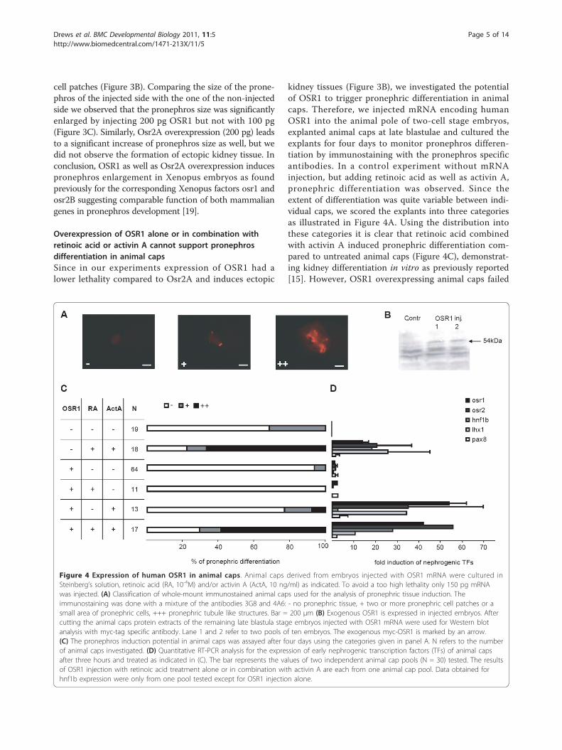
Overexpression of OSR1 alone or in combination withretinoic acid or activin A cannot support pronephrosdifferentiation in animal capsSince in our experiments expression of OSR1 had alower lethality compared to Osr2A and induces ectopic
kidney tissues (Figure 3B), we investigated the potentialof OSR1 to trigger pronephric differentiation in animalcaps. Therefore, we injected mRNA encoding humanOSR1 into the animal pole of two-cell stage embryos,explanted animal caps at late blastulae and cultured theexplants for four days to monitor pronephros differen-tiation by immunostaining with the pronephros specificantibodies. In a control experiment without mRNAinjection, but adding retinoic acid as well as activin A,pronephric differentiation was observed. Since theextent of differentiation was quite variable between indi-vidual caps, we scored the explants into three categoriesas illustrated in Figure 4A. Using the distribution intothese categories it is clear that retinoic acid combinedwith activin A induced pronephric differentiation com-pared to untreated animal caps (Figure 4C), demonstrat-ing kidney differentiation in vitro as previously reported[15]. However, OSR1 overexpressing animal caps failed
Figure 4 Expression of human OSR1 in animal caps. Animal caps derived from embryos injected with OSR1 mRNA were cultured inSteinberg’s solution, retinoic acid (RA, 10-4M) and/or activin A (ActA, 10 ng/ml) as indicated. To avoid a too high lethality only 150 pg mRNAwas injected. (A) Classification of whole-mount immunostained animal caps used for the analysis of pronephric tissue induction. Theimmunostaining was done with a mixture of the antibodies 3G8 and 4A6: - no pronephric tissue, + two or more pronephric cell patches or asmall area of pronephric cells, +++ pronephric tubule like structures. Bar = 200 μm (B) Exogenous OSR1 is expressed in injected embryos. Aftercutting the animal caps protein extracts of the remaining late blastula stage embryos injected with OSR1 mRNA were used for Western blotanalysis with myc-tag specific antibody. Lane 1 and 2 refer to two pools of ten embryos. The exogenous myc-OSR1 is marked by an arrow.(C) The pronephros induction potential in animal caps was assayed after four days using the categories given in panel A. N refers to the numberof animal caps investigated. (D) Quantitative RT-PCR analysis for the expression of early nephrogenic transcription factors (TFs) of animal capsafter three hours and treated as indicated in (C). The bar represents the values of two independent animal cap pools (N = 30) tested. The resultsof OSR1 injection with retinoic acid treatment alone or in combination with activin A are each from one animal cap pool. Data obtained forhnf1b expression were only from one pool tested except for OSR1 injection alone.
Drews et al. BMC Developmental Biology 2011, 11:5http://www.biomedcentral.com/1471-213X/11/5
Page 5 of 14
to differentiate and this was not improved by addingretinoic acid or activin A. Furthermore, OSR1 overex-pression in animal caps treated with retinoic acid andactivin A led to a similar extent of pronephric differen-tiation compared to uninjected, but treated animal caps.Quantitative RT-PCR analysis after three hours showedno induction of the early transcription factors uponOSR1 expression alone or in combination with retinoicacid, whereas treatment of OSR1 injected caps with acti-vin A led to an induction of osr1, osr2 and lhx1 (Figure4D), as observed frequently by activin A treatment alone(Figure 1C). As a control we verified the presence of theOSR1 protein translated from the injected OSR1 mRNAby Western blots (Figure 4B). Taken together, OSR1alone or in combination with retinoic acid or activin Adoes not have the potential to induce pronephros differ-entiation in animal caps.
LHX1 and/or PAX8 or HNF1B are not sufficient to inducepronephros differentiation in animal capsIt is known that embryonic overexpression of Xenopuslhx1 or pax8 induces enlargement of pronephros andcoexpression of both factors leads to a synergistic effect[25]. This enlargement of the pronephros we alsoobserved by unilateral injection of the human transcrip-tion factors, since coinjected LHX1 and PAX8 result ina pronephros size of 163 +/- 42% compared to the con-trol side (N = 23, p-value = 3 × 10⁻⁷). However, in con-trast to experiments using the Xenopus factors [23,25],no ectopic pronephric tissue formation was seen withthe human factors. To investigate the pronephros differ-entiation potential of human LHX1 and PAX8 in animalcaps, we injected mRNA encoding these proteins, eitheralone or in combination, into the animal pole of two-cell stage embryos and analysed the animal caps byimmunostaining after four days. Both LHX1 and PAX8alone or in combination could not induce pronephrosdifferentiation in animal caps at day four (Figure 5A).Measuring the level of the transcripts encoding the fivenephrogenic transcription factors after 3 hours weobserved no change (data not shown). The missing pro-nephros formation in animal caps overexpressing LHX1and PAX8 is consistent with unpublished data reviewedrecently [14]. To prove successful injection, we con-firmed in all embryos green fluorescence derived fromthe coinjected GFP mRNA. In addition, for LHX1 injec-tions into animal caps we deduce functional relevantamounts of LHX1 protein, as the lhx1 target gene cer-berus (cer1) [38] was 6- or 64-fold induced in two inde-pendent experiments (data not shown).Since differential addition of retinoic acid and activin
A to animal caps has shown that activin A aloneinduces osr2, lhx1 and frequently also osr1, but neverhnf1b that requires retinoic acid in addition (Figure 1C),
we wondered whether HNF1B injection might replaceretinoic acid to get pronephric induction in animal caps.However, injection of HNF1B mRNA into the animalpole at the two-cell stage, failed to induce pronephricdifferentiation in the explanted animal caps, even if cul-tured in the presence of activin A (Figure 5B). In con-clusion, LHX1, PAX8 and HNF1B failed to inducepronephros differentiation in animal caps.
Overexpression of HNF1B induces in animal caps genesimportant for nephrogenesisSince numerous target genes of hnf1b have been defined[39] or postulated [40] in various mammalian systems,we wonder whether these genes are activated in animalcaps representing embryonic stem cells. Thus, wesearched for orthologs present in Xenopus ESTs andselected 26 genes reported to be expressed early inXenopus development or in the developing pronephros[41]. Analysing animal caps of HNF1B injected embryosby quantitative RT-PCR revealed after seven hours aclear increase in transcripts encoding the early nephro-genic transcription factors lhx1, osr2 and osr1, but alsoof those encoding the transcription factors hnf1a, hnf4a
PAX8
LHX1
PAX8+LHX1
HNF1B
HNF1b+ActA
N
33
52
8
18
24
20 40 60 80 100
A
B
- + ++
% of pronephric differentiation
15
44
RA+ActA
RA+ActA
20 40 60 80 100
% of pronephric differentiation
Figure 5 Expression of human LHX1, PAX8 and HNF1B inanimal caps. Animal caps of LHX1 and/or PAX8 (250 pg alone or125 pg each) or HNF1B (150 pg) injected embryos were cultured inSteinberg’s solution for three hours and analysed after four days bywhole-mount immunostaining using a mixture of the antibodies3G8 and 4A6. Activin A (ActA, 10 ng/ml) and retinoic acid (RA, 10-4M) were added as given. (A) and (B) The pronephric tissueinduction in animal caps treated as indicated on the left was scoredusing the categories given in Figure 4A. The number of animal caps(N) is given.
Drews et al. BMC Developmental Biology 2011, 11:5http://www.biomedcentral.com/1471-213X/11/5
Page 6 of 14
and tfe3 as well as the signalling molecules wnt11b andgdnf (Table 1). After fourteen hours we found as adelayed response induction of the transcripts encodingthe transcription factor pax2 and esterase D (esd). Atthis later time point decreased level of transcriptsencoding the RNA binding protein rbms1 and the fibro-blast growth factor receptor (fgfr4c) were found imply-ing secondary effects. As lhx1 transcripts are induced byhnf1b, we also tested five transcripts of genes known tobe targeted by lhx1. Indeed, cerberus (cer1) and chordin(chrd) were induced, but three other lhx1 targets werenot (Table 1). In fact, goosecoid (gsc) was downregu-lated after fourteen hours. Taken together, HNF1B canactivate in animal caps several genes involved in kidneydevelopment, but some genes considered to be HNF1Btargets in mammals are not influenced.
HNF1B regulates lhx1 transcription by an HNF1 bindingsite in the lhx1 promoterThe Xenopus lhx1 gene is known to be regulated inearly embryogenesis by activin A via an activin responseelement (ARE) present in the first intron [42,43]. Sinceour data show that HNF1B induces lhx1 transcripts inanimal caps without activin treatment, we explored adirect activation of the lhx1 gene via HNF1B. By insilico analysis with JASPAR [44] we identified potentialHNF1 binding sites (Figure 6C) in the promoter regionand the first intron of the Xenopus lhx1 gene (Figure6A). To determine functional HNF1 binding sites, wetested four luciferase reporter constructs carrying var-ious fragments of the lhx1 gene (Figure 6A). These con-structs were transiently transfected into a HEK293 cellline containing tetracycline-inducible HNF1B [45]. Asshown in Figure 6A, the construct Ex-5:B containing theentire lhx1 gene was inducible by HNF1B and by analy-sis of various deletion constructs regulation by HNF1Bcould be pinned down to the HNF1 site of the promoterarea (compare Ex1(-120/+3) versus Ex1(-117/+3) in Fig-ure 6A). Clearly, partial mutation (three of twelve basepairs) of the HNF1 site of the promoter (Figure 6C)abolished HNF1B regulation completely. HNF1B trans-activation via the HNF1 binding site in the lhx1 promo-ter is about 70% (Figure 6A) compared to the effectseen with the HNF4A P2 promoter, a well studiedHNF1B target [46,47].To extend these findings to Xenopus embryonic cells we
tested some constructs in animal caps that were derivedfrom controls or HNF1B injected eggs. All constructsretaining the HNF1 binding site were transactivated byinjected HNF1B, including the minimal construct Ex1(-120/+3), whereas the reporter Ex1(-117/+3) containing
Table 1 Activation of genes by HNF1B in animal caps
gene symbol fold induction
7 h(N = 5)
14 h(N = 4)
early nephrogenic TFs lhx1 9.9 ↑ 1.5
osr2 4.3 ↑ 2.2
osr1 3.7 ↑ 2.5
pax8 2.4 1.7
hnf1b 1.3 0.9
genes involved in nephrogenesis hnf1a 13.7 ↑ 7.1
wnt11b 4.6 ↑ 1.3
wnt11 1.6 1.1
pax2 1.1 10.7 ↑
gdnf 1.2 ↑ 1.4
wnt9b 0.8 0.7
proximal tubule genes prodh2 95.9 1.7
hnf4a 22.9 ↑ 3.4
anxa13 5.3 1.4
slc22a6 3.4 25.6
cpn1 2.3 3.1
tmem27 2.2 3.5
slc5a2 1.9 2.3
tfe3 1.8 ↑ 1.2
rpl35a 1.5 1.5
gjb1 1.3 1.0
trps1 1.2 0.8
ube3a 1.1 0.8
esd 1.0 1.3 ↑
rbms1 1.0 0.2 ↓
slc7a8 1.0 1.5
fgfr4a 0.9 0.7
c8a 0.9 1.1
ncor1 0.9 1.1
slc4a7 0.9 1.1
fgfr4c 0.7 0.5 ↓
lhx1 target genes cer1 421.3 ↑ 2.9
chrd 9.0 ↑ 7.8
pcdh8.2 5.1 2.0
gsc 3.5 0.1 ↓
otx2 1.3 1.3
HNF1B (150 pg) injected animal caps were cultured for seven or fourteenhours in Steinberg’s solution and analysed by quantitative RT-PCR for geneexpression of the early nephrogenic transcription factors, of genes involved innephrogenesis [78], of proximal tubule genes [40] and of the lhx1 targetgenes chrd [82], cer1 [38], gsc [82], otx2 [83] and pcdh8.2 [84] using theprimers given in Additional file 2. Based on the low sample numbers (N = 4or 5), genes were defined as induced or repressed, if all induction values wereeither higher or lower for a given probe. Distinct induction or reduction isindicated by an upward or downward arrow, respectively. N is the number ofpools with 30 animal caps.
Drews et al. BMC Developmental Biology 2011, 11:5http://www.biomedcentral.com/1471-213X/11/5
Page 7 of 14
ATG
-5000 Luc
ATG
-5000 Luc
ATG
-350 Luc
ATG
-3000 Luc
ATG
Luc
ATG
Luc
Luc
AREHNF1 HNF1
Figure 6 Functional identification of HNF1 binding sites in the promoter region of lhx1 in HEK293 cells and in animal caps. (A) HEK293(HNF1B) cells [45] were transfected with lhx1 gene firefly luciferase fusion constructs and HNF1B expression was induced by adding doxycycline.On the left panel schematic drawings of transfected lhx1 promoter luciferase fusion constructs (not to scale) are given. Lines represent promoterand intron regions, boxes are exons with protein coding (filled) or untranslated (open) regions (adapted from [42]). Arrows mark potential HNF1binding sites identified by JASPAR [44]. The arrow head marks the ARE (activin response element) in intron 1 [42]. The Ex1(-120/+3) constructcontains the complete HNF1 binding site in the promoter region of lhx1, whereas the binding site in the Ex1(-117/+3) construct is partiallydeleted (compare panel C). The HNF-4a P2-285 construct contains the P2-promoter of the HNF-4a gene which is regulated by HNF1B [46]. Thefold change of luciferase activity of transfected constructs in doxycycline induced HEK293(HNF1B) versus untreated cells is given using the renillaluciferase reporter pRL-Con as an internal control. The Student’s t-test was used and *, ** and *** refer to p-value of ≤ 0.05, ≤ 0.01 and ≤ 0.001,respectively and n.s. means not significant. N is the number of independent transfections made. (B) The luciferase reporter constructs (50 pgeach) were tested in animal caps of controls or HNF1B (150 pg) injected embryos. The luciferase activity was measured in pools of four animalcaps after four hours cultivation in Steinberg’s solution. To calculate the increase of luciferase activity in HNF1B injected animal caps the ratio offirefly luciferase (FL) to renilla luciferase (RL) was used (FL/RL). The vertical line in the middle represents the mean and the small bars thestandard deviation. Since experiments with different pools of animal caps were not comparable in quantitative terms, we used the Mann-Whitney-test to score significant differences. * and ** refer to p-value of ≤ 0.05 and ≤ 0.01, respectively and n.s. means not significant. N is thenumber of animal cap pools tested. (C) The upper panel shows the sequence logo of HNF1 binding site given in JASPAR [44] and the lowerpanel the sequences of the HNF1 binding site (capital letters) in Ex1(-120/+3) and Ex1(-117/+3) constructs. The vector sequence is indicated byitalics.
Drews et al. BMC Developmental Biology 2011, 11:5http://www.biomedcentral.com/1471-213X/11/5
Page 8 of 14
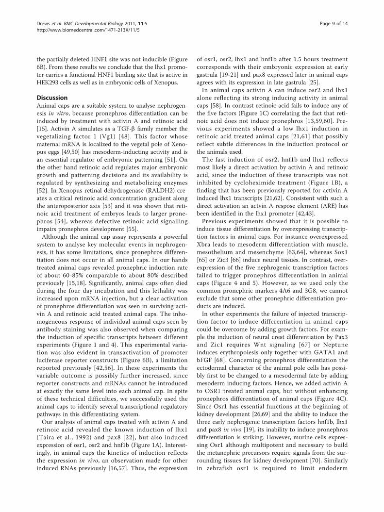
the partially deleted HNF1 site was not inducible (Figure6B). From these results we conclude that the lhx1 promo-ter carries a functional HNF1 binding site that is active inHEK293 cells as well as in embryonic cells of Xenopus.
DiscussionAnimal caps are a suitable system to analyse nephrogen-esis in vitro, because pronephros differentiation can beinduced by treatment with activin A and retinoic acid[15]. Activin A simulates as a TGF-b family member thevegetalizing factor 1 (Vg1) [48]. This factor whosematernal mRNA is localized to the vegetal pole of Xeno-pus eggs [49,50] has mesoderm-inducting activity and isan essential regulator of embryonic patterning [51]. Onthe other hand retinoic acid regulates major embryonicgrowth and patterning decisions and its availability isregulated by synthesizing and metabolizing enzymes[52]. In Xenopus retinal dehydrogenase (RALDH2) cre-ates a critical retinoic acid concentration gradient alongthe anteroposterior axis [53] and it was shown that reti-noic acid treatment of embryos leads to larger prone-phros [54], whereas defective retinoic acid signallingimpairs pronephros development [55].Although the animal cap assay represents a powerful
system to analyse key molecular events in nephrogen-esis, it has some limitations, since pronephros differen-tiation does not occur in all animal caps. In our handstreated animal caps revealed pronephric induction rateof about 60-85% comparable to about 80% describedpreviously [15,18]. Significantly, animal caps often diedduring the four day incubation and this lethality wasincreased upon mRNA injection, but a clear activationof pronephros differentiation was seen in surviving acti-vin A and retinoic acid treated animal caps. The inho-mogeneous response of individual animal caps seen byantibody staining was also observed when comparingthe induction of specific transcripts between differentexperiments (Figure 1 and 4). This experimental varia-tion was also evident in transactivation of promoterluciferase reporter constructs (Figure 6B), a limitationreported previously [42,56]. In these experiments thevariable outcome is possibly further increased, sincereporter constructs and mRNAs cannot be introducedat exactly the same level into each animal cap. In spiteof these technical difficulties, we successfully used theanimal caps to identify several transcriptional regulatorypathways in this differentiating system.Our analysis of animal caps treated with activin A and
retinoic acid revealed the known induction of lhx1(Taira et al., 1992) and pax8 [22], but also inducedexpression of osr1, osr2 and hnf1b (Figure 1A). Interest-ingly, in animal caps the kinetics of induction reflectsthe expression in vivo, an observation made for otherinduced RNAs previously [16,57]. Thus, the expression
of osr1, osr2, lhx1 and hnf1b after 1.5 hours treatmentcorresponds with their embryonic expression at earlygastrula [19-21] and pax8 expressed later in animal capsagrees with its expression in late gastrula [25].In animal caps activin A can induce osr2 and lhx1
alone reflecting its strong inducing activity in animalcaps [58]. In contrast retinoic acid fails to induce any ofthe five factors (Figure 1C) correlating the fact that reti-noic acid does not induce pronephros [13,59,60]. Pre-vious experiments showed a low lhx1 induction inretinoic acid treated animal caps [21,61] that possiblyreflect subtle differences in the induction protocol orthe animals used.The fast induction of osr2, hnf1b and lhx1 reflects
most likely a direct activation by activin A and retinoicacid, since the induction of these transcripts was notinhibited by cycloheximide treatment (Figure 1B), afinding that has been previously reported for activin Ainduced lhx1 transcripts [21,62]. Consistent with such adirect activation an actvin A respose element (ARE) hasbeen identified in the lhx1 promoter [42,43].Previous experiments showed that it is possible to
induce tissue differentiation by overexpressing transcrip-tion factors in animal caps. For instance overexpressedXbra leads to mesoderm differentiation with muscle,mesothelium and mesenchyme [63,64], whereas Sox1[65] or Zic3 [66] induce neural tissues. In contrast, over-expression of the five nephrogenic transcription factorsfailed to trigger pronephros differentiation in animalcaps (Figure 4 and 5). However, as we used only thecommon pronephric markers 4A6 and 3G8, we cannotexclude that some other pronephric differentiation pro-ducts are induced.In other experiments the failure of injected transcrip-
tion factor to induce differentiation in animal capscould be overcome by adding growth factors. For exam-ple the induction of neural crest differentiation by Pax3and Zic1 requires Wnt signaling [67] or Neptuneinduces erythropoiesis only together with GATA1 andbFGF [68]. Concerning pronephros differentiation theectodermal character of the animal pole cells has possi-bly first to be changed to a mesodermal fate by addingmesoderm inducing factors. Hence, we added activin Ato OSR1 treated animal caps, but without enhancingpronephros differentiation of animal caps (Figure 4C).Since Osr1 has essential functions at the beginning ofkidney development [26,69] and the ability to induce thethree early nephrogenic transcription factors hnf1b, lhx1and pax8 in vivo [19], its inability to induce pronephrosdifferentiation is striking. However, murine cells expres-sing Osr1 although multipotent and necessary to buildthe metanephric precursors require signals from the sur-rounding tissues for kidney development [70]. Similarlyin zebrafish osr1 is required to limit endoderm
Drews et al. BMC Developmental Biology 2011, 11:5http://www.biomedcentral.com/1471-213X/11/5
Page 9 of 14
differentiation to allow kidney development [71]. SinceOSR1 did not improve the differentiation potential ofactivin A and retinoic acid treated animal caps (Figure4C), other signalling molecules are missing. In this con-text it may be relevant that ectopic kidney tissues inOSR1 (Figure 3B), osr2 (Tena et al., 2007) or lhx1 andpax8 [23,25] overexpressing embryos were found exclu-sively close to the pronephros. This suggests that signalsin the region of pronephros anlage are needed. Mostrecently Wnt11b has been proposed as such a signal [5].In our experiments we overexpressed either the human
or murine transcription factors in order to distinguishthe activity of the endogenous gene from the injectedRNA, because previous experiments have shown equiva-lence between Xenopus and human hnf1b. This we con-firmed in principle for OSR1 and Osr2A (Figure 3) aswell as for coinjected LHX1 and PAX8 (see text). How-ever, in contrast to the Xenopus factors, human LHX1coinjecetd with PAX8 cannot induce ectopic pronephrictissue and such subtle differences may limit the use ofmammalian factors in Xenopus embryos.Investigating the influence of HNF1B on 26 potential
hnf1b target genes we could show the activation of ninegenes in injected animal caps (Table 1). The inducedgenes include the transcription factor lhx1, hnf1a, hnf4aand tfe3 suggesting the activation of various transcrip-tional cascades. This assumption is supported by theobservation that the lhx1 target genes cer1 [38] and chrd[72], both with important roles in nephrogenesis, werealso induced. Since other lhx1 target genes were not acti-vated (Table 1), we assume that some lhx1 target genesare inhibited by HNF1B expression. It is striking thatcer1 is induced at very high levels and peaked off withinseven hours indicating a transient activation of cer1 bylhx1 which itself is only transiently induced by HNF1B(Table 1). The activation of hnf1a was expected, asHNF1B expression in the Xenopus embryos activateshnf1a [24] and the hnf1a promoter contains a functionalHNF1 site [73]. Since HNF1B induces the expression ofosr1 and osr2 we deduce a positive feedback loop, as osr1and osr2 are able to induce hnf1b [19]. Furthermore, anincreased expression of the signalling molecules wnt11b[5] and gdnf [74] were found, both of which play a role innephrogenesis. Similarly, the downregulation of the fibro-blast growth factor receptor (fgfr4c) may be relevant, asfgfr4c down-regulation is needed to allow pronephrosdevelopment [75]. The delayed induction of two otherimportant molecules in nephrogenesis, pax2 [76] and esd[77], implied an indirect activation. The fact that severalgenes crucial for pronephros development and expressedlater in more differentiated pronephric tissues were notactivated in animal caps by HNF1B, may either reflectthat hnf1b acts differentially in Xenopus compared to
mammals or more likely other signals are missing in theundifferentiated animal cap. Albeit our data show thatHNF1B regulates in embryonic cells many genes poten-tially essential for pronephros development.A function of Hnf1b in early nephrogenesis has most
recently been reported also in mice by tetraploid anddiploid embryo complementation to overcome earlyembryonic lethality of homozygous knock-out mice [78].Significantly, in these mice Lhx1 was shown to be regu-lated by Hnf1b and to contain an HNF1 binding site inthe far upstream promoter. Thus, the regulation of lhx1by hnf1b seems to be evolutionary conserved, as a 10-fold increase in the expression level of lhx1 in HNF1Bdifferentiated animal caps is seen (Table 1) and thisinduction is mediated by an HNF1 binding site in theXenopus lhx1 promoter region (Figure 6).
ConclusionsIn this study using animal cap assays we provide insightinto the network of the early nephrogenic transcriptionfactors in pronephros development and the genesinduced by HNF1B, especially (Figure 7). Significantly,we identified a functional HNF1 binding site in the lhx1promoter. However, none of the early nephrogenic tran-scription factors has the ability to induce pronephrosdifferentiation in animal caps.
0 1.5 7 1413 h
ActA +
RA
osr1
osr2
hnf1b
lhx1
pax8
osr1osr2hnf1ahnf4a
wnt11bgdnftfe3
lhx1 cer1chrd
pax2esd
HNF1B
0 1.5 7 1413 h
ActA +
RA
osr1
hnf1b
pax8
osr1osr2hnf1ahnf4a
wnt11bgdnftfe3
lhx1 cer1chrd
pax2esd
rbms1fgfr4cgsc
Figure 7 Induction of the early nephrogenic transcriptionfactors and other genes in animal caps. osr1, osr2, hnf1b and lhx1are induced already after 1.5 hours treatment with activin A (ActA)and retinoic acid (RA) suggesting that they are direct targets,whereas pax8 is first expressed after thirteen hours implying anindirect activation. osr2 and lhx1 (highlighted in grey) are inducedafter three hours by activin A alone. HNF1B overexpressed in animalcaps induces after seven hours osr1, osr2, hnf1a, hnf4a, wnt11b, gdnfand tfe3. After fourteen hours pax2 and esd are increased and rbms1,fgfr4c and gsc are decreased suggesting secondary effects. HNF1Binduces the expression of lhx1 by an HNF1 binding site in itspromoter region (arrow). Furthermore, the lhx1 target genes cer1 andchrd are induced in HNF1B injected animal caps possibly via lhx1.
Drews et al. BMC Developmental Biology 2011, 11:5http://www.biomedcentral.com/1471-213X/11/5
Page 10 of 14
MethodsPlasmidsThe myc-Rc/CMVHNF1B expression vector has beendescribed previously [24]. The mouse Osr2A plasmidwas kindly provided by S. Kawai (Osaka UniversityGraduate School of Dentistry, Osaka, Japan). The full-length open reading frame of the human gene OSR1(IRATp970D0444D6; RZPD, Gemany) was clonedinto pCS2+MT [79] to generate N-terminal myc-tagfusion proteins using 5’-GCTCTAGAGATGGGCAG-CAAAACCTTGCC-3’ and 5’-GCTCTAGATTAG-CATTTGATCTTGGAGGTTTT-3’ as forward andreverse primers, respectively. The XbaI restriction sitesfor cloning are underlined and the construct was veri-fied by sequencing. The full-length open reading framesof the human PAX8 (RC200651) and LHX1 (RC210977)in pCMV6 were obtained from OriGene Technologies.The pRL-Con renilla luciferase construct [80] and the
HNF-4a P2-285 pGL3-Basic reporter plasmid [46] havebeen described. The lhx1 gene luciferase fusion con-structs Ex-1:A, Ex-2:C, Ex-2:D and Ex-5:B [42] werekindly provided by M.L. Rebbert (NICHD, USA). Thelhx1 reporter construct harbouring the HNF1 bindingsite in the promoter region (Ex-2:C) was used for addi-tional constructs. XhoI/HindIII DNA fragments contain-ing the intact or mutated HNF1 binding site in thepromoter region to the transcription start generated byPCR using the primers Ex1(-120/+3): 5’-CCGCTCGAGGCTTAATGGTT-3’ (forward), Ex1(-117/+3): 5’-CCGCTCGAGGGTTTACCAG-3’ (forward) and 5’-CCCAAGCTTTCCCTTTGGTTAT-3’ (reverse) wereinserted into the XhoI and HindIII digested pGL3-Basicvector (Promega). The restriction sites for cloning areunderlined. Both constructs were verified by sequencing.
mRNA injectionThe expression vectors encoding OSR1, Osr2A, HNF1B,LHX1 and PAX8 proteins and the GFP encoding expres-sion vector (pCSGFP2) were linearized and in vitro tran-scribed with RNA polymerases (Nielsen and Shaprio,1986). The restriction enzymes and RNA polymerasesused are given in Additional file 1. Capped mRNA encod-ing the different proteins together with 100 pg of cappedgreen fluorescent protein (GFP) mRNA as internal controlwere injected into one blastomere of the two-cell stageand after two days, the injected side was scored under astereofluorescence microscope for the presence of GFP. Incase of animal explants, the capped mRNA together withGFP is injected into the animal region of Xenopusembryos in each blastomere of the two-cell stage.
Animal cap assaysXenopus late blastulae, stage 9 [36], were de-jellied bytreatment with 2% cysteine hydrochloride in water. The
presumptive ectoderm (animal cap) was isolated withloops of 20 μm platinum wire heated to about 450°C fora few microseconds using the Gastromaster (XenotekEngineering, Belleville, USA). The explants were incu-bated for three hours in Steinberg’s solution (58 mMNaCl, 0.67 mM KCl, 0.34 mM Ca(NO3)2, 0.83 mMMgSO4, 3 mM HEPES, pH 7.8) containing recombinanthuman activin A (10 ng/ml; Sigma, A4941) and all-transretinoic acid (10-4M; Sigma, R2625) or only in Stein-berg’s solution for controls and for explants frommRNA injected embryos. After three-times washingwith Steinberg’s solution the explants were cultured inSteinberg’s solution at 20°C until they were equivalentto stage 40-42 in normal embryos (four days) and usedfor whole-mount immunostaining.
Quantitative RT-PCRRNA from pools of 30 animal caps was isolated withpeqGold RNAPure (PeqLab) followed by phenol/chloro-form extraction. For cDNA synthesis the High CapacitycDNA Reverse Transcription Kit (Applied Biosystems)was used. SYBR-Green real-time PCR was performed ona 7900HT Sequence Detection System (Applied Biosys-tems) using Power-SYBRGreen Mix (Applied Biosys-tems). Templates were determined in duplicate and forprimers used see Additional file 2. Results are normal-ized to ornithin decarboxylase (odc) expression levels. Inall cases water only and reverse transcriptase negativecontrols failed to produce specific products. The foldinduction of the early transcription factors was obtainedby comparison of treated and untreated animal caps.
Whole-mount immunostainingWhole-mount immunostaining of four day cultured ani-mal caps or animals at the swimming larval stage weredone as described [81]. The difference between theinjected and the non-injected sides of embryos was eval-uated by measuring the whole area using the lateralview with the widest diameter from the dorsal to theventral side of the immunostained pronephros includingthe pronephric tubules and the anterior part of the pro-nephric duct. The measurements were made using Axio-Vision 4.6 software (Carl Zeiss Imaging Solutions), andthe non-injected side was used as a reference for eachanimal. The values representing kidney size obtainedfrom each mRNA injected embryo were compared tovalues obtained from GFP control-injected embryos(data adapted from [23]). Significant differences werescored using the Student’s t-test to calculate p-values.
Cell culture and transient transfection assaysThe HEK293 (HNF1B) cell line [45] contains a tetracy-cline-inducible HNF1B transgene. In a 96-well plate(17,500 cells/well) 30 ng of the promoter constructs
Drews et al. BMC Developmental Biology 2011, 11:5http://www.biomedcentral.com/1471-213X/11/5
Page 11 of 14
were cotransfected using FuGeneHD (Roche) with 0.05ng of renilla luciferase plasmid pRL-Con for normaliza-tion of transfection efficiencies. Four hours after trans-fection HNF1B expression was induced by the additionof 1 μg/ml doxycycline. Twenty-four hours after trans-fection firefly and renilla luciferase activities were mea-sured in triplicate with the Dual-Luciferase ReporterAssay (Promega).
Luciferase reporter assays in animal caps50 pg reporter constructs and 150 pg HNF1B mRNAwere coinjected into the animal region of Xenopusembryos at the two-cell stage together with renilla luci-ferase as internal control. Animal caps were cut, cul-tured in Steinberg’s solution for four hours and pools offour caps were assayed by the Dual-Luciferase ReporterAssay (Promega). As the absolute levels of luciferaseactivity varied between pools of animal caps (data notshown, also described previously [42]), the Mann-Whitney-test was used to score significant differences.
Additional material
Additional file 1: Table S1: Expression vectors with restrictionenzymes and RNA polymerases used for RNA synthesis.
Additional file 2: Table S2: List of primers used for quantitativeRT-PCR.
AcknowledgementsWe thank Christoph Waldner for critical reading of the manuscript, ElizabethA. Jones for the antibodies 3G8 and 4A6, Martha L. Rebbert for the lhx1luciferase reporter constructs and S. Kawai for the Osr2A plasmid. We alsothank Ludger Klein-Hitpass for instruction on quantitative RT-PCR analysisand Tanja Boes for statistical advice.Parts of this work were supported by the Deutsche Forschungsgemeinschaft[Graduiertenkolleg 1431/1].
Authors’ contributionsCD carried out all the experiments with the animal caps and the injectedembryos and drafted the manuscript. SS made the transfection assays in cellculture. GUR conceived the study, participated in its design andcoordination and helped to finish the manuscript. All authors read andapproved the final manuscript.
Received: 25 August 2010 Accepted: 31 January 2011Published: 31 January 2011
References1. Dressler GR: Advances in early kidney specification, development and
patterning. Development 2009, 136:3863-3874.2. Vize PD, Carroll TJ, Wallingford JB: Introduction: Embryonic Kidneys and
other nephrogenic models. In The Kidney: From Normal Development toCongenital Disease. Edited by: Vize PD, Woolf AS, Bard JBL. Amsterdam:Academic Press; 2003:1-6.
3. Seufert DW, Brennan HC, DeGuire J, Jones EA, Vize PD: Developmentalbasis of pronephric defects in Xenopus body plan phenotypes. Dev Biol1999, 215:233-242.
4. Mauch TJ, Yang G, Wright M, Smith D, Schoenwolf GC: Signals from trunkparaxial mesoderm induce pronephros formation in chick intermediatemesoderm. Dev Biol 2000, 220:62-75.
5. Tételin S, Jones EA: Xenopus Wnt11b is identified as a potentialpronephric inducer. Dev Dyn 2009, 239:148-159.
6. Carroll TJ, Wallingford JB, Vize PD: Dynamic patterns of gene expression inthe developing pronephros of Xenopus laevis. Dev Genet 1999, 24:199-207.
7. Ryffel GU: What can a frog tell us about human kidney development.Nephron Exp Nephrol 2003, 94:e35-e43.
8. Jones EA: Xenopus: a prince among models for pronephric kidneydevelopment. J Am Soc Nephrol 2005, 16:313-321.
9. Chan T, Asashima M: Growing Kidney in the Frog. Nephron Exp Nephrol2006, 103:e81-e85.
10. Sive H, Grainger RM, Harland RM: Early development of Xenopus laevis, Alaboratory manual Cold Spring Harbor, New York: Cold Spring HarborLaboratory Press; 2000.
11. Heasman J: Morpholino oligos: making sense of antisense? Dev Biol 2002,243:209-214.
12. Brändli AW: Towards a molecular anatomy of the Xenopus pronephrickidney. Int J Dev Biol 1999, 43:381-395.
13. Okabayashi K, Asashima M: Tissue generation from amphibian animalcaps. Curr Opin Genet Dev 2003, 13:502-507.
14. Asashima M, Ito Y, Chan T, Michiue T, Nakanishi M, Suzuki K, Hitachi K,Okabayashi K, Kondow A, Ariizumi T: In vitro organogenesis fromundifferentiated cells in Xenopus. Dev Dyn 2009, 238:1309-1320.
15. Moriya N, Uchiyama H, Asashima M: Induction of pronephric tubules byactivin and retinoic acid in presumptive ectoderm of Xenopus laevis.Dev Growth Differ 1993, 35:123-128.
16. Uochi T, Asashima M: Sequential gene expression during pronephrictubule formation in vitro in xenopus ectoderm. Dev Growth Differ 1996,38:625-634.
17. Brennan HC, Nijjar S, Jones EA: The specification and growth factorinducibility of the pronephric glomus in Xenopus laevis. Development1999, 126:5847-5856.
18. Osafune K, Nishinakamura R, Komazaki S, Asashima M: In vitro induction ofthe pronephric duct in Xenopus explants. Dev Growth Differ 2002,44:161-167.
19. Tena JJ, Neto A, de lC-M, Bras-Pereira C, Casares F, Gomez-Skarmeta JL:Odd-skipped genes encode repressors that control kidney development.Dev Biol 2007, 301:518-531.
20. Demartis A, Maffei M, Vignali R, Barsacchi G, De Simone V: Cloning anddevelopmental expression of LFB3/HNF1 beta transcription factor inXenopus laevis. Mech Dev 1994, 47:19-28.
21. Taira M, Jamrich M, Good PJ, Dawid IB: The LIM domain-containinghomeo box gene Xlim-1 is expressed specifically in the organizer regionof Xenopus gastrula embryos. Genes Dev 1992, 6:356-366.
22. Heller N, Brändli AW: Xenopus Pax-2/5/8 orthologues: novel insights intoPax gene evolution and identification of Pax-8 as the earliest marker forotic and pronephric cell lineages. Dev Genet 1999, 24:208-219.
23. Wu G, Bohn S, Ryffel GU: The HNF1beta transcription factor has severaldomains involved in nephrogenesis and partially rescues Pax8/lim1-induced kidney malformations. Eur J Biochem 2004, 271:3715-3728.
24. Wild W, Pogge v, Strandmann E, Nastos A, Senkel S, Lingott-Frieg A,Bulman M, Bingham C, Ellard S, Hattersley AT, Ryffel GU: The mutatedhuman gene encoding hepatocyte nuclear factor 1beta inhibits kidneyformation in developing Xenopus embryos. Proc Natl Acad Sci USA 2000,97:4695-4700.
25. Carroll TJ, Vize PD: Synergism between Pax-8 and lim-1 in embryonickidney development. Dev Biol 1999, 214:46-59.
26. Wang Q, Lan Y, Cho ES, Maltby KM, Jiang R: Odd-skipped related 1 (Odd1) is an essential regulator of heart and urogenital development. DevBiol 2005, 288:582-594.
27. Lan Y, Ovitt CE, Cho ES, Maltby KM, Wang Q, Jiang R: Odd-skipped related2 (Osr2) encodes a key intrinsic regulator of secondary palate growthand morphogenesis. Development 2004, 131:3207-3216.
28. Lan Y, Kingsley PD, Cho ES, Jiang R: Osr2, a new mouse gene related toDrosophila odd-skipped, exhibits dynamic expression patterns duringcraniofacial, limb, and kidney development. Mech Dev 2001,107:175-179.
29. Gresh L, Fischer E, Reimann A, Tanguy M, Garbay S, Shao X, Hiesberger T,Fiette L, Igarashi P, Yaniv M, et al: A transcriptional network in polycystickidney disease. EMBO J 2004, 23:1657-1668.
30. Shawlot W, Behringer RR: Requirement for Lim1 in head-organizerfunction. Nature 1995, 374:425-430.
Drews et al. BMC Developmental Biology 2011, 11:5http://www.biomedcentral.com/1471-213X/11/5
31. Mansouri A, Chowdhury K, Gruss P: Follicular cells of the thyroid glandrequire Pax8 gene function. Nat Genet 1998, 19:87-90.
32. Torres M, Gomez-Pardo E, Dressler GR, Gruss P: Pax-2 controls multiplesteps of urogenital development. Development 1995, 121:4057-4065.
33. Bouchard M, Souabni A, Mandler M, Neubuser A, Busslinger M: Nephriclineage specification by Pax2 and Pax8. Genes Dev 2002, 16:2958-2970.
34. Smith JC, Price BM, Green JB, Weigel D, Herrmann BG: Expression of aXenopus homolog of Brachyury (T) is an immediate-early response tomesoderm induction. Cell 1991, 67:79-87.
35. Kawai S, Kato T, Inaba H, Okahashi N, Amano A: Odd-skipped related 2splicing variants show opposite transcriptional activity. Biochem BiophysRes Commun 2005, 328:306-311.
36. Nieuwkoop PD, Faber J: Normal table of Xenopus laevis (Daudin)Amsterdam, The Netherlands: Elsevier/North-Holland Publishing Co; 1975.
37. Vize PD, Jones EA, Pfister R: Development of the Xenopus pronephricsystem. Dev Biol 1995, 171:531-540.
38. Yamamoto S, Hikasa H, Ono H, Taira M: Molecular link in the sequentialinduction of the Spemann organizer: direct activation of the cerberusgene by Xlim-1, Xotx2, Mix.1, and Siamois, immediately downstreamfrom Nodal and Wnt signaling. Dev Biol 2003, 257:190-204.
40. Brunskill EW, Aronow BJ, Georgas K, Rumballe B, Valerius MT, Aronow J,Kaimal V, Jegga AG, Grimmond S, McMahon AP, et al: Atlas of geneexpression in the developing kidney at microanatomic resolution. DevCell 2008, 15:781-791.
41. Unigene: Unigene. 2010 [http://www.ncbi.nlm.nih.gov/unigene].42. Rebbert ML, Dawid IB: Transcriptional regulation of the Xlim-1 gene by
activin is mediated by an element in intron I. Proc Natl Acad Sci USA1997, 94:9717-9722.
43. Watanabe M, Rebbert ML, Andreazzoli M, Takahashi N, Toyama R,Zimmerman S, Whitman M, Dawid IB: Regulation of the Lim-1 gene ismediated through conserved FAST-1/FoxH1 sites in the first intron. DevDyn 2002, 225:448-456.
44. Portales-Casamar E, Thongjuea S, Kwon AT, Arenillas D, Zhao X, Valen E,Yusuf D, Lenhard B, Wasserman WW, Sandelin A: JASPAR 2010: the greatlyexpanded open-access database of transcription factor binding profiles.Nucleic Acids Res 2010, 38:D105-D110.
45. Senkel S, Lucas B, Klein-Hitpass L, Ryffel GU: Identification of targetgenes of the transcription factor HNF1beta and HNF1alpha in ahuman embryonic kidney cell line. Biochim Biophys Acta 2005,1731:179-190.
46. Thomas H, Jaschkowitz K, Bulman M, Frayling TM, Mitchell SM, Roosen S,Lingott-Frieg A, Tack CJ, Ellard S, Ryffel GU, et al: A distant upstreampromoter of the HNF-4alpha gene connects the transcription factorsinvolved in maturity-onset diabetes of the young. Hum Mol Genet 2001,10:2089-2097.
47. Wirsing A, Johnstone KA, Harries LW, Ellard S, Ryffel GU, Stanik J,Gasperikova D, Klimes I, Murphy R: Novel monogenic diabetes mutationsin the P2 promoter of the HNF4A gene are associated with impairedfunction in vitro. Diabet Med 2010, 27:631-635.
48. White JA, Heasman J: Maternal control of pattern formation in Xenopuslaevis. J Exp Zoolog B Mol Dev Evol 2008, 310:73-84.
49. Rebagliati MR, Weeks DL, Harvey RP, Melton DA: Identification andcloning of localized maternal RNAs from Xenopus eggs. Cell 1985,42:769-777.
50. Weeks DL, Melton DA: A maternal mRNA localized to the vegetalhemisphere in Xenopus eggs codes for a growth factor related to TGF-beta. Cell 1987, 51:861-867.
51. Birsoy B, Kofron M, Schaible K, Wylie C, Heasman J: Vg 1 is an essentialsignaling molecule in Xenopus development. Development 2006,133:15-20.
52. Niederreither K, Dolle P: Retinoic acid in development: towards anintegrated view. Nat Rev Genet 2008, 9:541-553.
53. Chen Y, Pollet N, Niehrs C, Pieler T: Increased XRALDH2 activity has aposteriorizing effect on the central nervous system of Xenopusembryos. Mech Dev 2001, 101:91-103.
54. Taira M, Otani H, Jamrich M, Dawid IB: Expression of the LIM classhomeobox gene Xlim-1 in pronephros and CNS cell lineages of Xenopusembryos is affected by retinoic acid and exogastrulation. Development1994, 120:1525-1536.
55. Cartry J, Nichane M, Ribes V, Colas A, Riou JF, Pieler T, Dolle P, Bellefroid EJ,Umbhauer M: Retinoic acid signalling is required for specification ofpronephric cell fate. Dev Biol 2006, 299:35-51.
56. Weber H, Holewa B, Jones EA, Ryffel GU: Mesoderm and endodermdifferentiation in animal cap explants: identification of the HNF4-bindingsite as an activin A responsive element in the Xenopus HNF1alphapromoter. Development 1996, 122:1975-1984.
57. Uochi T, Asashima M: XCIRP (Xenopus homolog of cold-inducible RNA-binding protein) is expressed transiently in developing pronephros andneural tissue. Gene 1998, 211:245-250.
58. Asashima M, Kinoshita K, Ariizumi T, Malacinski GM: Role of activin andother peptide growth factors in body patterning in the early amphibianembryo. Int Rev Cytol 1999, 191:1-52.
59. Chan TC, Ariizumi T, Asashima M: A model system for organ engineering:transplantation of in vitro induced embryonic kidney.Naturwissenschaften 1999, 86:224-227.
60. Ariizumi T, Asashima M: In vitro induction systems for analyses ofamphibian organogenesis and body patterning. Int J Dev Biol 2001,45:273-279.
61. Chan TC, Takahashi S, Asashima M: A role for Xlim-1 in pronephrosdevelopment in Xenopus laevis. Dev Biol 2000, 228:256-269.
62. Tadano T, Otani H, Taira M, Dawid IB: Differential induction of regulatorygenes during mesoderm formation in Xenopus laevis embryos. DevGenet 1993, 14:204-211.
63. Cunliffe V, Smith JC: Ectopic mesoderm formation in Xenopus embryoscaused by widespread expression of a Brachyury homologue. Nature1992, 358:427-430.
64. Tada M, O’Reilly MA, Smith JC: Analysis of competence and of Brachyuryautoinduction by use of hormone-inducible Xbra. Development 1997,124:2225-2234.
65. Nitta KR, Takahashi S, Haramoto Y, Fukuda M, Onuma Y, Asashima M:Expression of Sox1 during Xenopus early embryogenesis. BiochemBiophys Res Commun 2006, 351:287-293.
66. Nakata K, Nagai T, Aruga J, Mikoshiba K: Xenopus Zic3, a primary regulatorboth in neural and neural crest development. Proc Natl Acad Sci USA1997, 94:11980-11985.
67. Sato T, Sasai N, Sasai Y: Neural crest determination by co-activation ofPax3 and Zic1 genes in Xenopus ectoderm. Development 2005,132:2355-2363.
68. Huber TL, Perkins AC, Deconinck AE, Chan FY, Mead PE, Zon LI: neptune, aKruppel-like transcription factor that participates in primitiveerythropoiesis in Xenopus. Curr Biol 2001, 11:1456-1461.
69. James RG, Kamei CN, Wang Q, Jiang R, Schultheiss TM: Odd-skippedrelated 1 is required for development of the metanephric kidney andregulates formation and differentiation of kidney precursor cells.Development 2006, 133:2995-3004.
70. Mugford JW, Sipila P, McMahon JA, McMahon AP: Osr1 expressiondemarcates a multi-potent population of intermediate mesoderm thatundergoes progressive restriction to an Osr1-dependent nephronprogenitor compartment within the mammalian kidney. Dev Biol 2008,324:88-98.
71. Mudumana SP, Hentschel D, Liu Y, Vasilyev A, Drummond IA: odd skippedrelated1 reveals a novel role for endoderm in regulating kidney versusvascular cell fate. Development 2008, 135:3355-3367.
72. Mitchell T, Jones EA, Weeks DL, Sheets MD: Chordin affects pronephrosdevelopment in Xenopus embryos by anteriorizing presomiticmesoderm. Dev Dyn 2007, 236:251-261.
73. Zapp D, Bartkowski S, Holewa B, Zoidl C, Klein-Hitpass L, Ryffel GU:Elements and factors involved in tissue-specific and embryonicexpression of the liver transcription factor LFB1 in Xenopus laevis. MolCell Biol 1993, 13:6416-6426.
74. Kyuno J, Jones EA: GDNF expression during Xenopus development. GeneExpr Patterns 2007, 7:313-317.
75. Colas A, Cartry J, Buisson I, Umbhauer M, Smith JC, Riou JF: Mix.1/2-dependent control of FGF availability during gastrulation is essential forpronephros development in Xenopus. Dev Biol 2008, 320:351-365.
76. Goode DK, Elgar G: The PAX258 gene subfamily: a comparativeperspective. Dev Dyn 2009, 238:2951-2974.
77. Loughna S, Bennett P, Gau G, Nicolaides K, Blunt S, Moore G:Overexpression of esterase D in kidney from trisomy 13 fetuses. Am JHum Genet 1993, 53:810-816.
Drews et al. BMC Developmental Biology 2011, 11:5http://www.biomedcentral.com/1471-213X/11/5
78. Lokmane L, Heliot C, Garcia-Villalba P, Fabre M, Cereghini S: vHNF1functions in distinct regulatory circuits to control ureteric bud branchingand early nephrogenesis. Development 2010, 137:347-357.
79. Rupp RA, Snider L, Weintraub H: Xenopus embryos regulate the nuclearlocalization of XMyoD. Genes Dev 1994, 8:1311-1323.
80. Pillai RS, Bhattacharyya SN, Artus CG, Zoller T, Cougot N, Basyuk E,Bertrand E, Filipowicz W: Inhibition of translational initiation by Let-7MicroRNA in human cells. Science 2005, 309:1573-1576.
81. Bohn S, Thomas H, Turan G, Ellard S, Bingham C, Hattersley AT, Ryffel GU:Distinct molecular and morphogenetic properties of mutations in thehuman HNF1beta gene that lead to defective kidney development. J AmSoc Nephrol 2003, 14:2033-2041.
82. Agulnick AD, Taira M, Breen JJ, Tanaka T, Dawid IB, Westphal H: Interactionsof the LIM-domain-binding factor Ldb1 with LIM homeodomainproteins. Nature 1996, 384:270-272.
83. Mochizuki T, Karavanov AA, Curtiss PE, Ault KT, Sugimoto N, Watabe T,Shiokawa K, Jamrich M, Cho KW, Dawid IB, et al: Xlim-1 and LIM domainbinding protein 1 cooperate with various transcription factors in theregulation of the goosecoid promoter. Dev Biol 2000, 224:470-485.
84. Hukriede NA, Tsang TE, Habas R, Khoo PL, Steiner K, Weeks DL, Tam PP,Dawid IB: Conserved requirement of Lim1 function for cell movementsduring gastrulation. Dev Cell 2003, 4:83-94.
doi:10.1186/1471-213X-11-5Cite this article as: Drews et al.: The nephrogenic potential of thetranscription factors osr1, osr2, hnf1b, lhx1 and pax8 assessed inXenopus animal caps. BMC Developmental Biology 2011 11:5.
Submit your next manuscript to BioMed Centraland take full advantage of:
• Convenient online submission
• Thorough peer review
• No space constraints or color figure charges
• Immediate publication on acceptance
• Inclusion in PubMed, CAS, Scopus and Google Scholar
• Research which is freely available for redistribution
Submit your manuscript at www.biomedcentral.com/submit
Drews et al. BMC Developmental Biology 2011, 11:5http://www.biomedcentral.com/1471-213X/11/5