Page 1
TEMPORAL PREPARATION AND TUMOR PATIENTS 1
This is the unedited, author’s version of a paper which was published in Neuropsychologia. Please cite this paper as follows: Vallesi A., Mussoni A., Mondani M., Budai R., Skrap M., Shallice T. (2007). The neural basis of temporal preparation: insights from brain tumor patients. Neuropsychologia, Vol. 45(12), pp. 2755-63. DOI:10.1016/j.neuropsychologia.2007.04.017.
I DEDICATE THIS WORK TO ALESSANDRO MUSSONI (SECOND AUTHOR), A DEAR FRIEND
AND COLLEAGUE WHO ISN’T AMONG US ANYMORE.
Running Head: TEMPORAL PREPARATION AND TUMOR PATIENTS
THE NEURAL BASIS OF TEMPORAL PREPARATION: INSIGHTS FROM
BRAIN TUMOR PATIENTS
Antonino Vallesi1#, Alessandro Mussoni1, Massimo Mondani2, Riccardo Budai3, Miran Skrap2,
Tim Shallice1,4
1Cognitive Neuroscience Sector, SISSA, Trieste, Italy,
2Neurosurgery Department, Hospital S. Maria della Misericordia, Udine, Italy,
3Neurology Department, Hospital S. Maria della Misericordia, Udine, Italy,
4Institute of Cognitive Neuroscience, UCL, London, UK.
#Address correspondence to (current contact information):
Antonino Vallesi
Rotman Research Institute - Baycrest Centre for Geriatric Care
3560 Bathurst St., Toronto, ON, Canada, M6A 2E1
Telephone: (416) 785-2500 ext. 3509
Fax: (416) 785-2862
E-mail: [email protected] or [email protected]
Page 2
TEMPORAL PREPARATION AND TUMOR PATIENTS 2
Abstract
When foreperiods (FPs) of different duration vary on a trial-by-trial basis equiprobably but
randomly, the RT is faster as the FP increases (variable FP effect), and becomes slower as the FP on
the preceding trial gets longer (sequential effects). It is unclear whether the two effects are due to a
common mechanism or to two different ones. Patients with lesions on the right lateral prefrontal
cortex do not show the typical FP effect, suggesting a deficit in monitoring the FP adequately [Stuss
et al. (2005), Neuropsychologia, 43, 396-417]. The aim of this study was twofold: 1) to replicate
this neuropsychological result testing cerebral tumor patients before and after surgical removal of
the tumor located unilaterally in the prefrontal, premotor or parietal cortex, respectively; 2) to
investigate whether the sequential effects would change together with the FP effect (supporting
single-process accounts) or the two effects can be dissociated across tumor locations (suggesting
dual-process views). The results of an experiment with a variable FP paradigm show a significant
reduction of the FP effect selectively after excision of tumors on right prefrontal cortex. On the
other hand, the sequential effects were reliably reduced especially after surgical removal of tumors
located in the left premotor region, despite a normal FP effect. The latter dissociation between the
two effects supports a dual-process account of the variable FP phenomena. This study demonstrates
that testing acute cerebral tumor patients represents a viable neuropsychological approach for the
fractionation and localisation of cognitive processes.
Keywords: Foreperiod Effect; Frontal Lobe; Non-Specific Preparation; Prefrontal Cortex;
Sequential Effects; Temporal Processing.
Page 3
TEMPORAL PREPARATION AND TUMOR PATIENTS 3
In cognitive terms, preparation is the ability to prepare an optimized response to forthcoming
stimuli. It can take advantage of human capacity of anticipating future events, reducing uncertainty
about them, and thus optimizing processes necessary for responding to them (Brunia & Van Boxtel,
2000). In particular, unspecific preparation over time usually implies the reduction of uncertainty
about ‘when’ a response (regardless of ‘what’ specific response) should be executed. This capacity
is used in everyday life. In soccer, for instance, a goalkeeper does not know in advance when an
opponent will kick the ball towards the goal; as time elapses, however, the probability that the other
player will decide to kick the ball increases and the goalkeeper has to increase his readiness
consequently. In a more common situation, when a driver waits for the traffic light to turn green,
especially if she/he is in a hurry, her/his right foot is more and more prepared to push the
accelerator as time goes on with the traffic light still displaying red.
Experimentally, temporal preparation has been extensively investigated in studies manipulating
the foreperiod (FP) duration, that is, the waiting time between a warning stimulus and an imperative
stimulus requiring a response. Since the seminal study by Woodrow (1914), it has been consistently
shown that, when a range of FPs is randomly drawn from a rectangular distribution, so that every
FP has the same a priori probability of occurring on any trial, RTs are slower for shorter FPs and
faster for longer ones. This is the so-called variable FP effect (Drazin, 1961; Karlin, 1959;
Woodrow, 1914; see Niemi & Näätänen, 1981, for a review).
When each FP in the range occurs equally often across trials, it is impossible to predict the
exact moment at which the imperative stimulus will occur on each trial. However, the elapsing time
itself provides information about the next occurrence of the stimulus (Elithorn & Lawrence, 1955).
Indeed, as time flows during the FP without the imperative stimulus occurring, the conditional
probability of the imperative stimulus being presented in the next time-interval increases. The
cognitive system presumably monitors this changing conditional probability in order endogenously
increase response preparation (e.g., Elithorn & Lawrence, 1955; Näätänen, 1970).
Page 4
TEMPORAL PREPARATION AND TUMOR PATIENTS 4
However, despite its simplicity, this account has a limitation, in that it does not explain the
pattern of sequential FP effects usually obtained in this paradigm (Karlin, 1959; Woodrow, 1914):
RTs on the current trial (FPn) are slower when preceded by a longer FP on the previous trial (FPn-1)
than when preceded by an equally long or shorter one. Such effects are usually asymmetric, being
mainly present on the shortest FPn in a block of trials, and so producing a typical FPn x FPn-1
interaction in the RT data. Notably, the asymmetry in the sequential effects may contribute to the
negative slope of the FP-RT function. If this would be the case, any account explaining the
asymmetric sequential effects, explains in fact also the FP effect.
Recently, a non strategic account has been proposed by Los and colleagues explaining both the
FP and the asymmetric sequential effects by means of common conditioning laws (Los, Knol, &
Boers, 2001; Los & van den Heuvel, 2001; but see Alegria, 1975; Drazin, 1961; Karlin, 1959, for
alternative strategic accounts). On this account, a conditioned level of activation corresponds to
each possible FP. On any trial, this activation level is increased for the FP that actually occurs
(reinforcement), unchanged for longer FPs, and decreased for shorter ones (extinction). This final
assumption is motivated by a supposed need to avoid to respond before the onset of the imperative
stimulus. This need is supposedly strong when the current FP is longer than the preceding one (Los
& van den Heuvel, 2001, p. 372; Näätänen, 1971). It follows that the conditioned strength of
activation corresponding to the longest FPs can never decrease, since no even longer FP can occur.
Hence, the sequential effects, if present, should be asymmetrically biased towards the shortest FP.
This single-process view has the advantage of making the FP effect a direct consequence of the
asymmetric sequential effects, because the RT on the current trial is influenced by the conditioning
mechanisms occurred on the previous trial.
Besides of this enduring interest of cognitive psychology in investigating the nature of the
processes underlying preparation over time (e.g., Correa, Lupianez, & Tudela, 2006; Los & van den
Heuvel, 2001; Los & Agter, 2005; Niemi & Näätänen, 1981), there is a renewed interest in
elucidating which brain areas may be responsible for such processes (e.g., Coull & Nobre, 1998;
Page 5
TEMPORAL PREPARATION AND TUMOR PATIENTS 5
Janssen & Shadlen, 2005; Lewis & Miall, 2003; Stuss et al., 2005; Vallesi, Shallice, & Walsh,
2007).
In a recent neuropsychological study, Stuss and colleagues (Stuss et al., 2005) found that right
lateral prefrontal patients were selectively impaired in a variable FP task, as they did not show the
classical FP effect. Worth mentioning, these patients were not impaired in a similar RT task with a
fixed FP presentation. According to the traditional account concerning conditional probability
monitoring (e.g., Näätänen, 1970; Niemi & Näätänen, 1981), right prefrontal patients fail to check
whether a stimulus has occurred over a few seconds, and are not able to increase their readiness to
respond as time goes on (Stuss et al., 2005). This account fits a range of neuropsychological (e.g.,
Picton, Stuss, Shallice, Alexander, & Gillingham, 2006; Rueckert & Grafman, 1996; Wilkins,
Shallice, & McCarthy, 1987) and functional imaging studies (e.g., Coull, Frith, Buchel, & Nobre,
2000; Henson, Shallice, & Dolan, 1999), which assign a monitoring role to the right dorsolateral
prefrontal cortex (hereafter DLPFC; see Fletcher & Henson, 2001; Shallice, 2002; 2004 for
reviews; cf. Posner & Peterson, 1990).
Another possible explanation for the deficit of right frontal patients, however, may be that the
FP effect vanishes as a consequence of reduced or absent sequential effects. The conditioning
single-process account, indeed, would predict this possibility (Los & van den Heuvel, 2001). On
this view, the FP effect is entirely a side effect of the conditioning mechanisms operating on the
preceding trial and generating the asymmetric sequential effects. Unfortunately, sequential effects
were not investigated in Stuss and colleagues’ study (Stuss et al., 2005). Therefore, it is not possible
to disentangle this possibility directly from the data reported in that study.
A recent TMS study (Vallesi et al., 2007) replicated the neuropsychological finding (Stuss et
al., 2005) on healthy participants. As results showed, when right DLPFC was temporarily inhibited
by the TMS, a reduction in the FP effect was observed with respect to a pre-TMS baseline and with
the stimulation of other control areas, such as the left DLPFC and the right angular gyrus. That
study also checked the sequential effects, which were however not influenced in magnitude by the
Page 6
TEMPORAL PREPARATION AND TUMOR PATIENTS 6
TMS of any of the three areas under study. In other words, the FP effect was reduced in the
presence of normal size sequential effects. To our knowledge, no study has found the opposite
dissociation, namely reduced or absent sequential effects in the presence of an unchanged FP effect.
Thus, it is not possible to know from that study whether the two effects derive from entirely
independent processes, as the possibility exists that the asymmetric sequential effects are a
necessary but not sufficient condition for the occurrence of a normal-size FP effect. In other words,
the FP effect may have been reduced because of the impairment of an unknown process, whose
contribution to the FP effect may be additional to that made by the sequential effects. On the other
hand, the presence of a normal FP effect in the absence of asymmetric sequential effects, if found,
could be taken as evidence for an independence of the processes underlying the two effects,
according to the logic of double dissociations (Shallice, 1988).
In this study, an approach similar to that developed by Stuss and colleagues (e.g., Stuss,
Shallice, Alexander, & Picton, 1995; Stuss et al, 2005) was adopted to analyse attentional deficits
derived from lesions in different cortical areas. On this approach, a careful task analysis may
provide valuable insights about the fractionation of cognitive functions (Stuss, 2006). This approach
was specifically employed here on a cohort of patients with unilateral brain tumors performing a
variable FP task. An anatomically-driven analysis was performed on patients grouped into different
anatomical regions, according to the tumor location. As it arises from the brief review above, an
open issue, which still remains to be investigated, is the neural locus of the sequential effects. For
this reason, investigation of the neural bases of the FP phenomena has been extended, in this study,
to lesions outside the prefrontal cortex. Therefore, the six tumor locations of patients tested here
were: right and left prefrontal, right and left premotor, right and left parietal. Prefrontal patients
have been tested with the specific purpose of replicating previous neuropsychological and TMS
studies on the role of lateral prefrontal cortex in the variable FP effect (Stuss et al., 2005; Vallesi et
al., 2007). The investigation of patients with tumors in premotor and parietal regions was justified
by the fact that several imaging studies on temporal preparation or temporal processing have
Page 7
TEMPORAL PREPARATION AND TUMOR PATIENTS 7
consistently shown activations of areas within these regions (e.g., Basso, Nichelli, Wharton,
Peterson, & Grafman, 2003; Coull et al., 2000; Lewis & Miall, 2003; Macar et al., 2002).
A clear advantage of the study of tumor patients with respect to other categories of
neuropsychological patients is that baseline performance may be measured within-subject before
tumor resection. As it is still unclear whether and to what extent tumors, especially high-grade ones,
have deleterious effects on the cognitive system, we also investigated whether the baseline
performance of tumor patients on the variable FP paradigm was already defective, due to the tumor
per se, by comparing it with the performance of a control group of hospitalized (orthopaedic)
patients without any cerebral disease.
Method
Assignment to Patient Group
The pre-operative location of the tumor was determined using a digital format T1-weighted
MRI scan obtained 1-2 days before surgery. The post-operative MR scans were available 3-4
months after surgery, about one month from the end of the radiotherapy. As by this time the area of
removed brain tissue was partially replaced by healthy brain, pre-operative MR scans have been
used for localisation purposes. Each patient’s lesion was referred to an anatomical template image
AAL (Automated Anatomical Labeling; Tzourio-Mazoyer et al., 2002), that is a macroscopic
anatomical partition of Montreal Neurological Institute (MNI) volume (Collins et al., 1998).
MRIcro software was used to extrapolate a 3D representation of the lesion from digital MR scans
(Rorden & Brett, 2000). The tumor contour was drawn as a region of interest (ROI) on each sagittal
slide. Afterwards, the scans and ROIs were normalised using Statistical Parametric Mapping
(SPM2, Wellcome Department of Cognitive Neurology, London, UK) with a human-assisted
process. In collaboration with the neurosurgeon and, for low grade tumors, also with the
neuroradiologist, who did not know the behavioral results, the tumor boundary was limited to the
brain tissue effectively removed during the surgical operation, therefore excluding the oedema.
Page 8
TEMPORAL PREPARATION AND TUMOR PATIENTS 8
Patients were assigned to the parietal group if the tumor involved the parietal and occipito-
parietal cortices or posterior temporal cortex (posterior to BA 4). Patients with tumors in either or
both the motor and premotor areas (BA 4 and 6) have been included in the premotor group. Patients
with tumors involving areas anterior to BA 6 have been included in the prefrontal group. Patients
with tumors located in the anterior portion of Sylvian fissure, fronto-insular and fronto-temporal
areas have been excluded.
Patient selection
One-hundred and eleven patients had initially been tested with tumors of the following types:
gliomas, mav, meningiomas and metastases. Fifty-three patients have been excluded from the
analysis reported in the current study for the following reasons: they were left-handed (2 cases), the
operation was for a recurrence of the tumor (4 cases), they were only available for testing in one of
the two sessions (11 cases), they had multiple metastatic lesions (2 cases), the lesions involved
white matter almost entirely (2 cases) or were intra-ventricular (2 case), bilateral (8 cases),
predominantly insular with frontal-temporal involvement (11 cases), involved roughly equally two
of the three brain regions under study (7 cases), because of marked diffused cognitive deficits (1
case), because of the absence of a 3D scan (1 case), because the patient suffered from alcoholism (1
case) or mental retardation (1 case).
The remaining 58 patients were divided into 6 groups with the following sample sizes: 6 left
prefrontal, 14 right prefrontal, 8 left premotor, 7 right premotor, 9 left parietal, 14 right parietal (see
Figure 1). The histological examination of the tumors of the included patients were: 20 high grade
gliomas, 20 low grade gliomas, 15 meningiomas, 3 metastasis. Mean tumor volume was 36.4 ml
(on a total of 1352 ml), SD 29.8 ml.
Patients having tumors which show pronounced involvement of a defined region but a small
involvement of other critical regions have been included in the study. This was the case for the 8
following patients: tumors of 3 right prefrontal patients extended to right premotor regions; tumor
Page 9
TEMPORAL PREPARATION AND TUMOR PATIENTS 9
of another right prefrontal patient extended to the anterior portion of Sylvian fissure; one left
prefrontal patient had an involvement of the anterior portion of Sylvian fissure; tumor of another
left prefrontal patient had compressive effects on a small portion of the right hemisphere (however,
only the tumor in the left hemisphere was surgically removed); tumors of 2 premotor patients, one
left and one right, involved a small amount of left and right prefrontal cortex, respectively.
Occasionally patients had oedema involving other critical brain regions under study: two right
parietal patients had oedema in the premotor and motor areas; one right premotor patient had an
involvement of parietal and prefrontal cortex; two right prefrontal patients had oedema involving
premotor areas.
Insert Figure 1 about here
A control group of 12 hospitalized orthopedic patients without neurological problems or
cognitive impairment (Corrected Mini-Mental State Examination > 24) was also tested in order to
check for learning effects, and for the baseline performance of tumor patients on the pre-surgery
session. The demographic characteristics of each patient group are reported in Table 1.
When the 7 groups were compared in one-way ANOVAs, there was no significant differences
between the groups with respect to age [F(6, 63) = 1.38, p = .23] and to years of education [F(6, 63)
= 1, p = .4]. Among the 6 groups of tumor patients, there was a tendency towards significance for
location on lesion volume [F(5, 52) = 2.19, p = .07]. The lesion volume for the premotor groups
tended to be smaller than that for the parietal and prefrontal groups. Specific t-tests showed that left
and right premotor patients had a significantly smaller lesion with respect to the right prefrontal
patients (for both contrasts, p < .05). For all the other contrasts between each premotor group and
each other group, the p value ranged between .052 and .12. There was no effect of hemisphere (left
vs. right) in the lesion volume (t-test for independent samples, p = .44). Forty participants
underwent surgery under general anesthesia, whereas the other 18 were awake during operation.
Page 10
TEMPORAL PREPARATION AND TUMOR PATIENTS 10
Preliminary analyses did not reveal any effect of interaction between gender, volume size or
anesthesia, on the one side, and the variable FP phenomena and the testing session, on the other
side. Therefore, data were collapsed with respect to these factors. The study has been performed in
accordance with the ethical standards laid down in the 1964 Declaration of Helsinki and was
previously approved by SISSA ethical committee.
Insert Table 1 about here
Stimuli and Procedure
Each patient was tested individually with her/his gaze ~55 cm from the screen. Patients were
tested twice: 1-3 days before operation and 2-6 days after it. The control participants were also
tested twice with a comparable time-range between the two testing sessions (i.e., 4-8 days) but
without any surgical intervention in between. In addition to the test reported here, tumor patients
carried out 20 other neuropsychological tests: 5 on perception, 5 on praxis and 8 on executive
functions and working memory, 1 on optic ataxia, and 1 on neglect. For the variable FP task,
participants are required to fixate a cross in the centre of a 15” VGA monitor (composed by 2 black
lines, 4 cm each). The onset of the fixation cross served as a warning signal. The cross was
displayed on the screen until the FP expired. The imperative stimulus was a central yellow rectangle
(width: 5.5, height: 4 cm). Participants were instructed to press the spacebar as soon as they would
see the rectangle. The imperative stimulus disappeared when the response was detected. The FPs
between the cross onset and the rectangle onset were: 3, 4, 6 and 7 sec, respectively. These
relatively long FPs were chosen in order to use similar experimental conditions as those used by
Stuss and colleagues (Stuss et al., 2005), who administered a very similar FP range to the frontal
patients. The 4 FPs were administered randomly and equiprobably across trials. The inter-trial
interval between the response detection and the next fixation onset was 1 sec. All the stimuli were
presented against a white background. During each session, the experiment consisted of 36 trials (9
Page 11
TEMPORAL PREPARATION AND TUMOR PATIENTS 11
per each FP) presented in a different pseudo-random order for each patient. A familiarization phase
with 4 trials (one per each FP) preceded the test phase. The recorded variable was the RT.
Data Analysis
RTs outside the 100-3000 ms range, the first trial of the test block and data from the initial
familiarization phase were excluded from analyses. Initial analyses comprehend all the 7 groups,
typically using a 7x2x2x2 mixed ANOVA. This ANOVA involved patient group as the only
between-subject factor (left and right prefrontal, left and right premotor, left and right parietal, and
controls), and 3 within-subject factors: FPn (short vs. long, i.e., 3-4 vs. 6-7 sec), FPn-1 (3-4 sec vs. 6-
7 sec), and testing session (first and second session, which means pre- vs. post-surgery for tumor
patients). A mixed ANOVA with tumor type (high grade, low grade, meningioma, methastasis) and
lesion area as the between-subjects factors, and testing session, FPn, and FPn-1 as the within-subject
factors did not give any effect of tumor type. Therefore we collapsed this factor in the following
analyses.
Results
Excluded trials. Less than 0.6% of trials were discarded because of RTs being outside the 100-
3000 ms range. This percentage tended to be significantly different across patient groups, as
demonstrated by a non-parametric Kruskal-Wallis test [H(6, N: 70) = 12.4, p = .054]. This could be
due to the fact that virtually no trial was excluded for the controls and the premotor groups.
However, the percentage of excluded trials was low also in the other 4 groups (0.7, 0.6, 1.3 and
1.5%, for the left and right parietal, and left and right prefrontal groups, respectively).
Reaction Times. The results are presented in Figures 2 and 3. The overall ANOVA produced
the following significant effects. The main effect of FPn was significant [F(1, 63) = 87.2, p < .001],
indicating that RTs were slower on the short FPn than on the long one (i.e., the classical FP effect).
Page 12
TEMPORAL PREPARATION AND TUMOR PATIENTS 12
The main effect of FPn-1, concerning basic sequential effects, was also significant [F(1, 63) = 47.5, p
< .001]: RTs were slower after a long FPn-1 than after a short one. The effect of FPn-1 was modulated
by the testing session [F(1, 63) = 6.9, p = .01], being stronger in the first testing session than in the
second one. In agreement with the standard findings in the area (e.g., Drazin, 1961), the sequential
effects were asymmetric as indicated by a significant FPn x FPn-1 interaction [F(1, 63) = 5, p < .05].
However, the latter two interactions were better qualified by a tendency toward significance of the
testing session x FPn x FPn-1 interaction [F(1, 63) = 3.7, p < .056]. This tendency suggested that
asymmetric sequential effects were present in the first session, but absent in the second session, a
pattern mainly observed on the short FPn.
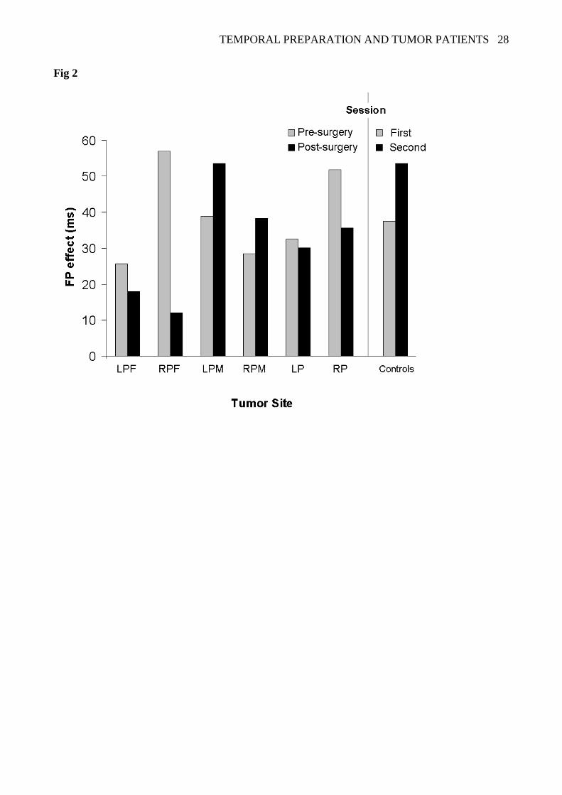
More critically, the patient group x testing session x FPn interaction was also significant
[F(6, 63) = 2.5, p < .05]. Visual inspection of Figure 2 suggests that this interaction was due to a
reduction of the FP effect selectively after removal of tumoral tissue in right lateral PFC. In order to
corroborate this observation statistically, separate ANOVAs were conducted for each group with
testing session, FPn and FPn-1, as repeated measures. As predicted (cf. Stuss et al., 2005), the testing
session x FPn interaction was significant for the right prefrontal patients [F(1, 13) = 8.2, p = .01],
due to a reduction of the FP effect after surgery (12 ms) with respect to the pre-surgery effect (57
ms). We further checked if there was a correlation between this effect and lesion size. Neither the
pre- nor the post-surgery FP effect in right prefrontal patients correlated with lesion size1. It should
be noted that the testing session x FPn interaction was not significant for all the other five tumor
patient groups (p = .73, .36, .44, .88, .12, for the left prefrontal, left and right premotor, left and
right parietal groups, respectively).
Insert Footnote 1 about here
Insert Figure 2 about here
Page 13
TEMPORAL PREPARATION AND TUMOR PATIENTS 13
These separate ANOVAs had also been carried out to find the source of the testing session x
FPn-1 and, more relevant, of the testing session x FPn x FPn-1 interactions in the overall ANOVA.
Although these interactions are not significantly modulated by the patient group in the overall
ANOVA, visual inspection of Figure 3 suggests that the premotor and prefrontal groups are
principally responsible for these effects. This was only partially confirmed as the testing session x
FPn-1 interaction was a tendency for the right prefrontal group (p = .06) and for the left premotor
group (p = .08).
Insert Figure 3 about here
To find the source of the testing session x FPn x FPn-1 interaction, which is critical for
determining the locus of the asymmetric sequential effects, we chose a Bonferroni correction of a
critical significance level of .0083 (i.e., .05 divided by the 6 tumor patient groups). The motivation
for the use of a Bonferroni correction was twofold: first, the 3-way interaction was only a trend in
the overall ANOVA; second, we did not have a precise a priori prediction as far as the locus of the
asymmetric sequential effects was concerned. The only individual patient group which showed a
significant testing session x FPn x FPn-1 interaction, when analyzed separately from the other groups,
was the left premotor one [session x FPn x FPn-1 3-way interaction: F(1, 7) = 22.1, p = .002]: the
asymmetric sequential effects, which were present before surgery mainly on the short FPn, had
disappeared after it. This was observed in this patient group despite the standard FP effect being
present with the same magnitude before and after the operation, as shown by a significant main
effect of FPn [F(1, 7) = 55.4, p < .001], which was not modulated by the testing session (session x
FPn interaction, p = .36). The main effect of testing session was also reliable in this group [F(1, 7) =
31.7, p < .001], due to RTs being slower after the operation than before, which could conceivably
arise from a motor effect, given that these patients were all right-handed.
Page 14
TEMPORAL PREPARATION AND TUMOR PATIENTS 14
It should be noted that this 3-way testing session x FPn x FPn-1 interaction was far from
significant in all the other five tumor patient groups, the p values being .27, .94, .72, .38, .79, for the
left and right prefrontal, right premotor, left and right parietal groups, respectively. However, when
the post-surgery performance of each tumor patient group was contrasted to that of the same second
session in the control group in a 2x2x2 mixed ANOVA (between-subjects factor: patient group;
within-subject factors: FPn, FPn-1), the 2-way group x FPn-1 interaction (concerning basic sequential
effects) was significant not only for the left premotor group (p = .007), but also for the left and right
prefrontal groups (p = .04, .03, respectively; not significant if Bonferroni corrected), and there was
a strong trend for the right premotor group (p = .055). However, the interaction was not significant
for the left and right parietal groups (p = .12 and .40, respectively). This interaction shows that, the
basic post-surgery sequential effects, when evaluated separately from the asymmetry of the effects
(as revealed by the FPn x FPn-1 interaction), were in fact smaller in all the frontal groups as
compared to the controls. Critically, the left premotor patients were the only group differing in the
asymmetric aspect of the sequential effects as compared to the controls, as indicated by the
significant 3-way interaction [group x FPn x FPn-1 interaction, F(1, 18) = 8.9, p = .008]2.
Insert Footnote 2 about here
Discussion
In this study, we aimed to investigate the variable FP phenomena in tumor patients, when tested
before and after surgical removal of tumors which were located in different cortical areas. The most
important finding was a reduction in the FP effect after surgical removal of tumors of the right
prefrontal cortex. This finding corroborates recent studies on FP phenomena obtained in chronic
patients with predominantly other etiologies such as stroke (Stuss et al., 2005; see also Picton et al.,
2006), and in healthy participants undergoing inhibitory TMS over right DLPFC (Vallesi et al.,
2007).
Page 15
TEMPORAL PREPARATION AND TUMOR PATIENTS 15
Although obtained in such a simple experimental task, the FP effect is generally considered as a
marker of high-level monitoring processes (e.g., Näätänen, 1970; Niemi & Näätänen, 1981; Stuss et
al., 2005; but see Los & van den Heuvel, 2001). On this view, this result supports the hypothesis
that right lateral prefrontal cortex is the seat of the critical process producing the FP effect, that is
monitoring of the increasing conditional probability of stimulus occurrence along the FP (e.g.,
Näätänen, 1970). The neuropsychological work by Stuss and colleagues (Stuss et al., 2005) helps
clarifying that monitoring of the conditional probability of the stimulus occurrence is the process
impaired in right prefrontal patients, and not keeping track of elapsing time per se. When the
conditional probability of stimulus occurrence was kept constant by using an interval fixed within a
block instead of a variable FP paradigm, the performance of the right prefrontal group was
comparable to that of the controls. In contrast, with this fixed FP paradigm, the superior medial
frontal group was the only group who was impaired. Monitoring of conditional probability was not
relevant with a fixed FP paradigm, where time intervals are constant within a block. Moreover, as
simple and choice RT tasks were used, monitoring of elapsing time was also not required. On the
other hand, when the task demands require monitoring of temporal information, either implicitly (as
in the current study) or explicitly, as it is the case for time estimation and reproduction tasks,
evidence for an involvement of right lateral prefrontal cortex (usually dorsolateral) has been found
in neuropsychological (e.g., Harrington, Haaland, & Knight, 1998; Koch, Oliveri, Carlesimo, &
Caltagirone, 2002), TMS (Jones, Rosenkranz, Rothwell, & Jahanshahi, 2004; Koch, Oliveri,
Torriero, & Caltagirone, 2003), and imaging studies (Lewis & Miall, 2003; Rao, Mayer, &
Harrington, 2001), also when working memory demands were controlled (Smith, Taylor, Lidzba, &
Rubia, 2003), although these studies generally involved different ranges of time intervals from that
used in the current one.
Unlike the previous neuropsychological work (Stuss et al., 2005), the current study additionally
investigated the effect of the preceding FP, which is known to give rise to sequential effects: RTs
are slower for long FPn-1 than for short ones; these effects are typically asymmetric in that they
Page 16
TEMPORAL PREPARATION AND TUMOR PATIENTS 16
occur specifically only when the current FP is a short one. In the overall ANOVA, there was a
reduced effect of the FP occurring on the preceding trial (i.e., basic sequential effects), when
performance in the second session was compared to that in the first session. This effect is difficult
to interpret from the localisational point of view, as we did not find clear statistical evidence for the
specificity of the tumor site in the overall ANOVA or in more specific analyses. These analyses,
indeed, showed that the basic post-surgery sequential effects were reduced (significantly or as a
tendency) in the four frontal groups, even if not in the two parietal groups.
More critically, examining the behavior of the left premotor group provides additional
information about the localisation of the asymmetric sequential effects and their underlying
cognitive mechanisms. Despite the presence of an unchanged FP effect, the asymmetric sequential
effects disappeared after operation; this was supported statistically when performance was
compared within-group with the pre-surgery performance (i.e., a significant session x FPn x FPn-1
interaction) and between-groups with the second session of the controls (i.e., significant group x
FPn x FPn-1 interaction). In particular, there was no RT reduction after a short FPn-1 in the post-
surgery session of the left premotor patients. This result may be interpreted as suggesting a pre-
motoric/motoric locus of a facilitatory effect when a short FP had occurred in the previous trial.
Left premotor areas are indeed directly involved in the preparation of the manual key-press, which
is the response required in the task. Supporting this hypothesis, an electrophysiological study on
monkey premotor and motor cortex (Riehle & Requin, 1993) revealed that activity of neurons
within this region correlate with performance speed in tasks with a preparation period. During the
delay period of a delayed-reach task, moreover, micro-stimulation of neurons within premotor
cortex lead to a highly-specific lengthening in reach RT (Churchland & Shenoy, 2006).
Nevertheless, one cannot draw firm conclusions about the localisational of sequential effects
because of the lack of interaction with the other patient groups in the overall ANOVA. Indeed, there
are suggestions from the findings that reduced basic sequential effects may be present in all the
Page 17
TEMPORAL PREPARATION AND TUMOR PATIENTS 17
frontal groups. Therefore, these findings concerning the left premotor localisation of sequential
effects should be seen as a suggestion for further studies.
However, functional conclusions can be drawn even in the absence of strong anatomical
localisation. Indeed, this finding represents the second component of a double dissociation between
FP and sequential effects. On the one hand, Vallesi and colleagues (Vallesi et al., 2007) found a
reduction in the FP effect as a result of inhibitory TMS on the right DLPFC in the absence of a
modulation in the sequential effects. On the other hand, here it has been shown that sequential
effects disappear after surgery in left premotor patients despite an intact FP effect. This pattern
supports dual-process accounts of the FP phenomena (e.g., Vallesi et al., 2007; Vallesi & Shallice,
in press; see also Los & Agter, 2005), and is much difficult to account for in a single-process
account (Los & van den Heuvel, 2001).
The functional meaning of the sequential effects, therefore, needs to be revised. According to
the dual-process account put forward by Vallesi and colleagues (Vallesi et al., 2007; Vallesi &
Shallice, in press), the sequential effects may be due to a tonic arousal modulation by the FPn-1. As
maintaining a high level of preparation for a long FP is effortful, a long FPn-1 decreases arousal
(refractoriness) and lengthens RTs on trial n, whereas a short FPn-1 increases arousal (facilitation)
and produces relatively faster RTs on trial n (see Los & Heslenfeld, 2005, for electrophysiological
evidence). This arousal modulation is especially detectable on the shortest current FP (i.e.,
asymmetric sequential effects), when the compensatory effect of the monitoring the conditional
probability of stimulus occurrence cannot take place. After tumor removal in frontal patients here,
and especially left premotor patients, the second process (facilitation) seems to be impaired, so that
RTs on a short FPn do not benefit from a short FPn-1, conceivably because the brain area where this
arousal modulation should produce its effects (i.e., left premotor cortex) is not working properly
due to the surgical lesion.
The effects of tumor per se on cerebral functionality are still almost unknown. However, there
are a few studies investigating cognitive functioning of brain tumor patients before any treatment
Page 18
TEMPORAL PREPARATION AND TUMOR PATIENTS 18
and surgical intervention, which found cognitive deficits caused by the presence of tumor (e.g.,
Rabbit & Page, 1998; Tucha, Smely, Preier, & Lange, 2000). Therefore, a baseline evaluation of
cognitive abilities before surgery is methodologically desirable in any study of tumor patients
undergoing surgery. To that purpose, the use of a matched control group of orthopedic patients
allowed us to exclude, at least before surgery, any particular deficit of our sample of tumor patients
in performing the variable FP paradigm.
A critical aspect of the present results is that the effects found are selective and are generally
robust across etiologies. Indeed, resection of a right prefrontal tumour gives the same reduction in
the FP effect as in a cohort of patients primarily suffering from stroke in the same region (Stuss et
al., 2005). From a methodological point of view, this study supports the one by Shallice and
colleagues on optic ataxia (Shallice, Mussoni, D’Agostini, & Skrap, submitted), demonstrating that
the effects of operation for resection of tumors can be a valuable method for localizing cognitive
processes.
In conclusion, the present findings confirm the studies on the anatomical basis of the FP effect
(Stuss et al., 2005; Vallesi et al., 2007), suggesting that this effect can be used as a measure of the
functionality of right lateral prefrontal cortex, and additionally provide surprising new
neuropsychological insights on the sequential effects. The latter are best explained by a dual-
process account of the FP phenomena. Finally the findings strongly support the utility of using
acute brain tumor patients as a source of evidence about the localisation and fractionation of
cognitive functions.
Page 19
TEMPORAL PREPARATION AND TUMOR PATIENTS 19
References
Basso, G., Nichelli, P., Wharton, C.M., Peterson, M., & Grafman, J. (2003). Distributed neural
systems for temporal production: a functional MRI study. Brain Res.Bull., 59, 405-411.
Brunia, C.H.M. & Van Boxtel, G.J.M. (2000). Motor preparation. In Cacioppo J.T., L.G. Tassinary,
& G.G. Berntson (Eds.), Handbook of psychophysiology (2nd ed. ed., pp. 507-532). Cambridge,
UK: Cambridge University Press.
Churchland, M.M. & Shenoy, K.V. (2006). Delay of movement caused by disruption of cortical
preparatory activity. J Neurophysiol..
Collins, D.L., Zijdenbos, A.P., Kollokian, V., Sled, J.G., Kabani, N.J., Holmes, C.J. et al. (1998).
Design and construction of a realistic digital brain phantom. IEEE Trans.Med Imaging, 17, 463-
468.
Correa, A., Lupianez, J., & Tudela, P. (2006). The attentional mechanism of temporal orienting:
determinants and attributes. Exp Brain Res., 169, 58-68.
Coull, J.T., Frith, C.D., Buchel, C., & Nobre, A.C. (2000). Orienting attention in time: behavioural
and neuroanatomical distinction between exogenous and endogenous shifts. Neuropsychologia,
38, 808-819.
Coull, J.T. & Nobre, A.C. (1998). Where and when to pay attention: the neural systems for
directing attention to spatial locations and to time intervals as revealed by both PET and fMRI. J
Neurosci., 18, 7426-7435.
Drazin, D.H. (1961). Effects of foreperiod, foreperiod variability, and probability of stimulus
occurrence on simple reaction time. J Exp Psychol., 62, 43-50.
Elithorn, A. & Lawrence, C. (1955). Central inhibition: Some refractory observations. Quarterly
Journal of Experimental Psychology, 11, 211-220.
Fletcher, P.C. & Henson, R.N. (2001). Frontal lobes and human memory: insights from functional
neuroimaging. Brain, 124, 849-881.
Page 20
TEMPORAL PREPARATION AND TUMOR PATIENTS 20
Harrington, D. L., Haaland, K. Y., & Knight, R. T. (1998). Cortical networks underlying
mechanisms of time perception. J Neurosci., 18, 1085-1095.
Henson, R.N., Shallice, T., & Dolan, R.J. (1999). Right prefrontal cortex and episodic memory
retrieval: a functional MRI test of the monitoring hypothesis. Brain, 122 ( Pt 7), 1367-1381.
Hommel, B., Proctor, R.W., & Vu, K.P. (2003). A feature-integration account of sequential effects
in the Simon task. Psychol.Res..
Ivry, R.B. & Spencer, R.M. (2004). The neural representation of time. Curr.Opin.Neurobiol., 14,
225-232.
Janssen, P. & Shadlen, M.N. (2005). A representation of the hazard rate of elapsed time in macaque
area LIP. Nat.Neurosci., 8, 234-241.
Jones, C. R., Rosenkranz, K., Rothwell, J. C., & Jahanshahi, M. (2004). The right dorsolateral
prefrontal cortex is essential in time reproduction: an investigation with repetitive transcranial
magnetic stimulation. Exp.Brain Res., 158, 366-372.
Karlin L. (1959). Reaction time as a function of foreperiod duration and variability. J Exp Psychol.,
58, 185-191.
Koch, G., Oliveri, M., Carlesimo, G. A., & Caltagirone, C. (2002). Underestimation of time
perception after repetitive transcranial magnetic stimulation. Neurology, 59, 1658-1659.
Koch, G., Oliveri, M., Torriero, S., & Caltagirone, C. (2003). Underestimation of time perception
after repetitive transcranial magnetic stimulation. Neurology 60, 1844-1846.
Lewis, P.A. & Miall, R.C. (2003). Distinct systems for automatic and cognitively controlled time
measurement: evidence from neuroimaging. Curr.Opin.Neurobiol., 13, 250-255.
Los, S.A. & Agter, F. (2005). Re-weighting sequential effects: estimating intentional and
unintentional contributions to nonspecific preparation across different distributions of
foreperiod. Perception & Psychophysics, in press.
Los, S.A. & Heslenfeld, D.J. (2005). Intentional and unintentional contributions to nonspecific
preparation: electrophysiological evidence. J Exp Psychol.Gen., 134, 52-72.
Page 21
TEMPORAL PREPARATION AND TUMOR PATIENTS 21
Los, S.A. & van den Heuvel, C.E. (2001). Intentional and unintentional contributions to nonspecific
preparation during reaction time foreperiods. J Exp Psychol.Hum.Percept.Perform., 27, 370-
386.
Macar, F., Lejeune, H., Bonnet, M., Ferrara, A., Pouthas, V., Vidal, F. et al. (2002). Activation of
the supplementary motor area and of attentional networks during temporal processing.
Exp.Brain Res., 142, 475-485.
Näätänen, R. (1970). The diminishing time-uncertainty with the lapse of time after the warning
signal in reaction-time experiments with varying foreperiods. Acta Psychologica, 34, 399-419.
Niemi, P. & Näätänen, R. (1981). Foreperiod and simple reaction time. Psychol. Bull., 89, 133-162.
Picton, T. W., Stuss, D. T., Shallice, T., Alexander, M. P., & Gillingham, S. (2006). Keeping time:
effects of focal frontal lesions. Neuropsychologia, 44, 1195-1209.
Posner, M.I. & Peterson, S.E. (1990). The attention system of the human brain. Annu.Rev.Neurosci.,
13, 25-42.
Rabbit, J.E., Page, M.S. (1998). Selected complications in neuro-oncology patients. Semin Oncol
Nurs, 14, 53–60.
Rao, S. M., Mayer, A. R., & Harrington, D. L. (2001). The evolution of brain activation during
temporal processing. Nat.Neurosci., 4, 317-323.
Riehle, A. & Requin, J. (1993). The predictive value for performance speed of preparatory changes
in neuronal activity of the monkey motor and premotor cortex. Behav.Brain Res., 53, 35-49.
Rorden, C. & Brett, M. (2000). Stereotaxic display of brain lesions. Behav.Neurol, 12, 191-200.
Rueckert, L. & Grafman, J. (1996). Sustained attention deficits in patients with right frontal lesions.
Neuropsychologia, 34, 953-963.
Shallice, T. (1988). From Neuropsychology to Mental Structure. Cambridge, UK: Cambridge
University Press.
Shallice, T. (2002). Fractionation of the Supervisory System. In D.T.Stuss & R. T. Knight (Eds.),
Principles of Frontal Lobe Function (pp. 261-277). Oxford: University Press.
Page 22
TEMPORAL PREPARATION AND TUMOR PATIENTS 22
Shallice, T. (2004). The fractionation of supervisory control. In M.S.Gazzaniga (Ed.), The Cognitive
Neurosciences (III ed., Cambridge: Mass: MIT Press.
Smith, A., Taylor, E., Lidzba, K., & Rubia, K. (2003). A right hemispheric frontocerebellar network
for time discrimination of several hundreds of milliseconds. Neuroimage, 20, 344-350.
Soetens, E., Boer, L.C., & Hueting, J.E. (1985). Expectancy or automatic facilitation? Separating
sequential effects in two-choice reaction time. J Exp Psychol.Hum.Percept.Perform., 11, 598-
616.
Stuss, D.T. (2006). Frontal lobes and attention: processes and networks, fractionation and
integration. J Int.Neuropsychol.Soc., 12, 261-271.
Stuss, D.T., Alexander, M. P., Shallice, T., Picton, T. W., Binns, M. A., Macdonald, R. et al.
(2005). Multiple frontal systems controlling response speed. Neuropsychologia, 43, 396-417.
Stuss, D.T., Shallice, T., Alexander, M. P., & Picton, T. W. (1995). A multidisciplinary approach to
anterior attentional functions. Ann.N.Y.Acad.Sci., 769, 191-211.
Tucha, O., Smely, C., Preier, M., & Lange, K.W. (2000). Cognitive deficits before treatment among
patients with brain tumors. Neurosurgery, 47, 324-333.
Tzourio-Mazoyer, N., Landeau, B., Papathanassiou, D., Crivello, F., Etard, O., Delcroix, N. et al.
(2002). Automated anatomical labeling of activations in SPM using a macroscopic anatomical
parcellation of the MNI MRI single-subject brain. Neuroimage, 15, 273-289.
Vallesi A. & Shallice T. (in press). “Developmental dissociations of preparation: deconstructing
variable foreperiod phenomena”. Journal of Experimental Psychology: Human Perception and
Performance.
Vallesi, A., Shallice, T., & Walsh, V. (2007). Role of the prefrontal cortex in the foreperiod effect:
TMS evidence for dual mechanisms in temporal preparation. Cerebral Cortex, 17, 466-474.
Wilkins, A.J., Shallice, T., & McCarthy, R. (1987). Frontal lesions and sustained attention.
Neuropsychologia, 25, 359-365.
Woodrow, H. (1914). The measurement of attention. Psychological Monographs, 5, 1-158.
Page 23
TEMPORAL PREPARATION AND TUMOR PATIENTS 23
Footnotes
1When lesion size and FP effects in the post-surgery session are compared across patient groups, a
significant negative correlation is observed (r = -.94), indicating that the FP effect decreases as the
lesion volume increases at the group level. However, when the Pearson correlation analysis is
carried out within each tumor group, that is between post-surgery FP effect of each patient within
each group and her/his lesion size, no significant correlation is observed for any group. The r (and
p) values were: .11 (p =.7), -.42, (p = .4) etc. , .17 (p = .72), .01 (p =.98), -.33 (p = .25), -.37 (p =
.33), for the right and left prefrontal, left and right premotor, left and right parietal groups,
respectively. These results suggest that lesion size alone cannot account for the reduction of the FP
effect.
2Each tumor patient group was also contrasted with each other in a 2x2x2 mixed ANOVA for the
post-surgery session, with region as the between-subjects factor, and FPn and FPn-1 as the within-
subject factors. The 3-way interaction was significant when the left premotor group was contrasted
with the right parietal one [F(1, 20) = 6.56, p = .018], and there was a similar tendency when the left
premotor group was contrasted with the left parietal one (p = .07). This interaction was not
significant for any other pair of groups.
Page 24
TEMPORAL PREPARATION AND TUMOR PATIENTS 24
Acknowledgements
This research was partially supported by a grant from PRIN to TS and Raffaella Rumiati. AM
was supported by a grant from Regione Friuli Venezia Giulia to SISSA, 2005/2006
“Neuropsicologia clinica delle funzioni esecutive e prassiche”. The authors are also thankful to the
members of the Neurosurgical Department, Ospedale S.M. Misericordia, Udine, for their
helpfulness throughout the study.
Page 25
TEMPORAL PREPARATION AND TUMOR PATIENTS 25
Table 1
Main Demographical Characteristics of the seven Patient Groups included in the study.
Mean Agea Mean Educationa
Gender Anaesthesia Tumor Volumeb
Tumor Type Sample size
Group (min-max) (SD) F M G L (SD) HG LG Mng Mt
Left Prefrontal 45 (33-62) 11 (4) 3 3 5 1 3.3 (1.5) 0 2 4 0 6
Right Prefrontal 45 (23-72) 12 (4) 5 9 12 2 3.6 (2.2) 4 6 4 0 14
Left Premotor 45 (31-60) 11 (3) 5 3 2 6 1.1 (0.8) 4 3 1 0 8
Right Premotor 39 (18-58) 12 (3) 2 5 2 5 1.3 (1) 2 4 1 0 7
Left Parietal 53 (31-70) 9 (3) 3 6 6 3 3 (2.4) 5 2 1 1 9
Right Parietal 54 (30-70) 10 (4) 6 8 13 1 3 (2.7) 5 3 4 2 14
Controls 47 (23-73) 11 (4) 6 6 --- --- --- --- --- --- --- 12
Notes. aIn years. bIn percentage of the total volume. SD = standard deviation; F = female; M = male; G = general; L = local. HG = high grade
glioma; LG = low grade glioma; Mng = meningioma; Mt = metastasis.
Page 26
TEMPORAL PREPARATION AND TUMOR PATIENTS 26
Figure Captions
Figure 1. Display of the tumor overlap for the 6 groups of tumor patients. The percentage of
overlapping tumors in each voxel is illustrated using a grey-scale within the region of interest: the
lighter is a point on that scale, the higher the percentage of patients within that group with that
voxel damaged. The white colour indicates voxels with maximal percentage of tumors within each
patient group. Maximal percentage of overlap was 67, 43, 63, 43, 56, 36, for the left and right
prefrontal, left and right premotor and left and right parietal groups, respectively. The z-coordinates
of each transversal section in Montreal Neurological Institute space are -8, 0, 8, 16, 24, 32, 40, 50,
60, 70. LPF = left prefrontal; RPF = right prefrontal; LPM = left premotor; RPM = right premotor;
LP = left parietal; RP = right parietal. See supplementary Figure 1, for a color version of the Figure.
Figure 2. The foreperiod effect (reaction time difference between foreperiods of 3-4 and 6-7
seconds) as a function of patient group and testing session. FP = foreperiod. Tumor group labels as
for figure 1.
Figure 3. The sequential effects as a function of patient group and testing session. Short = 3-4
seconds. Long = 6-7 seconds. FP = foreperiod. Error bars indicate the standard error of the mean.
Page 27
TEMPORAL PREPARATION AND TUMOR PATIENTS 27
Fig 1
Page 28
TEMPORAL PREPARATION AND TUMOR PATIENTS 28
Fig 2
Page 29
TEMPORAL PREPARATION AND TUMOR PATIENTS 29
Fig 3