+ All Categories
Home > Documents > The Novel Desmin Mutant p.A120D Impairs …...inn cce ll lines and of ff thee isolatted recommbbi...

The Novel Desmin Mutant p.A120D Impairs …...inn cce ll lines and of ff thee isolatted recommbbi...

Date post: 24-Feb-2020
Category:
Upload: others
View: 2 times
Download: 0 times
Share this document with a friend
37
DOI: 10.1161/CIRCGENETICS.113.000103 1 The Novel Desmin Mutant p.A120D Impairs Filament Formation, Prevents Intercalated Disk Localization and Causes Sudden Cardiac Death Running title: Brodehl et al.; Characterization of a novel desmin mutation Andreas Brodehl, PhD 1,9 *; Mareike Dieding, MSc 2 *; Bärbel Klauke, PhD 1 ; Eric Dec, MD 3 ; Shrestha Madaan, MD 3 ; Taosheng Huang, MD, PhD 4 ; John Gargus, MD, PhD 3 ; Azra Fatima, PhD 5 ; Tomo Šaric, MD, PhD 5 ; Hamdin Cakar, PhD 6 ; Volker Walhorn, PhD 2 ; Katja Tönsing, PhD 2 ; Tim Skrzipczyk, MSc 1 ; Ramona Cebulla, TN 1 ; Désirée Gerdes, TN 1 ; Uwe Schulz, MD 1 ; Jan Gummert, MD 1 ; Jesper Hastrup Svendsen, MD, DMSc 7,8 ; Morten Salling Olesen, PhD 7,8 ; Dario Anselmetti, PhD 2 ; Alex Hørby Christensen, MD, PhD 7,8 ; Virginia Kimonis, MD 3 ; Hendrik Milting, PhD 1 1 Erich & Hanna Klessmann Inst for Cardiovascular Research & Development (EHKI), Heart & Diabetes Center NRW, Ruhr Univ Bochum, Bad Oeynhausen; 2 Experimental Biophysics & Applied Nanoscience, Faculty of Physics & Bielefeld Inst for Biophysics & Nanoscience (BINAS), Bielefeld Univ, Bielefeld, Germany; 3 Division of Genetics & Metabolism, Dept of Pediatrics, Univ of California, Irvine, CA; 4 Division of Human Genetics, Dept of Pediatrics, Cincinatti Children’s Hospital, Cincinnati, OH; 5 Inst for Neurophysiology, Medical Center, Univ of Cologne, Cologne; 6 Physikalisch-Technische Bundesanstalt (PTB), Braunschweig, Germany; 7 Dept of Cardiology, Rigshospitalet, Copenhagen Univ Hospital, 8 The Danish National Research Foundation Centre for Cardiac Arrhythmia, Copenhagen, Denmark; 9 Present address: Libin Cardiovascular Inst of Alberta, Dept of Cardiac Sciences, Univ of Calgary, Calgary, AB, Canada *contributed equally Correspondence: Andreas Brodehl, PhD Hendrik Milting, PhD Department of Cardiac Sciences E. & H. Klessmann Inst for Cardiovascular Libin Cardiovascular Institute of Alberta Research & Development (EHKI) University of Calgary Heart and Diabetes Center NRW 3280 Hospital Drive NW Ruhr-University T2N4Z6 Calgary, AB Bochum, D-32545 Bad Oeynhausen Canada Germany Tel: +1-403-210-7322 Tel: +49-5731-973510 Fax: none Fax: +49-5731-972476 E-mail: [email protected] E-mail: [email protected] Journal Subject Codes: [16] Myocardial cardiomyopathy disease, [89] Genetics of cardiovascular disease, [137] Cell biology/structural biology, [178] Aggregation d d dse se se sen, n, n n, M M M MD, D, D, D D D D DMS MS MS MSc c c c 7, 7, 7, 7,8 8 8 8 ; ; ; Chris s s ste tens nsen en, , MD MD MD MD, , Ph i i i i i 3 d ik il i h 1 H C U n G m d e g pitalet Copenhagen Univ Hospital The Danish National Research Foundation Centre for Cardi Vi V V V rginia Kimonis, MD 3 ; He He Hend n rik Milting, PhD hD hD D 1 Han n nna a a a Klessmann n In n nst f for r r Ca a a ard rd rd r iovasc cul l lar r Re e ese earch h & De De De D velo lopm pm men nt (EH HKI) I) I), He H art t t & & & & Di Di Diabet t tes C Univ iv v v B B B Boc oc oc ochu hu hu h m, m m m B B Bad ad ad d O O Oey ey eynh nh nh n ause se se s n ; ; ; 2 2 2 2 Ex E E E pe e e eri ri ri r me me me ment nt ntal al a B B B Bio o ioph ph ph phys ys ysic ic ic ics s s s & & & & Ap Ap Appl pl pl plie ie ie ied d d Na Na Na Nano o o osc sc sc scie ie ienc nc nc nce, F F F Fac ac ac acul ul l ulty ty ty t o o of nst for Biophysics s s & & & & Nanosc c cie ie e nce (BINAS), Bie ele le lefeld Uni i i iv, v, v Bielefeld, , G G G Ger e many; 3 Division of G m, Dept of Pediatr r r ric ic ic ics, s, s, U U U Uni ni niv v v of o of o C C Cal al al alif if if i or or or rni ni ni nia, a, a, I I I Irv rv rv rvin in ine, e e C C CA A A A ; 4 4 4 4 Di D vi vi vi visi i si on on on on o of f f Hu Hu Hu Huma ma man n n Ge Ge Ge Gene n ne netics, Dept of Ped Children’s Hosp ital, Cincinnati, O H; 5 5 5 5 Inst for Neurop hy siolog y, Medical Center, Univ of Colo e e e ; 6 Physikal lis is s isch ch ch ch - - - Te Te e Tech ch c chni ni ni nisc s he he he e B B B Bun un n unde de de desa s s ns ns ns nsta a talt lt t lt ( ( ( (PT PT T PTB) B) B) B), , Br Br Br Brau u uns ns s nsch c ch chwe we we weig ig ig, Ge Ge Ge Germ rm m rman an an any; y; y; 7 De De De Dep p p p t t t t of of of Cardiolog g p p pit ital alet et Co Cope penh nhag agen en U Uni niv v Ho Hosp spit ital al 8 8 Th The e Da Dani nish sh N Nat atio iona nal l Re Rese sear arch ch F Fou ound ndat atio ion n Ce Cent ntre re f for or C Car ard di by guest on November 9, 2013 http://circgenetics.ahajournals.org/ Downloaded from
Transcript

DOI: 10.1161/CIRCGENETICS.113.000103

1

The Novel Desmin Mutant p.A120D Impairs Filament Formation, Prevents Intercalated Disk Localization and Causes Sudden Cardiac Death

Running title: Brodehl et al.; Characterization of a novel desmin mutation

Andreas Brodehl, PhD1,9*; Mareike Dieding, MSc2*; Bärbel Klauke, PhD1; Eric Dec, MD3;

Shrestha Madaan, MD3; Taosheng Huang, MD, PhD4; John Gargus, MD, PhD3;

Azra Fatima, PhD5; Tomo Šaric, MD, PhD5; Hamdin Cakar, PhD6; Volker Walhorn, PhD2;

Katja Tönsing, PhD2; Tim Skrzipczyk, MSc1; Ramona Cebulla, TN1; Désirée Gerdes, TN1;

Uwe Schulz, MD1; Jan Gummert, MD1; Jesper Hastrup Svendsen, MD, DMSc7,8;

Morten Salling Olesen, PhD7,8; Dario Anselmetti, PhD2; Alex Hørby Christensen, MD, PhD7,8;

Virginia Kimonis, MD3; Hendrik Milting, PhD1

1Erich & Hanna Klessmann Inst for Cardiovascular Research & Development (EHKI), Heart & Diabetes Center NRW, Ruhr Univ Bochum, Bad Oeynhausen; 2Experimental Biophysics & Applied Nanoscience, Faculty of Physics & Bielefeld Inst for Biophysics & Nanoscience (BINAS), Bielefeld Univ, Bielefeld, Germany; 3Division of Genetics & Metabolism, Dept of Pediatrics, Univ of California, Irvine, CA; 4Division of Human Genetics, Dept of Pediatrics,

Cincinatti Children’s Hospital, Cincinnati, OH; 5Inst for Neurophysiology, Medical Center, Univ of Cologne, Cologne; 6Physikalisch-Technische Bundesanstalt (PTB), Braunschweig, Germany; 7Dept of Cardiology,

Rigshospitalet, Copenhagen Univ Hospital, 8The Danish National Research Foundation Centre for Cardiac Arrhythmia, Copenhagen, Denmark; 9Present address: Libin Cardiovascular Inst of Alberta, Dept of Cardiac

Sciences, Univ of Calgary, Calgary, AB, Canada*contributed equally

Correspondence: Andreas Brodehl, PhD Hendrik Milting, PhD Department of Cardiac Sciences E. & H. Klessmann Inst for Cardiovascular Libin Cardiovascular Institute of Alberta Research & Development (EHKI)University of Calgary Heart and Diabetes Center NRW3280 Hospital Drive NW Ruhr-University T2N4Z6 Calgary, AB Bochum, D-32545 Bad OeynhausenCanada GermanyTel: +1-403-210-7322 Tel: +49-5731-973510 Fax: none Fax: +49-5731-972476 E-mail: [email protected] E-mail: [email protected]

Journal Subject Codes: [16] Myocardial cardiomyopathy disease, [89] Genetics of cardiovascular disease, [137] Cell biology/structural biology, [178] Aggregation

dddsesesesen,n,nn, MMMMD,D,D,D DDDDMSMSMSMScccc7,7,7,7,8888;;;

Chrisssstetensnsenen, ,,, MDMDMDMD, , ,, Ph

i i i i i 3 d ik il i h 1

H CUn Gm d

e gpitalet Copenhagen Univ Hospital The Danish National Research Foundation Centre for Cardi

ViVVV rginia Kimonis, MD3; HeHeHendn rik Milting, PhDhDhDD1

Hannnnaaa a Klessmann n Innnst fforrr Caaaardrdrdr iovascculllarr Reeeseearchh & DeDeDeD velolopmpmmennt (EHHKI)I)I), HeH arttt &&&& DiDiDiabetttes CUnivivvv BBBBocococochuhuhuh m,mmm BBBadadadd OOOeyeyeynhnhnhn auseseses n;;;; 2222ExEEE peeeeriririr mememementntntalala BBBBiooiophphphphysysysicicicicssss & & & & ApApApplplplplieieieiedd d NaNaNaNanooooscscscscieieiencncncnce, FFFFacacacaculullultytytyt ooof nst for Biophysicsss & &&& Nanoscccieieence (BINAS), Bieelelelefeld Uniiiiv,v,v Bielefeld,, GGGGere many; 3Division of Gm, Dept of Pediatrrrricicicics,s,s, UUUUnininiv v v ofoofo CCCalalalalifififi orororrninininia,a,a, IIIIrvrvrvrvininine,ee CCCAAAA; 4444DiD vivivivisiisionononon oof ff HuHuHuHumamaman n n GeGeGeGenennenetics, Dept of Ped Children’s Hospppital,,, Cincinnati, ,, OH;; 5555Inst for Neuroppphyysiologggy,y,y, Medical Center,,, Univ of Coloeee; 6Physikallisissischchchch---TeTeeTechchcchninininiscs hehehee BBBBununnundedededesass nsnsnsnstatataltlttlt ((((PTPTTPTB)B)B)B), , BrBrBrBrauuunsnssnschcchchweweweweigigig, GeGeGeGermrmmrmanananany;y;y; 7DeDeDeDeppppt t tt ofofof Cardiologgpppititalaletet CoCopepenhnhagagenen UUninivv HoHospspititalal 88ThThee DaDaninishsh NNatatioionanall ReReseseararchch FFououndndatatioionn CeCentntrere fforor CCararddi

by guest on November 9, 2013http://circgenetics.ahajournals.org/Downloaded from

anselm
Textfeld
Circulation: Cardiovascular Genetics 6, 615-623, 2013

DOI: 10.1161/CIRCGENETICS.113.000103

2

Abstract:

Background - The intermediate filament protein desmin is encoded by the gene DES and

contributes to the mechanical stabilization of the striated muscle sarcomere and cell contacts

within the cardiac intercalated disk. DES mutations cause severe skeletal and cardiac muscle

diseases with heterogeneous phenotypes. Recently, DES mutations were also found in patients

with arrhythmogenic right ventricular cardiomyopathy (ARVC). Currently, the cellular and

molecular pathomechanisms of the DES mutations leading to this disease are not exactly known.

Methods and Results - We identified the two novel variants DES-p.A120D (c.359C>A)

and -p.H326R (c.977A>G), which were characterized by cell culture experiments and atomic

force microscopy. Family analysis indicated a broad spectrum of cardiomyopathies with a

striking frequency of arrhythmias and sudden cardiac deaths. The in vitro experiments of

desmin-p.A120D evidenced a severe intrinsic filament formation defect causing cytoplasmic

aggregates in cell lines and of the isolated recombinant protein, respectively. Model variants of

codon 120 indicated that ionic interactions contribute to this filament formation defect. Ex vivo

analysis of ventricular tissue slices revealed a loss of desmin staining within the intercalated disk

and severe cytoplasmic aggregate formation whereas z-band localization was not affected. The

functional experiments of desmin-p.H326R did not demonstrate any differences from wild type.

Conclusions - Due to the functional in vivo and in vitro characterization DES-p.A120D has to be

regarded as a pathogenic mutation, whereas DES-p.H326R is a rare variant with unknown

significance, respectively. Presumably, the loss of the desmin-p.A120D filament localization at

the intercalated disk explains its clinical arrhythmogenic potential.

Key words: cardiomyopathy, desmosome, death, sudden, desmosomes arrhythmia, desmin, intermediate filaments, intercalated disk

p

ioioioomymymymyopopopopatatatathihihihieseseses wwwwiiiith h hh a

itro exxxxpepepepeririririmememementntntnts s s s ofofofof

A120D evidenced a severe intrinsic filament formation defect causing cytoplasm

in cell lines and of the isolated recombinant rotein, re ectively. Model variant

i

ventricular tissue slices revealed a loss of desmin staining within the intercalate

experiments of desmin-p.H326R did not demonstrate any differences from wild

A120D0D0D eeeviviviv dedededenccedeee a severe intrinsic filamemementn formation defffececect causing cytoplasm

inn cccell lines aandndnd oof ff ththththee isisisisolololatattatededed rrrecommmbbinaannt pprorotetetteininin, rerresppecece tttiveelylylyly. MoMMoModededeel vavavaririririanaa t

innnndidididicacc ted thhhhata iooonicc iiinterereracactionons coontttrrrir bbutte too thththis ffilamamament fofoormmaaationnn ddedd fefeect. EEx

ventricular tissssueueue ssliceces ss rerer vevevealededed aa losooss ofoof ddesmiimin n n sts aiaa ningggg wwwititithin the intercalate

cytyy oppplasmic aggggggregagg te ffformation whehhh reas z bb-bband ddd llol calililiza itiion was not affected.

eeexpxpererimimimenentsts oof fff dededed smsmininin-pp.H.H.H323232326R6R6RR dddidididd nnotott ddddememononsttstraratetet aanyny dddififififfefefef rerencnceses fffroromm wiwiwildldld

by guest on November 9, 2013http://circgenetics.ahajournals.org/Downloaded from

DOI: 10.1161/CIRCGENETICS.113.000103

3

The intermediate filament (IF) protein desmin is encoded by the gene DES and contributes to the

mechanical stabilization of the sarcomeres and cell contacts within the cardiac intercalated disk

(ID). Desmin is the predominant IF-protein of striated muscles. It belongs to the type III IF-

proteins characterized by a uniform assembly mechanism. In the first step of the in vitro

assembly two coiled-coil dimers form an antiparallel tetramer 1. These tetramers are the essential

building blocks of the IF. Eight tetramers anneal in lateral orientation into unit length filaments

(ULFs). In the longitudinal elongation step these ULFs are assembled and radially compacted

into IF 2. Since the first reports on DES-mutations 3–5 it became obvious that DES-mutations

cause skeletal myopathies and different forms of cardiomyopathies 6,7.

In the meantime more than 60 different DES-mutations distributed over the whole

sequence are known, which lead in the majority of cases to filament formation defects with

deposition of cytoplasmic desmin aggregates 8,9. However, the pathomechanisms of desmin

aggregation leading to skeletal or cardiac myopathies are mechanistically not understood in

detail. Moreover, aggregate formation of mutant desmins does not explain per se the

arrhythmogenic phenotype of some cardiomyopathies.

Recently, different DES-mutations were also identified in patients with ARVC 10–15.

ARVC is an inherited cardiomyopathy clinically characterized by arrhythmias and predominately

right ventricular dilatation leading to cardiac syncope, heart failure, or even sudden cardiac death

16. It is well established that mutations in the genes coding for desmosomal plaque proteins cause

ARVC 17–19 and rare forms of dilated cardiomyopathy (DCM) 20. In the cardiac muscle desmin is

found in costamers, the z-disk and -connected via plaque proteins- to the cardiac desmosome

within the ID. The molecular processes contributing to the destabilization of the ID through

desmin filaments are fragmentarily understood. Especially, it is not known, how and which of

us that DES-SS mutatitititionooo

h

re known, which lead in the ma rity of cases to filament formation defects with

o n

n leading to skeletal or cardiac myopathies are mechanistically not understood in

eo er aggregate formation of m tant desmins does not e plain the

he mememeananantititit memm mmmooore than 60 different DEEESSS-m-m-mutations distribbbuuuted over the wholeSSSS

reeee kkknown, whiici hhh leeadadadad iinnn n ttht e mamm jorrityyy oof ccasses s totooo fffilllammmeent ffof rmrmrmatatation dedededefeeecccts wiwiwiith

of cyytototooplplplplaasmimiicc dddedesmsminini aggggggrerereregatesss 8,8,8,8,9999. HHoHoweweveveverrr, thehehe patatathohohohomemechc annnnisisisms oofff dededd smsminii

n leading gg to skelelll tal lll or cardidididiac myoyy papp thhhhiiies are me hchhhanisii tiiiicalllllllly yy not understood in

at ffo atiio fof t t dde iin ddo ot llaiin hth

by guest on November 9, 2013http://circgenetics.ahajournals.org/Downloaded from

DOI: 10.1161/CIRCGENETICS.113.000103

4

the desmin mutations impair the connection of the IF-system to the cardiac desmosome.

In this study, we report a novel pathogenic DES-mutation (c.359C>A, p.A120D), which

appears to interfere particularly with the connection of desmin IF to the ID. Furthermore, we

investigate if the DES-variants p.A120D and p.H326R (c.977A>G) affect the IF formation using

ectopic expression cell culture systems and atomic force microscopy (AFM). These data reveal

that desmin-p.A120D but not desmin-p.H326R inhibits the longitudinal assembly step

confirming its pathogenic potential.

Material and Methods

Clinical description of the patients

In family A the 34 years old female index patient (III:24) presented with atrial flutter, variable

atrioventricular conduction (Fig. S1) and dilated atria. The average ventricular frequency was 64

beats per minute (bpm) and the atrial frequency was 120 bpm. In the electrocardiogram some

polymorphic premature ventricular contractions (PVCs) with a frequency of 45 – 111 bpm were

detected (Fig. S1). The cardiological evaluation including 2D, M-mode, spectral and color

Doppler was performed. These investigations reveal normal left ventricular systolic function (left

ventricular ejection fraction of 67%), a borderline concentric left ventricular hypertrophy.

Nevertheless, the left atrium was severely dilated and the right atrium was also dilated. She is a

member of a large family with dilated cardiomyopathy (DCM) and several sudden cardiac deaths

(SCD) (Fig. 1A). The patient had no signs of a myopathy but received an implantable

cardioverter defibrillator (ICD). She had one sister (III:21) and two brothers (III:22, III:23) who

died from SCD as teenagers (aged 13, 17 and 13 years, respectively). Her father (II:13) and

grandfather (I:3) died due to cardiomyopathy aged 33 and 45 years. Patient III:24 lost three aunts

(II:2 aged 34 years, II:4 aged 42 years and II:10 aged 50 years) by SCD. Another four members

scription of the patients

A the 34 ears old female index tient (III:24) resented with atrial flutter, varia

c w

minute (bpm) and the atrial frequency was 120 bpm. In the electrocardiogram som

ic premature ventricular contractions (PVCs) with a frequency of 45 – 111 bpm

scrippptitiononon of fff thhe ee patients

A tththeee 34 yearss oollld fffemmmalalalale e e ininindededed xx pappatiennntt (IIII::24)4)4) prereressennntedded www titth atttatriririr alalal fffflululul ttttttererer, vavavaria

cullarararar ccconononondududuuctctctioioion nnn (F(F(Figigigg... S1111) ) ) anananand ddd diiiilalalal teteteeddd d atatatriririria... TTTThehehee aaaaveveveerararar gegegee vvvvenenenntrtrtrtriccccululululararrar ffffrerereququququenenenencycycyc wwrrr

minute (bpm) andndndnd ttthehehehe aatrtrtrriaiaiai ll frfrfrfreqeqequeueueuencncnccy yyy wawawaas ss 12121220 00 bppppmmm.m IIIIn nnn ththhheeee elelellececectrtrtrrococococardiogram som

icc prpremematatururee veventntririicuculalarr coconttntraracttctioioi nsns ((PVPVPVCsCsC ) ) wiwiiththth aa fffrereququenencycy oof ff 45455 – 111111 bpbpmm

by guest on November 9, 2013http://circgenetics.ahajournals.org/Downloaded from

DOI: 10.1161/CIRCGENETICS.113.000103

5

of family A died suddenly at age 13 (III:3, III:4, III:15) and at age 30 years (III:16). One paternal

aunt (II:6) of the index patient (III:24) suffering from Ebstein anomaly (EA) was transplanted.

All children of II:6 were without cardiac disease. The index patient (III:24) and her aunt (II:6)

were heterozygous for the DES mutation p.A120D (c.359C>A). Her son (IV:3) was wild-type

and presented no signs of a muscle disease. The mutation was not found in the children of patient

II:6. One cousin of the index patient (III:7) was positive for the DES mutation p.A120D. The

ECG of patient (III:7) showed normal sinus rhythm with prolonged PR interval, T wave

abnormalities and AV block. Furthermore, patient III:7 was examined by echocardiography and

magnetic resonance imaging (MRI). The patient revealed normal left ventricular size and normal

left ventricular systolic function (left ventricular ejection fraction 64% by Biplane). There were

no evidences for left ventricular hypertrophy. In summary, the spectral Doppler showed normal

pattern of LV diastolic filling. Normal right ventricular size and systolic function were detected.

Of note, the right atrium was severely dilated, similar to his cousin (patient III:24, family A).

Nevertheless, because of the young age of this patient and the remarkable similar phenotype

compared to the index patient (III:24) it is expected that the clinical symptoms will potentially

increase during the next decades. For that reason the patient received a pacemaker/ICD.

In family B (Fig. 1B) the index patient (IV:1) had experienced palpitations from an age of

25 years. Clinical workup showed a borderline ARVC phenotype based on non-sustained

ventricular tachycardia of left bundle branch block (LBBB) morphology, a positive signal

averaged-ECG, and a suspicious family history. Echocardiography and MRI were normal.

Review of her father’s (III:2) medical history revealed that he had been evaluated in the early

eighties due to syncope. Workup had shown frequent premature ventricular contractions (PVC)

of LBBB morphology. He died suddenly playing golf aged 38 years. The probands uncle (III:3)

by echocardiograaaaphphphphy

ventriiiic llullar siziii e annnnddd d n

ular systolic function (left ventricular ejection fraction 64% by Biplane). There w

es for left ventricular pertro y. In summary, the spectral Doppler showed no

L e

e right atrium was severely dilated, similar to his cousin (patient III:24, family A

ss beca se of the o ng age of this patient and the remarkable similar phenot p

ularrr sssysysystottotolililiic cc fuuunnnction (left ventricular ejeeeectctction fraction 64%%%% by Biplane). There w

essss fffor left venttriiiculalalaarrr hyyhyyppep rtrorrophphphyy. Innn ssummmmarry,y,y,, tttthehehe speppectraal DoDooppppp leerrr r shsss owowowedddd nno

LV dididdiasasasstotototolililic fififilllllliiining.g NNNNoro mamamallll riright ttt veveventnntnt iiricucullalarr sisisizezezeze aaanddndnd sssyyysystotott lililicc funcncncnctititiion werere e ddedetett

e rigght atrium was severelylyl ddddilililat dded,,, siiii imiiilllar to his cou iisin (p((p( atieiii nt IIIII:24,, family yy A

bbe fof thhe ff hthiis atiient dnd thhe kkablbl isi imilla hph ot

by guest on November 9, 2013http://circgenetics.ahajournals.org/Downloaded from

DOI: 10.1161/CIRCGENETICS.113.000103

6

had experienced syncope during running at the age of 38 years and was diagnosed with a non-

ischemic cardiomyopathy (ejection fraction 25%) and non-sustained ventricular tachycardia. His

ejection fraction improved to 50% on therapy (Angiotensin-converting-enzyme inhibitor, beta

blocker, digoxin, spironolactone, ICD) but he developed chronic atrial fibrillation. All three

individuals (IV:1, III:2, III:3) were tested positive or obligate carriers of the DES-p.H326R

(c.977A>G) variant. The probands brother (IV:2) was phenotype and genotype negative (current

age 37 years). No clinical or genetic data were available on individuals II:1 and II:2 as they had

died more than 30 years ago. Individual III:5 was diagnosed with a DCM and PVCs before

experiencing sudden cardiac arrest aged 36 years. Individual IV:4 was initially examined at the

age of nine years due to PVCs and non-sustained ventricular tachycardia. Later, her left ventricle

was dilated (left ventricular end diastolic diameter 69 mm) and ejection fraction was at the

lowest 25% but improved to 55% with treatment (Angiotensin-converting-enzyme inhibitor, beta

blocker, amiodarone, PVC ablation, ICD). Blood was available from individual IV:4 and she did

not carry the DES-p.H326R variant. None of the patients had signs of peripheral muscular

involvement.

Genetic analysis and mutation detection

Genomic DNA was isolated and purified from the affected individuals using the illustra blood

genomicPrep Mini Spin Kit (GE Healthcare, Chalfont St. Giles, UK). The BigDye® Terminator

v1.1 Cycle Sequencing Kit (Applied Biosystems, CA, USA) and an ABI310 Genetic Analyser

(Applied Biosystems, CA, USA) were used for sequencing according to the manufacturer´s

instructions. The sequences were analyzed with the Variant Reporter Software v1.0 (Applied

Biosystems, CA, USA). The allele frequencies of novel variants were determined in 394 healthy

control individuals using the TaqMan SNP Genotyping Assay (Applied Biosystems, CA, USA).

CM and PVCs befofofoforerrr

inittiaiii lllllllly examiiiinedededed at

n

d

% but improved to 55% with treatment (Angiotensin-converting-enzyme inhibito

miodarone, PVC ablation, ICD). Blood was available from individual IV:4 and sh

he DES p H326R ariant None of the patients had signs of peripheral m sc larS

yeararars s dudududue e tottt PPPVCVVV s and non-sustained veveventnntricular tachycy arrdiddid a. Later, her left ven

d ((l( eeeft ventriculalalar ennddd d did asasasa toliiiicc diiiaameeteer 69 mmm) )) ananaa d eeejeectiononn fffrararactioonnn n wawawasss at thhheh

% buttt iiimpmpmpmprroveed dd ttoto 5555%%5%5% wititithhhh ttttreatmtmttmenenenenttt (A(A(AAngngngioioii tetetennnnsininin-conononnveve trtrtiiining-enenenenzzzymee iii hnhnhibibiibititito

miodarone,,, PVCCC ablblbllation,,, ICDCDCDC ).).) BBBllol oddd was availabbble fffrom indiddd viiiddudd al IV:4 and sh

he DEDESS HH32326R6R iiant NNo ff hth iti ts hh dad iig ff iri hph lal llarSS

by guest on November 9, 2013http://circgenetics.ahajournals.org/Downloaded from

DOI: 10.1161/CIRCGENETICS.113.000103

7

The consent of all participants was obtained and the study was approved by the ethics committee.

Cloning and site-directed mutagenesis

The sequence variants were inserted into the plasmids pET100D-Desmin 12 and pEYFP-N1-

Desmin 21 using appropriate primers and the QuickChange Lightning Kit (Agilent Technologies,

Santa Clara, USA) according to the manufacturer’s instructions (see Table S1). The DES coding

regions were verified by sequencing using the BigDye® Terminator v1.1 Cycle Sequencing Kit

(Applied Biosystems, CA, USA). The reference cDNA of human desmin (NM_001927.3) was

used for comparison.

Cell culture

H9c2-, HeLa-, C2C12- and SW-13-cells (LGC Standards, Middlesex, USA) were cultured in

DMEM (Invitrogen, Carlsbad, USA) supplemented with 10% fetal calf serum (FCS), 4.5 g/L

Glucose and Penicillin/Streptomycin. The HL-1 cells (kindly provided by W.C. Claycomb) were

cultured in Claycomb medium (Sigma-Aldrich, St. Louis, USA) supplemented with 10% FCS,

2 mM L-Glutamine, 100 nM Norepinephrine and Penicillin/Streptomycin 22. Cardiomyocytes

derived from human induced pluripotent stem cells (hiPS-CM) were generated and cultured as

previously described 23,24. Lipofectamin 2000 (Invitrogen, Carlsbad, USA) was used to transfect

the cells according to the manufacturer’s protocol.

Immunohistochemistry and fluorescence microscopy

Transfected cells were fixed with methanol (15 min, -20°C) and were then permeabilized with

0.1% Triton X 100 (20 min, RT). After blocking with 1% BSA/PBS the cells were incubated

with 7.5 μg/mL anti-desmin antibodies (R&D Systems, Minneapolis, USA) or 25 μg/mL anti-

vimentin antibodies (Sigma-Aldrich, Saint Louis, USA) over night at 4°C and were gently

washed with 1% BSA/PBS. Then the cells were incubated with Cy3-conjugated anti-goat-IgG or

L

nvitrogen, Carlsbad, US sup emented with 10% fetal calf serum CS 4.5 g/

d Penicillin/Streptomycin. The HL-1 cells (kindly provided by W.C. Claycomb)

F

l tamine 100 nM Norepinephrine and Penicillin/Streptom cin 22 Cardiom oc t

La-,,, CCC2C2C2C2C1212212--- annnd ddd SW-13-cells (LGC Staandndndn aards, Middlesex,x, UUUUSA) were cultured

vvitrrrrogen, Carlllsbbbaddd, UUSUSU A)A)A)A supupupplemmennnted wiw thhh 1110%0%0% feeetaal caalfff seeerrurum (F(F(F(FCSSS))),) 44.5.555 g/

d Penininicicicicilllllll iiin/SSSttrtrepepeptotot mymycin.nn TTTThhhhe HHHHLLL-1111 cecellllllllss (k(k(k(kininindldldldly yy ppprovovovididididededd bbbby W.WWW C.CCC CCClalal ycycomomb)bb

Clayyycomb meddddiiuii m (S(S(S( igiigi ma AA-Aldldldd iirichhh,,, SStS . LoLL iiuiis,,, UUUSASASAA) )) ) supppppplell mented with 10% F

l ta imi 101000 MnM NN iin hhriin dd PPe ini icilllliin/S/Str to icin 2222 CCa drdiio t

by guest on November 9, 2013http://circgenetics.ahajournals.org/Downloaded from

DOI: 10.1161/CIRCGENETICS.113.000103

8

anti-mouse-IgG secondary antibodies (1:400, Jackson Immuno Research, West Grove, PA, USA)

for 1 h at RT. The nuclei were stained with 1 μg/mL 4',6-diamidino-2-phenylindole (DAPI, 5

min, RT). The cells were washed with PBS and fluorescence images were recorded with an

Eclipse TE2000-U microscope (Nikon, Tokyo, Japan) equipped with a Digital sight DS-2MV

CCD-camera (Nikon, Tokyo, Japan), YFP, Cy3 and DAPI filter sets (AHF, Tübingen, Germany)

and an oil immersion objective (Plan Apochromat 60x/1.40 Oil; Nikon, Tokyo, Japan).

The paraffin embedded sections (5 μm) were deparaffinized and rehydrated with a

standard technique using Xylene and Ethanol. The heart tissue was stained with primary

antibodies over night at 4°C (see Table S2) and afterwards with secondary antibodies for one

hour at room temperature.

Desmin expression and purification

Bacteria (BL21-Star-DE3) transformed with desmin expression constructs (see Table S1) were

cultured in LB-medium supplemented with ampicillin (100 μg/mL). The desmin expression was

induced with Isopropyl- -D1-thiogalactopyranoside (IPTG, 1 mM), when the A600 nm reached a

value of 0.6-0.8. After 4 h incubation at 37°C, bacteria were harvested by centrifugation and

were frozen at -80°C. The inclusion bodies were isolated as earlier described 12. Finally, the

proteins were dissolved (8 M Urea, 20 mM Tris-HCl, 100 mM NaH2PO4, pH 8.0) and supplied

to a HiTrap DEAE Sepharose Fast Flow column (GE Healthcare, Chalfont St Giles, UK) using

the Aktapurifier system (GE Healthcare, Chalfont St Giles, UK). Recombinant desmin was

eluted by a linear salt gradient (0-0.35 M NaCl), fractions were pooled and supplied to 5 mL

Ni2+-NTA (Qiagen, Hilden, Germany) over night at 4°C. The column was washed with buffer (8

M Urea, 20 mM Tris-HCl, 10 mM imidazole, pH 8.0) until A280 nm decreased to a constant value

below 0.01. Recombinant desmin molecules were eluted with imidazole containing buffer (8 M

ained with pprimaryryyy

dary antitititibbbbodidididies fffforororor on

m

p

B w

n

th Isoprop l D1 thiogalactop ranoside (IPTG 1 mM) hen the A reach

m tttememempepeperararatututturee.

prreresssss ion and pupuurifficccationonono ii

BL21111-StStStS ararara DD-DE3E3E3))) tttrtranan fsfsformememedddd withhhh ddddesesss imimiinn exexprrresesesessisisioonon cccononononststtruruccts (s(s(s(seeeeee Tababbllele SSSS1)1)1) w

LB-medium supppplplllemented ddd iiwithhhh ampppiici ililili lililil n (1(1(1( 00 μμμg/g/// LmL).)).) TThhheh ddddesmin exprpp ession

thh II ll D1D1 thihi lal to isidde ((IPIPTGTG 11 MmM)) hhe hth AA hh

by guest on November 9, 2013http://circgenetics.ahajournals.org/Downloaded from

DOI: 10.1161/CIRCGENETICS.113.000103

9

Urea, 20 mM Tris-HCl, 300 mM imidazole, pH 6.9). The fractions containing more than 95%

recombinant desmin were pooled and stored at -80°C. The desmin concentration was determined

-1 cm-1, www.expasy.org).

Proteolysis and mass spectrometry

The identity of purified desmin variants was proven by mass spectrometry. The desmin variants

(20 μg, 100 mM Tris-HCl, pH 8.5) were incubated with 0.08 mg DTT (30 min, 60°C) and were

digested with trypsin or Lys-C (Sigma-Aldrich, St. Louis, USA) according to the manufacturer’s

instructions. The lyophilized peptides were dissolved in 0.1% formic acid and identified by

peptide mass fingerprinting using ESI-LC/MS and were confirmed by ESI-LC/MS-MS using a

micrOTOF-Q hybrid mass spectrometer (Bruker, Bremen, Germany). A Jupiter 5u C18 (2.0x150

mm, 300 Å) reverse phase column was used for chromatography. The spectrometer was run in

multiple reactions monitoring mode (20-25 eV) for MS-MS analysis.

Atomic force microscopy

After a stepwise dialysis into buffer without urea (5 mM Tris-HCl, 1 mM DTT, pH 8.4) the

filament formation of recombinant desmin was initiated by addition of an equal volume of

sodium chloride buffer (200 mM NaCl, 45 mM Tris-HCl, pH 7.0) and subsequent heating to

37°C for 1 h as previously described 25

applied to freshly cleaved mica substrates (Plano, Wetzlar, Germany), rinsed with deionized

water to remove unbound desmin and dried under a gentle flow of nitrogen. Topographic AFM

imaging was done with a Multimode AFM and Nanoscope IIIa controller (Bruker, Santa

Barbara, USA) as previously described 21.

acid and identified d d d bybybbff

ESII-LCLCLCLC/M/M/M/MSSSS MMM-MSSSS uusususin

-Q hybrid mass spectrometer (Bruker, Bremen, Germany). A Jupiter 5u C18 (2.0

Å n

a

c

p ise dial sis into b ffer itho t rea (5 mM Tris HCl 1 mM DTT pH 8 4) the

-Q hhhhybybybbririririd d d mamm ssssss spectrometer (Bruker, BBBrreremmen, Germany)... AAAA Jupiter 5u C18 (2.0

Å) reeeve erse phase colluuumn wwaw s usuu edd forrr cchromomattogogoggrarararapphyyy. Thee ssspeeeccctrommmmeetee err wwasss rrun

actionsnsnss mmmmonitttororiiiningg momode (((20202020-25 eVVeVeV)))) ffofor r MSMSMSMS-MSMMSMS aaannnalylylysisisisis.s

ce microscopppy yy

ii ddiiall isi iinto bb ffffe ii hth t ((55 MmM TT iri HHClCl 11 MmM DDTTTT HpH 88 44)) hth

by guest on November 9, 2013http://circgenetics.ahajournals.org/Downloaded from

DOI: 10.1161/CIRCGENETICS.113.000103

10

Results

Genetic analysis

We identified two unreported heterozygous sequence variants p.A120D (c.359C>A) and

p.H326R (c.977A>G) in the DES gene (Fig. 1C-D). Both variants were not present in 394

healthy control individuals. Furthermore, both variants were not present in publicly available

databases and in more than 12,500 alleles from the NHLBI Exome Sequencing Project (ref

http://evs.gs.washington.edu/EVS/).

The index patient (III:24) of family A (Fig. 1A) was tested for mutations in ARVC-

related genes DSG2, DSC2, JUP, PKP2, DSP, LMNA, TMEM43, PLN and in addition by a gene

panel including LDB3/ZASP, TNNT2, SGCD, ACTC1, MYH7, TPM1, TNNI3, TAZ, TTR, MYBC3

and LAMP2, which represent the most frequent DCM genes. We identified in the index patient of

family A (III:24) the PKP2-variant (c.1577C>T, p.T526M), which was earlier defined as a non-

pathogenic single nucleotide polymorphism 26,27. In the NHLBI Exome Sequencing Project

(ESP) the PKP2 variant (c.1577C>T, p.T526M) was found 48x in 12,958 alleles (ref

http://evs.gs.washington.edu/EVS/), which is by far above the expected prevalence of the

mutation if this was relevant for the disease. The Danish index patient (IV:1) of family B (Fig.

1B) has also been screened for mutations in above listed ARVC-genes without positive findings.

The p.A120D is part of the initial helix motive in coil 1 of the desmin protein and is

absolutely conserved in different species (Fig. 2A-B). This mutation is localized in a b-position

of the heptade sequence and therefore in close proximity to N116. The other sequence variant

p.H326R is localized at the heptade's f-position in coil 2 of the rod domain (Fig. 2B-C).

However, this amino acid is not completely conserved among IF-proteins (Fig. 2C) complicating

its pathogenic interpretation.

mutations in ARVCVCVCVC-

N andddd iiiin dddaddidididitititition bbbbyyyy a N

d M

2 t

t

PKP2 ariant (c 1577C>T p T526M) as fo nd 48 in 12 958 alleles (ref

dinnnng g g LDLDLDL B3B3B3B3/Z/// ASASASSP, TNNT2, SGCD, ACTCTCTC1,11 MYH7, TPM1,, TTTTNNI3, TAZ, TTR, M

2,,, wwwhich represeeent thhhhe momomom st ffffrereqququeent DDCM M geeneneees.s.s.s. WWWeee iidenntiiifiedededd in ththththeee innnddded xxx ppat

II:24444))) ththththeee PKPKPKP2P2P22-varariiiaiant (((ccc.1515151577C>C>C>C>TTTT, p.TTTT52525226M)M)MM), whhwhwhiccchhhh wawass eearlrlllieieieierrr ddddefififinened dd asas a

singlg e nucleotideddd ppp llolymyy orphphisii m 26262626 222,27777. IIn thehh NNHLLLBIBII EExome SSSSeqqquencing gg Projject

PKPPKP2P2 iri t ((c 1157577C7C T>T T5T52626M)M) ff dnd 4488 ii 1212 995858 llll lel (( ff

by guest on November 9, 2013http://circgenetics.ahajournals.org/Downloaded from

DOI: 10.1161/CIRCGENETICS.113.000103

11

Filament or aggregate formation of the desmin variants in transfected cells

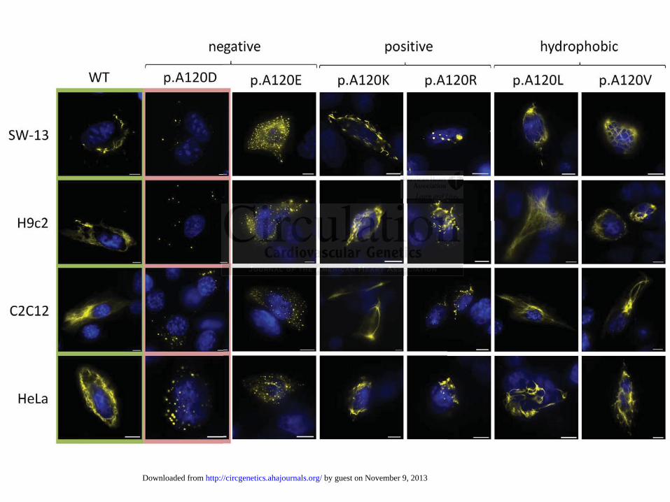
We investigated the filament formation of the different DES variants in transfected SW-13, HL-

1, H9c2, C2C12, HeLa and hiPS-CM. In these transfection experiments desmin p.H326R formed

in all cell lines filaments comparable to the wild-type (Fig. 3). In contrast desmin-p.A120D

accumulated into cytoplasmic aggregates independently of the transfected cell line (Fig. 3).

We further investigated by immunohistochemistry if the endogenous IFs were impaired

by coexpression of the desmin mutants. These analysis revealed that the expression of desmin-

p.A120D influences the assembly of endogenous desmin in the muscle cell lines HL-1, H9c2 and

C2C12, indicating a dominant inhibiting defect (Fig. 4). Desmin and vimentin form hetero-

filaments, when coexpressed within the same cell 28. Therefore, we used endogenously vimentin

expressing HeLa cells to investigate if the desmin mutants induce a coaggregation with vimentin.

In contrast to desmin-p.H326R and -WT the p.A120D mutant induced a partially coaggregation

with vimentin in transfected HeLa-cells. Nevertheless, the vimentin network was not strongly

affected by the expression of desmin-p.A120D.

In vitro desmin assembly using atomic force microscopy

To get better insights into the putative filament formation defects caused by these new desmin

variants, we purified the recombinant desmins by ion exchange and immobilized metal affinity

chromatography and analyzed the filament formation in vitro by AFM. In accordance with our

cell culture experiments we found that the variant p.H326R formed filaments similar to wild-type

desmin (Fig. 5). In contrast, desmin-p.A120D formed small accumulated fibrils (Fig. 5),

suggesting severe impairment of the filament elongation step by this mutation.

Investigation of different model mutants at position A120

Since it is not known, if the loss of the methylene group or the gain of the aspartate residue is

e cell lines HL-1, H9H9H9H c

vimenttttiiiin fffform hhheh teteteterrroro-

w 28 m

HeLa cells to investigate if the desmin mutants induce a coaggregation with vim

t a

n g

the e pression of desmin p A120D

wheeeen n n cococoexexexprprp esssesesed within the same cell 28... ThT erefore, we ussedeee endogenously vim

HHeLLLa cells to innnvestitititigatetetet if ththththe deddesmmminn mmuutanntsts iiindndnduccce a cooaaaggrgrgregatttioioioon wwwithhhh vvim

to ddddesmsmsmmininini -p.HHH32232326R6R6R aand -WTWTWTWT the ppp AAA.A12121220D0D00D mmutututtanananttt iiiindududuuceced dd aa partrtrtrtiaiaiallllllly cocoagaggrgregega

ntin in transfectedddd HHHeLLa-c lellllllsl . NNeN verthheh lllel ss, ,, thhhe viiiimentinii netwo kkrkk was not strongg

hth isi ff dde iin AA12120D0D

by guest on November 9, 2013http://circgenetics.ahajournals.org/Downloaded from

DOI: 10.1161/CIRCGENETICS.113.000103

12

causative for aggregation of desmin-p.A120D, we constructed different model variants

(p.A120E, p.A120K, p.A120R, p.A120V and p.A120L) to get more insights into the nature of

the amino acid residue, which causes the filament formation defect of desmin-p.A120D. In

transfected cells, the hydrophobic model mutants (p.A120V and p.A120L) formed filaments

similar to wild-type desmin (Fig. 6). In contrast, the exchange of A120 against positive or

negative amino acids induced desmin aggregation, with the exception of p.A120K (Fig. 6). In

summary, these experiments reveal the essential role of a hydrophobic amino acid at position

120 for the filament formation.

Immunohistochemistry in cardiac tissue

Ventricular myocardium of individual II:6 (family A) was available from heart transplantation

(Mount Sinai Hospital, Los Angeles, USA). Confirming the in vitro results of the cell culture and

AFM experiments with the mutant desmin-p.A120D we found a high density of desmin

aggregates within the ventricular myocardium, which was undetectable in the ventricles of

rejected donor hearts (Fig. 7).

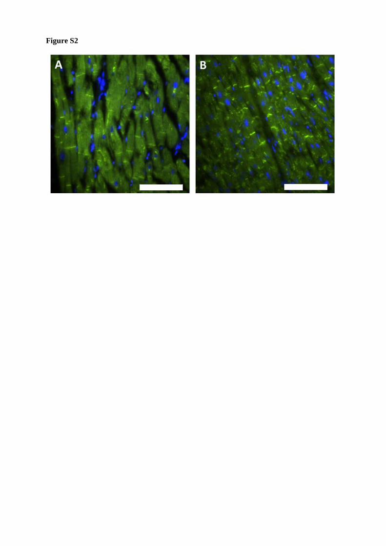
In addition, when the slices of the DES-p.A120D heart were costained for desmoplakin

colocalization of desmin and desmoplakin could not be observed. Thus, desmin was detectable in

the DES-p.A120D-heart of the mutation carrier in the z-bands and in prominent protein

aggregates but not within the ID (Fig. 7).

Recently, remodeling for plakoglobin (JUP) 29 as well as connexin-43 (Cx43) 30 was

described in AC patients. Therefore, we investigated if the localization of both proteins were

affected in the patient with the DES-p.A120D mutation. These results demonstrate that

plakoglobin as well as connexin-43 are localized in the ID similar like in healthy control persons

(Fig. S2-S3).

r myocardium of individual II:6 (family A) was available from heart transplantat

n u

r

nor hearts (Fig 7)

r mymymyocoocararardididiumuu ooof f individual II:6 (family A)A)AA was available fffrororom heart transplantat

naai HHHospital, LLosss AAAnnggellesesese , USSSSA)A)A). Confnnfirmiming tthehehe innn vvvittro reesssultltltsss of tttthehehehe cccelelelll cccultll u

rimentntntssss wiwiwiw th tttheheh mm tututana t dededesmsmsmsmin-ppp AAA.A1212121 0D0D0DD we fofoofoununundddd a hiihihi hhghgh ddddeensiiiitytytyty ooof deded smsminini

within the ventriiiiculllal r myyyocardidididium,,, hwhhhiiichh h was undded tectabbble iiiin hhthhe ventricles of

hh rt (F(Fiig 7)7)

by guest on November 9, 2013http://circgenetics.ahajournals.org/Downloaded from

DOI: 10.1161/CIRCGENETICS.113.000103

13

Discussion

At the end of the 1990’s the first mutations in the human DES gene were published 3,4. It became

obvious that mutations within the DES gene may lead to skeletal and/or cardiac myopathies with

a broad spectrum of pathological muscle phenotypes even within the same family 31. Of note,

about 74% of the desminopathy patients develop a cardiac phenotype like conduction disease,

arrhythmias or cardiomyopathies 6. Nevertheless, the molecular pathomechanisms leading to

desmin related arrhythmogenic cardiomyopathies are not well understood.

In recent years, mutations of DES associated with an ARVC-related phenotype were

found 10–15. In this study we identified and characterized the two novel heterozygous DES-

variants p.A120D and p.H326R.

The variant p.A120D is localized within the highly conserved IF-consensus motif at the

N-terminal segment of coil 1. The amino acid residue A120 is absolutely conserved in different

human IF-proteins and among species 32. Even lamin of Hydra attenuata contains this amino

acid 33,34. Interestingly, mutations in the homologous positions of the genes coding for keratins

K5 35,36, K10 37, K12 38, K86 39 and lamin A/C 40 cause severe clinical diseases of eye, skin and

muscle, respectively. The desmin variant p.A120D was not detectable in 788 control

chromosomes and revealed a severe filament formation defect in cell culture and AFM. These

data were confirmed by immunostaining of the failing ventricular myocardium of the affected

patient. Interestingly, the desmin-staining within the ID was undetectable in the mutation carrier.

It is known, that desmin is linked to the cardiac desmosome via the plaque protein desmoplakin

41. Based on yeast two hybrid analyses this protein interaction was claimed to be affected by the

desmin mutant p.I451M 41. However, the transgenic murine model of Mavroides et al. revealed

in contrast that this mutation affects the positioning of desmin to the z-bands but not in the

elated phenotype wewewewere

l heterozygous DEDEDEDESSSS-SS

A

variant p.A120D is localized within the h hly conserved IF-consensus motif at

segment of coil 1. The amino acid residue A120 is absolutely conserved in diffe

p n

nterestingl m tations in the homologo s positions of the genes coding for kera

A12220D0D0D aaandndndd pp.HHHH32333 6R.

vvvarrrriant p.A1222000D iiis locacacacallil zedddd wwwitthinnn thhe hhiighlhlhly y yy cocococ nnsereervved IFFF-cccooonsensnsnsn usss mmmotttiffff at

segmememeentntntnt of cocoillilil 111. ThThThThe ammminininino acidiidid resesese idididdueue AAAA12121220000 isisis absbsbsoolololututt llelely yy coooonsnsnsn erveeddd ininii dddifififfe

proteins and among gg spppecieii s 3233232. EvEE en llllaminiii offff HHyddyddra attenuatafff contains this amin

nte stiin lgl tatiio ii hth hho llo iitiio ff hth didi ff kk a

by guest on November 9, 2013http://circgenetics.ahajournals.org/Downloaded from

DOI: 10.1161/CIRCGENETICS.113.000103

14

intercalated disk 42. Thus, it remains an open question if a specific (sub-)domain of desmin is

linked to the ID via desmoplakin. The lack of desmin within the ID might explain the high

prevalence of malignant arrhythmias in the affected family A. Of note, the pedigree of this

family reveals a number of SCDs among teenaged family members, which might reflect the

arrhythmogenic potential of desmin-p.A120D. Recently, remodeling of plakoglobin and

connexin-43 were described in AC patients 29,30. Nevertheless, plakoglobin staining as well as

connexin-43 remodeling are controversially discussed in the literature since the absence could

not be detected in every patient 43–46. In this study, we demonstrate comparable amounts of

plakoglobin and connexin-43 within the ID in heart tissue of a healthy control person and of a

patient with the DES-p.A120D mutation. Based on these experiments we conclude that the

remodeling of plakoglobin and connexin-43 does not play a major role for the pathomechanisms

caused by this specific DES mutation.

The mutation p.A120D leads to an exchange of a hydrophobic amino acid side chain

against an acidic one. We hypothesized that a hydrophobic amino acid at position 120 is essential

for filament formation. For testing this hypothesis we constructed further model mutants with (i)

hydrophobic residues (p.A120L and p.A120V), (ii) negative (p.A120E) and (iii) positive charge

(p.A120R and p.A120K). Remarkably, ionic side chains at this position disturbed the filament

formation with the exception of the lysine residue. Whereas the experiments with hydrophobic

amino acid side chains reveal that even a larger mutant amino acid at this position does not

disturb filament formation. We conclude from these data that a steric hindrance of the side chain

can be excluded as a reason for the p.A120D filament formation defect. We assume that this

position within the desmin primary structure, which is in the neighborhood of the recently

published mutations p.N116S 12 and p.E114del 13, might be a hotspot for desminopathies

mparable amountssss oooof

y contrttt ollll person annnndd dd o

h

g n

h

mutation p.A120D leads to an exchange of a hydrophobic amino acid side chain

acidic one We h pothesi ed that a h drophobic amino acid at position 120 is es

h thehehehe DEDEDEDESSS-pp-p.AAA121211 0D mutation. Based on nn ththtt eese experimentss wwwe conclude that the SSS

g ooof f plakoglobiiinnn annd connnnnnn exinininin-4333 doesees noot plaaay y y a a a a mmajorr rolllel fooro the ppppaaathohohomemechchhhan

his spepepeecicicicififific DEDEDED SSSS mm tututata ion.nn SSSS

mutation p.pp A120202020D DD llel addds to an exchhhangegg offf f a hyydrddd oppphohh bibibibic aminii o acid side chain

icididi WW hh hth ii ded thhat hh ddr hhobibi iin idid t isi iti 112020 ii

by guest on November 9, 2013http://circgenetics.ahajournals.org/Downloaded from

DOI: 10.1161/CIRCGENETICS.113.000103

15

associated with arrhythmias. It could be speculated that the intra-molecular interaction of this

part of the desmin coil 1 with its head domain, might be affected, which was recently shown for

vimentin 47,48.

In summary, although a cosegregation analysis was not possible in family A due to the

lack of genomic DNA from suddenly deceased members, we conclude from our experiments that

DES-p.A120D is indeed a disease causing mutation with a high potential for SCD.

The amino acid p.H326 is conserved among vertebrate desmins. However, arginine at this

position is also found in the human homologue vimentin. In addition this allele was not found in

788 control chromosomes or in the Washington Exome Data. To assess the pathogenic potential

of this variant we investigated in vitro the influence of p.H326R on filament formation and

performed a cosegregation analysis within the family. When tested in cell culture experiments

this recombinant desmin variant did not reveal any filament formation defect, which was also

supported by AFM of the purified recombinant desmin. Nevertheless, we cannot exclude that

other relevant functions of desmin, like biomechanical properties or protein-protein interactions,

i.e. with desmoplakin, might be disturbed by this variant. Of note, the variant did not completely

cosegregate within family B. In summary, we regard p.H326R as a rare variant of unknown

pathogenic significance.

Acknowledgements: We thank all participating patients. The authors are grateful to Birte Bohms for excellent technical assistance. Furthermore we thank Dr. Dawn Lombardo (University if Irvine) for her help in clinical investigations. In addition, we thank Dr. Bianca Werner (Heart and Diabetes Centre NRW, Germany) for providing the HeLa cells, Dr. Christine Mummery (Leiden University Medical Centre, Netherlands) for providing the END2 cells and Dr. William Claycomb (New Orleans, USA) for providing the HL-1 cells.

Funding Sources: H.M. was kindly funded by the Erich and Hanna Klessmann Foundation, Gütersloh, Germany and by FoRUM-grant (F649-2009) of the Ruhr-University Bochum. This work was also supported by the grant from the BMBF to T.Š. (grant no: 01 GN 0824). J.H.S. was

his allele was not fofofofou

ss the pppatttthhhhoge inic popopopote

a d

a e

binant desmin variant did not reveal any filament formation defect, which was al

by AFM of the purified recombinant desmin. Nevertheless, we cannot exclude th

ant f nctions of desmin like biomechanical properties or protein protein interac

ant wewewe iiinvnvnvesesestigagagated in vitro the influenceee of p.H326R on filililaaament formation and

a coooosegregatioonnn annallllys sisiss wwithihihih n tthhe ffafammilyy.. WhWhWhenenenen ttestetted inn cccellll cccultuuuureee eeexxxperrrimme

binantntnt ddddesesese min n vavaririiananttt did dd noononotttt reveeeallalal aaaanyny ffffilililamamennenentttt fofoformrmmattatatiiioionn dddedefectctctct, whicici hhhh wawass ala

byy AFM of the pupp riiiififififi dedd recombibibiinant dddesmiiin. NNeverthhhhellless,,, we cannot exclude th

nt ff ctiio ff dde iin lilikke bbiio hch iic lal rtiie teiin teiin iint

by guest on November 9, 2013http://circgenetics.ahajournals.org/Downloaded from

DOI: 10.1161/CIRCGENETICS.113.000103

16

funded by The Danish National Research Foundation, The John and Birthe Meyer Foundation, The Research Foundation of the Heart Centre, Copenhagen University Hospital and The Arvid Nilsson Foundation.

Conflict of Interest Disclosures: None.

References

1. Potschka M, Nave R, Weber K, Geisler N. The two coiled coils in the isolated rod domain of the intermediate filament protein desmin are staggered. A hydrodynamic analysis of tetramers and dimers. Eur J Biochem. 1990;190:503–508.

2. Herrmann H, Strelkov SV, Burkhard P, Aebi U. Intermediate filaments: primary determinants of cell architecture and plasticity. J Clin Invest. 2009;119:1772–1783.

3. Goldfarb LG, Park KY, Cervenáková L, Gorokhova S, Lee HS, Vasconcelos O, et al.Missense mutations in desmin associated with familial cardiac and skeletal myopathy. Nat Genet. 1998;19:402–403.

4. Muñoz-Mármol AM, Strasser G, Isamat M, Coulombe PA, Yang Y, Roca X, et al. A dysfunctional desmin mutation in a patient with severe generalized myopathy. Proc Natl Acad Sci USA. 1998;95:11312–11317.

5. Li D, Tapscoft T, Gonzalez O, Burch PE, Quiñones MA, Zoghbi WA, et al. Desmin mutation responsible for idiopathic dilated cardiomyopathy. Circulation. 1999;100:461–464.

6. van Spaendonck-Zwarts, K Y, van Hessem L, Jongbloed, J D H, de Walle, H E K, Capetanaki Y, van der Kooi, A J, et al. Desmin-related myopathy. Clin Genet. 2011;80:354–366.

7. Cao L, Hong D, Zhu M, Li X, Wan H, Hong K. A novel heterozygous deletion-insertion mutation in the desmin gene causes complete atrioventricular block and mild myopathy. ClinNeuropathol. 2013;32:9–15.

8. Bär H, Mücke N, Kostareva A, Sjöberg G, Aebi U, Herrmann H. Severe muscle disease-causing desmin mutations interfere with in vitro filament assembly at distinct stages. Proc Natl Acad Sci USA. 2005;102:15099–15104.

9. Kostareva A, Sjoberg G, Gudkova A, Smolina N, Semernin E, Shlyakhto E, et al. Desmin A213V substitution represents a rare polymorphism but not a mutation and is more prevalent in patients with heart dilation of various origins. Acta Myol. 2011;30:42–45.

10. van Tintelen JP, van Gelder IC, Asimaki A, Suurmeijer AJH, Wiesfeld ACP, Jongbloed JDH, et al. Severe cardiac phenotype with right ventricular predominance in a large cohort of patients with a single missense mutation in the DES gene. Heart Rhythm. 2009;6:1574–1583.

p y

sconcelelelelosososos OOOO,,,, etetetet aaaallll....mutations in desmin associated with familial cardiac and skeletal myopathy Nat8

Mn c9

p te for idiopathic dilated cardiomyopathy. ;

mutatioonsnsns in nn deesmss in associated with familialaa cardiac and skekeeletal myopathy. Nat 8;19191919:4:4:402–4–4–4400030 .

MMMMárárárrmommm l AM, StStrrrassseeer GGGG, Isamat MM, CoCCoulomombeee PPPPA,AA,A Yanaang YY, RRRocococca X,,, eeete alll. AAAnal ll dededed smsmsmmininin mmmmuutu atatatatioioion ininiin aaaa pppatatatatieieieient wwwwitittithhh h seseseveveveverereree ggggenennnererereralalallizizizi edededd mmmmyoyoyoopapapapaththththy.y.y.y PrPPP oooco NNNNaaata l ll AcAA998;95:11312–1–1–113131 17.

pppscscscofofofttt TTT, GGGGonononzazazalelelezzz O,O,O, BBBurururchchch PPPE,E,E,E, QQQQuiuiuiñoñoñonenenesss MAMAMAA,,, ZoZoZoghghghg bibibibi WWWA,A,A, etetet aaalll. DeDeDesmsmsmininin mmmututute foforr ididioiopapathththicic dddililatatteded ccarardidiiomomyoyopapathththy.y. CiCiC rcrcululatattioioi nn.. 19191999999999;1;1;1100000000:4:4:446161616 –4–4–44646464..

by guest on November 9, 2013http://circgenetics.ahajournals.org/Downloaded from

DOI: 10.1161/CIRCGENETICS.113.000103

17

11. Otten E, Asimaki A, Maass A, van Langen IM, van der Wal A, Jonge N de, et al. Desmin mutations as a cause of right ventricular heart failure affect the intercalated disks. Heart Rhythm.2010;7:1058–1064.

12. Klauke B, Kossmann S, Gaertner A, Brand K, Stork I, Brodehl A, et al. De novo desmin-mutation N116S is associated with arrhythmogenic right ventricular cardiomyopathy. Hum MolGenet. 2010;19:4595–4607.

13. Vernengo L, Chourbagi O, Panuncio A, Lilienbaum A, Batonnet-Pichon S, Bruston F, et al.Desmin myopathy with severe cardiomyopathy in a Uruguayan family due to a codon deletion in a new location within the desmin 1A rod domain. Neuromuscul Disord. 2010;20:178–187.

14. Hedberg C, Melberg A, Kuhl A, Jenne D, Oldfors A. Autosomal dominant myofibrillar myopathy with arrhythmogenic right ventricular cardiomyopathy 7 is caused by a DES mutation. Eur J Hum Genet. 2012;20:984-985.

15. Lorenzon A, Beffagna G, Bauce B, Bortoli M de, Li Mura IEA, Calore M, et al. Desmin mutations and arrhythmogenic right ventricular cardiomyopathy. Am J Cardiol. 2013;111:400–405.

16. Azaouagh A, Churzidse S, Konorza T, Erbel R. Arrhythmogenic right ventricular cardiomyopathy/dysplasia: a review and update. Clin Res Cardiol. 2011;100:383–394.

17. Gerull B, Heuser A, Wichter T, Paul M, Basson CT, McDermott DA, Lerman BB, et al.Mutations in the desmosomal protein plakophilin-2 are common in arrhythmogenic right ventricular cardiomyopathy. Nat Genet. 2004;36:1162–1164.

18. Heuser A, Plovie ER, Ellinor PT, Grossmann KS, Shin JT, Wichter T, et al. Mutant desmocollin-2 causes arrhythmogenic right ventricular cardiomyopathy. Am J Hum Genet.2006;79:1081–1088.

19. Gerull B, Kirchner F, Chong JX, Tagoe J, Chandrasekharan K, Strohm O, et al. Homozygous Founder Mutation in Desmocollin-2 (DSC2) Causes Arrhythmogenic Cardiomyopathy in the Hutterite Population. Circ Cardiovasc Genet. 2013;6:327–336.

20. Elliott P, O'Mahony C, Syrris P, Evans A, Rivera Sorensen C, Sheppard MN, et al.Prevalence of desmosomal protein gene mutations in patients with dilated cardiomyopathy. Circ Cardiovasc Genet. 2010;3:314–322.

21. Brodehl A, Dieding M, Cakar H, Klauke B, Walhorn V, Gummert J, et al. Functional characterization of desmin mutant p.P419S. Eur J Hum Genet. 2013;21:589–590.

22. Claycomb WC, Lanson NA, Stallworth BS, Egeland DB, Delcarpio JB, Bahinski A, et al.HL-1 cells: a cardiac muscle cell line that contracts and retains phenotypic characteristics of the adult cardiomyocyte. Proc Natl Acad Sci USA. 1998;95:2979–2984.

alore MMMM, tttet allll. DeDDD smsmsmsminJ CarrdidididiololololJ 2222010101013;3;3;3;111111111:11 4

ap

B li

aggh A, Churzidsdsdse SS,S KKKKonnnnooro za TTT, EErbeeel R. AArrhyhyhythhhmommm gggennic rrighgghtt t vvventttriririricucuculalaar patatthyhhyhy/dysplplplasaa ia::: a rereeviewww aandd uupdppdateee. CClinn Resese Carardiolool. 20011;11100000 :383838383–3339994.

B, Heuser A, WiWWW chchhhteteteter r r T,T,T,T, PPPauauauaul l ll M,M,M,M, BBBBasasasassososon nn CTCTCTT, McMcMccDeDeDeermrmrmrmotototottt t t DADADAD ,, , LeLeLeLerrmr an BB, et alin the desmosomalll prpp ot iiein plplakkkoppphhih liliilin-2222 are common iiin arrhhhhytytythhhmh ogggenic rigght ccardiomyyopopopopatatathyhyhyhy.. NaNaNaNat GeGeGeGeneneenet.t.tt. 222000000004;4;4;36363636:1:11:116116162–2–2–2–11111164646464..

by guest on November 9, 2013http://circgenetics.ahajournals.org/Downloaded from

DOI: 10.1161/CIRCGENETICS.113.000103

18

23. Brodehl A, Hedde PN, Dieding M, Fatima A, Walhorn V, Gayda S, et al. Dual Color Photoactivation Localization Microscopy of Cardiomyopathy-associated Desmin Mutants. J BiolChem. 2012;287:16047–16057.

24. Fatima A, Xu G, Shao K, Papadopoulos S, Lehmann M, Arnáiz-Cot JJ, et al. In vitro modeling of ryanodine receptor 2 dysfunction using human induced pluripotent stem cells. Cell Physiol Biochem. 2011;28:579–592.

25. Kreplak L, Bär H, Leterrier JF, Herrmann H, Aebi U. Exploring the mechanical behavior of single intermediate filaments. J Mol Biol. 2005;354:569–577.

26. Quarta G, Muir A, Pantazis A, Syrris P, Gehmlich K, Garcia-Pavia P, et al. Familial evaluation in arrhythmogenic right ventricular cardiomyopathy: impact of genetics and revised task force criteria. Circulation. 2011;123:2701–2709.

27. den Haan, A Dénise, Tan BY, Zikusoka MN, Lladó LI, Jain R, et al. Comprehensive desmosome mutation analysis in north americans with arrhythmogenic right ventricular dysplasia/cardiomyopathy. Circ Cardiovasc Genet. 2009;2:428–435.

28. Traub P, Kühn S, Grüb S. Separation and characterization of homo and hetero-oligomers of the intermediate filament proteins desmin and vimentin. J Mol Biol. 1993;230:837–856.

29. Asimaki A, Tandri H, Huang H, Halushka MK, Gautam S, Basso C, et al. A new diagnostic test for arrhythmogenic right ventricular cardiomyopathy. N Engl J Med. 2009;360:1075–1084.

30. Saffitz JE. Arrhythmogenic cardiomyopathy and abnormalities of cell-to-cell coupling. Heart Rhythm. 2009;6:S62-5.

31. Bergman, Jorieke E H, Veenstra-Knol HE, van Essen, Anthonie J, van Ravenswaaij, et al.Two related Dutch families with a clinically variable presentation of cardioskeletal myopathy caused by a novel S13F mutation in the desmin gene. Eur J Med Genet. 2007;50:355–366.

32. Kapinos LE, Schumacher J, Mücke N, Machaidze G, Burkhard P, Aebi U, et al.Characterization of the head-to-tail overlap complexes formed by human lamin A, B1 and B2 "half-minilamin" dimers. J Mol Biol. 2010;396:719–731.

33. Erber A, Riemer D, Hofemeister H, Bovenschulte M, Stick R, Panopoulou G, et al.Characterization of the Hydra lamin and its gene: A molecular phylogeny of metazoan lamins. JMol Evol. 1999;49:260–271.

34. Herrmann H, Strelkov SV. History and phylogeny of intermediate filaments: now in insects. BMC Biol. 2011;9:16.

35. Bolling MC, Lemmink HH, Jansen GHL, Jonkman MF. Mutations in KRT5 and KRT14 cause epidermolysis bullosa simplex in 75% of the patients. Br J Dermatol. 2011;164:637–644.

aaaallll. CoCoCoCompmpmpmprerererehehehehensnsnsnsiiiviveeeec righthththt ventrttt iciii lularrrr

P, Kühn S, Grüb S. Separation and characterization of homo and hetero-oligome

kh

J09;6:S62 5

P, KKKKüüühü n S, GGGGrüüürübbbb S... SSSSepepepparaaa atatatioioioion nnn anananand dd chchchcharaa accteerizaazatitititiononono oooofff f hohohomomoomo andddd hhhhetetettereee o-o ololoo igigigomomomomeddddiaatttet filament ppprotteiiiins ddddeese mimimiminn anaand vvvimmenntiin. JJJ J MoMoMol BBBiool. 11999933;3 22230:8383838 7–––8885666.

ki A, TTTanananandrdrdri H,HH HHHHuauangng H, HaHaHaHallllushhhhkakkaka MMMMKKK, GGGauautatatammmm S,SS, BBBassasassoo CCCC, et aaaallll. AAAA nenew didididiagagnnhythmogenic righghghght t t veveveventntntriririr cucuculalalalar r r r cacacaardrdrdr ioioioomymymym opopopatatatthyhyhyh . N N N N EnEnEnEnglglglg JJJJ MeMeMeMed.d.d 2222000000009;360:1075–1

JJJE. Arrhyttythmhmhmhmogoggogeneneenicicicic cccarararardiddidiomomomomyoyoyopappapaththhthy yy y anannand d dd ababababnonoormrmrmrmalalalititititieii s sss ofoofof ccccelelelell-l-l-totototo c-c-celelelell ll cocc upling..0909 6;6 S:S6262 55

by guest on November 9, 2013http://circgenetics.ahajournals.org/Downloaded from

DOI: 10.1161/CIRCGENETICS.113.000103

19

36. Hamada T, Kawano Y, Szczecinska W, Wozniak K, Yasumoto S, Kowalewski C, et al.Novel keratin 5 and 14 gene mutations in patients with epidermolysis bullosa simplex from Poland. Arch Dermatol Res. 2005;296:577–579.

37. Yang JM, Yoneda K, Morita E, Imamura S, Nam K, Lee ES, et al. An alanine to proline mutation in the 1A rod domain of the keratin 10 chain in epidermolytic hyperkeratosis. J InvestDermatol. 1997;109:692–694.

38. Takahashi K, Takahashi K, Murakami A, Okisaka S, Kimura T, Kanai A. Heterozygous Ala137Pro mutation in keratin 12 gene found in Japanese with Meesmann's corneal dystrophy. Jpn J Ophthalmol. 2002;46:673–674.

39. Winter H, Vabres P, Larrègue M, Rogers MA, Schweizer J. A novel missense mutation, A118E, in the helix initiation motif of the type II hair cortex keratin hHb6, causing monilethrix. Hum Hered. 2000;50:322–324.

40. Brown CA, Lanning RW, McKinney KQ, Salvino AR, Cherniske E, Crowe CA, et al. Novel and recurrent mutations in lamin A/C in patients with Emery-Dreifuss muscular dystrophy. Am J Med Genet. 2001;102:359–367.

41. Lapouge K, Fontao L, Champliaud M, Jaunin F, Frias MA, Favre B, et al. New insights into the molecular basis of desmoplakin- and desmin-related cardiomyopathies. J Cell Sci.2006;119:4974–4985.

42. Mavroidis M, Panagopoulou P, Kostavasili I, Weisleder N, Capetanaki Y. A missense mutation in desmin tail domain linked to human dilated cardiomyopathy promotes cleavage of the head domain and abolishes its Z-disc localization. FASEB J. 2008;22:3318–3327.

43. Arbustini E, Pasotti M, Pilotto A, Pellegrini C, Grasso M, Previtali S, et al. Desmin accumulation restrictive cardiomyopathy and atrioventricular block associated with desmin gene defects. Eur J Heart Fail. 2006;8:477–483.

44. Basso C, Pilichou K, Thiene G. Is it time for plakoglobin immune-histochemical diagnostic test for arrhythmogenic cardiomyopathy in the routine pathology practice? Cardiovasc Pathol.2013;22:312-313.

45. Tavora F, Zhang M, Cresswell N, Li L, Fowler D, Franco M, Burke A. Quantitative Immunohistochemistry of Desmosomal Proteins (Plakoglobin, Desmoplakin and Plakophilin), Connexin-43, and N-cadherin in Arrhythmogenic Cardiomyopathy: An Autopsy Study. Open Cardiovasc Med J. 2013;7:28–35.

46. Paul M, Wichter T, Gerss J, Arps V, Schulze-Bahr E, Robenek H, et al. Connexin expression patterns in arrhythmogenic right ventricular cardiomyopathy. Am J Cardiol. 2013;111:1488–1495.

47. Aziz A, Hess JF, Budamagunta MS, Voss JC, FitzGerald PG. Site-directed spin labeling and

E, CrC owe CACACACA,, et aaaallll. Nmuscccculululularararar ddddysysysystrtrtrtropopopophhhhy

.

g sl

4

dn desmin tail domain linked to human dilated cardiom th omotes cleav eomain and abolishes its Z disc locali ation FASEB J 2008;22:3318 3327

. 20000001;1;1;1010101 2:2:2:3533 9–9–9–9 367.

gee e KKK, Fontao LL, CChaammmpliiiiaaua d MM,MM JJJaauniinn F, FFriasas MMMAA,A FFFaavre BBB, eteete al. NeNeNeNewww iinini siiiighhhhtslar r r babbbasis offf dddessmmmoplpllakinnn- and d ddeeesmminnn---relaatted d d ccarddiomymymyopppaathhhieses. J CeCeCellll ScScSci.

4974–444 494949498585858 .

dis M,,, Panagggopppoulllol u P,P,P, KKKostavasiiillil II,,, WWeW iiislllel der N,N,, CCCCapppetan kkakiii i Y.YY A missense n desmin ttaiaiaiaillll dododomamammainininin lininninkekkeked dd d totototo hhhumumumumanananan ddddililililatataatedededed cccararrardiddidiommomomyoyooyopapapapaththhthy yyy prprrpromomoomototototeseses cleavagge

iai dd bab loliishhe iit ZZ didi ll lalii atiio FAFASESEBB JJ 22000088;2222 3:3313188 33332727

by guest on November 9, 2013http://circgenetics.ahajournals.org/Downloaded from

DOI: 10.1161/CIRCGENETICS.113.000103

20

electron paramagnetic resonance determination of vimentin head domain structure. J Biol Chem.2010;285:15278–15285.

48. Hess JF, Budamagunta MS, FitzGerald PG, Voss JC. Characterization of structural changes in vimentin bearing an epidermolysis bullosa simplex-like mutation using site-directed spin labeling and electron paramagnetic resonance. J Biol Chem. 2005;280:2141–2146.

49. Cetin N, Balci-Hayta B, Gundesli H, Korkusuz P, Purali N, Talim B, et al. A novel desmin mutation leading to autosomal recessive limb-girdle muscular dystrophy: distinct histopathological outcomes compared with desminopathies. J Med Genet. 2013;50:437–443.

50. McDonald KK, Stajich J, Blach C, Ashley-Koch AE, Hauser MA, Schuelke M. Exome Analysis of Two Limb-Girdle Muscular Dystrophy Families: Mutations Identified and Challenges Encountered. PLoS ONE. 2012;7:e48864.

51. McLaughlin HM, Kelly MA, Hawley PP, Darras BT, Funke B, Picker J. Compound heterozygosity of predicted loss-of-function DES variants in a family with recessive desminopathy. BMC Med Genet. 2013;14:68.

52. Schröder R, Goudeau B, Simon MC, Fischer D, Eggermann T, Clemen CS, et al. On noxious desmin: functional effects of a novel heterozygous desmin insertion mutation on the extrasarcomeric desmin cytoskeleton and mitochondria. Hum Mol Genet. 2003;12:657–669.

53. Henderson M, Waele L, Hudson J, Eagle M, Sewry C, Marsh J, et al. Recessive desmin-null muscular dystrophy with central nuclei and mitochondrial abnormalities. Acta Neuropathol.2013;125:917–919.

Figure Legends

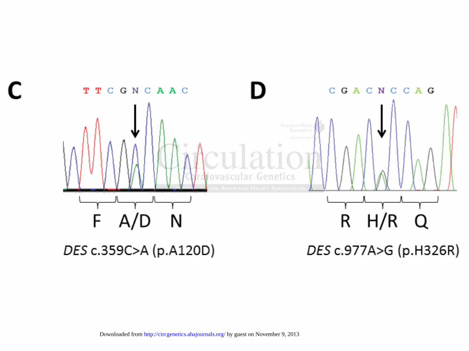
Figure 1. Identification and verification of the DES-variants. Pedigrees of family A (A) and

family B (B). Squares represent males and circles females. Deceased individuals are indicated by

slashes. The index patients are marked by a red arrow. Genotypes are shown by present (+) or by

absent (-) of the heterozygous DES mutations (p.A120D, family A; p.H326R family B).

Electropherograms showing the heterozygous alleles c.359C>A, p.A120D (C) and c.977A>G,

p.H326R (D). Converted codons lead to the protein changes p.A120D (C) and p.H326R (D).

ckckckckerererer JJJJ. CoCoCoCompmpmpmpououououndndndnd withh recessiiiive

e onm 9

s ny l

9

errrr RRRR, Gouddddeaeee u u u B,BBB SSSSimimimimonooo MMMMC,C,C,C FFFFisisisi chchchhererere D, EEggeeermrmrmrmanaa n n n n T,TT, CCCleleelememmm n n n n CSCSCSCS,,,, et aaaallll. OnOnOnOn noncccctiiono al effectss of aa nnnovevevev ll l hetetet rooozzygooouus ddesmimiminn inininssertttioon mumuutaaatititiion oooon n n thhheee meeeriiririccc desmininin ccyyyty osskeeeletototonn and d mimmitoccchoonddriia. HuHHH mm MMoMoll Geenet. 2222003;3;3;3;11211 :::657–6––669

son M, Waele LLLL, HuHuHuHudsdsdsononono JJJJ, EaEaEaE glglglgle e e e M,M,M,M, SSewewewryryryy CCC, MaMaMaMarsrsrsrsh h hh J,J,J,J, etetete aaaal.l.l. RRRRececece essive desminysyy troppphyy with centrall l nucleiiii a dnddd mitochhhoh dnddd iiriial abnbbb ormalilililities. AcAAA ta Neuropapp thol17–919.

by guest on November 9, 2013http://circgenetics.ahajournals.org/Downloaded from

DOI: 10.1161/CIRCGENETICS.113.000103

21

Figure 2. Localization of the identified DES-mutations within the desmin sequence. (A)

Schematic domain structure of the desmin protein and the distribution of known disease causing

mutations. Some DES mutations affecting the splice sites are not shown (e.g. 49,50). *This

sequence variants were also identified in healthy control persons 9. #p.K201RfsX20 and p.R429X

were both identified in the same patient 51. **p.A360P and p.N393I were both identified in the

same patients 3. ##In the same ARVC patient a pathogenic PKP2 mutation (p.T816RfsX10) was

identified 15. ***The mutation p.K240del was originally described as an insertion mutation 52.

However, the authors corrected the sequence analysis in 2007. ###Two siblings compound

heterozygous for the mutations p.T76fsX21 and p.E108X were described 53. (B, C) Alignment of

the desmin/vimentin sequences of Homo sapiens (hs), Mus musculus (mm), Rattus norvegicus

(rn), Xenopus laevis (xl) and Dario rerio (dr). The heptad sequence is highlighted in yellow and

the positions of the mutations identified in this manuscript are marked in red.

Figure 3. Impairment of filament formation by desmin mutants in transfected cells.

Representative fluorescence images of transfected SW-13, H9c2, HL-1, C2C12 and HeLa cells

and hiPS-CM expressing desmin-eYFP constructs (yellow). hiPS-CM were identified by staining

for sarcomeric -actinin using AlexaFluor555-conjugated secondary antibodies (red). Nuclei

were stained with DAPI (blue). Scale bars represent 10 μm.

Figure 4. Endogenously expressed IF-proteins in cells transfected with desmin mutants.

Representative fluorescence images of transfected HL-1, H9c2, C2C12 and HeLa cells

expressing desmin-eYFP constructs (green), immunostained for desmin (HL-1, H9c2 and

C2C12) or vimentin (HeLa) using Cy3-conjugated secondary antibodies (red). The nuclei were

stained with DAPI (blue). Scale bars represent 10 μm.

siblings compounnnnd d d d

bed 53535353. ((((BBBB, CCCC)))) AAAlA igigigignnnnm

/ i

w

n

mpairment of filament formation by desmin mutants in transfected cells.

/vimmmmenenentitititinnn sesesequuueeences of Homo sapiens (hhhhs)s)ss ,, Mus musculus (m(m(mmm), Rattus norvegi

puuuus lllaevis (xl) aannnd DaDaariooo o rrer riiio oo (dddr)). ThTThe hhepptadadad sssseqeqee uuencnnce iss hhhiggghlhhlh ighthththtededede iiinnn yeelllllllow

ns offff ttthehehehe mmmutatattiioionsns iiidddedentifififieieieedddd in tttthihhihisss mammm nunuscscririiptptptt arerere marararrkkkeked dd iiinin redededed.

mmmpapairirmementnt ooff fififilallamementnt ffforormamatitionon bbyy dedesmsminin mmututtanantstst iinn trttranansffsfececttetedd cecelllls.s.

by guest on November 9, 2013http://circgenetics.ahajournals.org/Downloaded from

DOI: 10.1161/CIRCGENETICS.113.000103

22

Figure 5. Filament formation of recombinant mutant desmin. Desmin was expressed in E. Coli,

purified, prepared on mica, and measured under ambient conditions in AFM tapping mode of

operation . Representative AFM topography images of desmin-wt, -p.A120D and p.H326R are

(bottom row) . Distinct filament structures could be discerned for desmin-wt and –p.H326R with

typical (averaged) dimensions of 500 nm (length), 30 nm (width) and 3-6 nm (height). The

apparent width of 30 nm is consistent with a real filament diameter of 8-10 nm that is broadened

by artifacts due to a finite AFM tip radius of ~ 20 nm. The reduced height is attributed to surface

capillary force effects. In contrast, desmin-p.A120D exhibited complete loss of filament

structure, presenting more globular structures with typical size of 85 nm. Representative AFM

topography images are shown.

Figure 6. Analysis of model mutants for desmin-p.A120D in different cell lines. Representative

fluorescence images of transfected SW-13, H9c2, C2C12 and HeLa cells expressing indicated

mutant desmin-eYFP constructs (yellow). Nuclei were stained with DAPI (blue). Scale bars

represent 10 μm.

Figure 7. Immunohistological analysis of cardiac tissue heterozygous for the DES-p.A120D

mutation. (A-C) Representative fluorescence images of a control sample from a human non-

failing control heart, showing normal localization of desmin (red) and desmoplakin (green) at the

intercalated discs and at the z-bands. (D-F) Representative paraffin sections of cardiac tissue of

patient II:6 demonstrate strong accumulation of desmin (white arrowheads). Of note, the desmin

localization is completely lost at the ID. Scale bars represent 10 μm.

igght is attributed to o o o ssu

ete loss offf f fifififilalll mentntntnt

resenting more globular structures with typical size of 85 nm. Representative AF

y

A t

ce images of transfected SW-13, H9c2, C2C12 and HeLa cells expressing indica

resesesentntntininininggg momm rerere globular structures with tytytypip cal size of 85 nnmmm. Representative AF

y iiimmmam ges are shhhooownn.

Analysis of modedededel ll mumumum tatataantntntn ss fofofofor rr dedededesmsmsmminininin-p-p-pp.A.A.A12121220D00 iiiinnn n didididiffffff ererrreneenent tt cececelllllll llllininini es. Represent

ceee iimamagegess ofoff ttraransnsfefecttctededd SSWWW-1-113,3, HHH9c9c9 2,2, CCC2C2C2C12112 aandnd HHHeLeLaa cecellllllss exexprpresessisingng iindndicicaa

by guest on November 9, 2013http://circgenetics.ahajournals.org/Downloaded from

by guest on November 9, 2013http://circgenetics.ahajournals.org/Downloaded from

by guest on November 9, 2013http://circgenetics.ahajournals.org/Downloaded from

by guest on November 9, 2013http://circgenetics.ahajournals.org/Downloaded from

by guest on November 9, 2013http://circgenetics.ahajournals.org/Downloaded from

by guest on November 9, 2013http://circgenetics.ahajournals.org/Downloaded from

by guest on November 9, 2013http://circgenetics.ahajournals.org/Downloaded from

by guest on November 9, 2013http://circgenetics.ahajournals.org/Downloaded from

by guest on November 9, 2013http://circgenetics.ahajournals.org/Downloaded from

by guest on November 9, 2013http://circgenetics.ahajournals.org/Downloaded from

SUPPLEMENTAL MATERIAL Table S1: Designation of constructs and methods of cloning Primers (5’-3’): (1) agactcgaggccgtcaccatgagccaggcctactcgtccagcc; (2) acaggatccccgagcacttcatgctgctgctgtgtgg; (3) gctcaatgaccgcttcgacaactacatcgagaagg; (4) ccttctcgatgtagttgtcgaagcggtcattgagc; (5) gctcaatgaccgcttcgtcaactacatcgagaagg; (6) ccttctcgatgtagttgacgaagcggtcattgagc; (7) ggagctcaatgaccgcttcctaaactacatcgagaaggtgc; (8) gcaccttctcgatgtagtttaggaagcggtcattgagctcc; (9) agctcaatgaccgcttcgagaactacatcgagaaggtg; (10) caccttctcgatgtagttctcgaagcggtcattgagct; (11) ggagctcaatgaccgcttcaagaactacatcgagaaggtgc; (12) gcaccttctcgatgtagttcttgaagcggtcattgagctcc; (13) agctcaatgaccgcttccgcaactacatcgagaagg; (14) ccttctcgatgtagttgcggaagcggtcattgagct; (15) gatgatggaataccgacgccagatccagtcctcca; (16) tgtaggactggatctggcgtcggtattccatcatc; (17) caccatgagccaggcctactc; (18) ttagagcacttcatgctgctgct;

Construct No. Construct designation Source of cDNA/ method of cloning Restriction sides Primers

1 pEYFP-N1-Desmin PCR of pCMV6-AC-Desmin# / TOPO-TA XhoI, BamHI 1, 2

2 pEYFP-N1-Desmin-p.A120D SDM of pEYFP-N1-Desmin XhoI, BamHI 3, 4

3 pEYFP-N1-Desmin-p.A120V SDM of pEYFP-N1-Desmin XhoI, BamHI 5, 6

4 pEYFP-N1-Desmin-p.A120L SDM of pEYFP-N1-Desmin XhoI, BamHI 7, 8

5 pEYFP-N1-Desmin-p.A120E SDM of pEYFP-N1-Desmin XhoI, BamHI 9, 10

6 pEYFP-N1-Desmin-p.A120K SDM of pEYFP-N1-Desmin XhoI, BamHI 11, 12

7 pEYFP-N1-Desmin-p.A120R SDM of pEYFP-N1-Desmin XhoI, BamHI 13, 14

8 pEYFP-N1-Desmin-p.H326R SDM of pEYFP-N1-Desmin XhoI, BamHI 15, 16

10 pET100D-Desmin PCR of pCMV6-AC-Desmin# / TOPO-TA --- 17, 18

11 pET100D-Desmin-p.A120D SDM of pET100D-Desmin --- 3, 4

12 pET100D-Desmin-p.H326R SDM of pET100D-Desmin --- 15, 16

# Origene Technologies, Rockville, USA

Table S2: Overview about the antibodies used for IHC

Antibody Manufacturer Specification Used concentration (over night, 4°C)

anti Desmin R&D Systems Inc. (Minneapolis, MN, USA) AF3844, polyclonal goat IgG 1:40 (25 ng/µL)

anti Desmoplakin Acris Antibodies GmbH (Herford, Germany) AM09122SU-N, monoclonal mouse IgG2b undiluted

anti Plakoglobin (JUP) Acris Antibodies GmbH (Herford, Germany) BM5100, monoclonal mouse IgG2b 1:5 (10 ng/µL)

anti Connexin-43 Abcam (Cambridge, UK) AB11370, polyclonal rabbit IgG 1:500 (1.22 ng/µL)

Supplemental Figure Legends:

Figure S1. Electrocardiogram (ECG) of patient III:24 (family A) with DES-p.A120D.

The atrial-ventricular conduction is variable (2:1 and 3:1 conduction). Atrial heart rate is

approximately 120 bpm. The ventricular heart rate is approximately 64 bpm. Left axis

deviation. Left anterior hemiblock is present with slow progression of R wave and

pronounced S in the cheast leads. No premature ventricular beats (but polymorphic forms of

PVB in 24 h ECG). QRS duration 91 msec. QT/QTc in the normal range.

Figure S2. Immunohistological plakoglobin staining of cardiac tissue heterozygous for

the DES-p.A120D mutation.

(A) Representative IHC analysis of paraffin sections of cardiac tissue of patient II:6

demonstrate no reduced expression of plakoglobin at the intercalated disc. (B) Representative

fluorescence images of a control sample from a human non-failing control heart. Plakoglobin

was stained with FITC-conjugated secondary antibodies (green). The nuclei were stained with

DAPI (blue). Scale bars represent 100 µm.

Figure S3. Immunohistological connexin-43 staining of cardiac tissue heterozygous for

the DES-p.A120D mutation. (A) Representative IHC analysis of paraffin sections of cardiac

tissue of patient II:6 exclude a severe remodeling of connexin-43 at the intercalated disc. (B)

Representative fluorescence images of a control sample from a human non-failing control

heart. Connexin-43 was stained with Cy3-conjugated secondary antibodies (red). The nuclei

were stained with DAPI (blue). Scale bars represent 100 µm.

Figure S1

Figure S2

Figure S3


Recommended