30
Màster Biodiversitat The opisthokonts Iñaki Ruiz-Trillo, Institut Biologia Evolutiva & Dept. Genètica UB.

Màster Biodiversitat
The opisthokonts Iñaki Ruiz-Trillo, Institut Biologia Evolutiva & Dept. Genètica UB.

Rhodophyta
Dyphyllatida
Acrasis
Choa
nofla
gella
ta
Oxymonads
Labyrinthulomycetes
Apicomplexa
Glaucophyta
Haptophyta
RWKHU�DOJDOïOLNH
Mes
omyc
etoz
oa=
Icht
hyos
pore
aFi
laste
rea
Fung
i
Malawimonas
Naegleria
Diatoms
JaNobids
Arch
amoe
bae
Centrohelida
Guttulinopsis
Ancyromonads
Foraminifera
Met
azoa
Trimastix
Kinetoplastida
Myx
ogast
ria
Phaeophyceae
Dictyo
stelid
a
Telonemia
Cryptophyta
Chrysophyceae
Rigifili
da
Euglenida
RWKHU�IXQJL�OLNH
Oomycetes
Breviat
es
other
Amoebo
zoa
Radiolaria
Retortamonada
Parabasalia
Blastocystis
Xanthophyceae
Nucle
ariid
a
CharophytaEmbryophyta (land plants)
Dinoflagellata
Corallochytrium
Ciliates
Diplomonada
Chlorarachniophytes
Chlorophyta
Apus
omon
ads
Opisthokonta
Amoebozoa
Archaeplastida
Amorphea
Diaphoretickes
Metamonada
Discoba
Rhizaria
Stramenopiles
Alveolata
Excavata
Fig. 1

Rhodophyta
Dyphyllatida
Acrasis
Choa
nofla
gella
ta
Oxymonads
Labyrinthulomycetes
Apicomplexa
Glaucophyta
Haptophyta
RWKHU�DOJDOïOLNH
Mes
omyc
etoz
oa=
Icht
hyos
pore
aFi
laste
rea
Fung
i
Malawimonas
Naegleria
Diatoms
JaNobidsAr
cham
oeba
e
Centrohelida
Guttulinopsis
Ancyromonads
Foraminifera
Met
azoa
Trimastix
KinetoplastidaM
yxog
astria
Phaeophyceae
Dictyo
stelid
a
Telonemia
Cryptophyta
Chrysophyceae
Rigifili
da
Euglenida
RWKHU�IXQJL�OLNH
Oomycetes
Breviat
es
other
Amoebo
zoa
Radiolaria
Retortamonada
Parabasalia
Blastocystis
Xanthophyceae
Nucle
ariid
a
CharophytaEmbryophyta (land plants)
Dinoflagellata
Corallochytrium
Ciliates
Diplomonada
Chlorarachniophytes
Chlorophyta
Apus
omon
ads
Opisthokonta
Amoebozoa
Archaeplastida
Amorphea
Diaphoretickes
Metamonada
Discoba
Rhizaria
Stramenopiles
Alveolata
Excavata
Fig. 1the opisthokonts

the opisthokonts
opistho means “posterior”!Kontos means “pole” (flagellum)

Homo NKMDSTEPPYSQKRYE ... GDNMLEPSANMPWFKGWKVTR------KDGNASGTTLLEALDCILPP
Drosophila NKMDSTEPPYSEARYE ... GDNMLEPSEKMPWFKGWSVER------KEGKAEGKCLIDALDAILPP
Brugia NKMDSTEPAFSEARFN ... GDNMLEPSPNMPWFKGWNVER------KEGNASGKTLLEALDAVIPP
Lottia NKMDSTEPPYSESRFD ... GDNMLEKSQKMPWWKQWKIEQKD-EKGNMQTVTGETLSDALDSIQPP
Dugesia NKMDSTEPPFSEPRFD ... GDNMIDESSNMPWYKGWEITRKN-AKKEEIKTTGRTLLDALDSLEPP
Trichoplax NKMDSTEPPYSEARYN ... GDNMIEESTNMKWFKGWSVER------KEGNASGKTLFEALDAILPP
Nematostella NKMDSTEPPYSEARFK ... GDNMLEKSENMPWFKQWTIERVDPATKKEANASGVTLFEGLDSILPP
Geodia NKMDSTEPPYSQARYD ... GDNMLEESPNMKWFKGWNVER------KEGNASGKTLFNPLDSILPP
Monosiga NKMDSTEPPYSESRFN ... GDNMIEASEKLPWYKGWEITR------KDGNAKGKTLLEALDAIIPP
Corallochytrium NKMDSIK--YSKDRFD ... GDNMIEASTNMDWYKGWE---------KDGSVGGKTLIEALDAVSPP
Capsaspora NKMDSIK--FAEERYN ... GDNMLEASENMPWFKGWTIER------KEGNASGKTLIEALDAISPP
Ministeria NKMDSIK--YDEARFT ... GDNMLDASTNMPWYKGWEVDRD-----KNGKASGKTLIDALDAVLPP
Amoebidium NKMDSIK--FAQDRFN ... GDNMVEPTDNMPWYKGWEVER------KEGNATGKTLLEALDAILPP
Ichthyophonus NKMDSVK--YSEDRFK ... GDNMVAPTENMPWYKGWTCER------KEGNTSGFTLLEALDNIQAP
Ustilago NKMDTTK--YSEDRFN ... GDNMIEPTKEMPWYKGWERET------KAGKVSGKTLLDAIDAIEPP
Neurospora NKMDTTQ--WSQTRFE ... GDNMLEPSTNCPWYKGWEKET------KAGKATGKTLLEAIDAIEPP
Mucor NKMDTTK--WSQDRYN ... GDNMLDESTNMPWFKGWNKET------KAGSKTGKTLLEAIDAIEPP
Allomyces NKMDMVD--WSEARFK ... GDNLLTPSANMPWYQGWSRQSK-----DGTVKTGMTLIEAMDAVDPP
Batrachochytrium NKMDTNK--WSEERFN ... GDNMLEPSANMPWFKGWTKET------KAGTSTGKTLLNAIDSIEAP
Spizellomyces NKMDSDPAPYKKERYD ... GDNLLKKSEKMSWYQGQEVTAL-----SGKKVKVHTLLDALNDFEMP
Glugea NKVDTIDEKNRISRFD ... GINIVEKGDKFEWFKGWKPVSG-----AG--DSIFTLEGALNSQIPP
Fonticula NKMDSCQ--YSEARFT ... GDNMIEPTTNMSWWKGFEITR------GSAKLTGLTLLDALNHIEPP
Nuclearia NKMDTCK--YSEERFN ... GDNMLEATPNMPWFKNWEIER------KSGKVTGKTLVDALDAIEPP
Amastigomonas NKMDADSVQFSQQRFE ... GDNMLEPSSNMSWWT------------------GPTLLEALDSIKAP Planomonas NKMDDKSVNYSKARFD ... GDNMTEPSANMPWYS------------------GPTLLGALDACEVP
Apusomonas NKMDDKTVKYSKDRYE ... GDNMMEPSPQMGWWK------------------GGTLLEALDAITPP
Entamoeba NKMDAIQ--YKQERYE ... GDNMIEPSTNMPWYK------------------GPTLIGALDSVTPP
Dictyostelium NKMDEKSTNYSQARYD ... GDNMLERSDKMEWYK------------------GPTLLEALDAIVEP
Physarum NKMDEKSVNWSQARYD ... GDNMLEKSANLPWYK------------------GPTLLEALDQITEP
Acanthamoeba NKMDNVN--WAENRYN ... GDNMVDRTDKMPWYK------------------GPTLLEALDDIKPP
Arabidopsis NKMDATTPKYSKARYD ... GDNMIERSTNLDWYK------------------GPTLLEALDQINEP
Porphyra NKMDDKNVNWSKERYE ... GDNMLEKSTNMPWYK------------------GPCLLEALDNCDPP
Phytophthora NKMDDSSVMYGQARYE ... GDNMIDRSSNMPWYK------------------GPYLLEALDNLNAP
Toxoplasma NKMDSCN--YSEDRFN ... GDNMVEKSTNMSWYK------------------GKTLVEALDTMEAP
Paramecium NKMDEKTVNYAQGRYD ... GDNMLEKSANFGWYK------------------GPTLLEALDAVTPP
Euglena NFKDDKTVKYSQARYE ... GDNMIEASENMGWYK------------------GLSLIGALDNLEPP
Leishmania NKMDDKTVTYAQSRYD ... GDNMIEKSDNMPWYK------------------GPTLLDALGMLEPP
Naegleria NKFDDTSVNYAEKRYD ... GDNMIEKSDKMGWYK------------------GPCLLDALDNLIEP
Acrasis NKMDDKSVQYKEDRYK ... GDNMLEKSTNMPWYK------------------GPTLLEALDALEPP
Trichomonas NKMDDKTVNYNKARFD ... GDNMTEKSPNMPWYN------------------GPYLLEALDSLQPP
Giardia NKMDDGQVKYSKERYD ... GDNIMEKSDKMPWYE------------------GPCLIDAIDGLKAP
Dinenympha NKMDDKSVNWAESRYN ... GDNMLDRSTNMPWYK------------------DPILFDALDLLEVP
Sulfolobus NKMDLTEPPYDEKRYK ... GDNITHRSENMKWYN------------------GPTLEEYLDQLELP
Thermoplasma NKMDATEPPFSEKRFN ... GDNVTKPSPNMPWYK------------------GPSLLQALDAFKVP
153 168 198 238
METAZOA
FUNGI
METAZOAN ALLIES
ARCHAEAOXYMONADIDADIPLOMONIDIDAPARABASALIDEA
HETEROLOBOSEA
EUGLENOZOA
ALVEOLATAHETEROKONTA
PLANTAE
AMOEBOZOA
APUSOZOA
FUNGI ALLIES
OPISTHOKONTA
Molecular synapomorphy: aprox. 12 amino acid insertion in gene EF1-alpha
Steenkamp et al. 2006. Mol Biol Evol 23: 93-106.!Sebé-Pedrós et al. 2010. PNAS 107:10142-10147.

Please cite this article in press as: Shadwick, J.D.L., Ruiz-Trillo, I., A genomic survey shows that the haloarchaeal type tyrosyl tRNAsynthetase is not a synapomorphy of opisthokonts. Eur. J. Protistol. (2011), doi:10.1016/j.ejop.2011.10.003
ARTICLE IN PRESSEJOP-25229; No. of Pages 5
J.D.L. Shadwick, I. Ruiz-Trillo / European Journal of Protistology xxx (2011) xxx–xxx 3
Fig. 1. Maximum likelihood tree of the tyrRS gene of the dataset containing archaea and eukaryotes only. Colors of the branches representgroups to which the taxa belong (purple = archaea, red = opisthokonts, and blue = non-opisthokont eukaryotes). The percentage of maximumlikelihood bootstrap values and Bayesian posterior probabilities (BV/PP) are to left of each node above or below the branch or indicated withan arrow if there is not enough space. Support values of 100%/1.0 are represented with a black oval. Values less then 50%/0.5 are representedwith a “–” or not reported. Several nodes are represented in cartoon form to save space. In panel B, a schematic tree of the putative phylogeneticrelationships between the different amoebozoan taxa used in this study is shown based on Minge et al. (2009) and Parfrey et al. (2010).
haloarchaeal tyrosil-tRNA is NOT a synapomorphy of opisthokonts
Shadwick, J. D. & Ruiz-Trillo, I. Eur J Protistol 48, 89-93 (2012).
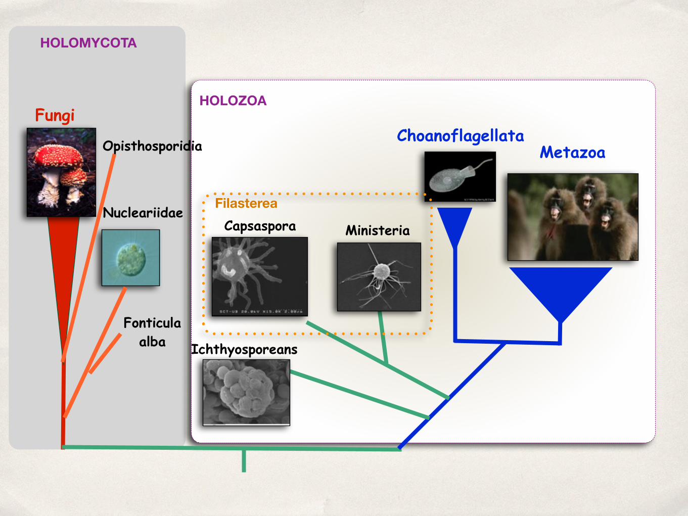
HOLOZOA
Metazoa
FungiChoanoflagellata
Nucleariidae
Ichthyosporeans
Fonticula alba
Capsaspora Ministeria
Filasterea
HOLOMYCOTA
Opisthosporidia

Holomycota
Fungi
Nucleariidae
Fonticula alba
Opisthosporidia

Nucleariidae
free-living heterotrophic amoebas!aprox. 11 species identified.!some have branching filopodia.!some are free-living algal predators.!uni- or multinucleated.!some have intracell bacterial symbionts.!

Fonticula alba
Amoeboid protist that forms a multicellular body by aggregation of several individuals
Brown, M. W. et al. Mol Biol Evol 26, 2699-2709 (2009).

Opisthosporidia
-Cryptomycota (Rozellomycota)-parasites of fungi-like!-Aphelida-parasites of algaea!-Microsporidia-mostly parasite of animals
A-C Rozella; D-G Aphelida
Karpov et al. 2014. Front Microbiol 5: 112.
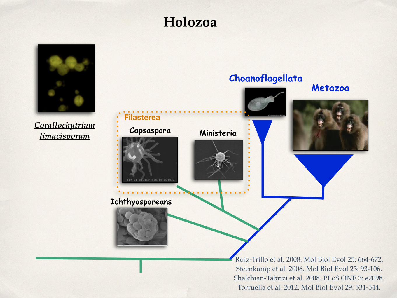
MetazoaChoanoflagellata
Ichthyosporeans
Capsaspora Ministeria
Filasterea
Holozoa
Corallochytrium limacisporum
Ruiz-Trillo et al. 2008. Mol Biol Evol 25: 664-672.!Steenkamp et al. 2006. Mol Biol Evol 23: 93-106.!
Shalchian-Tabrizi et al. 2008. PLoS ONE 3: e2098.!Torruella et al. 2012. Mol Biol Evol 29: 531-544.!

Choanoflagellatea
around 140 species!marine or fresh-water!flagellate!heterotrophic!some form simple colonies
King N (2005) Curr Biol 15: R113-R114.!Carr et al. 2008. Proc Natl Acad Sci U S A 105: 16641-16646.!
Nitsche et al. 2011. . J Eukaryot Microbiol 58: 452-462.

Choanoflagellatea
Codosigidae (“naked”)!Salpingoecidae-theca!Acanthaecidae-basket (silica)
Pula, Italy (39100N, 9110E) in June 2007. Parvicorbicula pedun-culata (Fig. 6) was collected from The English Channel, Hastings(501510N, 01340E) in September 2006, Salpingoeca sp. (Mallorca)(Fig. 1) was collected from surface water at the Bay of Piedra(391550N, 3180E) in May 2006, and Salpingoeca sp. (Vietnam)(Fig. 2 and 3) collected from the River Mekong at the island BinhHoa Phuoc (101190N, 106120E) in May 2005.
DNA extraction and sequencing. Cultures of Salpingoeca sp.(American Type Culture Collection [ATCC] 50931) and Salpin-goeca sp. (ATCC 50938—deposited under the name Salpingoecaminuta) were obtained from the ATCC and genomic DNA ex-tracted from 50-mL cultures following the protocol of Carr et al.(2006). A partial fragment of SSU rDNA was amplified fromSalpingoeca sp. (ATCC 50931) using the primers A and B of
Fig. 1–11. Light and electron microscopical images confirming identity of newly sequenced species included in this text.1. Salpingoeca sp. (Mal-lorca). Flask-shaped cell with flagellum (f) and relatively long collar (c). Rigid theca (arrow t) surrounds cell and is pedicellate (stalked) (p). Bar 5 5mm.2, 3. Salpingoeca sp. (Vietnam). Flask-shaped cell surrounded by theca (arrow t). Bar 5 5 mm. 4. Salpingoeca tuba. Highly elongated cell with long collar(c) located in long relatively narrow tube-shaped theca (arrows t). Bar 5 5mm. 5. Diaphanoeca sp. Cell with long collar (c) bearing an accumulation ofcostal strips (arrows) suspended in voluminous lorica (arrow l) with narrower neck. A pedicellate stalk (p) at base of lorica secures lorica to substratum.Bar 5 10mm. 6. Parvicorbicula pedunculata. Cell located within stalked lorica whose chamber contains 10 longitudinal costae and two transverse costaeone comprising the anterior ring. Junctions between longitudinal costae and costal strips of the anterior ring are of the ‘‘T’’ variety (arrowheads).Bar 5 5mm. 7. Acanthocorbis unguiculata. Cell within lorica containing approximately 15 longitudinal costae projecting forwards as spines (somecollapsed). Inner layer of disturbed helical costae. Bar 5 2mm. 8. Stephanoeca paucicostata. Empty lorica comprising an outer layer of 14 longitudinalcostae and an inner layer of helical costae at base of lorica and three transverse costae, one at base of upper chamber, one in mid-chamber and an anteriorring. Bar 5 5 mm. 8 Inset: S. paucicostata. Light micrograph of living cell. Bar 5 5mm. 9. Stephanoeca apheles. Lorica similar to S. paucicostata (seeFig. 8) but without intermediate transverse costa in upper chamber and limited helical costae in posterior chamber. Bar 5 2mm. 10. Stephanoeca norrisii.Empty lorica, comprising an outer layer of longitudinal costae, some extending from base to the anterior ring and some limited to anterior chamber. Innerlayer of helical costae in posterior chamber and a series of transverse rings at anterior end of lorica. Bar 5 2mm. 10 Inset: S. norrisii. Light micrograph ofliving cell. Bar 5 5mm. 11. Stephanoeca cauliculata. Stalked lorica containing an outer layer of approximately 15 longitudinal costae, which reveal a left-handed rotation in the stalk and posterior chamber. Inner layer of helical costae in posterior chamber and two transverse costae, one at mid-chamber leveland an anterior ring. Bar 5 5 mm. 11 Inset: S. cauliculata. Light micrograph of living cell. Bar 5 10 mm.
453NITSCHE ET AL.—CHOANOFLAGELLATE PHYLOGENY
Nitsche, F. et al. . J Eukaryot Microbiol 58, 452-462 (2011).

Choanoflagellatea
colony Salpingoeca rosetta
Fairclough, S. R. et al. Curr Biol 20, R875-6 (2010).

Choanoflagellatea
Fairclough, S. R. et al. Curr Biol 20, R875-6 (2010).

Filasterea
only two known species!ameoboid, without flagella

Filasterea: Capsaspora owczarzaki
Hertel ET AL. 2002. Int J Parasitol 32: 1183-1191.

Capsaspora owczarzaki attached/filopodial stage
Sebe-Pedros et al. 2013. Elife 2: e01287.!

Capsaspora owczarzaki cystic/floating stage
Sebe-Pedros et al. 2013. Elife 2: e01287.!

Capsaspora owczarzaki aggregative stage
Sebe-Pedros et al. 2013. Elife 2: e01287.!

Filasterea: Ministeria vibrans

Ichthyosporea
also known as DRIPs (acronym of Dermocystidium, rossete agent, Ichthyophonus and Psorospermium) or Mesomycetozoa.!!
some form colonies!most are animal parasites or endosymbionts
Mendoza et al. 2002. Annu Rev Microbiol 56: 315-344.!Glockling et al. 2013. Fungal Ecology 6: 237-247.!

16 Aug 2002 13:56 AR AR168-MI56-14.tex AR168-MI56-14.SGM LaTeX2e(2002/01/18) P1: GJC
THE MESOMYCETOZOEANS 321
Figure 2 Depiction of the putative life cycle of members of the orders Dermocystida(left) and Ichthyophonida (right). Members of the order Dermocystida develop spheri-cal cells with endospores (stage 1). In vitro the released endospores (stage 2) give riseto uniflagellated zoospores (stage 3). When the zoopores (infecting units) infect thehost, they encyst (stage 4) and increase in size (stages 4, 5) and undergo cleavage intoendospores (stage 1). The endospores can also be directly released within the host’sinfected tissues, and the cycle is repeated inside the hosts (stages 1, 4, 5, 1). Mem-bers of the order Ichthyophonida develop spherical (Ichthyophonus spp.) or ovoid cellsin infected tissues (Psorospermium haekeli) or on its hosts (Amoebidium parasiticum)(stage 1). In vitro (stage 1) the hatching of spore receptacles occurs from the ovoid cells(stage 2); the receptacles containing spores (stage 3) then rupture and release their en-dospores (stage 4), which develop intomotile amoeboid cells (infecting units) (stage 5).The amoeboid cell reach the hosts and develops into a small receptacle (stage 6) thatlater generates a hard cell wall with endospores (stages 7, 1). The endospores can alsobe released within their hosts, repeating the cycle (stages 1, 6, 7, 1). Note that in thegenus Ichthyophonus, the development of hyphae that produce spherical cells withendopores (in vitro) is a feature, so far, not encountered in the other members of theorder (62, 64, 83). For the order Ichthyophonida the cell cycle was adapted from Vogt& Rugg (92).
used to describe their phenotypic stages would be of importance in studying themembers of this class. In this review, however, we continue to use the traditionalnomenclature until a consensus is reached regarding their terminology.We provide a brief description of the genera that are currently in the class Me-
somycetozoea, highlighting their similarities and differences, with the intentionof covering the latest known developments of the mesomycetozoeans and intro-ducing the microbiological community to this novel class of microorganisms andtheir morphological, pathogenic, and phylogenetic relationships.
Ann
u. R
ev. M
icro
biol
. 200
2.56
:315
-344
. Dow
nloa
ded
from
arjo
urna
ls.an
nual
revi
ews.o
rgby
DA
LHO
USI
E U
NIV
ERSI
TY o
n 04
/12/
07. F
or p
erso
nal u
se o
nly.
Ichthyosporea
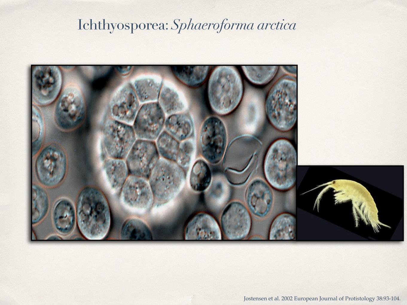
Jostensen et al. 2002 European Journal of Protistology 38:93-104.
Ichthyosporea: Sphaeroforma arctica

Ichthyosporea: Sphaeroforma arctica
http://www.youtube.com/watch?v=NwQk6xsqLJo

the ichthyosporean Creolimax fragrantissima
Marshall et al. 2008. Protist 159:415-433.

the ichthyosporean Creolimax fragrantissima
http://www.youtube.com/watch?v=4ba7arVBKvQ

Corallochytrea
Corallochytrium limacisporum
so far, just one species described: Corallochytrium limacisporum, found in coral reefs in India!!
free-living!marine
16 Aug 2002 13:56 AR AR168-MI56-14.tex AR168-MI56-14.SGM LaTeX2e(2002/01/18) P1: GJC
336 MENDOZA ⌅ TAYLOR ⌅ AJELLO
Figure 12 (a) Single, diad, and tetrad spherical vegetative cells of Corallochytriumlimacisporum (X 1500). (b) Vegetative and elongated limax amoeba cells ofC. limacis-porum from cultures (arrows) (2000) (courtesy of S. Raghu-Kumar).
Dermocystida and Ichthyphonida. These two phylogenetic groups were found todiffer from each other, not only on the basis of their 18S SSU rDNAs and internaltranscriber spacers sequences but also on the morphological stages of their lifecycles (Figure 12). For instance, the production of uniflagellate zoospores wasthe main feature of the Dermocystidium species and the rosette agent, both inthe order Dermocystida. Flagellated cells are not known in R. seeberi, but as men-tioned above, thematter is currently under investigation (Table 2). In contrast, mostmembers of the order Ichthyophonida (Amoebidium, Ichthyophonus, and Psoros-permium spp.) developmotile, amoeba-like cells. No such simple dichotomy existswhen it comes to the morphology of mitochondrial cristae. Both R. seeberi andDermocystidium spp. in the Dermocystida have mitochondria with flat cristae. Therosette agent was reported to have tubular mitochondrial cristae (4). However, theelectronmicrographs of the rosette agent’smitochondria described byArkush et al.(4) had features compatible with flat mitochondrial cristae. Recent morphologicalstudies of the rosette agent’s mitochondria confirmed that it possesses flat cristae(Figure 11c). Alone among the mesomycetozoeans, I. hoferi seems to have tubularmitochondrial cristae. It may be that the morphology of cristae easily changed overevolutionary time or that interpretation of mitochondrial morphology is difficultin this group. As Zettler et al. (101) argued, the finding of different mitochondrialcristae types among microbes considered to be monophyletic should be carefullyevaluated together with other morphological and physiological characteristics toavoid misinterpretations.
CLOSE RELATIVES OF THE MESOMYCETOZOEA ANDTHE EVOLUTION OF TRAITS COMMON TO ALL
The closest members of the mesomycetozoeans are Corallochytrium limacispo-rum and Nuclearia spp. in the classes Corallochytrea and Cristidiscoidea, and thechoanoflagellates (Table 1). Study of these taxa may help us understand the origins
Ann
u. R
ev. M
icro
biol
. 200
2.56
:315
-344
. Dow
nloa
ded
from
arjo
urna
ls.an
nual
revi
ews.o
rgby
DA
LHO
USI
E U
NIV
ERSI
TY o
n 04
/12/
07. F
or p
erso
nal u
se o
nly.
Corallochytrium limacisporum
Raghu-kumar et al. 1987. Indian J Mar Sci 16: 122-125.!Cavalier-Smith & Allsopp. 1996. European Journal of Protistology 32: 306-310.

the opisthokonts


