The Orbits—Anatomical Features in View ofInnovative Surgical MethodsCarl-Peter Cornelius, MD, DDS1 Peter Mayer, MD, DDS1 Michael Ehrenfeld, MD, DDS1
Marc Christian Metzger, MD, DDS2
1Department of Oral and Maxillofacial Surgery, Ludwig MaximiliansUniversity, Munich, Germany
2Department of Oral and Maxillofacial Surgery, Albert LudwigsUniversity, Freiburg, Germany
Facial Plast Surg 2014;30:487–508.
Address for correspondence Carl-Peter Cornelius, MD, DDS,Department of Oral and Maxillofacial Surgery, Ludwig MaximiliansUniversity, Lindwurmstr 2a, 80337 Munich, Germany(e-mail: [email protected]).
The orbits are a pair of symmetrical mirror–imaged bonyhousings positioned in the craniofacial transition territory.They enclose and protect the ocular globes, complexly orga-nized organs of vision collecting and converting light intoelectrochemical signals to enter the anterior gate of the visualpathway, which extends from the photoreceptors in theretina back to the occipital brain lobe next to the calcarinesulcus. The eyes, retinae, optic nerves, chiasma, and optictracts are part of the central nervous systemormore preciselyanterior prolongations and constituents of the diencephalon.Binocular vision is based on the perception and fusion ofinformation from either side. The actual visual organ, the eyeor ocular globe, is surrounded by auxiliary structures main-taining or expanding its functionality such as the eyelids, thelacrimal apparatus, the extraocular muscles including theoptomotor nerves (cranial nerves—CN III, IV, and VI), theblood vessels, and the orbital fat body partitioned by a
delicate system of connective tissue septa.With the exceptionof the eyelids, the globes and the auxiliary structures areaccommodated within the bony orbital cavities.
The osseous orbital framework is connected by canals,fissures, and foramina to the middle cranial fossa as well as tothe internal nose, the ethmoid sinuses, the pterygopalatineand infratemporal fossa, and the outer skeletal surfaces of themidface and anterior cranial vault.
Apart from the optic nerve (CN II), numerous neural andvascular structures pass through the openings in the orbitalapex that is made up by the sphenoid which can be under-stood as kind of a backup structure for the whole midface.
It is pure coincidence that the orbit lends itself to a wordplay with its two last letters. Oddly enough, they are identicalwith the acronym for information technology. In fact, much ofthe ongoing revolution in craniofacial surgery started with ITapplications to optimize the assessment of orbital trauma and
Abstract The aim of this article is to update on anatomical key elements of the orbits in referenceto surgical innovations. This is a selective literature review supplemented with thepersonal experience of the authors, using illustrations and photographs of anatomicaldissections. The seven osseous components of the orbit can be conceptualized into asimple geometrical layout of a four-sided pyramid with the anterior aditus as a base andthe posterior cone as apex. All neurovascular structures pass through bony openings inthe sphenoid bone before diversification in the mid and anterior orbit. A set oflandmarks such as the optic and maxillary strut comes into new focus. Within thetopographical surfaces of the internal orbit the lazy S-shaped floor and the poster-omedial bulge are principal determinants for the ocular globe position. The inferome-dial orbital strut represents a discernible sagittal buttress. The periorbita and orbital softtissue contents—extraocular muscles, septae, neurovasculature—are detailed and putinto context with periorbital dissection.
its repair.1,2 These technologies in conjunction with thetremendous capabilities of endoscopically assisted surgeryof the paranasal sinuses and skull base3–6 have given newinsights into the details of orbital anatomy and have trans-formed the basic understanding of internal surface contoursin relation to volume, ocular globe position, and binocularfunction.7–13 Of course, fundamental anatomical facts andvariations have not changed, but refined ways of descriptionproviding unfamiliar facets and a newly developing anatomi-cal jargon among orbital surgeons make the differencenowadays.
Bony Orbits—Margin and Orbital Walls
The bony orbit or orbital cavity is conceived as a three-dimensional (3D) geometric assembly in form of a four-sidedpyramid with a triangular tip or apex. The base of thispyramid corresponds to the wide aperture or aditus orbitaeoriented frontolaterally to the face, whereas the narrowedapical end is pointing posteriorly toward the middle cranialfossa.14–16
The medial walls of the two orbital pyramids are arrangedalmost in parallel with a corridor of 2.5 cm width for theethmoid in between. The angle between each lateral andmedialwall is approximately 45degrees; hence, the extensionlines along the lateral walls of both the orbits meet at rightangles. The orbital axes which are imaginary bisecting linesbetween the medial and lateral walls converge toward theorbital apices.
The circumference of the orbital aperture is framed by asuperficial orbital margin. The margin is formed by themaxilla, the zygoma, as well as the frontal and lacrimalbones. This marginal rim is thick, prominent, and welldefined except for the medial side, which is discontinuousbecause of the interposition of the fossa for the lacrimal sacbetween its lower and upper part.17 The medial rim or nasalorbital margin consists of the frontonasal process of themaxilla, the lacrimal bone, and the maxillary process ofthe frontal bone. The frontonasal process extends upwardinto the anterior lacrimal crest and makes up the lowermedial margin leveling off toward the frontonasal sutureline. The supraorbital rim continues into the posterior lacri-mal crest in a more backward plane and creates a secondparallel bone ridge and the fossa lacrimalis in between afluted bevel.
The superior and inferior orbital margins curve distinctlyposterior, so that the lateral rim is least projecting in thewhole orbital circumference. The four walls of the humanorbit or the internal orbit, respectively, are formed by thefollowing seven bones: frontal, zygoma, maxilla, palatine,lacrimal, ethmoid, and sphenoid.
The roof of the orbit is composed largely of the orbital plateof the frontal bone anteriorly and of the lesser wing of thesphenoid (LWS) with a minor part in the posterior part. Thetriangular shape of the roof narrows toward the orbital apex.The anterior portion of the frontal bone contains the frontalsinuses, which can extend far up into the squamous part ofthe frontal bone and far back over the orbital roof when
extremely pneumatized. Thefloor of the anterior cranial fossaforms the endocranial side of both the orbital roofs.
The fossa for the lacrimal gland is a shallow depression inthe anterolateral aspect of the roof next to the zygomatico-frontal suture (ZFS). A small depression in the anteromedialportion of the roof, the trochlear fovea, is the site of attach-ment for the fibrocartilaginous ring (pulley) girdling thetendon of the superior oblique muscle.
The medial orbital wall is formed, again in the anteriorposterior direction, by the frontonasal maxillary process, thelacrimal bone, the lamina papyracea of the ethmoid bone,which is quadrangular in shape, and the anterolateral surfaceof the sphenoid body. The anterior and posterior ethmoidalforamina (EF) are located superiorly along the frontoethmoi-dal junction and indicate the level of the cribriform plate.
The lateral orbital wall consists of the lateral orbitalprocess of the zygoma constituting the anterior part andthe greater wing of the sphenoid (GWS) posteriorly. Thezygomaticosphenoid suture (ZSS) line is located in the thin-nest portion of the lateral wall, which becomes obvious inaxial cross sections. The axial cut through the GWS takes on athickening triangular shape in transit to the middle cranialfossa floor. The posterolateral GWS surface contributes to thetemporal and infratemporal fossa configuration. The ZSS is apertinent landmark for realignment of the fractured zygomafrom inside the orbit.18 Whitnall’s tubercle is situated on theorbital surface of the frontal process of the malar bone afew mm behind the orbital margin, 2–4 mm posterior to therim and 11 mmbelow the ZFS.19 The blunt eminence of 2 or 3mm—in generic nomenclature the marginal orbital tubercleon the zygoma—is the common attachment for the compo-nents of the lateral retinacular suspension complex, that is,the lateral canthal tendon or palpebral ligament, lateral hornof the levator aponeurosis, the so-called lateral check liga-ment of the lateral rectus muscle, and Lockwood suspensoryligament of the ocular globe inferiorly. In the anterolateralintraorbital surface of the lateral wall, anterior to the inferiororbital fissure (IOF) the zygomatico orbital foramina areexiting either with separate orifices or as a common grooveto the external bony surface of the zygoma. Within the bone,they continue as two canals to the zygomaticofacial andzygomaticotemporal foramina.
The “cranio-orbital” foramen also known by the names“lacrimal,”20,21 “meningo-orbital,” and “sphenofrontal” fora-men22 is located in the GWS anterolateral to the upper tip ofthe superior orbital fissure (SOF).
The floor of the orbit is shorter in its anteroposteriorextent than the three other orbital walls and terminates inthe IOF in front of the orbital apex that consequently turnsinto a triangular frontal cross section. For its most part, thefloor consists of the orbital plate of themaxilla supplementedby the tiny orbital plate of the palatine bone posteriorly andby the inferior orbital process of the zygoma anterolaterally.As a portray image of the orbital roof the floor takes atriangular shape, it is thin-walled and forms the superiorboundary of the maxillary sinus. The anteromedial cornerwhere the floor is continuous with the medial wall is perfo-rated by the nasolacrimal canal.
Facial Plastic Surgery Vol. 30 No. 5/2014
Anatomical Key Elements of the Orbits Cornelius et al.488
Dow
nloa
ded
by: I
P-P
roxy
Alb
ert-
Ludw
igs-
Uni
vers
ität F
reib
urg,
Alb
ert-
Ludw
igs-
Uni
vers
ität F
reib
urg.
Cop
yrig
hted
mat
eria
l.
The infraorbital groove opens in the middle section of theanterior border of the IOF posteriorly and runs forward in themidline of the orbital floor to convert into the infraorbitalcanal anteriorly. This canal usually projects into themaxillarysinus as a marked bony molding that ends at the facial antralwall with the infraorbital foramen.
Optic CanalThe medial wall and the roof of the orbit extend posteriorlyto the orbital end of the optic canal. The canal is 4 to 9.5 mmin diameter and 5.5 to 11.5 mm in length,6,23 and it takes apath with 45 degrees inward and 15 degrees upwardangulation through the junction of the sphenoid bodyand the LWS. The canal transmits the optic nerve, theophthalmic artery, and accompanying sympathetic nervefibers. The entrance or optic foramen opens in the superiormost and medial most corner of the orbital apex.15 Thecanal has an elliptical cross section whose major diameterlies in a horizontal plane, accordingly it is considered asbuilt up from sections of an ovoid outline. The bony layer ofthe upper arc corresponds to the anterior root of the LWSwhich is contiguous with the planum sphenoidale in theanterior cranial fossa. The inferomedial canal circumfer-ence is commonly formed by the sphenoid body. However,this pattern is subject to variation by sphenoethmoidal aircells, named Onodi cells. The Onodi cell, the most posteriorethmoid cell, may extend posteriorly and laterally into thesphenoid sinus with intimate contact to the optic nerve dueto excessive pneumatization and amissing bony protection.Laterally, the optic canal is separated from the SOF by theoptic strut. This is a bony bridge linking the base of theanterior clinoid process to the sphenoid body. Sometimes,the optic strut is addressed as the posterior LWS root.Occasionally, the optic strut itself presents with an extraforamen, which transmits the ophthalmic artery having alow origin from the internal carotid artery. This is referredto as ophthalmic foramen.
The optic strut is pneumatized from its inside, whatcreates the lateral opticocarotid recess in the posterior partof the sphenoid sinus. This is extending between the opticnerve superiorly and the intracavernous carotid arteryinferiorly.
Superior Orbital FissureThe SOF separates the posterior part of the lateral orbital wallfrom the roof, more precisely, it is a gap interposed betweenthe LWS and the GWS on the lateral side of the optic foramen.The fissure has a diagonal course sloping downward from thelateral apex to its medial base, where, it is bounded by thesphenoid body. Frequently, it is club-shaped24 with a narrowlateral top end and a wide medial bottom portion. However,various subtypes can be distinguished.25 The optic strut lies atthe superomedial border of the fissure in an angle positionbetween the upper and medial SOF edges. The orientation ofthe SOF deviates slightly from the coronal plane resulting
from a forward shift of the lateral apex. The SOF connects theorbit to the middle cranial fossa.
The annulus of Zinn26 (synonyms: common tendinousring, common annular tendon or Zinn’s ring, tendon of Lock-wood, and optic ring) is a circular array of connective tissueenfolding in the orbital apex. Zinn’s ring is the origin of thefour extraocular rectus muscles. The upper half of the ring,also named the superomedial foramen, consists of the supe-rior rectus muscle which encompasses the optic foramen andthe optic nerve. The laterally oriented half of the tendinousring, the superolateral foramen, is formed by the lateral,inferior, andmedial rectusmuscles and loops over thebottomportion of the SOF to demarcate the confines of the optomotorforamen, a compartment that inter alia transmits the oculo-motor (upper and lower division) and the abducens nerve.The medial side of the optomotor foramen is made up by theoptic strut. A small rectangular and flat or a pointed bony spurprotruding from the lateral SOF margin, the spina rectuslateralis serves as a site for the bony attachment of Zinnring.27 Inferiorly, the SOF terminates at a transverse bonyconfluence above the foramen rotundum, which is referred toas maxillary strut.20,21,28 Its edges along the lower SOF endproceed forward like the verts of a superficial half-pipewhichblends with the medial end of the IOF.
Inferior Orbital FissureThe IOF separates thefloor from the lateral wall of the orbit intheir posterior two-thirds. Its long axis is oriented in aposteromedial to anterolateral direction from the exocranialsurface of the maxillary strut to the tip of a loop in betweenthe anterior and the lateral orbital plate of the zygoma, theadjacent margins of the GWS, and the orbital plate of themaxilla. Posteromedially, the sphenoid body and the palatinebone contribute to the IOF formation. With the view fromabove, the IOF simply has the outline of a double-ended spoonwith a narrow intermediate grip. In fact, it is a rather complex3D space providing passageways and portals for vessels andnerves or fat pads to the pterygopalatine, infratemporal, andtemporal fossae. The narrowing (isthmus) in the center of theorbital opening of the IOF results from a crescent-shapedpromontory of the orbital floor lateral to the orbital plate ofthe palatine bone andmedial to the infraorbital groove that isprojecting posteriorly beyond the actual medial IOF rim(IOF isthmus promontory).
A smooth muscle going by several names such as, Müller’s(vestigial) muscle, periorbital muscle, or musculus orbitalis(Müller),29 is filling the IOF along the entire extent.28 It isunderstood as part of the orbital connective tissue system andthe periorbital lining. The orbital muscle of Müller yields aceiling which spans over the bony margins of the entire IOFlength and crosses over the maxillary strut to enter the SOF.
Ethmoidal ForaminaThe EF are arranged along the suture line between theethmoid and frontal bone. The number of EF varies, rangingfrom a single foramen over the prevalent double up to asextuple array.30 The most anterior and most posterior EF arecommonly defined as the anterior EF (AEF) and posterior EF
Facial Plastic Surgery Vol. 30 No. 5/2014
Anatomical Key Elements of the Orbits Cornelius et al. 489
Dow
nloa
ded
by: I
P-P
roxy
Alb
ert-
Ludw
igs-
Uni
vers
ität F
reib
urg,
Alb
ert-
Ludw
igs-
Uni
vers
ität F
reib
urg.
Cop
yrig
hted
mat
eria
l.
(PEF). The proximity of the PEF to the optic foramen is ofcritical significance in periorbital dissection to avoid opticnerve injury. Themean PEF–optic foramen distances reportedin the literature ranges between 4.3 and 9.15 mm.30
Cranio-Orbital ForamenThe cranio-orbital foramen COF is laying either in the GWSlateral to the narrow lateral end of the SOF or it is confluentwith it. The size, frequency of accessory foramina, and precisetransverse/vertical position of the COF has been analyzedquantitatively.31 Several variations, also in the arterialbranching pattern must be expected.
Supraorbital NotchThe exit of the supraorbital neurovascular bundle, the supra-orbital notch, or the foramen is located approximately on theborderline between the medial third and the lateral twothirds of the supraorbital rim. When a notch is present, afascial band completing the ring is a common finding.32
With a coronal approach appreciation is needed, that thesupraorbital nerve trunk beyond the orbital rim splits up toform two consistently present divisions: a superficial medialportion which pierces the frontalis muscle and fans out overits surface for sensory supply to the forehead and anteriormargin of the scalp and a deep lateral division that runs asuperolateral course across the forehead in a layer over thepericranium and deep to the galea aponeurotica plane to andin parallel to the superior temporal fusion line reach thefrontoparietal scalpmedially.33,34Only a subset of individualsreceives a dual innervation of the frontoparietal scalp fromboth the superficial and the deep division. Preservation of thefrontoparietal scalp sensation therefore requires a specialcoronal flap design to protect the deep division of thesupraorbital nerve.33
Nasolacrimal CanalThe nasolacrimal canal is formed by the maxilla, the lacrimalbone, and the lacrimal process of the inferior nasal concha. Itsurrounds the draining membranous lacrimal duct, is direct-ed downwards, backward and laterally thus producing a ridgein the anteromedial recess of the maxillary sinus. The canal isapproximately 12 to 15 mm long and has a diameter of 4 to5 mm. The lower opening is into the inferior nasal meatus.
Infraorbital foramenThe infraorbital foramen and the infraorbital canal belong tothe orbital part of themaxilla. Their spatial characteristics areabundantly documented in terms of distances of the foramenfrom the infraorbital rim, the facial midline, and the lateralrim of the piriform aperture but with awide range of differentvalues.35–37 The foramen is located between 7 and 10 mmbelow the infraorbital rim. Supernumerary foramina may bepresent.
Foramen rotundum—Maxillary Strut—Pterygoid CanalThe foramen rotundum penetrates the pterygoid process ofthe sphenoid bone below the medial portion of the SOF andlinks the middle cranial fossa with the pterygopalatine fossa
(PPF). The maxillary strut refers to the narrow bridge of boneseparating the foramen rotundum from the SOF.
The pterygoid canal, also known as Vidian canal is locatedinferomedial to the foramen rotundumandopens into the PPFanteriorly. The canal runs above the medial pterygoid platealong the junction with the sphenoid body.
Surface Contours, Topographical Shape, Dimensions,and Volume of the Internal OrbitDetailedmapping of the complex bony surfaces of the orbit, inparticular, the topography of the floor in transition to themedial wall including the so-called posterior medial bulgehas been taken to a new level by automated analysis ofcomputed tomographic (CT) data sets from routine exami-nations of patients with unaffected regional anato-my.12,13,38–43 In comparison to this, “big data mining”studies based on traditional manual caliper measurementsmust appear anachronistic. The visual impression of thepublished 3D orbital shapes and forms, however, elude exactnarrative description and will need direct inspection in therespective articles.
The orbital floor inclination on coronal plane, that is, theangle between the floor and a horizontal plane is steeper inmales than in females and decreases with age independent ofthe measurement in the anterior, middle, or posterior frontalplane. With aging, the lowest point of the orbital floor movesinto a posterior and inferior direction.38 The latter parameterprovides new estimates for the extent of the concavity rightbehind all orbital rims. This widening of the orbital cavity isbest appreciated in the sagittal sections of the conical orbit,aptly, it is referred to as the “postentry zone” according to thesequence in periorbital dissection of the floor.15 From thebottom of the postentry zone, the floor is continuouslyascending posteriorly up to the convex top of the orbitalplate of the palatine bone, which curves strongly downwardinto the rear end of the IOF. The term “lazy S-configuration”eventuates from the undulating shape of the orbital floor inparamedian sagittal planes medial to the infraorbital groove.The lazy S-shape revealed great variability with subtle tosteep curvatures as a function of the anteroposterior floorinclination.13 Along the maxillo-ethmoidal suture line, theorbital floor slopes medially upward and merges with themedial wall. The posterior terrain of this transition zone,which we identify as the IOF isthmus promontory may takeseveral forms from a flat, almost seamless silhouette, over anintermediate soft-edged terrace arrangement to a sharp bentbuckling.13 In abstraction of these variations, the inferolat-eral–medial wall transition directly behind the globe isclaimed to integrate a focal convex sinus bulging, which isconsidered the most essential support mechanism to main-tain the vertical position and anterior projection of the ocularglobe. It is called the “posteromedial bulge” or “the key area,”9
as this region is a main issue in orbital reconstruction. CT-databased 3D contour mapping and virtual molds of theorbital floor topography give some fundamental ideas whereto figure it out.12 It has become clear, however, that theposteromedial bulge remains a “fuzzy” surface structure, asits boundaries lack distinct anatomical criteria and are
Facial Plastic Surgery Vol. 30 No. 5/2014
Anatomical Key Elements of the Orbits Cornelius et al.490
Dow
nloa
ded
by: I
P-P
roxy
Alb
ert-
Ludw
igs-
Uni
vers
ität F
reib
urg,
Alb
ert-
Ludw
igs-
Uni
vers
ität F
reib
urg.
Cop
yrig
hted
mat
eria
l.
difficult to capture in automated digital shape analysis.40
As a consequence, the posteromedial bulge still needs to beoutlined manually in shape modeling based on surgicalexpertise and to transform it into an “embedded” region ofinterest. In males, larger and higher bulges were demon-strated than in females,12,13 but in the laterality evaluation,the topographical shapes were symmetrical between leftand right sides.12,40 The IOF isthmus promontory is thelateral offshoot of the posteromedial bulge. While the far-ended verge of the promontory is solid and coincides withthe orbital process of the palatine bone for the most part,the IOF isthmus promontory itself corresponds to theposterior recess of the maxillary sinus and ergo is hollow.The size of the orbital floor in terms of the parameters’width and length showed a limited number of differentgender-dependent patterns in general with smaller dimen-sions in females.12 In traumatic defect-like conditions, theprofile of the posteromedial bulge is commonly lost, butmost of the time the orbital plate of the palatine boneremains intact and can serve as a “posterior ledge” inreconstruction.44 The zygomatic/sphenoidal centerpieceof the internal lateral orbit displays a straight and evensurface plane from the anterior rim to the lateral edge of theSOF in an angle position of approximately 45 degrees to itsrespective medial wall. The length of the lateral orbital wallis approximately 1.0 cm shorter than the medial wall and itappears even shorter owing to its angulation and thebackward position of its outer rim.
The orbital cone angle characterizes the average medial/lateral wall angulation in the mid and posterior orbit, whichhas been evaluated in a 3D-based geometric measurementapproach using planes fitting the two walls. In stark contrastto the 45 degrees angulation just mentioned, the values for anEuropean white ethnicity ranged between 41 degrees and 63degrees.41 The dimensions and volumes of the orbit varygreatly because of ethnic, gender, and age differences, andescape from normative quantification.39 Although some val-ues typically found in adults may serve as initial orientation:the anterior opening measures 4 cm horizontally and 3.5 cmvertically, the depth anteroposteriorly is approximately 4.5 to5 cm—the overall orbital volume amounts to 30 mL, of which7 mL go to the ocular globe volume.15 For instance, theinterindividual variation of orbital volumes for an Europeanwhite ethnic group was lying between 20 to 29 mL (mean,24.4mL) in females and 22 to 32mL (mean, 26.8mL) inmales.The globe volume ranged from 6.5 to 9.3 mL in females andfrom 6.7 to 9.7 mL inmales.41 The sagittal orbital rim positionand angulation in relation to themedial rim (anterior lacrimalcrest) has been assessed in a two-dimensional and 3D fashionagain showing considerable gender-specific interindividualvariations.42
Inferomedial Orbital StrutThe inferomedial orbital strut (IOS) is rather a conceptualthan a discrete anatomic structure along the maxillo–eth-moid junction.45 In biomechanical terms and clinical par-lance, it is referred to as internal orbital buttress, a boneformation in sagittal direction, reinforcing the brinks of the
suture lines in the transition between the inferior and medialorbital wall. The IOS can be considered as one constituent of aset of three stabilizing sagittal ridges crossing the orbital floorin parallel—the reinforced medial bony IOF edge laterally, thebony surround of the infraorbital groove/canal intermediaryand the IOS medially.
Anterior Orbit—Midorbit—Posterior OrbitIt is a frequent practice to divide the orbit into three thirdsalong its anteroposterior extension (►Fig. 1). This threefolddivision is arbitrary after all and has no precise relation-ships or metric distances between acknowledged referencepoints or planes. The conical apex with its triangular crosssection is usually considered the posterior third of the orbitso that the floor extends over the remaining two thirds ofthe orbital depth. Regardless of any metric measurementsand a correctly scaled ratio of three, it appears ameaningfulapproach to implement the IOF as indicator to partition ananterior, a mid, and a posterior orbit. A frontal plane at thelevel of a tangent to the tip of the anterior loop of the IOFforms the boundary between the anterior and themidorbit,whereas a second frontal plane passing through the maxil-lary strut separates the midorbit from the posterior orapical orbit.
Soft Tissue Contents
PeriorbitaThe periorbita is the periosteal lining of the internal orbit andcovers the four orbital walls from the anterior aperture of theorbital cavity back to the conical apexwith the optic canal andthe SOF (►Fig. 2). The periorbital envelope provides protec-tion for the orbital soft tissue contents, in particular, it holdsthe orbital fat. The periorbita is not very adherent to the bonysurfaces except for some firm attachments to the suture lines,the trochlea, and the Whitnall tubercle. The inner side of theperiorbita facing toward the orbital cavity does not have asmooth surface, as the radiating septae of internal connectivetissue system attach there.
Anteriorly, the periorbita is continuous with the perioste-um over the orbital rims and the orbital septum in the upperand lower lids as well as the canthal structures (►Fig. 3). Thefusion zone between periorbita and periosteum is thickened,making up the arcus marginalis. This is the origin of theorbital septum, which corresponds to the deepest layer of theaponeurotic galea.33,46,47 Posteriorly in the orbital apex, theperiorbita communicates with the dura mater of the middlecranial fossa and the cavernous sinus walls through the opticcanal and the SOF. The periorbita covering the apex contrib-utes to the formation of Zinn’s ring in conjunction withfibrous components of the dura lining the SOF and opticcanal, the optic sheath,20,21 and the periosteum of the sphe-noid body.48
In the superolateral quadrant of the anterior orbit, theinferolateral surface of the orbital lobe of the lacrimal glandmay be invested by a fascia emanating from the adjacentperiorbita.47 Inferolaterally, the periorbita invaginates the IOFand bonds with the fibers of Müller’s orbital smooth muscle,
Facial Plastic Surgery Vol. 30 No. 5/2014
Anatomical Key Elements of the Orbits Cornelius et al. 491
Dow
nloa
ded
by: I
P-P
roxy
Alb
ert-
Ludw
igs-
Uni
vers
ität F
reib
urg,
Alb
ert-
Ludw
igs-
Uni
vers
ität F
reib
urg.
Cop
yrig
hted
mat
eria
l.
before its sheath duplication continues into the periostealcoating of the pterygopalatine and infratemporal fossae.
At the posterior lacrimal crest, the periorbita splits intotwo layers. A thin layer intervenes between the lacrimal fossa
and a thicker extension, the fascia lacrimalis, invests thelacrimal sac between the posterior and anterior lacrimalcrest.49,50 Along the frontoethmoidal junction, the exitingethmoidal neurovascular bundles are covered by sleeve-like
Fig. 1 Anterior Orbit, Midorbit, and Orbital Apex. (A) Upper left: Orbital apex–spatial overview with Zinn’s ring, extraocular muscles, and opticnerve. The optic canal, the SOF, and the posterior IOF represent the passageways between the middle cranial fossa and the orbit via the apex. (B)Upper right: Orbital apex–anterior coronal section breaking down the triangular bony cross section. The density of the orbital connective tissuesepta is less pronounced than in the midorbit. (C) Middle left:Midorbit–spatial overview with posterior sphere segment of ocular globe covered bythe according segments of the four rectus, the levator, and inferior oblique muscles. Superior oblique muscle segment adjacent to superomedialbony walls. Several bony landmarks determine this level: infraorbital groove, zygomaticosphenoid suture at the anterolateral circumference,ethmoidal foramina, lamina papyracea, and IOS/IOB. (D) Middle right: Midorbit–anterior aspect of coronal section just in front of the IOF.Throughout the midorbit the cross section is four-sided with a convex shifting of the inferomedial walls to the inside of the cavity. The floor and thelateral wall are separated with the beginning of the anterior IOF loop. The posterior end of the midorbit concurs with the orbital process of thepalatine bone. The thickness of the lateral wall varies considerably. The GWS makes up the major part of the lateral wall. It has a thick axial crosssection at the trigone over the temporal lobe extension of the middle cranial fossa and is thinned near the zygomaticosphenoid suture line. Theorbital connective tissue septa are most extensive and complex in the anterior orbit interconnecting the extraocular muscles and linking them tothe orbital walls. (E) Lower left: The anterior orbit corresponds to the segment in front of anterior IOF loop. The concave “postentry zone”represents the largest orbital circumference in the frontal plane. The anterior orbital floor is contiguous with the zygomatic orbital plate laterally.The tarsal plates and the orbicularis oculi close the front of the anterior orbital aperture. The insertion of the superior oblique muscle and therectus superior muscle is covered by the levator muscle which fans out. (F) Lower right: Anterior orbit frontal transview giving insight to the bonyand soft tissue structures of this level: lacrimal fossa for the lacrimal sac, inferior oblique muscle and its insertion, globe and insertions ofextraocular muscle, trochlear fossa and pulley for superior oblique tendon, tarsal plates, medial and lateral canthi, and Whitnall’s tubercle. Wholeset of illustrations originates from Fig. 4 of Jacquiéry C, Leiggener C, Cornelius CP, Kunz C. Aktuelle Behandlungsstrategien von knöchernenVerletzungen der Orbitae. OP Journal 2013;29 (2):136–146;Abb.1, c1–c6. Slightly modified and corrected. Redrawn and modified from Fig. 3A(Atlas of orbital surgery) of Rootman J, Stewart B, Goldberg R. Orbital Surgery: A Conceptual Approach. Philadelphia, PA/New York, NY: LippincottRaven; 1995:148.15 GWS, greater wing of sphenoid; IOB, internal orbital buttress; IOF, inferior orbital fissure; IOS, inferomedial orbital strut; SOF,superior orbital fissure.
Facial Plastic Surgery Vol. 30 No. 5/2014
Anatomical Key Elements of the Orbits Cornelius et al.492
Dow
nloa
ded
by: I
P-P
roxy
Alb
ert-
Ludw
igs-
Uni
vers
ität F
reib
urg,
Alb
ert-
Ludw
igs-
Uni
vers
ität F
reib
urg.
Cop
yrig
hted
mat
eria
l.
periorbital extensions following them into their respectiveforamina.
Small orbital vessel branches given off from the infraorbi-tal artery are penetrating the periorbita in the orbital floor toset up anastomotic connections with the muscular branchesof the ophthalmic artery at the level of the inferior obliqueand rectus muscles.51,52 The branches are encountered pos-terior to the postentry zone arising from the infraorbitalgroove, when the periorbita is lifted from the orbital floor(►Fig. 4).
While the inferior aspect of the periorbital envelope ex-tends gently upward into the medial side just like theunderlying bone formation, laterally it turns abruptly overthe medial border of the IOF. There can be tight adhesionsbetween the periorbita covering the infraorbital groove andthe epineurium of the nerve underneath it.
Intraorbital—Extraocular MusclesThere are six extraocular muscles (►Fig. 1A, C, E and ►Figs.
5–7), which are involved in coordinated voluntary ocular
motility, the four rectus muscles (superior, lateral, medial,and inferior), and the two oblique muscles (superior andinferior).53 The levator palpebrae superioris muscle acts aretractor of the upper lid. The four recti and the superioroblique muscles originate from or around the commonannular tendon (Zinn’s ring) (►Fig. 5). The levator palpebraearises from the orbital apex above the superior rectus. Itsorigin from the LWS is not considered a part of Zinn’s ring,albeit it proceeds forward together with the superior rectusand has a fused medial fascial sheath.
The superior oblique muscle arises from the periorbita onthe body of the sphenoid superomedial to the optic foramenand courses anteriorly and superiorly in close juxtaposition tothe superior medial bony walls. As it approaches the orbitalrim, the muscle narrows and becomes tendinous. The roundtendon passes through the trochlea, a fibrocartilaginouspulley that is secured to the trochlear fovea of the frontalbone. The trochlea redirects the tendon at an acute anglebackward, outward, and downward toward its insertion onthe posterosuperior surface of the sclera lateral to the
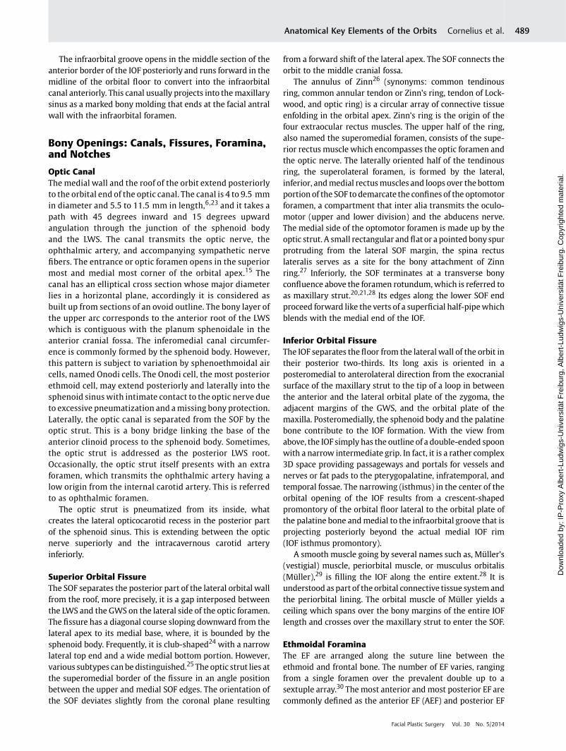
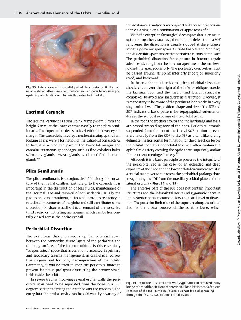
Fig. 2 Periorbita after removal of the lateral wall and roof of left orbit.Trigeminal ganglion and divisions with the anteromedial and antero-lateral triangle in between exposed in medial cranial fossa. Theophthalmic nerve (CN V1), the smallest of the three trigeminaldivisions, courses forward in the lower portion of the lateral wall of thecavernous sinus to reach the SOF.
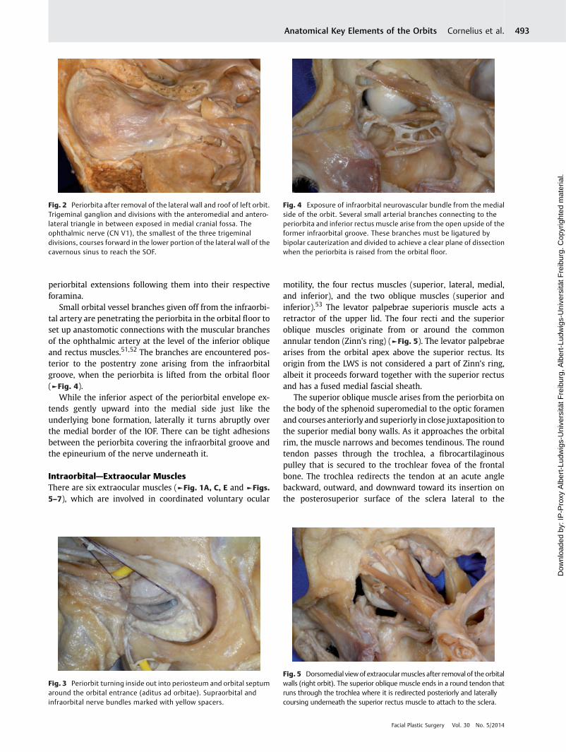
Fig. 3 Periorbit turning inside out into periosteum and orbital septumaround the orbital entrance (aditus ad orbitae). Supraorbital andinfraorbital nerve bundles marked with yellow spacers.
Fig. 4 Exposure of infraorbital neurovascular bundle from the medialside of the orbit. Several small arterial branches connecting to theperiorbita and inferior rectus muscle arise from the open upside of theformer infraorbital groove. These branches must be ligatured bybipolar cauterization and divided to achieve a clear plane of dissectionwhen the periorbita is raised from the orbital floor.
Fig. 5 Dorsomedial view of extraocularmuscles after removal of the orbitalwalls (right orbit). The superior oblique muscle ends in a round tendon thatruns through the trochlea where it is redirected posteriorly and laterallycoursing underneath the superior rectus muscle to attach to the sclera.
Facial Plastic Surgery Vol. 30 No. 5/2014
Anatomical Key Elements of the Orbits Cornelius et al. 493
Dow
nloa
ded
by: I
P-P
roxy
Alb
ert-
Ludw
igs-
Uni
vers
ität F
reib
urg,
Alb
ert-
Ludw
igs-
Uni
vers
ität F
reib
urg.
Cop
yrig
hted
mat
eria
l.
longitudinal axis of the globe. On its way there, the tendonfans out and passes below the superior rectus muscle. Theinferior oblique muscle is unique in that it originates from aspot just posterior to the infraorbital rim and lateral to theentrance into the nasolacrimal canal. It runs laterally andposteriorly following underneath the curvature of the globepassing between the inferior rectus muscle and the orbitalfloor toward its insertion on the posterolateral surface of thesclera underneath the lateral rectus muscle. The rectusmuscles follow the curvature of the globe posteriorly andtheir terminal tendonsfinally attach to the superior temporal,nasal, and inferior sclera anterior to the equator of the globe(►Fig. 6).
TrochleaThe trochlea is a complex structure consisting of four maincomponents, a grooved cartilaginous flange, a bursa-likesynovial space lining, the inner surface of the cartilaginoustrochlea that is created by a fibrillar highly vascular sheathand an outer dense fibrous condensation fixing the trochleafirmly to the fovea.54
Zinn’s Ring—Common Annular TendonZinn’s ring or the common annular tendon is composed oftwo tendinous portions, the inferior and superior tendon,which form a superolateral and superomedial annular fora-men. Zinn’s original description55 referred to the lower ringportion, which he named “de ligamento communi” or thecommon tendon. This inferior tendon is located inferior to theoptic foramen and serves as the origin for the lateral, inferior,and medial rectus muscles. These muscle form a gutter-like
track,48 which acts as a guide rail for the oculomotor nervedivisions (CN III), the nasociliary, and the abducens nerves(CN VI). The upper ring portion, the Lockwood tendon, insertson the superior margin of the optic foramen and gives rise tothe superior rectus and a variable superior head of the lateralrectus muscle. If the latter is present, it joins the main lateralrectus across the SOF. The upper ring tendon is fusedwith thedural sheath of the optic nerve in this region. At theirposterior extension, the two tendons or foraminal tubescommingle into a fibrous cone that reaches intracraniallythrough the SOF before insertion at the infraoptic tubercle orinside an infraoptic canal. These alternating structures arelocated in the body of the sphenoid beneath the optic strut.48
The optic strut lies inside Zinn’s ring.
Oculomotor ForamenThe oculomotor foramen is identical with the superolateralannular foramenwithin Zinn’s ring and represents the centralsector of the SOF.20,21,47 Besides CN III, CN VI, and thenasociliary nerve, it transmits the sensory and sympatheticroots of the ciliary ganglion.
Fig. 6 Dorsolateral view of extraocular muscles and globe (left orbit).Zinn’s ring (light green) and its two foraminal subcomponents aroundthe SOF and the anterior end of the optic canal are a funnel-structuredconnective tissue assembly in the orbital apex formed by the peri-orbita, the dural SOF lining and the optic nerve sheath. Detail (upperright corner): Anterior view of Zinn’s ring featuring the funnel structure.The lateral rectus muscle has a vertical attachment line to the scleraand globe. The insertion of the superior oblique muscle on the scleralies lateral to the vertical globe axis between the superior and lateralrectus muscles. The inferior oblique muscle traverses underneath thelateral rectus muscle and also inserts on the sclera between thesuperior and lateral rectus muscles. The nerves running outside Zinn’sring (CN IV, frontal, and lacrimal nerve) appear shining on its lateralsurface. Source: produced with ipad app-3D4medical Eye-Practical-3D4Medical.com, LLC. SOF, superior orbital fissure.
Fig. 7 Relationship of inferior rectus and inferior oblique muscles,posterior spherical segment of the globe preserved to show theinsertion of the inferior oblique to the sclera. Superior oblique andremaining rectus muscles reflected or removed. Optic nerve resected.The inferior oblique muscle originates from the orbital floor areaimmediately lateral to the nasolacrimal canal. The muscle is directedlaterally and posteriorly, passing underneath the inferior rectus mus-cle. The branch of the inferior division of CN III to the inferior obliquemuscle runs parallel to the lateral border of the inferior rectus muscle.The zygomatic nerve (CN V2) enters the orbit by passing through theIOF. The zygomatic nerve courses along the lateral orbital wall andbifurcates into the zygomaticofacial and zygomaticotemporal nerves.IOF, inferior orbital fissure.
Facial Plastic Surgery Vol. 30 No. 5/2014
Anatomical Key Elements of the Orbits Cornelius et al.494
Dow
nloa
ded
by: I
P-P
roxy
Alb
ert-
Ludw
igs-
Uni
vers
ität F
reib
urg,
Alb
ert-
Ludw
igs-
Uni
vers
ität F
reib
urg.
Cop
yrig
hted
mat
eria
l.
Connective Tissue System—Orbital SeptaeAn intimately linked web of extensive connective tissue septasurrounds the ocular globe and all other orbital structures,including the extraocular muscles and the orbital vascularand neural elements connecting them to the periorbita. Thisextremely complicated connective tissue framework consistsof the membranes, fascias, sheets, and ligaments which uniteto form chambers or pocket-like boundaries that are filledwith fat lobules of low viscosity (►Fig. 1B, D).56–59
Themechanical properties of the connective tissue strandsare necessary to maintain the spatial relationships, limitexcursions, and resist displacement with the purpose tosupport the coordination of the globe position during ocularmovements. For instance, a sideslip of the rectusmuscles overthe globe with extreme gaze shifts is prevented.
The connective tissue septa system within the orbit hasbeen extensively investigated and described by Koornneef.Although the system can be regarded as a functional unit,each eye muscle complex is embedded by a peculiar connec-tive tissue system with a special three-dimensionalarchitecture.56–60
In the anterior orbit and particularly in the midorbit(►Fig. 1D), the connective tissue system is most developed,whereas it is less dense in the orbital apex (►Fig. 1B), where itfuses with Zinn’s ring. Detailed descriptions, serial histologicslides, and illustrations can be found online at www.visible-orbit.org.
Tenon’s Capsule—Bulbar Sheath—Vagina BulbiThe ocular globe and the insertions of the extraocularmuscles are enveloped by a dense and elastic connectivetissuemembrane, known as Tenon’s capsule, bulbar sheath orvagina bulbi. The capsule begins shortly behind the sclero-corneal junction and extends over the globe to the opticnerve. The fluid-filled episcleral space separates the innersmooth surface of the bulbar sheath from outer surface of thesclera and is traversed by multiple fine trabeculae. Tenon’scapsule partitions the globe posteriorly from the intraconaland extraconal fat pads.61
Whitnall’s Superior Suspensory Ligament
Whitnall’s superior transverse ligament represents a fibrouscondensation of the fascial sheath of the levator palpebraemuscle. It extends from the fascial layers investing thetrochlea and the tendon of the superior oblique muscle tothe superolateral orbital corner next to the orbital lobe of thelacrimal gland. Whitnall ligament crosses the levator palpe-brae muscle posterior to the musculoaponeurotic junc-tion.62,63 The ligament converts the posterior vector of thelevator into a superior vector and prevents the upper lid frompulling away from the globe during elevation.
Medial Check Ligament
The medial check ligament is a fascial extension from thesheath of the medial rectus muscle and the Tenon capsule. Inconjunction with the medial horn of the levator palpebrae
muscle it attaches behind the posterior lacrimal crest, to theorbital septum medially and to the caruncle and plicasemilunaris.
Lateral Check Ligament
The lateral check ligament is a fascial connection from thelateral rectus sheath to Whitnall tubercle inside the lateralorbital rim. There are additional attachments to the orbitalseptum and the fornix of the conjunctiva.
The function of the check ligament is to limit the actions ofthe extraocular muscle in longitudinal direction and toprevent the eyeball from posterior or anterior displacementby contraction of the rectus or oblique muscles,respectively.47
Lockwood’s Inferior Ligament
Lockwood ligament stretches across the inferior orbit directlyanterior to the inferior oblique muscle extending from Whit-nall tubercle laterally to the medial canthal tendon. It isformed by the capsulopalpebral fascia originating from theinferior rectus muscle. The fascia splits to encompass theinferior oblique muscle and its two heads fuse anteriorly togive rise to the Lockwood’s ligament. Lockwood’s ligamentfunctions as sling support for the globe. The strip-like arcuateextension attaches to the inferolateral orbital rim andmergeswith the interpad septum separating the lateral and centralfat compartments of the lower lid.61 The capsulopalpebralfascia continues anteriorly getting involved inTenon’s capsuleand the inferior conjunctival fornix as well as the septalconnective tissue system at the bottom of the orbit.61
More anteriorly, the fascia fuses with the orbital septum 4to 5 mmbelow the inferior tarsal border and extends throughthe orbicularis oculi into the eyelid skin. The inferior tarsalmuscle, a smooth muscle lies posterior to the capsulopalpe-bral fascia and attaches at the base of the lower tarsus. Thecapsulopalpebral fascia and the tarsal muscle are acting aslower eyelid retractors.
Orbital fat
Fat is omnipresent within the confines of the orbital cavity. Itoccupies all spaces left by the periorbita, connective tissuesepta system, globe, muscles, neurovasculature, and glandu-lar structures. In correspondence to the interstices of theseptal framework, the consistency of the fat changes frommore dense and fibrous in the anterior orbit to larger lobulesposteriorly. The fat cushions the ocular globe and facilitates itswide range of movements. Distinct orbital fat pads lie poste-rior to the orbital septum, two fat pads in the upper and threein the lower eyelid. The two upper lid fat pads, the central, andthe medial or nasal fat pad are located in the preaponeuroticspace anterior of the levator. They are separated by a fascialextension, the interpad septum, from the trochlea. The lateralcompartment is filled with the lacrimal gland.
In the lower eyelid, the inferior oblique muscle divides thenasal and the central fat pad. The arcuate expansion of
Facial Plastic Surgery Vol. 30 No. 5/2014
Anatomical Key Elements of the Orbits Cornelius et al. 495
Lockwood inferior ligament spreads in between the centraland the lateral or temporal fat compartment.
Orbital Septum
The orbital septum is a distinct, multilayered though delicateconnective tissue membrane which expands from the arcusmarginalis, and the condensed periorbita around the bonymargins. It fuses with the levator aponeurosis in the uppereyelid and with the capsulopalpebral fascia below the tarsalplate in the lower eyelid. At the medial canthus it is discon-tinuous. The upper septal half originates from the posteriorlacrimal crest opposed to the lower portion, which arisesfrom the anterior lacrimal crest. The orbital septum repre-sents a barrier at the anterior base or the aditus of the orbitand separates facial from orbital structures. It is perforated bymultiple terminal vessel and nerve branches exiting theorbital cavity. The intraorbital part of the lacrimal drainagesystem is located in a space left by the diverging septalattachments to the superior and inferior lacrimal crest.64
Nerves of the Orbit
Five of the twelve cranial nerves supply the orbit (►Fig. 8):optic nerve (CN II), optomotor nerves to the extraocularmuscles—oculomotor (CN III), trochlear (CN IV), abducens(CN VI)—and the sensory nerves—first and second trigeminaldivision (CN V1 and CN V2).65 These nerves are comple-mented by secretomotor fibers from the facial nerve (CN VII)via the pterygopalatine ganglion and sympathetic fibers from
the paravertebral sympathetic chain and the superior cervicalganglion.
Optic Nerve
The optic nerve is composed of retinal ganglion cell axons andsupport cells. It passes from the globe and the orbit via theoptic canal to the optic chiasm with a total length of 45 to50 mm. Accordingly, it can be divided into four zones:intraocular, intraorbital, intracanalicular and intracranial.The retrobulbar part of the adult optic nerve was reportedto contain a mean of 1.16 million fibers (range: 777,000–1,679,000).66 This number decreased with advancing agewith a mean loss of approximately 4,000 per annum. CN IIcarries special somatic afferent (SSA) fibers, what means itconveys information from the special senses of vision. Theoptic nerve is invested within the three pachy- and leptome-ningeal layers. The subarachnoidal space along the intra-orbital and intracanalicular zones contains cerebrospinalfluid and communicates with the subarachnoidal spacearound the intracranial part and the brain stem.67
The dural sheath around the optic nerve binds with theperiorbita at the optic foramen and with the sclera. Theintraocular or scleral component of CN II is only 1 mm inlength and corresponds to the thickness of the sclera aroundthe optic disc. The perforations in the sclera (lamina cribrosa)are the passageway for the unmyelinated axons leaving theretina to form the optic nerve, where they are myelinated byoligodendrocytes. The intraorbital CN II portion is 4 mm indiameter and 25 to 30 mm long. It runs a serpentine course tocover the 20 mmspatial distance from the optic foramen to theposterior pole of the globe. This reserve length allows formovement and a certain limit of distension. The central retinalartery and an accompanying vein enter the nerve from infer-omedially some 5 to 15 mm posterior to the globe.68 Ingeneral, the intracanalicular CN II is 5 to 8 mm long andsupplied with pial branches of the ophthalmic artery whichruns along the inferolateral surface of the nerve. In continua-tion from the anterior end of the canal, the nerve passesthrough the superomedial annular foramen of Zinn’s ring.
The intracranial part approximately 10 mm long lies in thesubarachnoid cistern of the optic chiasm directly beneath thefrontal lobe and with the internal carotid at the lateralsurface.
Sensory Innervation of the Orbit
Trigeminal Nerve (CN V)The trigeminal nerve CN V represents the major sensorynerve of the face. Its sensory ganglion (synonym: Gasserianor semilunar ganglion) is seated in an impression near theapex of the petrous bone in the middle cranial fossa (►Fig. 8).
“Meckel’s cave” is a dural pocket that contains the sensoryand motor roots of the trigeminal nerve, the trigeminalganglion, and the trigeminal cistern.
The sensory nerves of the orbit come from the ophthalmictrigeminal division (V1) for the main part. The maxillarydivision (V2) contributes the zygomatic nerve and the
Fig. 8 Lateral view of right middle cranial fossa. Part of the durallining stripped off to reveal trigeminal ganglion, internal carotidartery and to open cavernous sinus. The cavernous sinus is locatedmedial to the trigeminal ganglion and extends downward to thelower border of the ophthalmic nerve (CN V1). The optic nerve andophthalmic artery pass through the optic canal and the medial partof the annular tendon. The oculomotor, trochlear, and ophthalmicnerves course in the lateral wall of the cavernous sinus. CN III islocated superiorly in the posterior cavernous sinus and shiftsinferiorly then. The intracranial course of CN IV is approximately 25-mm long before penetrating the dura and entering the cavernoussinus. In the cavernous sinus wall, CN IV passes forward between CNIII and the ophthalmic nerve branches. At the level of the SOF, itturns medially to cross above Zinn’s ring and continue on the levatormuscle.
Facial Plastic Surgery Vol. 30 No. 5/2014
Anatomical Key Elements of the Orbits Cornelius et al.496
Dow
nloa
ded
by: I
P-P
roxy
Alb
ert-
Ludw
igs-
Uni
vers
ität F
reib
urg,
Alb
ert-
Ludw
igs-
Uni
vers
ität F
reib
urg.
Cop
yrig
hted
mat
eria
l.
infraorbital nerve, which pass through the inferolateral orbit.The functional category of nerve fibers in these two divisionsis general sensory afferent (GSA).67
Ophthalmic Nerve—Trigeminal OphthalmicDivision
Trigeminal ophthalmic division (CN V1) is the smallest of thethree peripheral trigeminal divisions. In the lateral wall of thecavernous sinus CN V1 gives off the lacrimal, frontal andnasociliary nerves which pass through the SOF (►Fig. 9).The lacrimal nerve is the smallest branch of CN V1 and runsoutside Zinn’s ring, lateral to the frontal nerve, and above thesuperior ophthalmic vein. In the superolateral orbital quad-rant, it courses along the superior border of the lateral rectusmuscle toward the lacrimal fossa, through the gland and theorbital septum to the skin of the lateral upper eyelid. Thelacrimal nerve provides the end of the pathway for postgan-glionic secretomotor (parasympathetic) fibers from the pter-ygopalatine ganglion. They travel with the zygomatic andzygomaticotemporal nerve (CNV2) to join the lacrimal sensorynerve across a communicating ramus along the lateral orbitalwall and are distributed to the lacrimal gland. The frontal nerveis the largest branch of CN V1. It runs outside Zinn’s ring,medial to the lacrimal nerve and below the trochlear nerve (CNIV), and passes anteriorly between the superior surface of thelevator palpebrae and the periorbita. At the level of theposterior eyeball, the frontal nerve subdivides into twobranches, the supraorbital and supratrochlear. The supraorbit-al nerve continues forward on top of the levator muscletogether with the homonymous artery and exits the orbit atthe supraorbital notch or foramen. Its terminal superficial anddeep branches collect fibers from the upper eyelid, eyebrow,forehead, and scalp far beyond the vertex.33,34
The supratrochlear nerve passes anteromedially with thesupratrochlear artery above the trochlea and exits the orbitthrough a small frontal notch. It receives fibers from the
medial upper eyelid and brow, the lateral glabellar region andthe medial forehead. The nasociliary nerve (CN V1) passesthrough the superolateral foramen of Zinn’s ring to themedialside. Conceptually, it can be subdivided into a ciliary portionalong the lateral aspect of the optic nerve in a rowwith a nasalportion in the superomedial orbit.69 The ciliary portion keepslaterally in the orbital apex and provides a sensory root aswell as sympathetic fibers to the ciliary ganglion (►Fig. 10).Thefibers of bothmodalities pass the ciliary ganglionwithoutsynapsing. These fibers and the parasympathetic fibers leavethe ganglion within the short ciliary nerves, which run to theeyeball and distribute to the cornea, the ciliary body, and theiris.53 The two or three long ciliary nerves arise as thenasociliary nerve crosses over the optic nerve medially.They bypass the ciliary ganglion and enter the sclera aroundthe optic nerve intermingledwith the short ciliary nerves andconvey sympathetic fibers to the globe and the pupillarydilator. Moreover, they collect sensory fibers from the corneaand the iris. The sympathetic fibers within the short ciliarynerves are vasomotoric in contrast to the pupillodilator fibersin the long ciliary fibers.
The nasal portion represents the continuation of thenasociliary nerve in the medial part of the orbit betweenthe superior oblique and medial rectus muscle. In associationwith branches of the ophthalmic artery, it gives off theposterior and anterior ethmoidal nerve to the ethmoidal aircells, sphenoid sinuses, anterior nasal septum, and skin at the
Fig. 9 View of the SOF focusing on the oculomotor foramen. Thefrontal nerve (CN V1) and CN IV are displaced superomedially on top ofthe optic strut. The ophthalmic nerve has been retracted to show thenasociliary nerve arising from its medial side and passing amid thelateral side of the oculomotor foramen. CN VI passes through theforamen below the nasociliary nerve to enter the ocular (medial)surface of the lateral rectus muscle. The superior and inferior divisionsof CN III run on the medial side of the nasociliary and abducens nerves.SOF, superior orbital fissure.
Fig. 10 Lateral view of the orbital contents of Fig. 9 after opening theoculomotor foramen between the origin of the superior rectus andlateral rectus muscle. The lateral rectus has been reflected to exposethe ciliary ganglion inferolateral to the optic nerve. The inferior andsuperior CN III divisions course on top of each other as they passthrough the SOFand the oculomotor foramen on the medial side of thebranches of CN V1. The ciliary ganglion has parasympathetic (motor),sympathetic and sensory roots. The short ciliary nerves carry the fibersof each of these roots to the globe. The preganglionic parasympatheticfibers from the branch of the inferior oculomotor division to theinferior oblique muscle synapse within the ciliary ganglion, whereasthe postganglionic sympathetic and the sensory fibers, which conveycorneal sensation, transverse it. The long ciliary nerves bypass theciliary ganglion, they also convey sympathetic fibers (see the text fordetails). The inferior CN III division splits into three branches supplyingthe inferior rectus, medial rectus and inferior oblique muscles. CN VIenters the lateral rectus muscle from the ocular (medial) surface. Atthe level of the ciliary ganglion the nasociliary nerve, the ophthalmicartery, the superior ophthalmic vein pass between CN II, and thesuperior rectus muscle to approach the medial side of the orbit.
Facial Plastic Surgery Vol. 30 No. 5/2014
Anatomical Key Elements of the Orbits Cornelius et al. 497
Dow
nloa
ded
by: I
P-P
roxy
Alb
ert-
Ludw
igs-
Uni
vers
ität F
reib
urg,
Alb
ert-
Ludw
igs-
Uni
vers
ität F
reib
urg.
Cop
yrig
hted
mat
eria
l.
dorsum of the nose. The infratrochlear nerve is the anteriortermination that traverses the orbital septum below thetrochlea and receives fibers from the side of the nose, medialcanthus region, the lacrimal sac, and the caruncle.
Themaxillary nerve derives from themiddle of the trigeminalganglion and enters the pterygopalatine fossa (PPF) throughthe foramen rotundum. The zygomatic and infraorbitalbranches of CNV2 ascend into the orbit through the IOF.The zygomatic nerve divides into the zygomaticofacial andzygomaticotemporal branches on the inside of the lateralorbital wall. The zygomaticotemporal nerve delivers secreto-motor fibers to the lacrimal nerve before the departure ofboth branches from the orbit through the zygomatico-orbitalforamina. On the external surface of the zygoma, the branchesreceive sensory fibers from the skin of the cheek and temple.
The infraorbital nerve courses in the sagittal midplane ofthe orbital floor within the infraorbital groove and the canalultimately exiting at the infraorbital foramen. The terminalbranches draw fibers from the skin of the lower eyelid,conjunctiva, cheek, lateral side of the nose, and the upper lip.
Optomotor Innervation of the Orbit—Nervesto Extraocular Muscles
The oculomotor (CN III), trochlear (CN IV), and abducens (CNVI) collectively innervate the extraocular muscles. CN III alsocontributes a motor (parasympathetic) supply to the intraoc-ular muscles (pupillary constrictor and ciliary muscle) viasynapses in the ciliary ganglion. The optomotor nerves carrygeneral somatic efferent (GSE) fibers, CN III carries generalvisceral efferent (GVE) fibers in addition.67
Oculomotor Nerve (CN III)The oculomotor nerve supplies all the extraocular muscleswith the exception of the lateral rectus and superior oblique.The superior division sends branches to the superior rectusand levator palpebrae, the inferior division innervates theinferior and medial recti and the inferior oblique. CN III exitsventrally from the brain stem in front of the pons (interpe-duncular space) and runs through the lateral wall of thecavernous sinus lateral to the intracavernous ICA. It entersthe orbit through the medial part of the SOF and the super-omedial or oculomotor foramen. The nerve splits into its twodivisions within the SOF already. The superior division passesthe oculomotor foramen next to the tendinous attachment ofthe superior rectus and sends branches to the ocular (inferior)surfaces in the posterior third of the superior rectus andlevatormuscles. The inferior division divides into the followingthree roots: medial, central, and lateral. The medial rootcrosses beneath the optic nerve to reach the ocular (lateral)surface of the rectus medialis muscle in its posterior third. Thecentral root runs anteriorly to innervate the inferior rectusfrom its ocular (superior) surface, again posteriorly. The lateralroot is the longest and travels anteriorly along the lateral
border of the inferior muscle to enter the ocular (superior)surface of the inferior obliquemuscle near themidpoint. Alongthis course, the lateral root gives off the small motor (para-sympathetic) twig which is ascending to the ciliary ganglion atthe inferolateral side of the optic nerve. The preganglionicparasympathetic neurons are located in the Edinger–Westphalnucleus in themidbrain. After synapsing in the ciliaryganglion,the postganglionic fibers continue within the short ciliarynerves to the pupillary constrictor and ciliary muscles.
Trochlear Nerve (CN IV)The trochlear nerve is the single cranial nerve that exits on thedorsum of the brain stem from the inferior tectum. CN IV hasthe smallest caliber of the cranial nerves and travels thelongest intracranial course of approximately 40 mm curvingaround the cerebral peduncle above the pons and along thefree edge of the tentorium cerebelli. The dura is penetratedinferior and lateral to the entry point of CN III into thecavernous sinus. Within the lateral sinus wall, the trochlearnerve moves superiorly until it crosses CN III before theentrance into the SOF. The SOF is passed outside the annulartendon, in classical terms through the superolateral narrowportion, accompanied by the frontal and lacrimal branchesfrom the ophthalmic trigeminal division. These three nervesare the most superficial structures underneath the periorbitaon the topside of the orbital apex with CN IV in a super-omedial position. CN IV runs from temporal to nasal thenabove the levator palpebrae and continues to the orbital(superolateral) surface of the superior oblique muscle. CNIV is the only nervewhich does not enter its respectivemusclefrom the ocular (intraconal) surface.17
Abducens Nerve (CN VI)The abducens nerve exits the brain stem ventrally at thepontomedullary junction.
After an ascending prepontine intracranial course, it pier-ces the dura of the posterior cranial fossa on the clivus. Thisportal is in line with the opening of CN III and is located 10 to12 mm below it. After passing over the basilar plexus and theinferior petrosal sinus, it enters Dorello’s canal at the petrousapex, an osteofibrous conduit inside the venous confluence inthe petroclival area located below the petrosphenoid liga-ment. During the passage through the canal, the nerve makesan abrupt sharp angled bend before it finally arrives in thecavernous sinus. CN VI continues forward within the cavern-ous sinus and lateral to the posterior vertical segment of theinternal carotid artery and medial to the ophthalmic nerve(CN V1).6,17,50,70 Having passed the medial SOF portion andthrough the superolateral annular foramen CN VI arrives inthe orbital apex, where it turns laterally to end up spreadingat the innervation site on the ocular (medial) surface at themidpoint of the lateral rectus muscle.
Ciliary Ganglion
The ciliary ganglion is located between the lateral aspect ofthe optic nerve and the lateral rectus muscle close to theorbital apex (►Fig. 10). It is associated with roots from
Facial Plastic Surgery Vol. 30 No. 5/2014
Anatomical Key Elements of the Orbits Cornelius et al.498
Dow
nloa
ded
by: I
P-P
roxy
Alb
ert-
Ludw
igs-
Uni
vers
ität F
reib
urg,
Alb
ert-
Ludw
igs-
Uni
vers
ität F
reib
urg.
Cop
yrig
hted
mat
eria
l.
oculomotor (CN III) and the nasociliary (CNV1) nerve, as wellas with direct sympathetic rami from the internal carotidplexus. The ganglion receives the parasympathetic motorfibers from the inferior division of the oculomotor nerve(CN III) or more specifically from its longest branch supplyingthe inferior oblique muscle.15,71 The sensory root is suppliedby the nasociliary nerve (CN V1). The sympathetic fibersreach the ganglion from the intracavernous carotid arteryplexus via the SOF. The parasympatheticmotor fibers synapsein the ganglion, whereas the sensory and sympathetic fiberssimply traverse it. Five to six short ciliary nerves convey theparasympathetic, the sensory, and the sympathetic fibersfrom the ciliary ganglion to the globe.72
Sympathetic Pathways
The sympathetic pathways are responsible for pupillarydilation and for the innervation of the smooth muscles(Müller’s, superior tarsal muscle, Müller’s orbital muscle,and inferior eyelid tarsal muscle). The sympathetic axonsarising from the paravertebral chain synapse in the superiorcervical ganglion. From there, postganglionic fibers ascendthrough the pericarotid plexus along the ICA until they reachtheir targets.
The final sympathetic pathways into the orbit are stillunder investigation. There is agreement that they follow theICA through the foramen lacerum into the cavernous sinus,from where they pass through the SOF and the superolateralannular foramen to be carried on by the abducens (CN VI)intermittently and then by the ophthalmic/nasociliary nerve(CN V1).53,73 Others follow the course of CN III and CN IV.Direct routes to the ciliary ganglion and the ocular globealong with the ophthalmic artery have been described.53
Some sympathetic fibers from the nasociliary and the oculo-motor nerve course through the ciliary ganglion to enter theshort ciliary nerves “en passant” to the globe, others bypassthe ganglion in the long ciliary nerves. Although the sympa-thetic fibers within the short ciliary nerves provide vasocon-striction, the fibers within long ciliary nerves are thesuppliers to the pupillary dilatator muscle. Sympatheticinnervation to Müller’s superior tarsal muscle follows thesensory nerves and the arterioles through the levator palpe-brae. The sympathetic innervation to Müller’s orbital muscleand to inferior eyelid tarsal muscle derives from the ptery-gopalatine ganglion via the infraorbital nerve.17
Arterial Supply of the Orbit
The arterial system that supplies the ocular globe and itsauxiliary structures receives input from multiple vessels andvarious anastomoses interconnecting the external carotidartery (ECA) with the various branches of the ophthalmicartery, which are contiguous with the internal carotid system(ICA).15,74–76 Distinctive ECA/ICA collateralization exists be-tween the superficial temporal artery, its frontal branchesand the supraorbital and supratrochlear arteries. The maxil-lary artery makes contact with the anterior and posteriorethmoid arteries (PEA) via the sphenopalatine vessels and the
facial/angular arteries anastomose with the dorsal nasal andthe palpebral arteries.74–76 Other links are established be-tween the transverse facial artery, the deep temporal artery,and the lacrimal artery. Some ECA contribution to the arterialorbital supply is accomplished via the infraorbital artery andinconsistently an orbital branch of the middle meningealartery.
The infraorbital artery, a terminal branch of the maxillaryartery arrives in the orbit through the IOF and passes along theinfraorbital groove, before it enters the infraorbital canal to exitat the infraorbital foramen, where it meets with the angularartery and the inferior palpebral vessels. In the midorbit, closeupon the infraorbital groove, small branches anastomose withbranches from the inferior rectus and inferior oblique musclesand the orbital fat. During deep dissection of the periorbitaaway from the orbital floor it is important to watch out forthese anastomoses to section them after bipolar cauterization.The middle meningeal artery, another branch of the maxillaryartery, may anastomose with a recurrent meningeal branch ofthe lacrimal or of the adjacent ophthalmic artery.15,20,21,74–76
These meningeal or meningolacrimal recurrent branches passbackward through the lateral SOF or a particular foramen inthe GWS just anterior to the superolateral end of the SOF,named themeningo-orbital or cranio-orbital foramen (COF). Arare, but relevant variant is a singular OA origin from themiddle meningeal artery.21,77,78 During periorbital dissectiondeep in the lateral orbital, a meningeal branch traversing aGWS foramenmay be encountered and should be preserved incase of doubt about its provenance (major source of bloodsupply?).
Internal Carotid Artery
The intracranial ICA consists of an intracavernous and asupracavernous portion. The intracavernous ICA begins abovethe petrolingual ligament and ends in the anterior clinoidprocess or at the roof of the cavernous sinus. The supra-cavernous or cerebral part of the ICA extends subsequent tothe anterior vertical segment above the roof of the cavernoussinus (upper dural ring/carotid collar).6,73,79–81
Ophthalmic Artery
The OA is the primary source of arterial blood supply to theorbit. According to general layouts in textbooks and atlases,the OA is the first intracranial branch of the ICA, which isgiven off from the superomedial convexity of the supra-clinoid ICA segment in the subarachnoid space just abovethe dural roof of the cavernous sinus and below the opticnerve. The OA traverses the optic canal within the duralsheath on the undersurface of the optic nerve and followsan inferolateral course to enter the orbit through thesuperomedial foramen of Zinn’s ring. The OA may alsofollow a duplicate bony passage below the optic canal. Asa rarity, the OA can also arise from the clinoid ICA segmentor from the middle meningeal artery. In these instances, itpreferentially passes through the lateral SOF portion orthe COF.
Facial Plastic Surgery Vol. 30 No. 5/2014
Anatomical Key Elements of the Orbits Cornelius et al. 499
Dow
nloa
ded
by: I
P-P
roxy
Alb
ert-
Ludw
igs-
Uni
vers
ität F
reib
urg,
Alb
ert-
Ludw
igs-
Uni
vers
ität F
reib
urg.
Cop
yrig
hted
mat
eria
l.
Infrequently, the OAmay be duplicatedwith two vessels ofequal size or one normal-sized branch and an additionalhypoplastic collateral. The origins and pathways of the OAduplications toward and inside the orbit vary.21,74–76
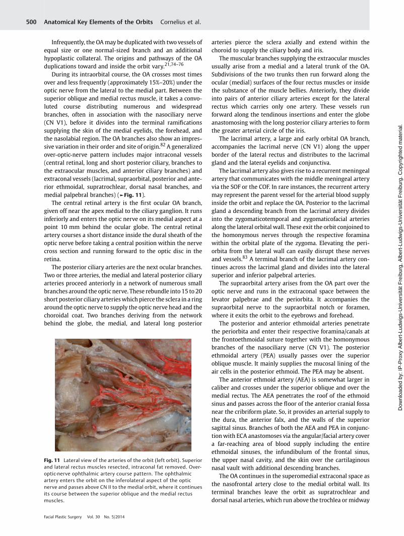
During its intraorbital course, the OA crosses most timesover and less frequently (approximately 15%–20%) under theoptic nerve from the lateral to the medial part. Between thesuperior oblique and medial rectus muscle, it takes a convo-luted course distributing numerous and widespreadbranches, often in association with the nasociliary nerve(CN V1), before it divides into the terminal ramificationssupplying the skin of the medial eyelids, the forehead, andthe nasolabial region. The OA branches also show an impres-sive variation in their order and site of origin.82 A generalizedover-optic-nerve pattern includes major intraconal vessels(central retinal, long and short posterior ciliary, branches tothe extraocular muscles, and anterior ciliary branches) andextraconal vessels (lacrimal, supraorbital, posterior and ante-rior ethmoidal, supratrochlear, dorsal nasal branches, andmedial palpebral branches) (►Fig. 11).
The central retinal artery is the first ocular OA branch,given off near the apex medial to the ciliary ganglion. It runsinferiorly and enters the optic nerve on its medial aspect at apoint 10 mm behind the ocular globe. The central retinalartery courses a short distance inside the dural sheath of theoptic nerve before taking a central position within the nervecross section and running forward to the optic disc in theretina.
The posterior ciliary arteries are the next ocular branches.Two or three arteries, the medial and lateral posterior ciliaryarteries proceed anteriorly in a network of numerous smallbranches around the optic nerve. These rebundle into 15 to 20short posterior ciliary arterieswhich pierce the sclera in a ringaround the optic nerve to supply the optic nerve head and thechoroidal coat. Two branches deriving from the networkbehind the globe, the medial, and lateral long posterior
arteries pierce the sclera axially and extend within thechoroid to supply the ciliary body and iris.
The muscular branches supplying the extraocular musclesusually arise from a medial and a lateral trunk of the OA.Subdivisions of the two trunks then run forward along theocular (medial) surfaces of the four rectus muscles or insidethe substance of the muscle bellies. Anteriorly, they divideinto pairs of anterior ciliary arteries except for the lateralrectus which carries only one artery. These vessels runforward along the tendinous insertions and enter the globeanastomosing with the long posterior ciliary arteries to formthe greater arterial circle of the iris.
The lacrimal artery, a large and early orbital OA branch,accompanies the lacrimal nerve (CN V1) along the upperborder of the lateral rectus and distributes to the lacrimalgland and the lateral eyelids and conjunctiva.
The lacrimal artery also gives rise to a recurrent meningealartery that communicates with the middle meningeal arteryvia the SOF or the COF. In rare instances, the recurrent arterymay represent the parent vessel for the arterial blood supplyinside the orbit and replace the OA. Posterior to the lacrimalgland a descending branch from the lacrimal artery dividesinto the zygomaticotemporal and zygomaticofacial arteriesalong the lateral orbital wall. These exit the orbit conjoined tothe homonymous nerves through the respective foraminawithin the orbital plate of the zygoma. Elevating the peri-orbita from the lateral wall can easily disrupt these nervesand vessels.83 A terminal branch of the lacrimal artery con-tinues across the lacrimal gland and divides into the lateralsuperior and inferior palpebral arteries.
The supraorbital artery arises from the OA part over theoptic nerve and runs in the extraconal space between thelevator palpebrae and the periorbita. It accompanies thesupraorbital nerve to the supraorbital notch or foramen,where it exits the orbit to the eyebrows and forehead.
The posterior and anterior ethmoidal arteries penetratethe periorbita and enter their respective foramina/canals atthe frontoethmoidal suture together with the homonymousbranches of the nasociliary nerve (CN V1). The posteriorethmoidal artery (PEA) usually passes over the superioroblique muscle. It mainly supplies the mucosal lining of theair cells in the posterior ethmoid. The PEA may be absent.
The anterior ethmoid artery (AEA) is somewhat larger incaliber and crosses under the superior oblique and over themedial rectus. The AEA penetrates the roof of the ethmoidsinus and passes across the floor of the anterior cranial fossanear the cribriform plate. So, it provides an arterial supply tothe dura, the anterior falx, and the walls of the superiorsagittal sinus. Branches of both the AEA and PEA in conjunc-tionwith ECA anastomoses via the angular/facial artery covera far-reaching area of blood supply including the entireethmoidal sinuses, the infundibulum of the frontal sinus,the upper nasal cavity, and the skin over the cartilaginousnasal vault with additional descending branches.
The OA continues in the superomedial extraconal space asthe nasofrontal artery close to the medial orbital wall. Itsterminal branches leave the orbit as supratrochlear anddorsal nasal arteries, which run above the trochlea or midway
Fig. 11 Lateral view of the arteries of the orbit (left orbit). Superiorand lateral rectus muscles resected, intraconal fat removed. Over-optic-nerve ophthalmic artery course pattern. The ophthalmicartery enters the orbit on the inferolateral aspect of the opticnerve and passes above CN II to the medial orbit, where it continuesits course between the superior oblique and the medial rectusmuscles.
Facial Plastic Surgery Vol. 30 No. 5/2014
Anatomical Key Elements of the Orbits Cornelius et al.500
Dow
nloa
ded
by: I
P-P
roxy
Alb
ert-
Ludw
igs-
Uni
vers
ität F
reib
urg,
Alb
ert-
Ludw
igs-
Uni
vers
ität F
reib
urg.
Cop
yrig
hted
mat
eria
l.
between the trochlea and the medial canthal tendon,respectively.53
Venous Orbital Outflow
The outflow pathways of the valveless orbital veins leadthrough the SOF and empty into the cavernous sinus. Theupstream orbital venous network has two distinct spatialdistributions with two principal veins: the superomedialorbit drained by the superior ophthalmic vein and the infero-lateral orbit drained by the inferior ophthalmic vein. Thesuperior ophthalmic vein is a vein with a rather constantcourse originating from a confluence of the supraorbital,nasofrontal, and angular veins. The angular vein is a subunitof the facial vein. The superior ophthalmic vein coursesbackward alongside the trochlea and from medially to lat-erally in relation to the superior rectus muscle. In the mid-orbit, it crosses above the optic nerve to reach the lateralborder of the superior rectus muscle and to enter into theorbital apex. It leaves the muscle cone between the heads ofthe superior and lateral rectus muscle and passes through thenarrow lateral part of SOF above the Zinn’s ring before itterminates below the ophthalmic nerve (CN V2) in theanteroinferior portion of the cavernous sinus. At the SOFlevel, the superior ophthalmic vein is usually joined by theinferior ophthalmic vein to form a common stem. This stemmerges with the spaces of the cavernous sinus which arespreading forward across the lower andmedialmargins of theSOF. The inferior ophthalmic vein forms as a plexus in theanterior floor and lateral orbital wall region. The venousplexus is located in between the inferior rectus muscle andthe optic nerve and can extend posteriorly close to the end ofthe muscle cone. Accordingly, the emerging inferior ophthal-mic vein is of different length and runs in parallel to thebranch of CN III supplying inferior oblique muscle.
The diameter of the superior ophthalmic vein rangesbetween 2 and 10 mm, while the caliber of the inferiorophthalmic vein is comparatively small. Apart from the outletinto the cavernous sinus via the superior ophthalmic vein,there also exist direct connections of the inferior ophthalmicvein through the inferior sector of the SOF below Zinn’s ringand communicating branches with the pterygoid venousplexus through the IOF.
The intraorbital tributaries of the superior ophthalmicvein are originating from the lacrimal gland, the extraocularmuscles in the vicinity (superior rectus, superior oblique,medial rectus, and lateral rectus), and the superior medialand lateral vortex veins (vena vorticosa) from the choroid andthe central retinal vein. The inferior ophthalmic vein receivesmuscular branches (inferior rectus and oblique), medial andlateral collateral veins connecting from the superior venoussystem, and branches from the lacrimal sac and the inferiorvortex veins.
Mostly, there are two ethmoidal veins. The anterior eth-moidal vein drains into the superior ophthalmic vein, where-as the posterior ethmoidal vein joins a venouswebbing underthe orbital roof. An infraorbital vein accompanying the in-fraorbital nerve and artery along the canal and groove in the
orbital floor interconnects with multiple twigs to the inferiorophthalmic vein and passes through the IOF to end in thepterygoid venous plexus in the infratemporal fossa.
As opposed to the veins elsewhere in the body, the veins ofthe orbit are not as closely associatedwith the arterial systemand showmore independent pathways with the exception ofthe anterior section of the superior ophthalmic vein thatcoincides with the ophthalmic artery.82 The orbital veinsexhibit considerable variability following an increasing order:superior ophthalmic vein < inferior ophthalmic vein < col-lateral veins < muscular branches. One remarkable variant isthe presence of a medial ophthalmic vein which correspondsto a semicircular vessel loop linking the anterior ethmoidal,the medial collateral, and the muscular vein from the medialrectus, which in this way indirectly drain into the superiorophthalmic vein.84 The entire orbital venous network issuspended in the connective tissue septa interspersing theintraorbital contents, just likemuscles and fat lobules.51,56–59
Lacrimal Apparatus
The lacrimal apparatus consists of a secretory, a distributary,and a drainage or excretory system.46,85 The function is toprovide a continuous tear film that moistens the conjunctivaand cornea, which is composed of an inner mucin layer, anintermediate watery layer, and an outer lipid layer.
The eyelids with their particular arrangement of theorbicularis oculi fibers are responsible for the coordinatedpropulsion and distribution of the tear film. The eyelidscontract progressively from temporal to nasal, propellingthe tear fluid over the eye obliquely downward toward thesuperior and inferior puncta for collection and drainage.67
Lacrimal Gland
The lateral horn of the levator aponeurosis partially separatesthemain lacrimal gland into a smaller inferior palpebral and alarger superior orbital lobe. Because of the parenchymalconnection of the lobe behind the lateral palpebral horn ofthe levator aponeurosis this separation is incomplete. Thegland is affixed to the anterolateral orbital roof by fibrousinterlobular septa with the orbital lobe lodged in the lacrimalfossa of the frontal bone. The palpebral lobe extends into thelateral aspect of the superior conjunctival fornix. The suspen-sion of the gland comes from attachments to the periorbita,the orbital septum, the conjunctiva, and the Whitnall liga-ment. All of the excretory ducts of the gland open into thesuperotemporal conjunctival fornix. Ducts from the orbitallobe join ducts from the palpebral part or traverse itindependently.
The blood supply and innervation of the lacrimal gland areprovided by the lacrimal artery and the nerve (CN V1). Thelacrimal vein drains into the superior ophthalmic vein.
Postganglionic parasympathetic secretomotor neurons(GVE) from the pterygopalatine ganglion reach the lacrimalgland via the zygomatic/zygomaticotemporal (CN V2) and thelacrimal nerve (communicating ramus). The respective pre-ganglionic parasympathetic neurons arise from the
Facial Plastic Surgery Vol. 30 No. 5/2014
Anatomical Key Elements of the Orbits Cornelius et al. 501
Dow
nloa
ded
by: I
P-P
roxy
Alb
ert-
Ludw
igs-
Uni
vers
ität F
reib
urg,
Alb
ert-
Ludw
igs-
Uni
vers
ität F
reib
urg.
Cop
yrig
hted
mat
eria
l.
geniculate ganglion (CN VII—intermediate nerve) and ap-proach the pterygopalatine ganglion as greater petrosal nerveand deep petrosal nerves. These nerves combine to thepterygoid or Vidian nerve, which enters the pterygopalatineganglion.
Sympathetic fibers originate from the superior cervicalganglion and follow the course of the arteries to the lacrimalgland.
The superior opening of the nasolacrimal canal is formed byindentations within the anteromedial orbital surface of themaxilla and the lacrimal bone. It is located next to theinfraorbital rim in the medial lower corner of the orbit andis the frontal starting point of the sutural junction betweenthe inferior and the medial orbital wall. The lacrimal drainagesystem consists of the lacrimal sac and its membranousdownward continuation, the nasolacrimal duct, which runsthrough the intraosseous canal and opens into the inferiornasal meatus underneath the inferior concha. The tear fluid iscollected by the lacrimal puncta and emptied into the lacrimalsac via the canaliculi.
The puncta are small round or transversely oval-shapedapertures at the margins of the eyelids near the medialcanthus. Both puncta face backward and become only visiblewhen the eyelids are everted. The puncta pass over into theupper and lower canaliculi. Each canaliculus has short vertical—ascending or descending—and a longer horizontal portion.The two horizontal portions run either separately or conjoint-ly into the sidewall of the upper lacrimal sac. The sac iswrapped by the lacrimal fascia—an extension of the perior-bita, which stretches between the anterior and the posteriorlacrimal crests.
Medial Canthal Ligament
The medial canthal ligament has an anterior and posteriorfibrous reflection.86–88 The anterior or superficial reflectionattaches to the anterior lacrimal crest and is the fixation forthe orbicularis muscle fibers. The posterior reflection is afibrous extension arising from the rear side of the junctionbetween the medial crura and the common tendon. It is deepto the lacrimal sac and inserts to the posterior lacrimal crestjust in front of Horner’s muscle.53 The posterior reflectionforms a tangential line with the curvature of the lids andsupports their apposition against the globe.64
Lateral Canthal Ligament
The lateral canthal ligament (LCL) is one of several structuralelements of a complex connective tissue fixation ensemble atthe lateral orbital rim.89 The LCL extends from the lateraltarsal ends to Whitnall’s lateral orbital. The precanthal liga-ment is a reinforced fibrous band of the orbital septum at thelevel of the canthus which runs from the tarsus to the outerperiosteal sleeve of the lateral orbital rim. A small lobule of
preaponeurotic (capsulopalpebral) fat fills the triangularspace between the septum anteriorly, the canthal ligamentposteriorly, and the lateral zygomatic orbital plate. Thisadipose tissue extension is known as Eisler’s fat pocket. Alongits superior border, the LCL is adjacent to the lateral horn ofthe levator aponeurosis and the inferolateral pole of thelacrimal gland.53,64,86 The posterior LCL portion is in closecontact with the lateral rectus muscle check ligament. Inferi-orly, the LCL is approximated by the lateral portion of Lock-wood’s inferior ligament. The inferolateralfibers ofWhitnall’ssuperior suspensory ligament connect to the LCL via intersti-tial fibrous septa in the lacrimal gland.
Retinacula
The connective structures associated with canthal ligamentsare summarized as retinacula. The medial retinaculum islocated behind the posterior lacrimal crest. It encompassesHorner’s muscle, the orbital septum, the medial horn of thelevator aponeurosis, the check ligaments of the medial rectusmuscle, and the medial portions of Whitnall’s and Lock-wood’s ligament.64 The lateral retinaculum, which attachesto Whitnall orbital tubercle, somewhat mirrors the medialretinaculum. It is composed of the LCL, the lateral horn of thelevator aponeurosis, the lateral rectus check ligaments, andthe lateral ends of Whitnall’s and Lockwood’s ligament.
Lid Retractors
The levator palpebrae superioris, Müller’s superior tarsalmuscle and the retractor muscles of the lower eyelid functionto open the eyelids and are antagonistic to the orbicularisoculi muscle (OOM), which is responsible for eyelid closure.
Levator Palpebrae Superioris
The levator palpebrae superioris and Müller’s superior mus-cle retract or elevate the upper eyelid. The levator palpebrae isa striated muscle, which is easily separable from the superiorrectus muscle apart from their medial border, where they areboth adherent to a common fascial sheath. The innervation ofboth muscles is by the superior CN III division. Whitnall’stransverse ligament, the condensation of the fascial sheathsaround the levator, is placed just posterior to the superiororbital rim or approximately 18 to 20 mm above the superiorborder of the tarsus (►Fig. 12). Below the level of Whitnall’sligament, the levator is widening horizontally into a broadfibrous aponeurotic sheath arching over the globe with twotendinous extremities, the lateral and medial horns. Theband-like lateral horn divides the lacrimal gland incompletelyinto the orbital and palpebral lobes before inserting onWhitnall’s lateral orbital tubercle as a component of thelateral retinaculum. Themedial horn passes over the superioroblique tendon, blends with the reflections of the medialcanthal tendon, and joins the medial retinacular structuresdeparting from the posterior lacrimal crest for osseous at-tachment. The cutaneous insertion of the aponeurosis iseffected by terminal fibers traversing the orbicularis muscle
Facial Plastic Surgery Vol. 30 No. 5/2014
Anatomical Key Elements of the Orbits Cornelius et al.502
Dow
nloa
ded
by: I
P-P
roxy
Alb
ert-
Ludw
igs-
Uni
vers
ität F
reib
urg,
Alb
ert-
Ludw
igs-
Uni
vers
ität F
reib
urg.
Cop
yrig
hted
mat
eria
l.
and forming a distinct supratarsal fold. A subset of deeperfibers attaches to the anterior surface of the upper tarsus.
However, the formation of a supratarsal eyelid fold is amatter of ethnic differences, and it depends on the verticalheight of the fusion line between the orbital septum and thelevator aponeurosis.90 In Asian, single’ eyelids without a foldor crease, the orbital septum fuses on the levator over themidor inferior tarsal plate. A protrusion of the aponeurotic fat padextending into the resulting pretarsal space prevents theterminal interdigitations of the aponeurotic fibers to anchorthe skin and create a crease indentation.91 In comparison toCaucasian upper, double eyelids are distinguished by dermalattachments of the aponeurotic fibers at or above the superiortarsal border demarcating the upper palpebral fold.
Müller’s Superior Tarsal Muscle
The superior tarsal muscle of Müller’s is an involuntarysmooth muscle innervated by sympathetic fibers. It is incor-porated in the undersurface of the levator palpebrae, isadherent to the conjunctiva posteriorly and inserts into thesuperior tarsal border. Additional smooth muscles fibers mayarise from Tenon’s capsule. The tarsal muscle is easily sepa-rable from the levator apart from the site of its origin near themusculoaponeurotic levator junction.
Lower Eyelid Retractors
The lower eyelid retractors consist of two principal layers, thecapsulopalpebral fascia and the inferior tarsal muscle. Fibrousextensions originate from the sheath of the inferior rectusmuscle to compose the head of the capsulopalpebral fascia.This head transforms into an envelope around the inferioroblique muscle. Anterior to the muscle the inferior andsuperior fascial portions conjoin to form Lockwood’s inferiorsuspensory ligament. The superior portion of the capsulo-palpebral fascia then extends posteriorly into the inferior
conjunctival fornix and converts into Tenon’s capsule. Theinferior portion fuses with the orbital septum anteriorly,connects through the orbicularis oculi muscle (OOM) to theskin and inserts more upwardly on the inferior border of thelower tarsus.92 The dermal attachments are the basis of thelower eyelid crease. A limited depression of the lower eyelidmargin occurs in downgaze to enable an unimpaired visualfield. This movement is essentially a synergic effect of theinferior rectus muscle contraction which is translated by thecapsulopalpebral head and the inferior fascial portion. Thesympathetic smooth inferior tarsal muscle is an accessorylower eyelid retractor. It originates and lies posterior to thecapsulopalpebral fascia and inserts at the base of the inferiortarsus.
Orbicularis Oculi Muscle
The OOM is a widespread array of muscle fibers that liesbeneath the skin and over the orbital septum and thetarsoligamentous sling. It is innervated by multiple (buccal,zygomatic, and frontal) branches of CN VII. The motor supplyof the levator palpebrae, its antagonist, is by the superiordivision of CN III.
The OOM is divided into three major concentric or ring-shaped bands, the orbital, preseptal, and pretarsal portions.The orbital orbicularis arises medially from origins along thesuperior and inferior orbital rims aswell as themedial canthaltendon and forms a wide loop around the lateral circumfer-ence of the orbital aperture. Superolaterally, the orbitalportion covers the corrugator supercilii muscle and theanterior temporoparietal fascia. In the inferior temporaland cheek area, the orbital orbicularis extends to the originof the masseter and the zygomaticus minor muscle at thesurface of the zygomatic body. Along the infraorbital rim andthe maxillary frontal process, the origins of the upper lip andnasal ala elevators are surrounded by the orbital orbicularis.86
The preseptal portion and the pretarsal OOM overlie theorbital septum and the tarsal plates. At the medial canthaltendon and around the lacrimal canaliculi and lacrimal sac,both the portions divide into two components, anterior orsuperficial and posterior or deep heads. Both superficialheads connect to the medial canthal tendon and beyond tothe anterior lacrimal crest and the nasofrontal maxillaryprocess. The deep head of the preseptal OOM portion, orJones’muscle, attaches posteriorly to the fascia of the lacrimalsac. The deep pretarsal orbicularis head, known as Horner’smuscle, pars lacrimalis or tensor tarsi, passes posterior to thecanaliculi and the lacrimal sac to attach to the posteriorlacrimal crest64 (►Fig. 13).
Laterally, the preseptal orbicularis from the lower andupper eyelid insert onto thehorizontal lateral palpebral raphewhich attaches to the zygoma. The pretarsal inferior andsuperior orbicularis parts join the LCL at Whitnall’s tubercle.The integrity of the Horner’s muscle connection to theposterior lacrimal crest determines the contact of the eyelidmargins in the inner canthal area on the globe surface. The lidmargins are pulled anteriorly away from the surface if themuscle is detached or disrupted.15
Fig. 12 Supporting elements and retractor muscle in the upper eyelid(right orbit). Skin layers around the orbit have been removed. Sub-orbicular dissection in the upper lid and preaponeurotic fat removal isshown. Incisions at the midpupillary line into the preseptal orbicularisand through tarsus up to the musculoaponeurotic junction to expandthem superiorly and in transverse direction, respectively. Superiorrectus muscle sling hooked upward, levator palpebrae locatedlaterally.
Facial Plastic Surgery Vol. 30 No. 5/2014
Anatomical Key Elements of the Orbits Cornelius et al. 503
Dow
nloa
ded
by: I
P-P
roxy
Alb
ert-
Ludw
igs-
Uni
vers
ität F
reib
urg,
Alb
ert-
Ludw
igs-
Uni
vers
ität F
reib
urg.
Cop
yrig
hted
mat
eria
l.
Lacrimal Caruncle
The lacrimal caruncle is a small pink bump (width 3 mm andheight 5 mm) at the inner canthus nasally to the plica semi-lunaris. The superior border is in level with the lower eyelidmargin. The caruncle is lined by a nonkeratinizing epitheliumlooking as if it were a formation of the palpebral conjunctiva.In fact, it is a modified part of the lower lid margin andcontains cutaneous appendages such as fine colorless hairs,sebaceous glands, sweat glands, and modified lacrimalglands.49
Plica Semilunaris
The plica semilunaris is a conjunctival fold along the curva-ture of the medial canthus, just lateral to the caruncle. It isimportant in the distribution of tear fluids, maintenance ofthe lacrimal lake and removal of ocular debris. Usually theplica is not very prominent, although it provides resiliency inrotational movements of the globe and still contributes someprotection. Phylogenetically, it is a remnant of the so-calledthird eyelid or nictitating membrane, which can be horizon-tally closed across the entire eyeball.
Periorbital Dissection
The periorbital dissection opens up the potential spacebetween the connective tissue layers of the periorbita andthe bony surfaces of the internal orbit. It is this essentially“subperiosteal” space that is commonly accessed in primaryand secondary trauma management, in craniofacial correc-tive surgery and for bony decompression of the orbits.Commonly, it will be tried to keep the periorbita intact toprevent fat tissue prolapses obstructing the narrow visualfield inside the orbit.
In severe trauma involving several orbital walls the peri-orbita may need to be separated from the bone in a 360degrees sector encircling the anterior and the midorbit. Theentry into the orbital cavity can be achieved by a variety of
transcutaneous and/or transconjunctival access incisions ei-ther via a single or a combination of approaches.93,94
With the exception for surgical decompression in an acuteoptic neuropathy (visual loss/afferent pupil defect) or in a SOFsyndrome, the dissection is usually stopped at the entranceinto the posterior apex space. Outside the SOF and Zinn ring,the dissectible space under the periorbita is considered safe.The periorbital dissection for exposure in fracture repairadvances starting from the anterior aperture at the rim leveltoward the apex posteriorly. The postentry concavities mustbe passed around stripping inferiorly (floor) or superiorly(roof) and backward.
In the anterior and the midorbit, the periorbital dissectionshould circumvent the origin of the inferior oblique muscle,the lacrimal duct, and the medial and lateral retinacularcomplexes to avoid any inadvertent disruption. Likewise, itis mandatory to be aware of the pertinent landmarks in everysingle orbital wall. The position, shape, and size of the IOF andSOF indicate a basic pattern for topographical orientationduring the surgical exposure of the orbital walls.
In the roof, the trochlear fovea and the lacrimal gland fossaare passed proceeding toward the apex. Periorbital strandssuspended from the top of the lateral SOF portion or evenmore laterally from the COF to the PEF as a tent-like foldingdelineate the horizontal termination for the dissection belowthe orbital roof. This periorbital fold will often contain theophthalmic artery crossing the optic nerve superiorly and/orthe recurrent meningeal artery.15
Although it is a basic principle to preserve the integrity ofthe periorbital sac in the case for an extended and deepexposure of thefloor and the lower orbital circumference, it isa crucial maneuver to cut across the periorbital prolongationsinvaginating the IOF from the maxillary orbital plate and thelateral orbital (►Figs. 14 and 15).
The anterior part of the IOF does not contain importantstructures and the infraorbital nerve and zygomatic nerve inthe posterior portion course below the usual level of dissec-tion. The posterior limitation of the exposure along the orbitalfloor is the orbital process of the palatine bone, which
Fig. 14 Exposure of lateral orbit with zygomatic rim removed. Bonybridge of orbital floor in front of anterior IOF loop left intact. Soft tissuecontents of the IOF—temporal/buccal (Bichat) fat pad spreadingthrough the fissure. IOF, inferior orbital fissure.
Fig. 13 Lateral view of the medial part of the anterior orbit. Horner’smuscle shown after combined transcaruncular lower fornix swingingeyelid approach. Plica semilunaris flap retracted medially.
Facial Plastic Surgery Vol. 30 No. 5/2014
Anatomical Key Elements of the Orbits Cornelius et al.504
Dow
nloa
ded
by: I
P-P
roxy
Alb
ert-
Ludw
igs-
Uni
vers
ität F
reib
urg,
Alb
ert-
Ludw
igs-
Uni
vers
ität F
reib
urg.
Cop
yrig
hted
mat
eria
l.
becomes the “posterior ledge” of preserved bone beyond anextensive floor defect. A reliable way to end up there, but inthe posterior maxillary recess, is to dissect along the lowermargin of the GWS in the lateral wall, which is rarelyfragmented. Once the posteromedial IOF has been reached,the periorbita is swept away medially over the posterior endof the infraorbital groove, the IOF isthmus promontory, andthe posterior ledge. Raising the periorbita medially allows tovisualize the infraorbital nerve and to release it in a controlledfashion.
The maxillary strut above the foramen rotundum wouldrepresent a salient bony landmark to terminate the floordissection posteriorly; however, it lies adjacent to the venousconnections through the inferior SOF and to the pterygoidplexus with no safety margin.
Coming along the lateral orbital wall the posteriorboundary for the exposure is the dural/periorbital reflec-tion at the lateral SOF margin. A few millimeters before theupper lateral arch of the SOF is approached a recurrentmeningeal artery may be encountered passing through COFat the sphenofrontal suture line. On the medial wall, themaximal extent of the deep dissection is marked by theposterior ethmoidal vessels. They come into view at thevertically running sphenoethmoid suture line anterior tothe optic foramen.
The pre/transcaruncular incision is often used to approachthe medial orbital wall. It proceeds posteriorly inside theperiorbita between the medial rectus muscle and Horner’smuscle until the lamina papyracea behind the posteriorlacrimal crest is reached (►Fig. 13). A vertical incision ismade and the periorbita is elevated to continue the exposurestraight below the frontoethmoid suture line to the AEF andPEF. The IOS in its full extent is best accessed via the pre/transcaruncular incision combined with a transconjunctivallower fornix approach and temporary detachment of theinferior oblique muscle.
In the literature, oftentimes the safe distances for the peri-orbital dissection from the intact outside rimof the adult orbit to
vital interior structures are specified inmillimeters.95Generally,a gauging depth of 25 mm along the lateral or inferior wall isregarded safe. A maximal distance of 30 mm from the supraor-bital rim and from the anterior lacrimal crest is admitted for asafe exposure of the roof or the medial wall, respectively.
More detailed measurements at the medial wall refer to theposition of the EF in relation to the anterior lacrimal crest andamong each other. On the average the distance is 24 mm to theAEF, another 12 mm beyond this to PEF and 6 mm between thePEF and the optic foramen. The rule of halves, that is, 24–12–6 mm, is the aide-mémoire for these lengths.15,96 In the heavilyinjured orbit or due to anatomic variations, the utility of thesevalues is questionable.97 Pushing the limits of dissection too fartoward the optic foramen probably does not give the full pictureof causative factors for CN II lesions. Indirect compression of CN IIin its intraorbital course in the tight spatial relationshipsnear theorbital apex may matter as much (►Fig. 16).
Conclusion
The orbits are craniofacial building blocks containing criticalocular, neural, and vascular structures. This article intends tooutline a ready-to-use surveyof the anatomyof the orbits andtheir contents to facilitate comprehension of the most rele-vant surgical details. Certainly, it is a truism to conclude thattherewill bemuchmore to discover, tofigure out, and to learnwith intensive immersion into the subject stimulated by theperspectives of technical innovations.
AcknowledgmentsThe authors thank the photographers, Rudolf Hertzig andGerhard Poetzel, OMFS Department, Ludwig MaximiliansUniversity, Munich, for their support. All anatomic dis-sections have been performed by the first author (C.-P. C)in the Anatomy Departments of the University of Ulm,University of Munich, Germany, and in the De Puy SynthesLaboratory, Westchester, PA.
Fig. 15 Another orbit—lateral orbital wall completely resected in-cluding SOF margin up to the periorbital/dural reflection. Contents ofIOF fully exposed up into cavernous sinus/pterygoid plexus. Micro-scissors demonstrate periorbital dissection cutting through the con-tents of IOF. Note: triangular cross IOF section consisting of periorbitalinvagination with fat pad inside andMüller muscle outside. IOF, inferiororbital fissure.
Fig. 16 Coronal section anterior to orbital apex. Spatula tip to elevatethe periorbita rests on palatine bone (posterior ledge). Note the closeapproximation and potential compressional effects of the instrumenton CN II.
Facial Plastic Surgery Vol. 30 No. 5/2014
Anatomical Key Elements of the Orbits Cornelius et al. 505
Dow
nloa
ded
by: I
P-P
roxy
Alb
ert-
Ludw
igs-
Uni
vers
ität F
reib
urg,
Alb
ert-
Ludw
igs-
Uni
vers
ität F
reib
urg.
Cop
yrig
hted
mat
eria
l.
DisclosureThe authors declare no conflicts of interest in relation tothe content of this article.
References
1 Schmelzeisen R, Gellrich NC, Schoen R, Gutwald R, Zizelmann C,Schramm A. Navigation-aided reconstruction of medial orbitalwall and floor contour in cranio-maxillofacial reconstruction.Injury 2004;35(10):955–962
2 Schramm A, Gellrich NC, Schmelzeisen R. Navigational surgery ofthe facial skeleton. Berlin Heidelberg: Springer Verlag; 2007
3 Snyderman CH, Harvey RJ. Skull base: meeting place for multidis-ciplinary collaboration. Otolaryngol Clin North Am 2011;44(5):xi–xii
4 Snyderman CH, Pant H, Carrau RL, Prevedello D, Gardner P, KassamAB. What are the limits of endoscopic sinus surgery?: the expand-ed endonasal approach to the skull base. Keio J Med 2009;58(3):152–160
5 Kassam AB, Gardner PA, eds. Endoscopic Approaches to the SkullBase. Progress in Neurological Surgery. Volume 26. Basel: S. KargerAG; 2012
6 Castelnuovo P, Dallan I, Tschabitscher M. Surgical Anatomy of theInternal Carotid Artery. Heidelberg, New York: Springer Verlag;2013
7 Manson PN, Grivas A, Rosenbaum A, Vannier M, Zinreich J, Iliff N.Studies on enophthalmos: II. The measurement of orbital injuriesand their treatment by quantitative computed tomography. PlastReconstr Surg 1986;77(2):203–214
8 Gruss JS, Phillips JH. Complex facial trauma: the evolving role ofrigid fixation and immediate bone graft reconstruction. Clin PlastSurg 1989;16(1):93–104
9 Hammer B. Orbital fractures. Diagnosis, Operative Treatment, Sec-ondary Corrections. Seattle Toronto Bern: Hofgrefe & Huber; 1995
10 Markiewicz MR, Dierks EJ, Bell RB. Does intraoperative navigationrestore orbital dimensions in traumatic and post-ablative defects?J Craniomaxillofac Surg 2012;40(2):142–148
11 Strong EB, Fuller SC, Chahal HS. Computer-aided analysis of orbitalvolume: a novel technique. Ophthal Plast Reconstr Surg 2013;29(1):1–5
12 Metzger MC, Schön R, Tetzlaf R, et al. Topographical CT-dataanalysis of the human orbital floor. Int J Oral Maxillofac Surg2007;36(1):45–53
13 Kamer L, Noser H, Schramm A, Hammer B. Orbital form analysis:problems with design and positioning of precontoured orbitalimplants: a serial study using post-processed clinical CT data inunaffected orbits. Int J Oral Maxillofac Surg 2010;39(7):666–672
14 Tessier P. Surgical anatomy and methods of examining the orbit.In: Tessier P, Rougier J, Wolfe SA, eds. Plastic Surgery of the Orbitsand Eyelids. NewYork, NY:Masson PublishingUSA, Inc; 1981:1–11
15 Rootman J, Stewart B, Goldberg R. Orbital surgery: a conceptualapproach. Section II, Atlas of orbital anatomy. Philadelphia, PA:Lippincott Raven; 1995:77–146
16 Bron AJ, Tripathi RC, Tripathi BJ. Wolff’s Anatomy of the Eye andOrbit, 8th Edition. London: Chapman and Hall Medical; 1997
17 DoxanasM, Anderson RL. Clinical Orbital Anatomy. Baltimore,MD:Williams and Wilkins; 1984
18 Rohner D, Tay A, Meng CS, Hutmacher DW, Hammer B. Thesphenozygomatic suture as a key site for osteosynthesis of theorbitozygomatic complex in panfacial fractures: a biomechanicalstudy in human cadavers based on clinical practice. Plast ReconstrSurg 2002;110(6):1463–1471, discussion 1472–1475
19 Whitnall SE. On a tubercle on the malar bone, and on the lateralattachments of the tarsal plates. J Anat Physiol 1911;45(Pt 4):426–432
20 Rhoton AL Jr, Natori Y. The Orbit and Sellar Region: MicrosurgicalAnatomy and Operative Approaches. New York, NY: ThiemeMedical; 1996
21 Rhoton AL Jr. Rhoton’s Cranial Anatomy and Surgical Approaches.Baltimore, MD: Lippincott Williams & Wilkins; 2007
22 Abed SF, Shams P, Shen S, Adds PJ, Uddin JM, Manisali M. Acadaveric study of the cranio-orbital foramen and its significancein orbital surgery. Plast Reconstr Surg 2012;129(2):307e–311e
23 Habal MB, Maniscalco JE, Rhoton AL Jr. Microsurgical anatomy ofthe optic canal: correlates to optic nerve exposure. J Surg Res 1977;22(5):527–533
24 Turvey TA, Golden BA. Orbital anatomy for the surgeon. OralMaxillofac Surg Clin North Am 2012;24(4):525–536
25 Reymond J, Kwiatkowski J, Wysocki J. Clinical anatomy of thesuperior orbital fissure and the orbital apex. J CraniomaxillofacSurg 2008;36(6):346–353
26 Zampieri F, Marrone D, Zanatta A. Should the annular tendon ofthe eye be named ’annulus of Zinn’ or ’of Valsalva’? Acta Oph-thalmol (Copenh) 2014; E-pub ahead of print
27 Bisaria KK, KumarN, PrakeshM, et al. The lateral rectus spine of thesuperior orbital fissure. J Anat 1996;189(Pt 1):243–245
28 De Battista JC, Zimmer LA, Theodosopoulos PV, Froelich SC, KellerJT. Anatomy of the inferior orbital fissure: implications for endo-scopic cranial base surgery. J Neurol Surg B Skull Base 2012;73(2):132–138
29 Müller H. Über einen glatten Muskel in der Augenhöhle desMenschen und der Säugetiere. Zeitschr Wissenschaftl Zool1858;9:541
30 Piagkou M, Skotsimara G, Dalaka A, et al. Bony landmarks of themedial orbital wall: an anatomical study of ethmoidal foramina.Clin Anat 2014;27(4):570–577
31 Celik S, Kazak Z, Ozer MA, Govsa F. Navigational area of the cranio-orbital foramen and its significance in orbital surgery. Surg RadiolAnat 2014; E-pub ahead of print
32 Fallucco M, Janis JE, Hagan RR. The anatomical morphology of thesupraorbital notch: clinical relevance to the surgical treatment ofmigraine headaches. Plast Reconstr Surg 2012;130(6):1227–1233,Discussion 1234
33 Knize DM. The Forehead and Temporal Fossa- Anatomy andTechnique. Philadelphia, PA: Lippincott Williams and Wilkins;2001
34 Knize DM. Lateral open forehead lift. Aesthet Surg J 2005;25(2):169–174
35 Kazkayasi M, Ergin A, Ersoy M, Bengi O, Tekdemir I, Elhan A.Certain anatomical relations and the precise morphometry ofthe infraorbital foramen—canal and groove: an anatomicaland cephalometric study. Laryngoscope 2001;111(4 Pt 1):609–614
36 Agthong S, Huanmanop T, Chentanez V. Anatomical variationsof the supraorbital, infraorbital, and mental foramina relatedto gender and side. J Oral Maxillofac Surg 2005;63(6):800–804
37 Chrcanovic BR, Abreu MH, Custódio AL. A morphometric analysisof supraorbital and infraorbital foramina relative to surgical land-marks. Surg Radiol Anat 2011;33(4):329–335
38 Nagasao T, Hikosaka M, Morotomi T, Nagasao M, Ogawa K,Nakajima T. Analysis of the orbital floor morphology. J Cranio-maxillofac Surg 2007;35(2):112–119
39 Kokemueller H, Zizelmann C, Tavassol F, Paling T, Gellrich NC. Acomprehensive approach to objective quantification of orbitaldimensions. J Oral Maxillofac Surg 2008;66(2):401–407
40 Noser H, Hammer B, Kamer L. A method for assessing 3D shapevariations of fuzzy regions and its application on human bonyorbits. J Digit Imaging 2010;23(4):422–429
41 Kamer L, Noser H, Schramm A, Hammer B, Kirsch E. Anatomy-based surgical concepts for individualized orbital decompressionsurgery in graves orbitopathy. I. Orbital size and geometry. Oph-thal Plast Reconstr Surg 2010;26(5):348–352
Facial Plastic Surgery Vol. 30 No. 5/2014
Anatomical Key Elements of the Orbits Cornelius et al.506
Dow
nloa
ded
by: I
P-P
roxy
Alb
ert-
Ludw
igs-
Uni
vers
ität F
reib
urg,
Alb
ert-
Ludw
igs-
Uni
vers
ität F
reib
urg.
Cop
yrig
hted
mat
eria
l.
42 Kamer L, Noser H, Kirsch E, Hammer B. Anatomy-based surgicalconcepts for individualized orbital decompression surgery ingraves orbitopathy. II. Orbital rimposition and angulation. OphthalPlast Reconstr Surg 2012;28(4):251–255
43 Borumandi F, Hammer B, Noser H, Kamer L. Classification of orbitalmorphology for decompression surgery in Graves’ orbitopathy:two-dimensional versus three-dimensional orbital parameters. BrJ Ophthalmol 2013;97(5):659–662
44 Kunz C, Cornelius CP, Audigé L, Ruddermann R, Buitrago-Téllez CH,Prein J. AO classification system for fractures of the craniomax-illofaxial skeleton: Level 3 orbital fractures. Cranial MaxillofacialTrauma & Reconstruction 2014; In press
45 Kim JW, Goldberg RA, Shorr N. The inferomedial orbital strut: ananatomic and radiographic study. Ophthal Plast Reconstr Surg2002;18(5):355–364
46 KikkawaDO, Lemke BN. Orbital and eyelid anatomy. In: DortzbachRK, ed. Ophthalmic Plastic Surgery. New York, NY: Raven Press;1994:1–29
47 Janfaza P, Rubin PAD, Azar N. Orbit. In: Janfaza P, Nadol JB, Galla RJ,Fabian RL, Montgomery WW, eds. Surgical Anatomy of the Headand Neck. Philadelphia, PA: Lippincott Williams &Wilkins; 2001:149–222
48 Froelich S, Abdel Aziz KM, van Loveren HR, Keller JT. The transitionbetween the cavernous sinus and the orbit. In: Dolenc VV, RogersL, eds. Cavernous Sinus. Chapter 1: Anatomy of the cavernoussinus. New York, NY: Springer; 2009:27–33
49 Lang J. Praktische Anatomie - Lanz Wachsmuth. Erster Band Teil1B- Kopf-Gehirn und Augenschädel. Berlin Heidelberg New York:Springer Verlag; 1979
50 Lang J. Skull Base andRelated Structures: Atlas of Clinical Anatomy.Stuttgart: Schattauer; 1995
51 Bergen MP. Microvessels in the human orbit in relation to theconnective tissue system. Acta Morphol Neerl Scand 1982;20(2):139–150
52 Rubin PA, Hatton MP, Bilyk JR. Orbital perforating branch of theinfraorbital artery: an important landmark in orbital surgery.Ophthal Plast Reconstr Surg 2005;21(6):447–449
53 Dutton JJ. Atlas of Clinical and Surgical Orbital Anatomy, 2nd ed,Saunders. 2011
54 Helveston EM, MerriamWW, Ellis FD, Shellhamer RH, Gosling CG.The trochlea. A study of the anatomy and physiology. Ophthal-mology 1982;89(2):124–133
55 Zinn IG. De musculi bulbi oculi, § III. De ligamento communi undeoriuntur musculi adducens, deprimens et abducentis caput al-terum. In: Zinn IG. Descriptio anatomica oculi humani, iconibusillustrata. Goettingae: A. Vandenhoeck; 1780:148–149
56 Koornneef L. Spatial Aspects of OrbitalMusculofibrous Tissue inMan.[PhD thesis]. University of Amsterdam: Swets & Zeitlinger; 1976
57 Koornneef L. New insights in the human orbital connective tissue.Result of a new anatomical approach. Arch Ophthalmol 1977;95(7):1269–1273
58 Koornneef L. The architecture of the musculo-fibrous apparatus inthe human orbit. Acta Morphol Neerl Scand 1977;15(1):35–64
59 Koornneef L. Orbital septa: anatomy and function. Ophthalmology1979;86(5):876–880
60 Botha CP, Willekens B, Klooster J, IJskes SG, Hötte G, Simonsz HJ.The Visible Human Orbit: Digital High-Resolution Conservation ofAnatomical Serial Sections of the Human Embryonal and AdultOrbit. In: Koornneef Los and de Haan, eds. Available at: www.visible-orbit.org2012. Accessed October 10, 2014
61 Manson PN, Clifford CM, Su CT, Iliff NT, Morgan R. Mechanisms ofglobal support and posttraumatic enophthalmos: I. The anatomyof the ligament sling and its relation to intramuscular cone orbitalfat. Plast Reconstr Surg 1986;77(2):193–202
62 Ettl A, Priglinger S, Kramer J, Koornneef L. Functional anatomy ofthe levator palpebrae superioris muscle and its connective tissuesystem. Br J Ophthalmol 1996;80(8):702–707
63 Ettl A, Zonneveld F, Daxer A, Koornneef L. Is Whitnall’s ligamentresponsible for the curved course of the levator palpebrae supe-rioris muscle? Ophthalmic Res 1998;30(5):321–326
64 McCord CD Jr, Codner MA, eds. Eyelid and Periorbital Surgery. StLouis, MO: Quality Medical Publishing; 2008
65 Kakizawa Y, Hongo K, Rhoton AL Jr. Construction of a three-dimensional interactivemodel of the skull base and cranial nerves.Neurosurgery 2007;60(5):901–910, discussion 901–910
66 Jonas JB, Schmidt AM, Müller-Bergh JA, Schlötzer-Schrehardt UM,Naumann GO. Human optic nerve fiber count and optic disc size.Invest Ophthalmol Vis Sci 1992;33(6):2012–2018
67 Schünke M, Schulte E, Schuhmacher U. Prometheus Lernatlas derAnatomie–Kopf und Neuroanatomie. Stuttgart: Georg ThiemeVerlag; 2006
68 Whitnall SE. Anatomyof the HumanOrbit and Accessory Organs ofVision. Reprint of original 1921 edition by Oxford University Press,London. Huntington, New York: Robert E. Krieger PublishingCompany; 1979
69 Martins C, Costa E Silva IE, Campero A, et al. Microsurgical anatomyof the orbit: the rule of seven. Anat Res Int 2011;2011:468727
74 Hayreh SS, Dass R. The ophthalmic artery. I. Origin and intracranialand intra-canalicular course. Br J Ophthalmol 1962;46(2):65–98
75 Hayreh SS, Dass R. The ophthalmic artery. II. Intra-orbital course.Br J Ophthalmol 1962;46(3):165–185
76 Hayreh SS. The ophthalmic artery. III. Branches. Br J Ophthalmol1962;46(4):212–247
77 Meyer F. Zur Anatomie der Orbitalarterien. Gegenbaurs MorpholJahrb 1888;12:414–418
78 Liu Q, Rhoton AL Jr. Middle meningeal origin of the ophthalmicartery. Neurosurgery 2001;49(2):401–406, discussion 406–407
79 Inoue T, Rhoton AL Jr, Theele D, Barry ME. Surgical approaches tothe cavernous sinus: a microsurgical study. Neurosurgery 1990;26(6):903–932
80 Seoane E, Rhoton AL Jr, de Oliveira E. Microsurgical anatomy of thedural collar (carotid collar) and rings around the clinoid segmentof the internal carotid artery. Neurosurgery 1998;42(4):869–884,discussion 884–886
81 Rhoton AL Jr. The middle cranial base and cavernous sinus. In:Dolenc VV. Rogers L, eds. Cavernous Sinus. Chapter 1: Anatomy ofthe Cavernous Sinus. Wien New York, NY: Springer; 2009:3–26
82 Hayreh SS. Orbital vascular anatomy. Eye (Lond) 2006;20(10):1130–1144
83 Govsa F, Celik S, Ozer MA. Orbital restoration surgery inthe zygomaticotemporal and zygomaticofacial nerves and im-portant anatomic landmarks. J Craniofac Surg 2009;20(2):540–544
84 Cheung N, McNab AA. Venous anatomy of the orbit. InvestOphthalmol Vis Sci 2003;44(3):988–995
85 Chastain JB, Sindwani R. Anatomy of the orbit, lacrimal apparatus,and lateral nasal wall. Otolaryngol Clin North Am 2006;39(5):855–864, v–vi
86 Zide BM, Jelks G. Surgical Anatomy of the Orbit. New York, NY:Raven Press; 1985
87 Zide BM, McCarthy JG. The medial canthus revisited—an anatomi-cal basis for canthopexy. Ann Plast Surg 1983;11(1):1–9
88 Zide BM. Surgical Anatomy Around the Orbit. The System of Zones.Philadelphia, PA: Lippincott Williams and Wilkins; 2006
Facial Plastic Surgery Vol. 30 No. 5/2014
Anatomical Key Elements of the Orbits Cornelius et al. 507
89 Knize DM. The superficial lateral canthal tendon: anatomic studyand clinical application to lateral canthopexy. Plast Reconstr Surg2002;109(3):1149–1157, discussion 1158–1163
90 Jeong S, Lemke BN, Dortzbach RK, Park YG, Kang HK. The Asianupper eyelid: an anatomical study with comparison to the Cauca-sian eyelid. Arch Ophthalmol 1999;117(7):907–912
91 Miyake I, Tange I, Hiraga Y. MRI findings of the upper eyelid andtheir relationship with single- and double-eyelid formation. Aes-thetic Plast Surg 1994;18(2):183–187
92 Hwang K, Kim DJ, Hwang SH, Chung IH. The relationship ofcapsulopalpebral fascia with orbital septum of the lower eyelid:an anatomic study under magnification. J Craniofac Surg 2006;17(6):1118–1120
93 Ellis E III, Zide MF. Surgical Approaches to the Facial Skeleton. 2nded. Philadelphia, PA: Lippincott Williams and Wilkins; 2006
94 Ellis E III. Surgical approaches to the orbit in primary andsecondary reconstruction. Facial Plast Surg 2014;30(5):537–544
95 Rontal E, Rontal M, Guilford FT. Surgical anatomy of the orbit. AnnOtol Rhinol Laryngol 1979;88(3, Pt 1):382–386
96 Lander MI, Terry O. The posterior ethmoid artery insevere epistaxis. Otolaryngol Head Neck Surg 1992;106(1):101–103
97 Lethaus B, Weigl S, Kloss-Brandstätter A, et al. Looking for land-marks in medial orbital trauma surgery. Int J Oral Maxillofac Surg2013;42(2):209–213
Facial Plastic Surgery Vol. 30 No. 5/2014
Anatomical Key Elements of the Orbits Cornelius et al.508