2/15/2017 1 The Paris System for Reporting Urinary Cytology: A Paradigm Shift Güliz A. Barkan, MD, FIAC Associate Professor of Pathology and Urology Loyola University Medical Center, Maywood, Illinois, USA [email protected]
Transcript
2/15/2017
1
The Paris System for Reporting Urinary Cytology: A Paradigm Shift
Güliz A. Barkan, MD, FIAC
Associate Professor of Pathology and Urology
Loyola University Medical Center, Maywood, Illinois, USA
Surgeons misunderstood pathologists’ reports 30% of the time. Powsner, SM. Costa J, Homer RJ. Clinicians are from Mars and pathologists are from Venus. Clinician Interpretation of Pathology Reports. Arch Pathol Lab Med 2000. 124:1040–1046
2/15/2017
5
Normal Urothelium
Hyperplasia Dysplasia
Low Grade Carcinoma High Grade Carcinoma Carcinoma in situ
Invasive Carcinoma
Papillary Pathway
80-90%
Non-Papillary Pathway
10-20%
9p-, 9q- p16
Genetically Stable FGFR3 (~85%)
Genetically Unstable p53 (~60%)
<10%
Recurrence Recurrence
RAS (?)
2/15/2017
6
Bladder cancer – more then one disease?
• ~ 75 % Non-Muscle-Invasive (Ta/T1)
– Good prognosis
– Recurrence
– 10%-15% progression (LG Ta - <1%)*
• ~ 25 % Muscle-Invasive (> T2)
– >60% overall survival
*Nielsen ME et al. Trends in Stage-Specific Incidence Rates for Urothelial Carcinoma of the Bladder In the United States: 1998-2006. Cancer 2014:120:86
2/15/2017
7
Question…. “Carcinoma”?
GU GI
2/15/2017
8
Classifications
WHO 1973
WHO/ISUP 2004
Papilloma
Papilloma
Grade I Grade III Grade II
Low Grade High Grade PUNLMP
~ 80-90% ~ 10-20% ~ 50-60%
URINE CYTOLOGY SENSITIVITY
2/15/2017
9
New paradigm
• Urine cytology is all about detecting High Grade Urothelial Carcinoma (HGUC)
• “Negative for High Grade Urothelial Carcinoma”
• AUC SHGUC HGUC
• LGUN – Low Grade Urothelial Neoplasm
Quality and Quantity Quantity
2/15/2017
10
What really matters?
High Grade Urothelial Carcinoma (HGUC)
I. Adequacy
II. Negative for HGUC
III. Atypical Urothelial Cells
IV. Suspicious for HGUC
V. High Grade Urothelial Carcinoma
VI. Low Grade Urothelial Neoplasm
VII. Other malignancies, both primary and secondary
VIII. Ancillary Studies
IX. Clinical management
X. Preparatory techniques relative to Urinary Tract samples
2/15/2017
11
Why “Paris”?
• 18th International Congress of Cytology, Paris, May, 2013 – “Paris Group” – all participants of two Urine Cytology Symposia
– Outline of the Paris System for Reporting Urinary Cytopathology that is based on consensus, wide participation and evidences
Matthew T. Olson , Güliz A. Barkan , Monique Courtade-Saïdi , Z. Laura Tabatabai , Yuji Tokuda , Toyonori Tsuzuki , and Christopher J. VandenBussche
• Presence of atypical or malignant cells
• Specimen type – Instrumented (Cellularity,
2600 cells, 2 urothelial cells/10HPF) (*)
– Voided (>30mL more likely “adequate”) (**)
• Obscuring elements (blood, lubricant, etc.)
(*) Prather J, Arville B, Chatt G, et al. Evidence-based adequacy criteria for urinary bladder barbotage cytology. Journal of the American Society of Cytopathology.4: 57-62. (**) VandenBussche CJ, Rosenthal DL, Olson MT. Adequacy in voided urine cytology specimens: The role of volume and a repeat void upon predictive values for high-grade urothelial carcinoma. Cancer Cytopathol. 2015.
2/15/2017
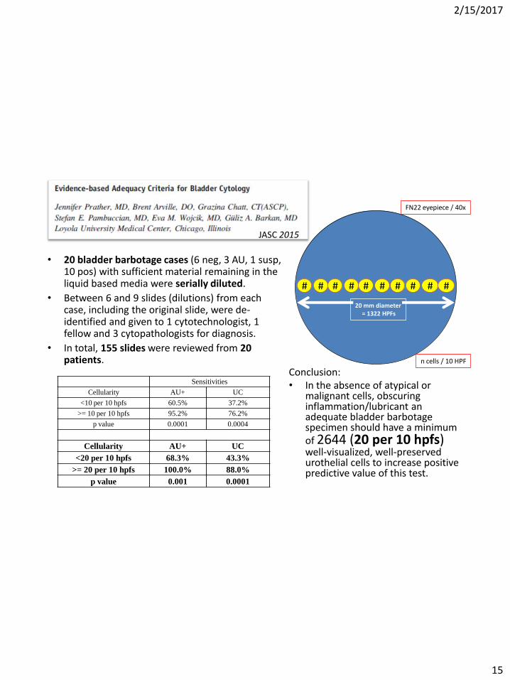
15
JASC 2015
# # # # # # # # # #
20 mm diameter = 1322 HPFs
n cells / 10 HPF
FN22 eyepiece / 40x
• 20 bladder barbotage cases (6 neg, 3 AU, 1 susp, 10 pos) with sufficient material remaining in the liquid based media were serially diluted.
• Between 6 and 9 slides (dilutions) from each case, including the original slide, were de-identified and given to 1 cytotechnologist, 1 fellow and 3 cytopathologists for diagnosis.
• In total, 155 slides were reviewed from 20 patients.
Sensitivities
Cellularity AU+ UC
<10 per 10 hpfs 60.5% 37.2%
>= 10 per 10 hpfs 95.2% 76.2%
p value 0.0001 0.0004
Cellularity AU+ UC
<20 per 10 hpfs 68.3% 43.3%
>= 20 per 10 hpfs 100.0% 88.0%
p value 0.001 0.0001
Conclusion: • In the absence of atypical or
malignant cells, obscuring inflammation/lubricant an adequate bladder barbotage specimen should have a minimum of 2644 (20 per 10 hpfs) well-visualized, well-preserved urothelial cells to increase positive predictive value of this test.
2/15/2017
16
Guidelines for estimating cellularity in urinary tract specimens
FN20 eyepiece 10 X objective
FN20 eyepiece 40 X objective
FN22 eyepiece 10 X objective
FN22 eyepiece 40 X objective
Prep Diameter (mm)
Area (mm2)
Number of fields at FN20, 10X
Number of cells/field for 2644 cells total
Number of fields at FN20, 40X
Number of cells/field for 2644 cells total
Number of fields at FN20, 10X
Number of cells/field for 2644 cells total
Number of fields at FN20, 40X
Number of cells/field for 2644 cells total
13 132.7 42.3 62.5 676 3.9 34.9 75.8 559 4.7
20 314.2 100 26.4 1600 1.7 82.6 32 1322 2
Adapted from the Bethesda System from Reporting Cervical Cytology, Editors Diane Solomon and Ritu Nayar, 2nd Ed, 2004, Chapter 1, pg 8
Should urine volume be a factor in specimen adequacy? (VandenBussche et al.)
-15,731 voided urine specimens, SurePath, >10 years -Inadequate cellularity during this period was purely subjective
Yes, especially if the volume is <20mL and no malignant cells are seen.
2/15/2017
18
2/15/2017
19
II. Negative for High-Grade Urothelial Carcinoma (Negative)
Dorothy L. Rosenthal, Michael B. Cohen, Hui Guan, Christopher L. Owens, Yuji Tokuda, and Eva M. Wojcik
Definition:
A sample of urine, either voided or instrumented, may be considered benign, i.e., NHGUC, if any of the following components are present in the specimen:
– Benign urothelial, glandular, and squamous cells
– Benign urothelial tissue fragments (BUTF) and urothelial sheets or clusters
– Post-therapy effect, including epithelial cells from urinary diversions
2/15/2017
20
Negative - Summary
• Negative for High Grade Urothelial Carcinoma
– This diagnostic category will include cases where “low grade urothelial carcinoma can not be excluded”
• If there is a cause for “atypia” i.e. urolithiasis, treatment related changes etc. – it is negative!
2/15/2017
21
“Negative, NOT atypia”
Wojcik EM: What should not be reported as atypia in urine cytology: JASC 2015;4;3;30-36
2/15/2017
22
• UTCy has a high (96.7%) NPV • NPV was highest in the hematuria patient group • NPV of UTCy in patients with history of UC was only 90%, suggesting that UTCy should be used in
conjunction with cystoscopy results to rule out recurrences of UC in this group of patients. Khan R, Hussain H, Pambuccian S, Wojcik, EM, Barkan GA: JASC November 2015
What is the NPV of UTC?
2/15/2017
23
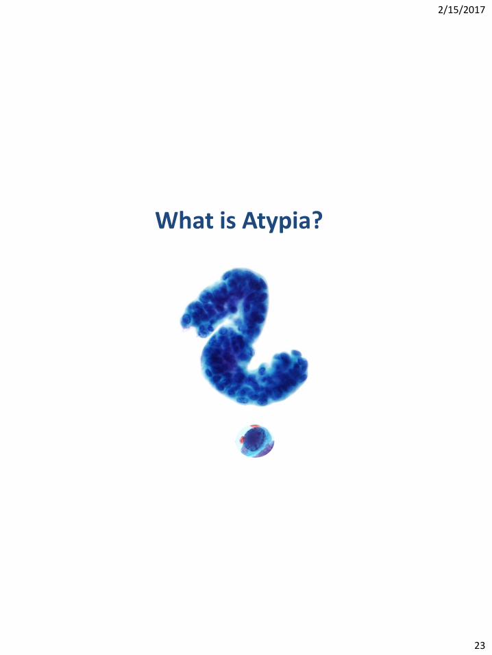
What is Atypia?
2/15/2017
24
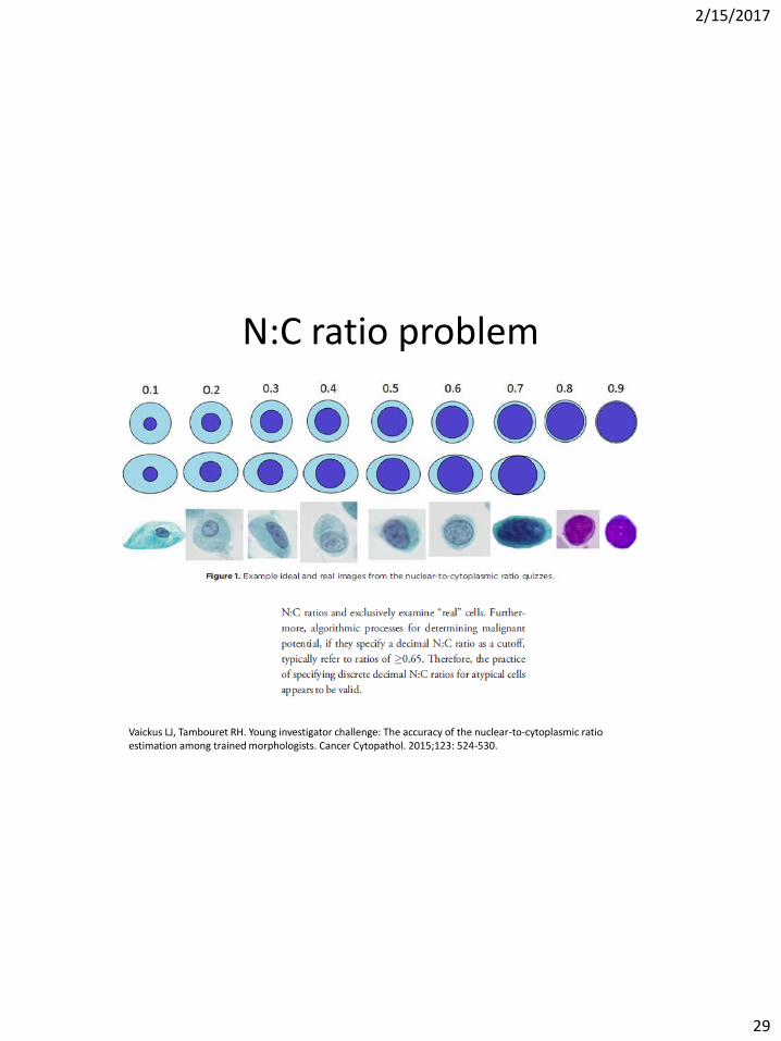
Atypia - Pubmed Search
“atypia” /“atypical” through a PubMed title search
Vaickus LJ, Tambouret RH. Young investigator challenge: The accuracy of the nuclear-to-cytoplasmic ratio estimation among trained morphologists. Cancer Cytopathol. 2015;123: 524-530.
N:C ratio problem
2/15/2017
30
So, when do I call atypia?
2/15/2017
31
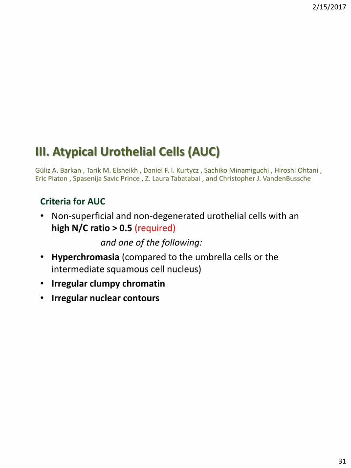
Criteria for AUC
• Non-superficial and non-degenerated urothelial cells with an high N/C ratio > 0.5 (required)
and one of the following:
• Hyperchromasia (compared to the umbrella cells or the intermediate squamous cell nucleus)
• Irregular clumpy chromatin
• Irregular nuclear contours
III. Atypical Urothelial Cells (AUC) Güliz A. Barkan , Tarik M. Elsheikh , Daniel F. I. Kurtycz , Sachiko Minamiguchi , Hiroshi Ohtani , Eric Piaton , Spasenija Savic Prince , Z. Laura Tabatabai , and Christopher J. VandenBussche
2/15/2017
32
AUC
N/C > 0.5 NO HYPERCHROMASIA, IRREGULAR MEMBRANES
N/C -0.5 HYPERCHOMASIA CLUMPY CHROMATIN
2/15/2017
33
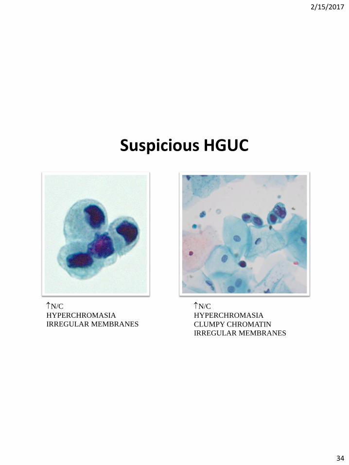
Criteria for Suspicious
• Non-superficial and non-degenerated urothelial cells with a high N/C ratio > 0.7 (required)
• Hyperchromasia (compared to the umbrella cells or the intermediate squamous cell nucleus) (required)
and one of the following:
• Irregular clumpy chromatin
• Irregular nuclear contours
IV. Suspicious for High-Grade Urothelial Carcinoma (Suspicious) Fadi Brimo , Manon Auger , Tarik M. Elsheikh , Hui Guan , Mitsuru Kinjo , Eric Piaton , Dorothy L. Rosenthal , Tatsuro Shimokama , and Rosemary H. Tambouret
2/15/2017
34
Suspicious HGUC
N/C
HYPERCHROMASIA
IRREGULAR MEMBRANES
N/C
HYPERCHROMASIA
CLUMPY CHROMATIN
IRREGULAR MEMBRANES
2/15/2017
35
Suspicious for HGUC vs. Positive HGUC Quantity matters..
“The number of atypical urothelial cells is an important criterion to classify urine cytology specimens into the ‘positive’ or the ‘suspicious’ categories. ..A cut-off number of >10 cells to render a definitive diagnosis of HGUCA seems valid from the clinical standpoint .”
Does the Number of Atypical Urothelial Cells Matter for distinguishing the “high-grade urothelial carcinoma (HGUCA)” from the “suspiciousfor HGUCA” cytological categories? Brimo, Fadi et al. Journal of the American Society of Cytopathology, 2015;4(4), 232 - 238
2/15/2017
36
SHGUC
Negative
AUC
2/15/2017
37
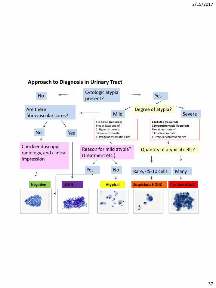
Cytologic atypia present?
No Yes
Check endoscopy, radiology, and clinical impression
Negative
Mild Severe Degree of atypia?
Quantity of atypical cells?
Atypical
Rare, <5-10 cells Many
Suspicious HGUC
Reason for mild atypia? (treatment etc.)
Positive HGUC
Approach to Diagnosis in Urinary Tract
1.N:C>0.5 (required) Plus at least one of: 2. Hyperchromasia 3.Coarse chromatin 4. Irregular chromatinic rim
Yes
1.N:C>0.7 (required) 2.Hyperchromasia (required) Plus at least one of: 3.Coarse chromatin 4. Irregular chromatinic rim
No
Are there fibrovascular cores?
Yes No
LGUN
2/15/2017
38
Negative
Suspicious HGUC
Positive HGUC
• Close follow up (within 1-3 months)
• Biopsy, Stage
• Follow up as Normal/ as needed
Atypical • Individualized
• Role of FISH?
IX. Clinical Management Marcus L. Quek , Trinity J. Bivalacqua , Ashish M. Kamat , and Mark P. Schoenberg
2/15/2017
39
The Paris System: Eds Rosenthal D, Wojcik EM, Kurtycz DFI,Springer 2015, Chapter 4
2/15/2017
40
Suspicious
Follow-up
Positive
Follow-up
Study Specimen
Type Clinical Indications Cases PPV Cases PPV
Joudi et al.
2016
Inst: 86%
Void: 14%
New Symptoms: 10%
Surveillance: 90% 150 55.3% 459 79.2%
Ton nu et al
2014
Ins: 29%
Void: 71%
New symptoms: 29%
Surveillance: 71% 191 79% 256 86%
VandenBussche
et al.
2013
N/A N/A 82 71% 143 77%
Piaton et al.
2013
Ins: 96%
Void: 4%
New Symptoms: 21%
Surveillance: 79% 185 37.8% 162 59.9%
Follow up on SHGUC and HGUC
Joudi A et al . Cancer 2016 Nov;124(11):811-819.
2/15/2017
41
V. High Grade Urothelial Carcinoma Momin T. Siddiqui , Guido Fadda , Jee-Young Han ,Christopher L. Owens , Z. Laura Tabatabai , and Toyonori Tsuzuki
HGUC Definition and Criteria
• Urine cytology cannot distinguish invasive HGUC from non-invasive HGUC or CIS.
• The background in CIS: clean without blood, abundant inflammation and cell debris
• HGUC: N/C ratio that is 0.7 or greater, nuclear hyperchromasia, irregular nuclear membranes and coarse chromatin
2/15/2017
42
HGUC Definition and Criteria
• A minimum of 5-10 viable malignant cells will qualify as HGUC.
• Depends on the specimen type and comfort level of the pathologist
• Upper urinary tract specimens will require at least 10 abnormal cells, whereas voided urine specimens may require a lesser number of cells to establish a definitive diagnosis of HGUC.
Morphologic Criteria For Atypia in Upper Urinary Tract Cytology: Should It Be Different than The Lower Urinary Tract Criteria of The Paris System of Reporting Urinary Tract Cytology (PSRUC)?
Fidan-Özbilgin Ö. et al. Modern Pathology 2016;29(Sup 2):98A
2/15/2017
45
Upper Tract Problem
The Performance of the Paris System for Reporting Urine Cytology (PSRUC) in Lower and Upper Tract Specimens: A Comparative Study of 358 Cases.
Brimo and Barkan et al. Modern Pathology 2016;29(Sup 2):92-93A
2/15/2017
46
VI. Low-Grade Urothelial Neoplasia (LGUN) Eva M. Wojcik, Tatjana Antic, Ashish Chandra, Michael B. Cohen, Zulfia McCroskey, Jae Y. Ro, and Taizo Shiraish
Is a consistent cytologic diagnosis of low-grade urothelial carcinoma in instrumented urinary tract cytologic specimens possible? A comparison between cytomorphologic features of low-grade urothelial carcinoma and non-neoplastic changes shows extensive overlap, making a reliable diagnosis impossible.
• The majority of the features described previously as diagnostic for LGPUC were observed almost equally in patients with or without biopsy-proven LGPUC, regardless of whether the specimens were from the upper or the lower urinary tract
• Mild nuclear membrane irregularity was present in 48% of LGPUC and 47.2% of negative controls (p=0.93); mild nuclear enlargement was observed in 42.9% of LGPUC patients and 49.1% negative controls (p=0.26)
McCroskey Z, Kliethermes S, Bahar B, Barkan GA, Pambuccian SE, Wojcik EM Journal of American Society of Cytopathology. 2014;4:90-97.
2/15/2017
48
Cytologic Criteria of Low Grade Urothelial Neoplasia (LGUN) (regardless of the specimen type: voided or
instrumented):
• Three-dimensional cellular papillary clusters (defined as clusters of cells with nuclear overlapping, forming "papillae") with fibrovascular cores with capillaries
2/15/2017
49
Cytologic Criteria of Low Grade Urothelial Neoplasia (LGUN):
(regardless of the specimen type: voided or instrumented)
Cell Block
2/15/2017
50
LGUN may be considered in correlation with cystoscopic or biopsy findings
Diagnosis - NHGUC
• Three-dimensional cellular clusters without fibrovascular cores
• Increased numbers of monotonous single (non-umbrella) cells
VII. Other Malignancies Primary and Metastatic and Miscellaneous Lesions
Rana S. Hoda , Stefan E. Pambuccian , Jae Y. Ro , and Sun Hee Sung
2/15/2017
52
Squamous Cell Carcinoma
2/15/2017
53
Small cell carcinoma Diffuse Large B cell Lymphoma
Leiomyosarcoma Melanoma
2/15/2017
54
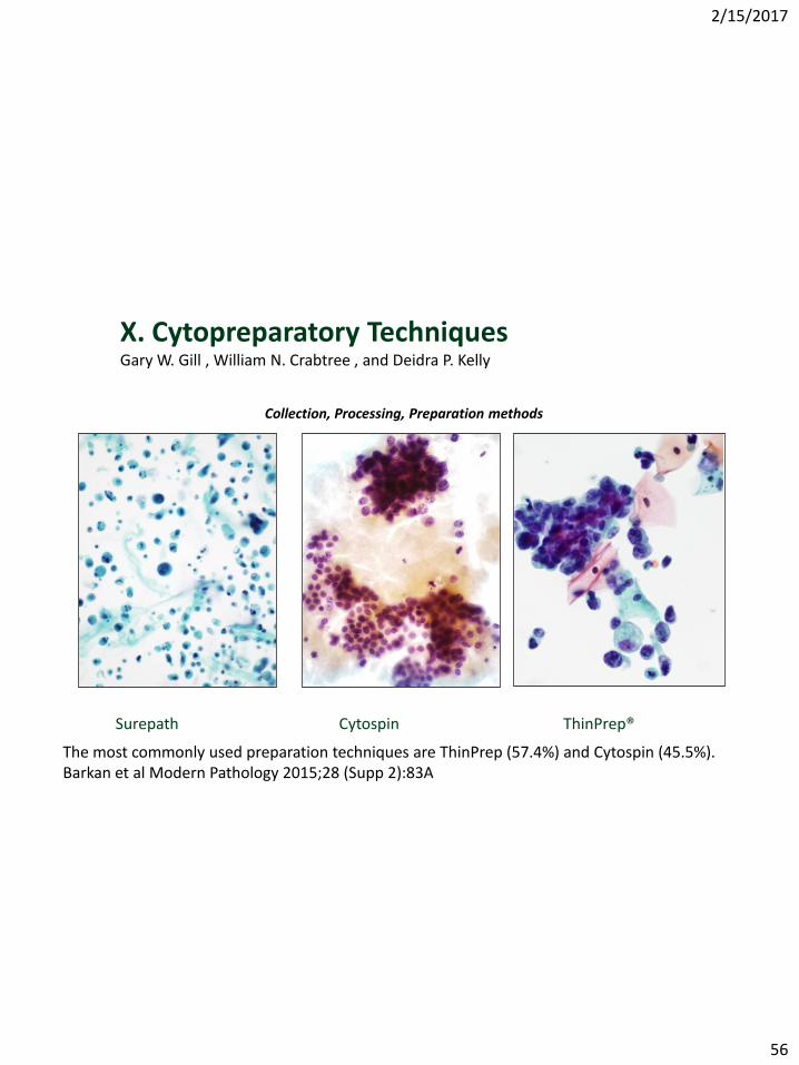
VIII. Ancillary Studies in Urinary Cytology
Lukas Bubendorf , Nancy P. Caraway, Andrew H. Fischer, Ruth L. Katz , Matthew T. Olson, Fernando Schmitt, Margareta Strojan Fležar , Theodorus H. Van Der Kwast, and Philippe Vielh
• Ancillary Tests: UroVysion FISH (Abbott Molecular Inc, Des Plaines, IL),ImmunoCyt (Scimedx, Denville, NJ), BTA stat (Polymedco, Cortlandt Manor, NY), and NMP 22 (Allere, Waltham, MA).
• When NOT to use the Ancillary studies: NHGUC, HGUC
• When ancillary studies may be of use: AUC (Selected conditions)
2/15/2017
55
UroVysion FISH in AUC
U-FISH in the setting of AUC: detection of HGUC
“A positive U-FISH result may heighten this degree of suspicion, but a negative U-FISH test cannot be used to limit the need for routine cystoscopic surveillance.”
The Value Of The UroVysion® FISH Assay In The Risk-Stratification Of
Patients With “Atypical Urothelial Cells” In Urinary Cytology Specimens


![Article - White Rose Research Onlineeprints.whiterose.ac.uk/113339/2/[Bladder_cancer]_Figures...TERT mutation and 9q LOH Flat dysplasia TP53 mutation, 9p LOH and 9q LOH T1 Invasive](https://static.documents.pub/doc/80x56/5f24abd2d3ccb012d859c51c/article-white-rose-research-bladdercancerfigures-tert-mutation-and-9q-loh.jpg)


