British Journal of Dermatology 1995: 133: 254-258. The pathogenesis of hidradenitis suppurativa: a closer look at apocrine and apoeccrine glands R.L.ATTANOOS. M.A.C.APPLETON AND A.G.DOUGLAS-JONES Department of Histopathology. University Hospital of Wales, Heath Park, Cardiff CF4 4XN. U.K. Accepted for publication 10 October 1994 Summary We undertook a retrospective pathological study of 118 skin resection specimens from 101 patients with hidradenitis suppurativa. Follicular occlusion was identified in all the specimens, regardless of disease duration (1 month to 18 years), but was not noted in the axillary and inguinal skin of controls. We therefore regard follicular occlusion as an early and important feature in the pathogenesis of the disease. The presence of apoeccrine glands in axillary skin provided an in vivo model to directly observe the effects of follicular occlusion on follicle inflammation and apocrine gland destruction. In the majority of cases, active folliculitis was associated with apocrinitis and apocrine destruction, whereas apoeccrine glands, which drain directly on to the epidermal surface, appeared intact and non-inflamed. These observations provide direct evidence in an in vivo model that follicular occlusion by keratinous material, with subsequent active folliculitis and secondary destruction of the skin adnexae and subcutis. occur as an integral step in the pathogenesis of hidradenitis suppurativa. Hidradenttis suppurativa is a chronic, inflammatory, scarring disease of apocrine sweat gland-bearing skin. The diagnosis of the disease is primarily clinical, based on the presence of multiple abscesses and sinuses, which often show a poor response to conventional antibiotics and a tendency to recur. The onset is predominantly after puberty, although it has been described in neonates. In women, premenstrual exacer- bation of the disease is not uncommon. A number of histopathological changes have been identified in resection specimens.^ but these are considered to be non-specific, and represent the late sequelae of the disease in many cases. The pathogenesis of the condition remains unclear. The anatomical distribution of the disease suggests that it is primarily a disorder of apocrine glands, and the term •apocrinitis' has been employed as a synonym for the condition. Indeed, the presence of distended apo- crine glands containing polymorph neutrophils was demonstrated in an early description of the disease by Brunsting in 1939.' More recent publications*'^ have suggested that follicular occlusion in apocrine gland- bearing skin is the likely primary event, followed by secondary suppuration and sinus formation. However, these notions are based on the end-stage histological appearances in hidradenitis suppurativa. and direct pathological evidence of disease evolution is lacking. The association with, and clinical similarity of hidra- denitis suppurativa to. acne conglobata. dissecting cellulitis of the scalp, and pilonidal sinus (follicular occlusion-related diseases), lends circumstantial sup- port to this view of the aetiology of the condition. Disturbances in androgen metabolism have high- lighted an endocrine component in the condition.^' but their part in its pathogenesis remains uncertain. The aim of the present study was to review the skin resection specimens from 101 patients with hidradenitis suppurativa of variable duration, describe the morpho- logical variation of the pathology at each site, and gain insight into the pathogenesis of the disease. To this end, particular attention was paid to the adnexae and, in the axillae, the apoeccrine sweat glands.^ The presence of apoeccrine sweat glands in axillary skin provides, for the first time, an in vivo model to study the direct effect of follicular occlusion on apocrine glands. The ducts of apocrine glands drain into the main follicle, and secretion reaches the skin surface by way of the hair shaft. Hence, follicular occlusion will cause obstruction of apocrine and sebaceous secretion. In contrast, apoeccrine glands structurally combine features of both apocrine and eccrine glands, with an apocrine secretory coil exhibiting 'decapitation' secretion, and a straight intradermal duct which opens directly on to the epidermal surface, circumventing the effect of 254 199S British Association of Dermatologists
Transcript
British Journal of Dermatology 1995: 133: 254-258.
The pathogenesis of hidradenitis suppurativa: a closer look atapocrine and apoeccrine glands
R.L.ATTANOOS. M.A.C.APPLETON AND A.G.DOUGLAS-JONESDepartment of Histopathology. University Hospital of Wales, Heath Park, Cardiff CF4 4XN. U.K.
Accepted for publication 10 October 1994
Summary We undertook a retrospective pathological study of 118 skin resection specimens from 101 patientswith hidradenitis suppurativa. Follicular occlusion was identified in all the specimens, regardless ofdisease duration (1 month to 18 years), but was not noted in the axillary and inguinal skin ofcontrols. We therefore regard follicular occlusion as an early and important feature in thepathogenesis of the disease. The presence of apoeccrine glands in axillary skin provided an in vivomodel to directly observe the effects of follicular occlusion on follicle inflammation and apocrinegland destruction. In the majority of cases, active folliculitis was associated with apocrinitis andapocrine destruction, whereas apoeccrine glands, which drain directly on to the epidermal surface,appeared intact and non-inflamed. These observations provide direct evidence in an in vivo modelthat follicular occlusion by keratinous material, with subsequent active folliculitis and secondarydestruction of the skin adnexae and subcutis. occur as an integral step in the pathogenesis ofhidradenitis suppurativa.
Hidradenttis suppurativa is a chronic, inflammatory,scarring disease of apocrine sweat gland-bearing skin.The diagnosis of the disease is primarily clinical, basedon the presence of multiple abscesses and sinuses,which often show a poor response to conventionalantibiotics and a tendency to recur. The onset ispredominantly after puberty, although it has beendescribed in neonates. In women, premenstrual exacer-bation of the disease is not uncommon.
A number of histopathological changes have beenidentified in resection specimens.^ but these areconsidered to be non-specific, and represent the latesequelae of the disease in many cases.
The pathogenesis of the condition remains unclear.The anatomical distribution of the disease suggests thatit is primarily a disorder of apocrine glands, and theterm •apocrinitis' has been employed as a synonym forthe condition. Indeed, the presence of distended apo-crine glands containing polymorph neutrophils wasdemonstrated in an early description of the disease byBrunsting in 1939.' More recent publications*'^ havesuggested that follicular occlusion in apocrine gland-bearing skin is the likely primary event, followed bysecondary suppuration and sinus formation. However,these notions are based on the end-stage histologicalappearances in hidradenitis suppurativa. and directpathological evidence of disease evolution is lacking.
The association with, and clinical similarity of hidra-denitis suppurativa to. acne conglobata. dissectingcellulitis of the scalp, and pilonidal sinus (follicularocclusion-related diseases), lends circumstantial sup-port to this view of the aetiology of the condition.Disturbances in androgen metabolism have high-lighted an endocrine component in the condition.^'but their part in its pathogenesis remains uncertain.
The aim of the present study was to review the skinresection specimens from 101 patients with hidradenitissuppurativa of variable duration, describe the morpho-logical variation of the pathology at each site, and gaininsight into the pathogenesis of the disease. To this end,particular attention was paid to the adnexae and, in theaxillae, the apoeccrine sweat glands.^ The presence ofapoeccrine sweat glands in axillary skin provides, forthe first time, an in vivo model to study the direct effect offollicular occlusion on apocrine glands. The ducts ofapocrine glands drain into the main follicle, andsecretion reaches the skin surface by way of the hairshaft. Hence, follicular occlusion will cause obstructionof apocrine and sebaceous secretion. In contrast,apoeccrine glands structurally combine features ofboth apocrine and eccrine glands, with an apocrinesecretory coil exhibiting 'decapitation' secretion, and astraight intradermal duct which opens directly on tothe epidermal surface, circumventing the effect of
254 199S British Association of Dermatologists
PATHOGENESIS OF HIDRADENITIS SUPPURATIVA 255
terminal follicular occlusion. Comparison of inflamma-tion and destruction in apocrine and apoeccrine glandsin hidradenitis suppurativa cases with control cadavericskin provided the basis for the study of the pathogenesisof this condition.
Methods
One hundred and eighteen resection specimens fromaxillary, inguinoperineal, perianal, mammary andvulval skin were reviewed simultaneously by twopathologists (R.L.A. and M.A.C.A.). Specimens werefixed in 10% formalin, and routinely processed. Five-micron sections were stained with haematoxylin andeosin for morphological assessment. Details regardingthe nature, distribution and extent of the inflammatoryceil infiltrate were noted. Granulomatous reactionswere classified into foreign-body or epithelioid types.Particular attention was paid to morphological abnor-malities of the adnexae. By careful serial sectioning,apocrine glands, eccrine glands, and the recentlydescribed apoeccrine glands were delineated.
The presence/absence of epidermal changes, particu-
larly follicular occlusion, hyperplasia and keratinocyteatypia was documented, and features such as epidermalcyst formation, sinus tracks, fibrosis and fat necrosiswere also noted. The histological changes were com-pared with control specimens of axillary and inguino-perineal skin taken from 10 cadavers with nodocumented history of hidradenitis suppurativa.
ResultsClinical results
The study group comprised 79 women and 22 men.Pifty-tbree patients (52-5%) had axillary disease, whichwas bilateral in 29 cases. Hidradenitis affecting theinguinoperineal and perianal skin was noted In 47and seven patients, respectively. Six female patientshad infra-mammary disease, and five had vuival skininvolvement. Multiple site disease was noted in 12cases. The duration of disease varied from 6 monthsto 18 years.
The control group comprised four women and sixmen.
1995 British Association of Dermatologists. British journal of Dermatobsfj. 133, 254-258
256 R.L.ATTANOOSeffli.
Pathological results
A detailed histopathological evaluation of the 118resection specimens is shown in Table 1.
In all of the study cases a significant degree offollicular plugging was identified: when prominent,this was manifested as large cystic lesions, many ofwhich contained refractile laminated keratin and hairshafts. Skin from all sites exhibited varying degrees ofinflammation and reparative fibrosis of both the dermisand subcutis. The presence of 'established' epidermalcysts was found to be less common. Acutely inflamedsinus tracks were also noted as a frequent feature,occurring predominantly in the extra-axillary sitesand in those subjects with long-standing disease.Adnexal inflammation, a common feature, was mostfrequently perifollicular in distribution, occurring in64% of axillary cases and 66% of inguinoperinealcases. In perianal, mammary and vulval skin, anactive folliculitis was noted in 71, 67 and 83% ofcases, respectively.
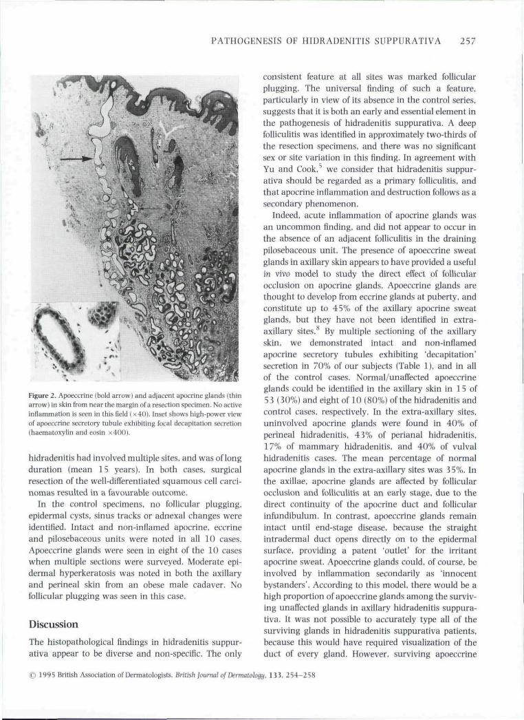
Active inflammation of the sweat glands was lesscommon in all sites, and where present was morefrequently eccrine than apocrine in distribution. At nosite was the inflammation solely seen around apocrineglands. Indeed, the axilla was the only site in whichactive destruction of apocrine glands by polymorphneutrophils was noted, occurring in 26% of cases.Peri-eccrine inflammation was identified in 32% ofaxillary specimens. When present, active apocrinitiswas invariably associated with severe acute inflamma-tion in the adjacent hair follicle {Fig. 1). In 1 5 of 5 3(30%) axillary specimens, unaffected secretory tubulesdisplaying 'decapitation' secretion (apocrine gland-type) were seen to drain directly on to the epidermalsurface via a straight intradermal duct lined by eccrineduct epithelium. Morphologically, these sweat glandscorresponded to so-called apoeccrine glands (Fig. 2).Apoeccrine glands were not identified in extra-axillarysites.
No chronological pattern to these changes could beidentified in any site, and there was no significantdifference between the sexes.
Foreign-body type granulomas containing refractilekeratinous material around ruptured hair follicles andsinus tracks were identified in approximately 2S% of allspecimens. No granulomatous inflammation near sweatglands was identified. The presence of discrete epithe-lioid granulomas in the deep dermis and subcutis, awayfrom the site of active inflammation, was infrequentlyseen {8% of all specimens). One female patient with
Figure 1. Abscess formation and active folliculitis, with adjacentinfiltration of apocrine glands by polymorph neutrophfls (haematoxy-lin and eosin x25O|.
discrete epithelioid granulomas had axillary hidradeni-tis suppurativa and concomitant pulmonary sarcoido-sis, and another with inguinoperineal hidradenitissuppurativa had colonic Crohn's disease. No systemicdisease was identified in the remaining patients whoseresection specimens showed epithelioid granulomas. Acareful search for tubercle bacilli and fungal organismswas undertaken In all granulomatous reactions devoidof foreign-body material.
The degree of epidermal change varied. Despite theuniversal presence of follicular plugging in all affectedsites, hyper- and parakeratosis were infrequentlyidentified. However, acanthosis was more common,especially in vulval and perianal skin. Similarly,pseudoepitheliomatous hyperplasia was relativelymore prominent in these sites, and in the inguinoper-ineai region. In one perianal and one vulval case ofchronic hidradenitis suppurativa. invasive squamouscell carcinoma had developed. In both patients the
1995 British Association of Dermatologists. British lourna! of Dermatology. 133. 254-258
PATHOGENESIS OF HIDRADENITIS SUPPURATIVA 257
Figure 2. Apoeccrine (bold arrow) and adjacent apocrine glands (thinarrow) in skin from near the margin of a resection specimen. No activeinflammation is seen in this field (x 40). Inset shows high-power viewof apoeccrine secretory tubule exhibiting focal decapitation secretion(haematoxylin and eosin x400).
hidradenitis bad involved multiple sites, and was of longduration (mean 15 years). In both cases, surgicalresection of the well-differentiated squamous cell carci-nomas resulted in a favourable outcome.
In the control specimens, no follicular plugging,epidermal cysts, sinus tracks or adnexal changes wereidentified. Intact and non-inflamed apocrine, eccrineand pilosebaceous units were noted in all 10 cases.Apoeccrine glands were seen in eight of the 10 caseswhen multiple sections were surveyed. Moderate epi-dermal hyperkeratosis was noted in both the axillaryand perineal skin from an obese male cadaver. Nofollicular plugging was seen in this case.
Discussion
The histopathological findings in hidradenitis suppur-ativa appear to be diverse and non-specific. The only
consistent feature at al! sites was marked follicularplugging. The universal finding of such a feature,particularly in view of its absence in the control series,suggests that it is both an early and essential element inthe pathogenesis of hidradenitis suppurativa. A deepfolliculitis was identified in approximately two-thirds ofthe resection specimens, and there was no significantsex or site variation in this finding. In agreement withYu and Cook, we consider that hidradenitis suppur-ativa should be regarded as a primary folliculitis, andthat apocrine inflammation and destruction follows as asecondary phenomenon.
Indeed, acute inflammation of apocrine glands wasan uncommon finding, and did not appear to occur inthe absence of an adjacent folliculitis in the drainingpilosehaceous unit. The presence of apoeccrine sweatglands in axillary skin appears to have provided a usefulin vivo model to study the direct effect of follicularocclusion on apocrine glands. Apoeccrine glands arethought to develop from eccrine glands at puberty, andconstitute up to 45% of the axillary apocrine sweatglands, but they have not been identified in extra-axillary sites. By multiple sectioning of the axillaryskin, we demonstrated intact and non-inflamedapocrine secretory tubules exhibiting 'decapitation'secretion in 70% of our subjects (Table 1), and in allof the control cases. Normal/unaffected apoeccrineglands could be identified in the axillary skin in 15 of53 (30%) and eight of 10 (80%) of the hidradenitis andcontrol cases, respectively. In the extra-axillary sites,uninvolved apocrine glands were found in 40% ofperineal hidradenitis. 43% of perianal hidradenitis,17% of mammary hidradenitis, and 40% of vulva!hidradenitis cases. The mean percentage of normalapocrine glands in the extra-axillary sites was 35%. Inthe axillae, apocrine glands are affected by follicularocclusion and folliculitis at an early stage, due to thedirect continuity of the apocrine duct and follicularinfundibulum. In contrast, apoeccrine glands remainintact until end-stage disease, because the straightintradermal duct opens directly on to the epidermalsurface, providing a patent 'outlet' for the irritantapocrine sweat. Apoeccrine glands could, of course, beinvolved by infiammation secondarily as 'innocentbystanders'. According to this model, there would be ahigh proportion of apoeccrine glands among the surviv-ing unaffected glands in axillary hidradenitis suppura-tiva. It was not possible to accurately type all of thesurviving glands in hidradenitis suppurativa patients,because this would have required visualization of theduct of every gland. However, surviving apoeccrine
1995 British Association of Dermatologists. British journal of Dermatology. 133. 254-258
258 R.L.ATTANOOS et al.
glands were seen in a significant proportion of hidra-denitis specimens (30%), and this observation lendssupport to the above model.
The stimulus for initial follicular occlusion remainsundetermined. Obesity is common in hidradenitis sup-purativa. and is likely to contribute to its pathogenesis.Obesity increases skin contact, and promotes bothepidermal desquamation and hyperkeratosis. Keratinhydration would be enhanced in the sweat gland-richregions of the body, and this has been shown to reducethe width of the pilosebaceous duct orifice, favouringporal occlusion.** Epidermal hyperkeratosis was noted inone obese control subject, but there was no evidence ofporal occlusion. This suggests that the mechanicalepidermal and follicular changes described above areinsufficient alone to explain the poral occlusion. Obesityis also known to alter the metabolism and activity of sexhormones, producing a state of relative androgenexcess, and inducing hirsutism in women. This mayproduce marked coarsening of the hair shaft andsubsequent follicular plugging. Hidradenitis has beensuccessfully controlled in some female patients by anti-androgen therapy.^"
In the present series, foreign-body-type granulomaswere found in 25% of the specimens in close relation toruptured hair follicles and epidermal cysts. Foamymacrophage-type granulomas have been describedin relation to apocrine sweat glands in the dermis, butwere not seen in our specimens. However, the experi-mental model used by Shelley and Cahn" incorporatedmanual depilation of the skin and the use of atropine-impregnated tape, both of which may have stimulatedan abnormal pathophysiological process. It is of interestthat discrete epithelioid granulomas in two patientswith systemic granulomatous disease were noted (onecase of sarcoidosis and the other of Crohn's disease).These findings have been published in our earlier
Squamous cell carcinoma arising in hidradenitissuppurativa is a recognized complication, which wasseen in two of 118 specimens (1-7%). Jackman^'observed four cases of squamous cell carcinoma in aseries of 12 5 perianal hidradenitis specimens, and all ofthese cases had long-standing disease (19-32 years)prior to the development of malignancy.
It is of interest that both our cases of squamous cellcarcinoma developed in non-axillary sites, suggestingthat malignant change in hidradenitis suppurativa is
more common in the perianal area. Squamous cellcarcinoma is an uncommon but recognized complica-tion in areas of chronic Injury or irritation, and occursin stasis ulcers, burn scars, and chronically inflamedfistulae.^^
We have demonstrated that occlusion of hair folliclesis an early and important aspect of the pathogenesis ofhidradenitis suppurativa. An active deep folliculitis wasobserved in all cases where apocrine gland inflamma-tion was present, indicating that apocrine destructionrepresented a secondary process. In a significantnumber of axillary cases (30%). intact apoeccrineglands were seen, which lends support to our in vivomodel for studying the direct effect of follicuiar occlu-sion on apocrine glands and their part in the patho-genesis of hidradenitis suppurativa.
References
1 Mortimer P. Hidradenitis suppurativa—diagnostic criteria. In:Acne and Related Disorders (Marks R. Plewig G. eds). London:Martin Dunitz Ltd. 1989: 359-60,
2 Lever WF. Schaumburg-Lever G. In: Histopathalogy ofihe Skin. 7thedn. Philadelphia: J.B. [Jppincott Co.. 1990: 322.
J Brunsting HA. Hidradenitis suppurativa: abscess of the apocrinesweat glands. Arch Derm Syphihl 1939: 59: 108-20.
4 Plewig G. Steger M. Acne inversa (alias acne triad, acne tetrad orhidradenitis suppurativa). In: Acne and Related Disorders (Marks R.Plewig G. eds). London: Martin Uunitz Ltd. 1989: 345-57.
5 Yu CCp Cook MG. Hidradenitis suppurativa: a disease of follicularepithelium rather than apocrine glands. Br ] Dermatol 1990: 122:763-9.
7 Harrison B|. Hughes LE. Enhanced peripheral androgen metabo-lism in patients with hidradenitis suppurativa, Br) Surg 1988; 75:600 (Abstr.).
8 Sato K, Leidal R, Sato F. Morphology and development of anapoeccrine sweat gland in human axillae. Am J Physiol 1987: 252:166-80.
9 Williams M. Cunliffe W(, Gould U. Effects of hydration on thepilosebaceous duct orifice. Br / Dermatol 1974: 90: 631-5 .
10 Mortimer PS. Dawber RPR, Gales MA et al A double-blindcontrolled crossover trial of cyproterone acetate in females withhidradenitis suppurativa. Br } Dermatol 1986: 115: 263-8 .
11 Shelley WB. Cahn MM. The pathogenesis of hidradenitis suppur-ativa in man. Arch Derm Syphilol 1955: 72: 562-5,
12 Attanoos RL. Applcton MAC. Hughes LE et a!. Granulomatoushidradenitis suppurativa and cutaneous Crohn's disease. Histo-pathology 1993: 2J: 111-15,
1 3 Jackman RJ. Hidradenitis suppurativa: diagnosis and manage-ment of perianai manifestations. Proc R Soc Med 1959: 52: 110-12.