The Pivotal Role of Insulin-Like Growth Factor I in Normal Mammary Development David L. Kleinberg, MD a,c, *, Mary Helen Barcellos-Hoff, PhD b Adult mammary development begins at puberty. A surge of estradiol (E 2 ) begins the process before menarche. In humans, breast development is referred to as thelarche. The early contributions of the various hormones important in mammary development have largely been studied in mice. The identification and isolation of estrogen, 1 prolactin (PRL), 2 and growth hormone (GH) 3 together with improved surgical tech- niques in mice permitted investigators to begin to understand the hormonal control of mammary development. The work began in the 1920s and continues today. Turner and Frank 4 showed that although estrogen alone stimulated mammary ductal morphogenesis, the combination of estrogen and progesterone caused lobu- loalveolar development similar to that seen during pregnancy. 5 Several investigators in the 1930s determined that the pituitary gland was essential for mammary devel- opment because mammary development did not occur in hypophysectomized animals even if estrogen was administered in high concentration. 6,7 Several years elapsed before pituitary hormones were sufficiently purified to help determine which of the pituitary hormones played a role in mammary development. 8–10 The laborato- ries of Lyons and colleagues 11 and Nandi 12 provided evidence that GH, together with estrogen, was responsible for ductal morphogenesis, whereas estrogen, a Department of Medicine (Endocrinology), New York University School of Medicine, 550 First Avenue, New York, NY 10016, USA b Departments of Radiation Oncology and Cell Biology, New York University School of Medicine, 566 First Avenue, New York, NY 10016, USA c Veterans Affairs Medical Center, 423 East 23rd Street, New York, NY 10010, USA * Corresponding author. Department of Medicine (Endocrinology), New York University School of Medicine, 550 First Avenue, New York, NY 10016. E-mail address: [email protected]KEYWORDS IGF-I Estradiol Ductal morphogenesis Progesterone TGF-b Endocrinol Metab Clin N Am 40 (2011) 461–471 doi:10.1016/j.ecl.2011.06.001 endo.theclinics.com 0889-8529/11/$ – see front matter Ó 2011 Published by Elsevier Inc.
Transcript
The Pivotal Role ofInsulin-Like GrowthFactor I in NormalMammaryDevelopment
David L. Kleinberg, MDa,c,*, Mary Helen Barcellos-Hoff, PhDb
Adult mammary development begins at puberty. A surge of estradiol (E2) begins theprocess before menarche. In humans, breast development is referred to as thelarche.The early contributions of the various hormones important in mammary developmenthave largely been studied in mice. The identification and isolation of estrogen,1
prolactin (PRL),2 and growth hormone (GH)3 together with improved surgical tech-niques in mice permitted investigators to begin to understand the hormonal controlof mammary development. The work began in the 1920s and continues today.Turner and Frank4 showed that although estrogen alone stimulated mammary
ductal morphogenesis, the combination of estrogen and progesterone caused lobu-loalveolar development similar to that seen during pregnancy.5 Several investigatorsin the 1930s determined that the pituitary gland was essential for mammary devel-opment because mammary development did not occur in hypophysectomizedanimals even if estrogen was administered in high concentration.6,7 Several yearselapsed before pituitary hormones were sufficiently purified to help determine whichof the pituitary hormones played a role in mammary development.8–10 The laborato-ries of Lyons and colleagues11 and Nandi12 provided evidence that GH, togetherwith estrogen, was responsible for ductal morphogenesis, whereas estrogen,
a Department of Medicine (Endocrinology), New York University School of Medicine, 550 FirstAvenue, New York, NY 10016, USAb Departments of Radiation Oncology and Cell Biology, New York University School ofMedicine, 566 First Avenue, New York, NY 10016, USAc Veterans Affairs Medical Center, 423 East 23rd Street, New York, NY 10010, USA* Corresponding author. Department of Medicine (Endocrinology), New York University Schoolof Medicine, 550 First Avenue, New York, NY 10016.E-mail address: [email protected]
Endocrinol Metab Clin N Am 40 (2011) 461–471doi:10.1016/j.ecl.2011.06.001 endo.theclinics.com0889-8529/11/$ – see front matter � 2011 Published by Elsevier Inc.
progesterone, GH, and PRL interacted to stimulate lobuloalveolar development inpreparation for lactation.
MAMMARY DEVELOPMENTDuctal Morphogenesis
At birth, the mammary gland is made up of a fat pad, with a small area of rudimentaryductal structures called ductal anlagen arising at the nipple.13,14 An increase in circu-lating E2 levels starts the process of ductal morphogenesis. GH-stimulated insulin-likegrowth factor (IGF) I, together with E2, stimulates formation of terminal end buds(TEBs). These are club-shaped multilayered organelles that bifurcate, and perhapstrifurcate. Eventually, the entire mammary fat pad is filled with a network of branchingducts.15–18 As the TEBs proliferate and extend, programmed cell death causes lumenformation within the ducts behind the TEBs.19 As the network extends to the limits ofthe mammary fat pad, TEB formation stops and further extension is halted. Transform-ing growth factor b is an inhibitory factor that prevents proliferation of the mammaryepithelium.20 Except for transient proliferative response to each estrous cycle,21–23
the mammary gland is quiescent until pregnancy. Although estrogen and proges-terone are critical for proliferation, it is clear that mammary epithelial cells differ in theirability to respond to these signals. During both ductal and lobuloalveolar mammarygrowth, the distribution of proliferating cells is heterogeneous, suggesting the involve-ment of local factors in dictating the specific response to systemic hormones.24–26
PARTICIPATION OF INDIVIDUAL HORMONESGH
Purer preparations of GH and PRL and the ability to make these or mutant forms ofPRL and GH by recombinant techniques allowed further insights into the relative rolesof these hormones in normal development. Ductal morphogenesis requires binding ofGH to the GH receptor in the stromal tissue.23,27 Both lactogenic and nonlactogenicGHs are capable of stimulating IGF-I messenger RNA (mRNA) in the mammary fatpad.28 Some GHs are lactogens, but PRLs are not somatotropic. IGF-I is formed inthe stromal compartment and presumably affects TEB formation by paracrinemeans.29 There is also evidence that IGF-I can be produced within the TEBs, but littleis known regarding the role of that pool of IGF-I in mammary development (estrogenreceptor [ER]).
IGF-I
Studies in mice suggest that all known actions of GH in mammary development aremediated by IGF-I. IGF-I has been shown to substitute for GH in mammary develop-ment in hypophysectomized animals.30 GH has no direct effect on TEB development,aside from stimulation of IGF-I. This finding was noted in oophorectomized IGF-I�/�
female mice.31,32 Neither E2 nor GH had any effect on ductal morphogenesis unlessanimals were also treated with IGF-I. In contrast, IGF-I alone was capable of stimu-lating some degree of ductal branching in the complete absence of GH, E2, andprogesterone.32 IGF-I binds to the IGF-I receptor as shown by stimulation of phos-phorylated insulin receptor substrate 1 (IRS-1). Most of the known effects of IGF-Iare mediated through the IGF-I pathway, which results in cell proliferation and inhibi-tion of apoptosis. Animals deficient in either GH or IGF-I fail to develop pubertalmammary glands when treated with estrogen, GH-deficient dwarf animals and IGF-I–deficient animals, including IGF-I�/� female animals, follow this pattern A schema forhormonally induced mammary development is presented in Fig. 1.23
Fig. 1. Systemic GH-induced IGF-I production and effect of GH on mammary development. GH-R, GH receptor; GHRH, GH-releasing hormone; SRIF,somatotropin release–inhibiting factor. (Reprinted from Wood TL, Furth PA, Lee AV. Growth hormone and insulin-like growth factor-I in the transitionfrom normal mammary development to preneoplastic mammary lesions. Endocr Rev 2009;30:51–74; with permission.)
Role
ofIGF-I
inNorm
alMammary
Deve
lopment
463
Kleinberg & Barcellos-Hoff464
E2
E2 plays a crucial role in mammary gland development and carcinogenesis. Theestrogen signal is known to be mediated initially by the ER, a member of the nuclearreceptor superfamily of transcription factors. In mammary glands, ER exists in 2molecular forms: ERa and ERb.33 Work from the laboratory of Gustafsson34 showedthat ERb frequently colocalizes with proliferation in human breast; however, studiesusing ERb null mutant mice have shown that it is dispensable for mammary growthand differentiation.33 In contrast, studies using the ER knockout mouse have shownthat both stromal and epithelial ERa are required for outgrowth of the mousemammary gland during puberty.35 ERa expression in the stroma and epithelium arenot necessarily concurrent during all developmental states. Stromal ERa is prominentin embryogenesis, in neonates, and near the nipple in the adult, whereas epithelial ERis absent during embryogenesis, induced in neonates, and uniformly distributed in theadult gland.36,37 ER-positive (ER1) cells constitute 20% to 30% of the luminal popula-tion and are widely distributed throughout the mouse, rat, and human epithelium.38
Importantly, estrogen primarily modulates the level of ER expression in mammaryepithelial cells in situ rather than the percentage of epithelial cells expressing ER inadult mammary gland.36,37
The regulation of steroid hormone receptor expression and frequency in mammaryglands has been reviewed by Shyamala and colleagues.36,37 In adult female mice,ovariectomy leads to an approximately 50% increase in ERa gene expression in tissueextracts,39 but this increase can be attributed to greater ER per cell when analyzed byimmunocytochemistry. Conversely, E2 administration leads to decreased stainingintensity in mammary epithelial cells of ovariectomized mice. Because ER regulatesthe expression of progesterone receptor, it is to be expected that ER1 cells are alsopositive for progesterone receptor as has been shown in adult human breast androdent mammary glands,40 but progesterone has no effect on ERa expression.Thus, the percentage of ERa-positive cells in the mouse mammary epithelium remainsconstant during these experimental manipulations. In xenografts of the human breast,the frequency of ERa-positive cells is likewise not affected by hormone treatment.41
In contrast to the uterus, Clarke and colleagues40 showed that ERa does notfrequently colocalize with DNA synthesis or with broad markers of proliferation inthe human breast. Additional studies have confirmed this finding in humans andhave shown that ER expression and proliferation usually do not frequently coincidein normal mouse or rat mammary glands.33,42–44 In mice, ERa and Ki-67, a markerof cells in cycle, colocalized in only 1.5% of the total luminal epithelial cells(Fig. 2).45 Anderson46 proposed that ER1 cells are sensors that indirectly, via growthfactors, regulate proliferation in ER-negative (ER�) effector cells. Several groups havereported data that support a model of paracrine stimulation of proliferation in ER� cellsby ER1 cells.Although E2 has no independent effect on ductal morphogenesis, it works by
enhancing the effect of IGF-I.30,32,47 When both are given together, full ductal morpho-genesis occurs, led by rapidly multiplying TEBs in which cell proliferation is active untilpubertal mammary development is complete. These observations were first made inhypophysectomized oophorectomized female rats47,48 and later in intact and oopho-rectomized IGF-I�/� female rats.31,32 In those experiments, E2 was given in physio-logic doses, whereas IGF-I was present in supraphysiologic concentrations in theform of des (1-3) IGF-I. This form is particularly potent because it binds poorly toIGF binding proteins. When combined with IGF-I, E2 increases activity through theIGF-I pathway to increase cell proliferation and phosphorylation of IRS-1. E2 had no
Fig. 2. Immunolocalization of nuclear ER (green) and Ki-67 (red) in the mouse mammarygland at estrous.45 Nuclei are counterstained blue. Nonnuclear green staining is nonspecificstaining. Note that ER and Ki-67 do not colocalize in the mammary epithelium.
Role of IGF-I in Normal Mammary Development 465
independent effect on stimulation of IGF-I mRNA, but the level IGF-I mRNA wasincreased when E2 was given along with human GH (Fig. 3).49 Thus, it seems thatthe major action of E2 is to enhance the effect of IGF-I at and downstream of itsreceptor. In addition, IGF-I has been shown to activate ERa,50 and there are otherknown mechanisms through which IGF-I and E2 cross talk.IGF-I mediation of the entire effect of GH in mammary development is shown in
Fig. 4. Neither E2 alone nor E2 together with human GH stimulated ductal morphogen-esis in mammary glands of IGF-I�/� female mice. In contrast, IGF-I together with E2
induced substantial ductal branching and following areas of TEB formation.
Fig. 3. (A) A mammary gland from an IGF-I�/� mouse treated for days with E2 (note the lackof development). (B) A mammary gland from a representative animal having been exposedto E2 1 human GH. Note the degree of ductal morphogenesis after 5 days of treatment withdes (1-3) IGF-I 1 E2. (Reprinted from Ruan W, Kleinberg DL. Insulin-like growth factor I isessential for terminal end bud formation and ductal morphogenesis during mammarydevelopment. Endocrinology 1999;140:5078; with permission; and Data from Ruan W,Catanese V, Wieczorek R, et al. Estradiol enhances the stimulatory effect of insulin-likegrowth factor-I (IGF-I) on mammary development and growth hormone-induced IGF-Imessenger ribonucleic acid. Endocrinology 1995;136:1296–302.)
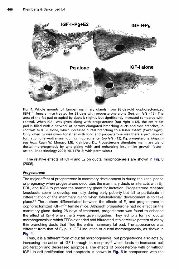
Fig. 4. Whole mounts of lumbar mammary glands from 98-day-old oophorectomizedIGF-I�/� female mice treated for 28 days with progesterone alone (bottom left �12). Thearea of the fat pad occupied by ducts is slightly but significantly increased compared withcontrol. When IGF-I was given along with progesterone (top right �12), the entire fatpad is filled with a network of narrow elongated branching ducts and side branches, incontrast to IGF-I alone, which increased ductal branching to a lesser extent (lower right).Only when E2 was given together with IGF-I and progesterone was there a profusion offormation of alveoli as seen during midpregnancy (top left �12). Pg, progesterone. (Reprin-ted from Ruan W, Monaco ME, Kleinberg DL. Progesterone stimulates mammary glandductal morphogenesis by synergizing with and enhancing insulin-like growth factor-Iaction. Endocrinology 2005;146:1170–8; with permission.)
Kleinberg & Barcellos-Hoff466
The relative effects of IGF-I and E2 on ductal morphogenesis are shown in Fig. 3(2005).
Progesterone
The major effect of progesterone in mammary development is during the luteal phaseor pregnancy when progesterone decorates the mammary ducts or interacts with E2,PRL, and IGF-I to prepare the mammary gland for lactation. Progesterone receptorknockouts seem to develop normally during early puberty but fail to participate indifferentiation of the mammary gland when lobuloalveolar development is to takeplace.51 The authors differentiated between the effects of E2 and progesterone inoophorectomized IGF-I�/� female mice. Although progesterone had no effect on themammary gland during 28 days of treatment, progesterone was found to enhancethe effect of IGF-I when the 2 were given together. They led to a form of ductalmorphogenesis in which TEBs extended and bifurcated into a treelike pattern of wispythin branching ducts that filled the entire mammary fat pad. The appearance wasdifferent from that of E2 plus IGF-I induction of ductal morphogenesis, as shown inFig. 4.Thus, it is a different form of ductal morphogenesis, but progesterone also acts by
increasing the action of IGF-I through its receptor,32 which leads to increased cellproliferation and decreased apoptosis. The effects of progesterone with or withoutIGF-I in cell proliferation and apoptosis is shown in Fig. 5 in comparison with the
Fig. 5. Effect of different combinations of E2, progesterone, and IGF-I on cell proliferation (leftpanel) and apoptosis (right panel). Pg, progesterone; TUNEL, terminal deoxynucleotidyl trans-ferase deoxyuridine triphosphate nick end labeling. (Reprinted from Ruan W, Monaco ME,Kleinberg DL. Progesterone stimulates mammary gland ductal morphogenesis by synergizingwith and enhancing insulin-like growth factor-I action. Endocrinology 2005;146:1170–8; withpermission.)
Role of IGF-I in Normal Mammary Development 467
relative effects of E2 and IGF-I. It is not clear whether progesterone serves a physio-logic role in pubertal ductal morphogenesis.Only when E2 is present with progesterone and IGF-I is alveolar differentiation
suggestive of pregnancy. IGF-I is also essential for the combination of E2 and proges-terone to permit lobuloalveolar differentiation.
POSTPUBERTAL EFFECTS OF GH AND IGF-I
In addition to IGF-I playing a crucial role in normal mammary development, adminis-tration of supraphysiologic concentrations of GH or IGF-I or overexpression can leadto hyperplasia during or after puberty. GH can and does cause hyperplasia inrodents32,52 and in primates.53 Overexpression of des (1-3) IGF-I or des (1-3) IGF-Ithat interacts with mutant p53 leads to accelerated mammary tumorigenesis,54,55 asdoes transgenic expression of a constitutively active IGF-IR.56
SUMMARY
In contrast to many other organs, including the prostate, mammary developmentoccurs almost entirely at the time of puberty. Development involves several hormonesand growth factors that are produced either in distant endocrine glands or locally in themammary gland. Although development starts with a surge of E2 levels, the participa-tion by other hormones and growth factors is essential.GH is one of the essential contributors. Although GH is produced in the pituitary
gland, it works in the stromal compartment of the mammary gland to induce produc-tion of IGF-I. IGF-I works by paracrine means in the glandular compartment of themammary gland. IGF-I activates the IGF-IR pathway that leads to proliferation ofthe ductal tree of the mammary gland through increased proliferation and decreasedapoptosis.E2 acts by enhancing the action of IGF-I on ductal morphogenesis. The 2 hormones
synergize so that ductal morphogenesis can be complete. E2 may act through ERa inthe glandular compartment by causing an effect in the stromal compartment thatallows synergy of the 2 hormones in the developing gland. The number of mammarygland cells that express ERa is fixed, but E2 increases expression in these cells. ERbdoes not seem to be necessary for growth and development of the mammary gland.
Kleinberg & Barcellos-Hoff468
The major role of progesterone is to interact with E2 and IGF-I to cause differentia-tion of the mammary gland during the luteal phase of the menstrual cycle and duringpregnancy when alveoli form. These structures eventually produce milk for lactation.Even though the steroid hormones are the proximal cause of mammary develop-
ment and function, IGF-I may be considered central to the process because neitherE2 nor progesterone can act in the absence of IGF-I. IGF-I is also required for theaction of other hormones and growth factors.
REFERENCES
1. Allen E, Francis BF, Robertson LL, et al. The hormone of the ovarian follicle; itslocalization and action in test animals, and additional points bearing upon theinternal secretion of the ovary. Am J Anat 1924;34:133–81.
2. Stricker S, Grueter F. Action du lobe anterieur de l’hypophyse sur la montee lai-teuse. Compt Rend Soc Biol 1928;99:1978–80 [in French].
3. Wilhelmi AE. Comparative biochemistry of growth hormone from ox, pig, horseand sheep pituitaries. In: Smith RW, Gaebler OH, Long CN, editors. Henry FordHospital International Symposium on the Hypophyseal Growth Hormone: natureand actions. New York: McGraw-Hill; 1955. p. 59–69.
4. Turner CW, Frank AH. The effect of the ovarian hormones theelin and corporinupon the growth of the mammary gland of the rabbit, vol. 174. Research BulletinMissouri Agricultural Experiment Station; 1932. p. 1–28.
5. Kleinberg DL, Ruan W. IGF-I, GH, and sex steroid effects in normal mammarygland development. J Mammary Gland Biol Neoplasia 2008;13:353–60.
6. Reece RP, Turner CW, Hill RT. Mammary gland development in the hypophysec-tomized albino rat. Proc Soc Exp Biol Med 1936;34:204–17.
7. Gardner WU. Growth of the mammary gland in hypophysectomized mice. ProcSoc Exp Biol Med 1940;45:835–7.
8. Li CH, Evans HM, Simpson ME. Isolation and properties of the anterior hypophy-seal growth hormone. J Biol Chem 1945;159:353–66.
9. Li CH. Growth and adrenocorticotropic hormones of anterior pituitary. HarveyLect Series 1950;46:181–217.
10. Li CH, Moskowitz M. Ultracentrifugation of hypophyseal growth hormone. J BiolChem 1949;178:203–5.
11. Lyons WR, Li CH, Johnson RE. The hormonal control of mammary growth andlactation. Recent Prog Horm Res 1958;14:219–54.
12. Nandi S. Hormonal control of mammogenesis and lactogenesis in the C3H/HeCrgl mouse. Berkeley (CA) and Los Angeles: University of California Press; 1959.
13. Daniel CW, Silberstein GB. Postnatal development of the rodent mammary gland.In: Neville MC, Daniel CW, editors. The mammary gland: development, regula-tion, and function. New York: Plenum Press; 1987. p. 1–36.
14. Richert MM, Schwertfeger KL, Ryder JW, et al. An atlas of mouse mammary glanddevelopment. J Mammary Gland Biol Neoplasia 2000;5:227–41.
15. Hinck L, Silberstein GB. Key stages in mammary gland development: themammary end bud as a motile organ. Breast Cancer Res 2005;7:245–51.
16. Silberstein GB. Postnatal mammary gland morphogenesis. Microsc Res Tech2001;52:155–62.
17. Howlin J, McBryan J, Martin F. Pubertal mammary gland development: insightsfrom mouse models. J Mammary Gland Biol Neoplasia 2006;11:283–97.
18. Sternlicht MD. Key stages in mammary gland development: the cues that regu-late ductal branching morphogenesis. Breast Cancer Res 2006;8:201.
Role of IGF-I in Normal Mammary Development 469
19. Humphreys RC. Programmed cell death in the terminal endbud. J MammaryGland Biol Neoplasia 1999;4:213–20.
20. Daniel CW, Robinson S, Silberstein GB. The role of TGF-beta in patterning andgrowth of the mammary ductal tree. J Mammary Gland Biol Neoplasia 1996;1:331–41.
21. Silberstein GB, Van Horn K, Hrabeta-Robinson E, et al. Estrogen-triggered delaysin mammary gland gene expression during the estrous cycle: evidence for a noveltiming system. J Endocrinol 2006;190:225–39.
22. Anderson TJ, Battersby S, King RJB, et al. Oral contraceptive use influencesresting breast proliferation. Hum Pathol 1989;20:1139–44.
23. Kleinberg DL, Wood TL, Furth PA, et al. Growth hormone and insulin-like growthfactor-I in the transition from normal mammary development to preneoplasticmammary lesions. Endocr Rev 2009;30:51–74.
24. Bresciani F. Topography of DNA synthesis in the mammary gland of the C3Hmouse and its control by ovarian hormones: an autoradiographic study. CellTissue Kinet 1968;1:51–63.
25. Daniel CW, Silberstein GB, Strickland P. Direct action of 17 beta-estradiol onmouse mammary ducts analyzed by sustained release implants and steroid auto-radiography. Cancer Res 1987;47:6052–7.
26. Christov K, Swanson SM, Guzman RC, et al. Kinetics of mammary epithelial cellproliferation in pituitary isografted BALB/c mice. Carcinogenesis 1993;14:2019–25.
27. Feldman M, Ruan W, Cunningham BC, et al. Evidence that the growth hormonereceptor mediates differentiation and development of the mammary gland. Endo-crinology 1993;133:1602–8.
28. Walden PD, Ruan W, Feldman M, et al. Evidence that the mammary fat pad medi-ates the action of growth hormone in mammary gland development. Endocri-nology 1998;139:659–62.
29. Wood TL, Richert MM, Stull MA, et al. The insulin-like growth factors (IGFs) andIGF binding proteins in postnatal development of murine mammary glands.J Mammary Gland Biol Neoplasia 2000;5:31–42.
30. Ruan W, Newman CB, Kleinberg DL. Intact and aminoterminally shortened formsof insulin-like growth factor I induce mammary gland differentiation and develop-ment. Proc Natl Acad Sci U S A 1992;89:10872–6.
31. Ruan W, Kleinberg DL. Insulin-like growth factor I is essential for terminal end budformation and ductal morphogenesis during mammary development. Endocri-nology 1999;140:5075–81.
32. Ruan W, Monaco ME, Kleinberg DL. Progesterone stimulates mammary glandductal morphogenesis by synergizing with and enhancing insulin-like growthfactor-I action. Endocrinology 2005;146:1170–8.
33. Saji S, Jensen EV, Nilsson S, et al. Estrogen receptors alpha and beta in therodent mammary gland. Proc Natl Acad Sci U S A 2000;97:337–42.
34. Jensen EV, Cheng G, Palmieri C, et al. Estrogen receptors and proliferationmarkers in primary and recurrent breast cancer. Proc Natl Acad Sci U S A2001;98:15197–202.
35. Mueller SO, Clark JA, Myers PH, et al. Mammary gland development in adultmice requires epithelial and stromal estrogen receptor alpha. Endocrinology2002;143:2357–65.
36. Shyamala G, Chou YC, Louie SG, et al. Cellular expression of estrogen andprogesterone receptors in mammary glands: regulation by hormones, develop-ment and aging. J Steroid Biochem Mol Biol 2002;1655:1–12.
Kleinberg & Barcellos-Hoff470
37. Shyamala G, Chou YC, Louie SG, et al. Cellular expression of estrogen andprogesterone receptors in mammary glands: regulation by hormones, develop-ment and aging. J Steroid Biochem Mol Biol 2002;80:137–48.
38. Petersen O, Hoyer P, van Deurs B. Frequency and distribution of estrogenreceptor-positive cells in normal, nonlactating human breast tissue. Cancer Res1987;47:5748–51.
39. Shyamala G, Schneider W, Guiot MC. Estrogen dependent regulation of estrogenreceptor gene expression in normal mammary gland and its relationship to estro-genic sensitivity. Receptor 1992;2:121–8.
40. Clarke RB, Howell A, Potten CS, et al. Dissociation between steroid receptorexpression and cell proliferation in the human breast. Cancer Res 1997;57:4987–91.
41. Laidlaw IJ, Clarke RB, Howell A, et al. The proliferation of normal human breasttissue implanted into athymic nude mice is stimulated by estrogen but notprogesterone [see comments]. Endocrinology 1995;136:164–71.
42. Russo IH, Russo J. Role of hormones in mammary cancer initiation and progres-sion. J Mammary Gland Biol Neoplasia 1998;3:49–61.
43. Zeps N, Bentel JM, Papadimitriou JM, et al. Estrogen receptor-negative epithelialcells in mouse mammary gland development and growth. Differentiation 1998;62:221–6.
44. Turgeon JL, Shyamala G, Waring DW. PR localization and anterior pituitary cellpopulations in vitro in ovariectomized wild-type and pr-knockout mice. Endocri-nology 2001;142:4479–85.
45. Ewan KB, Oketch-Rabah HA, Ravani SA, et al. Proliferation of estrogen receptor-alpha-positive mammary epithelial cells is restrained by transforming growthfactor-beta 1 in adult mice. Am J Pathol 2005;167:409–17.
46. Anderson E. Progesterone receptors—animal models and cell signaling in breastcancer: the role of oestrogen and progesterone receptors in human mammarydevelopment and tumorigenesis. Breast Cancer Res 2002;4:197–201.
47. Ruan W, Catanese V, Wieczorek R, et al. Estradiol enhances the stimulatory effectof insulin-like growth factor-I (IGF-I) on mammary development and growthhormone-induced IGF-I messenger ribonucleic acid. Endocrinology 1995;136:1296–302.
48. Kleinberg DL, Ruan W, Catanese V, et al. Non-lactogenic effects of growthhormone on growth and insulin-like growth factor-I messenger ribonucleic acidof rat mammary gland. Endocrinology 1990;126:3274–6.
49. Kleinberg DL. Endocrinology of lactation. In: DeGroot LJ, Jameson JL, editors.Endocrinology. Philadelphia: Elsevier; 2006. p. 3461–73.
50. Yee D, Chavez JB, Ruan WF, et al. Inhibition of normal mammary gland develop-ment and breast cancer growth by IGFBP1. Proc Am Soc Clin Oncol. AnnualMeeting, 2000 [abstract].
51. Lydon JP, DeMayo FJ, Funk CR, et al. Mice lacking progesterone receptor exhibitpleiotropic reproductive abnormalities. Genes Dev 1995;9:2266–78.
52. Kleinberg DL, Ameri P, Singh B. Pasireotide, an IGF-I action inhibitor, preventsgrowth hormone and estradiol-induced mammary hyperplasia. Pituitary 2011;14(1):44–52.
53. Ng ST, Zhou J, Adesanya OO, et al. Growth hormone treatment inducesmammary gland hyperplasia in aging primates. Nat Med 1997;3:1141–4.
54. Hadsell DL, Bonnette SG. IGF and insulin action in the mammary gland: lessonsfrom transgenic and knockout models. J Mammary Gland Biol Neoplasia 2000;5:19–30.
Role of IGF-I in Normal Mammary Development 471
55. Bonnette SG, Hadsell DL. Targeted disruption of the IGF-I receptor genedecreases cellular proliferation in mammary terminal end buds. Endocrinology2001;142:4937–45.
56. Carboni JM, Lee AV, Hadsell DL, et al. Tumor development by transgenic expres-sion of a constitutively active insulin-like growth factor I receptor. Cancer Res2005;65:3781–7.