THE PLANT FERREDOXINS AND THEIR RELATIONSHIP TO THE EVOLUTION OF FERREDOXINS FROM PRIMITIVE LIFE D. 0. HALL, R. CAMMACK and K. K. RAO Department of Botany, University of London King's College. London, UK ABSTRACT The first few ferredoxins were isolated, more or less simultaneously, from Clostridurn and spinach. These non-haem, ironcontaining oxidoreductases had almost the same low redox potential and both could catalyse the photo- reduction of NADP by chloroplasts. but differed in molecular weight and in the number of iron atoms they contained. Since that time nearly forty more ferredoxins have been isolated from all classes of organisms. These fall into four categories containing respectively 2, 4, 6 or 8 atoms of iron per mole. Numerous physical properties have been determined and studies on circular dichroism, electron spin resonance, proton magnetic resonance and Mossbauer spectra have been especially valuable in giving an insight into the varying nature of the active centre of the different ferredoxins. The amino acid sequences of fourteen ferredoxins have been determined and together with the variation in function permit phylogenetic relationships to be determined. The experi- mental work leading to these results is covered in this review. INTRODUCTION The name ferredoxin was originally suggested by Mortenson et a!' for an iron-containing protein which they isolated from the bacterium Cbs- tridium pasteurianuin and which functioned as an electron carrier in the phosphoroclastic reaction leading to hydrogen formation in this organism. About the same time Tagawa and Arnon2 isolated a non-haem iron protein with a low oxidation—reduction potential from spinach leaves which catalyzed the reduction of NADP to NADPH2 by illuminated chloroplasts. These authors also found that Cl. pasteurianum ferredoxin could substitute for the spinach protein in the photoreduction of NADP. Though the spinach non-haem iron protein differed from baterial ferredoxin in iron content, in molecular weight and in visible absorption spectrum, the similarity of the two proteins in their low red ox potentials (close to that of the hydrogen electrode) and in their interchangeability in the photo-reduction of NADP by chloro- plasts, caused Tagawa and Arnon to propose that the term ferredoxin be extended to include all non-haeme iron proteins with anoxidation—reduction potential more negative than NADP. Proteins similar to spinach ferredoxin had previously been reported in the literature under various names such as the methaemoglobin reducing factor, photosynthetic pyridine nucleotide reductase (PPNR), red enzyme of Chborella and so on.3 553
Transcript
THE PLANT FERREDOXINS AND THEIRRELATIONSHIP TO THE EVOLUTION OF
FERREDOXINS FROM PRIMITIVE LIFE
D. 0. HALL, R. CAMMACK and K. K. RAO
Department of Botany, University of LondonKing's College. London, UK
ABSTRACT
The first few ferredoxins were isolated, more or less simultaneously, fromClostridurn and spinach. These non-haem, ironcontaining oxidoreductaseshad almost the same low redox potential and both could catalyse the photo-reduction of NADP by chloroplasts. but differed in molecular weight and inthe number of iron atoms they contained. Since that time nearly forty moreferredoxins have been isolated from all classes of organisms. These fall intofour categories containing respectively 2, 4, 6 or 8 atoms of iron per mole.Numerous physical properties have been determined and studies on circulardichroism, electron spin resonance, proton magnetic resonance and Mossbauerspectra have been especially valuable in giving an insight into the varyingnature of the active centre of the different ferredoxins. The amino acid sequencesof fourteen ferredoxins have been determined and together with the variationin function permit phylogenetic relationships to be determined. The experi-
mental work leading to these results is covered in this review.
INTRODUCTION
The name ferredoxin was originally suggested by Mortenson et a!' foran iron-containing protein which they isolated from the bacterium Cbs-tridium pasteurianuin and which functioned as an electron carrier in thephosphoroclastic reaction leading to hydrogen formation in this organism.About the same time Tagawa and Arnon2 isolated a non-haem iron proteinwith a low oxidation—reduction potential from spinach leaves which catalyzedthe reduction of NADP to NADPH2 by illuminated chloroplasts. Theseauthors also found that Cl. pasteurianum ferredoxin could substitute forthe spinach protein in the photoreduction of NADP. Though the spinachnon-haem iron protein differed from baterial ferredoxin in iron content, inmolecular weight and in visible absorption spectrum, the similarity of the twoproteins in their low red ox potentials (close to that of the hydrogen electrode)and in their interchangeability in the photo-reduction of NADP by chloro-plasts, caused Tagawa and Arnon to propose that the term ferredoxin beextended to include all non-haeme iron proteins with anoxidation—reductionpotential more negative than NADP. Proteins similar to spinach ferredoxinhad previously been reported in the literature under various names such asthe methaemoglobin reducing factor, photosynthetic pyridine nucleotidereductase (PPNR), red enzyme of Chborella and so on.3
553
Tab
le 1
. Pr
oper
ties
o fe
rred
oxin
s
Fe o
r S
atom
s mol
1
Mol
ecul
ar
wei
ght
Num
ber
of
amin
o ac
ids
Red
ox
pote
ntia
l E
'0, m
V
Num
ber
of
elec
tron
s tr
ansf
erre
d
EPR
si
gnal
(r
ed. o
r ox
.)
Ref
s.
tem
p, K
8 Fe
ferr
edox
ins
Clo
stri
dium
8
6000
55
—
395
2 re
d.
76, 7
7, 2,
16,
78
(ana
erob
ic fe
rmen
tor)
15
Chl
orob
ium
8
6000
56
re
d.
79, 7
3
(gre
en p
hoto
synt
hesi
zer)
15
Chr
omat
ium
8
10 0
00
81
—49
0 2
red.
79
, 62,
80,
t (r
ed p
hoto
synt
hesi
zer)
15
6
Fe fe
rred
oxin
s A
zoto
bact
er T
ype
111
6 20
000
17
1 —
—
81
, 83
(a
erob
ic N
fixer
) 6—
7 13
000
ox
. 77
Rho
dosp
irill
um T
ype
1 6
8700
76
79
(r
ed f
acul
tativ
e pho
tosy
nthe
size
r)
4 Fe
ferr
edox
ins
Z
Des
ulph
otib
rio
4 60
00
56
—
1 re
d.
66
(sul
phat
e red
ucer
) <
30?
Bac
illus
pol
ymyx
a 4
9000
—
380
1 re
d.
67, 6
8
(fac
ulta
tive
N fi
xer)
<
30
Bee
f he
art m
itoch
ondr
ia
4 70
000
re
d.*
84
(sub
unit
of su
ccin
ate d
ehyd
roge
nase
) 10
3
Chr
omat
ium
HiP
IP
4 96
50
86
+ 3
50
1 ox
. 64
, 65,
85
(red
pho
tosy
nthe
size
r)
77
2 Fe
ferr
edox
ins
Bac
teri
al
Clo
stri
dium
EPR
pro
tein
2
24 0
00
1 re
d.
86. 4
6 (a
naer
obic
N2
fixe
r)
77
Clo
stri
dium
ozo
ferr
edox
in
2 27
500
—
—
re
d.
87
15?
Clo
stri
dium
hyd
roge
nase
2
60 00
0 —
88
, 89
E
.col
i 2
1260
0 —
re
d.
91.t
77
Rho
dosp
irill
um T
ype
11
2 75
00
67
—
-—
79
(red
pho
tosy
nthe
size
r:
facu
ltativ
e ae
robe
) A
zoto
bact
er T
ype
1 2
21 0
00
182
—-
1 re
d.
82
(aer
obic
N2
fixe
r)
104
Azo
toba
cter
Typ
e II
2
24 00
0 1
red.
82
, 90
(a
erob
ic N
2 fi
xer)
77
Ps
eudo
mon
as p
utid
a 2
12 50
0 11
4 —
240
1 re
d.
35, 54
(aer
obic
hyd
roxy
lato
r)
77
0 Pl
ant
Mic
rocy
stis
2
ii 50
0 98
-—
1
red.
92
(blu
e-gr
een
alga
) 77
0
Nos
toc
2 12
000
-—
—40
6 —
—
93
(blu
e-gr
een a
lga,
N2
fixe
r)
Scen
edes
mus
2
1150
0 96
—
1
red.
14
,t (g
reen
alg
a)
77
r E
quis
etui
n 2
1150
0 95
—
re
d.
504
(hor
seta
il, p
rim
itive
pla
nt)
77
—
Spi
nace
a 2
1150
0 97
—
430
1 re
d.
2,94
, 16,
29,
30
(hig
her
plan
t)
Ani
mal
B
eef
adre
nal
glan
ds
2 13
000
115
—34
0 1
red.
95
, 55
,45
(adr
enod
oxin
) 77
0
Bee
f he
art m
itoch
ondr
ia
2 26
000
-—
+ 22
0 1
red.
96
(Com
plex
III
Fe-
S pr
otei
n)
97
Z
* E
PR
sig
nal s
een i
n pu
rifi
ed s
ucci
nate
deh
ydro
gena
se.
t Our
unp
ublis
hed d
ata.
D. 0. HALL. R. CAMMACK AND K. K. RAO
In the early stages of the study of the ferredoxin two distinct types wererecognised, the bacterial ferredoxins and the chloroplast ferredoxins.However, as we shall see later, with the availability of information regardingthe chemical composition, amino acid sequence and physicochemical para-meters of fèrredoxins, and with the discovery of more than one ferredoxinin some organisms, it became necessary to revise this simple classification.Furthermore, proteins resembling ferredoxins in structure and compositionwere also obtained from mammalian sources. It is now recognised that theferredoxins belong to the group known as iron—sulphur proteins (proteinswhich contain one or more atoms of non-haem iron bonded to cysteine orinorganic sulphur atoms) which are ubiquitous in nature. Since they occurin primitive bacteria, algae, higher plants and animals, ferredoxins are verygood candidates for the study of biological evolution based on variations inamino acid sequences of homologous proteins. This technique has beenapplied in the phylogenetic classification of organisms from the primarystructures of several proteins including cytochromes c, haemoglobins andfibrinopeptides48.
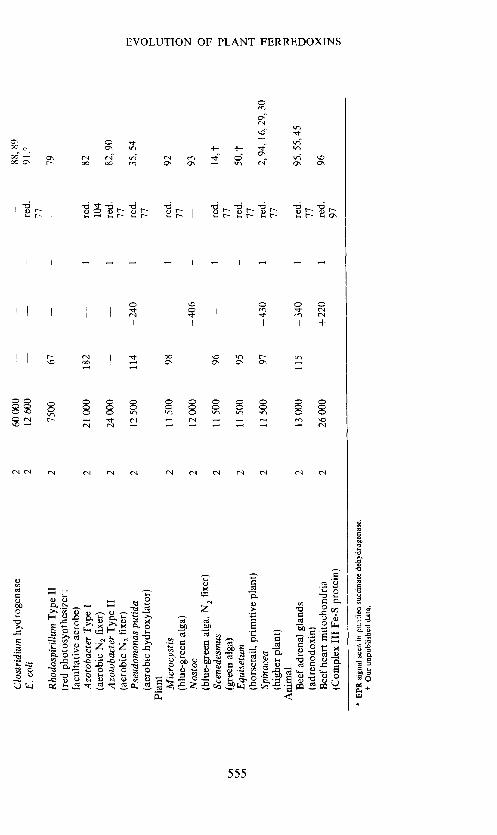
The main features of the ferredoxins are the presence of an active centrecontaining iron and sulphur, their low redox potential (E =c — 0.4 volts),their participation in biological electron transport, and the formation of acharacteristic epr signal centred around g = 1.94 (in the reduced state)at very low temperatures, e.g. liquid nitrogen or liquid helium. The types ofelectron transfer reactions in which they participate are quite diverse, suchas the Clostridia phosphoroclastic reaction, nitrogen fixation, nitrite reduc-tion, photosynthesis and mammalian steroid hydroxylation9' . Someproperties of the various ferredoxins are listed in Table 1.
EXTRACTION OF FERREDOXINS
The ferredoxins characteristically contain a preponderance of acidic aminoacids and therefore are readily adsorbed by the anion exchange resin DEAE-cellulose. This property is well utilized in the isolation and purification ofthese proteins. The main steps involved in the isolation procedure are:(1) preparation of a cell-free extract and centrifugation to get rid of insolubledebris: (2) adsorption of ferredoxins from the supernatant by DEAE-cellulose either on a column or by batchwise addition of the resin; (3)chromatographic elution of the ferredoxin from the DEAE-cellulose usinga buffered sodium chloride gradient; (4) ammonium sulphate fractionation;and (5) gel filtration on Sephadex G-75 or Biogel P-10'2' Commoncontaminants in ferredoxins are nucleic acid derivatives which can beremoved by protamine sulphate treatment, by treatment with nucleases orby chromatography on hydroxylapatite'4. Many ferredoxins are sensitivetowards heat and aerobic oxidation and so over long periods it is advisableto store these proteins in liquid nitrogen.
THE PLANT-TYPE FERREDOXINS
The plant-type ferredoxins are obtained from all oxygen-evolving photo-synthetic organisms, from the blue-green algae up to higher vascular plants.
556
Tab
le 2
. A
min
o ac
id co
mpo
litio
n 02
plan
t ter
re@
ornn
i
Lys
H
is
Arg
T
rp
Asx
T
hr
Ser
Gix
Pr
o G
ly
Ala
C
ys
Va!
M
et
lie
Leu
T
yr
Phe
Tot
al
Ref
.
Mic
rocy
stis
3
1 1
0 13
7
6 13
4
12
9 5
4
1 6
9 3
1 98
92
(blu
e-gr
een
alga
) A
nacy
stis
3
1 1
0 15
12
7
11
3 6
12
6 9
0 5
7
5
2
105
97
(blue-green al
ga)
Bum
elle
riop
sis
5
1 1
0 14
6
7 15
3
6
11
4
6
1
5
9
4
3
101
98
(yellow-green alga)
Scenedesmus*
4
1
1 0
12
10
8 10
4
7 10
6
5 1
3
7
4
3
96
14
<
(gre
en a
lga)
E
ugle
na
5
1
1 0
14
9 8
9 4
7 8
6 6
0 4
7 1
3 93
99
c
(gre
en alga)
Cladophora
7
1
2 0
10
8 7
16
3 7
12
2 7
2 3
9 4
1 10
1 10
0 0
(gre
en al
ga)
Equiseturn
4
1
1
0
9
7
8
16
4
9
6
4
6
1
5
8
2
4
95
50
(primitive va
scul
ar pl
ant)
-d
Poly
stic
hum
4
2
1
0
14
6 8
9 5
9
7 5
5 2
6
7
3
4
97
101
r (f
ern)
Z
eam
ays
3
2
1
1
13
5
8
14
4
8
8
4
10
0
5
8
5
1
100
102
—4
(mon
ocot
, C
4 pl
ant)
C
yper
us
6
1 1
1 10
4
7 14
4
9 8
4 9
1 4
10
4 1
98
103
(nut
sedg
e, C
4 pl
ant)
C
oloc
asia
* 5
1
1 1
10
6 8
15
4 9
6 5
10
0
4
6
4
2
97
104
(tar
o, m
onoc
ot)
a A
mar
anth
us
4 1
1
1 12
8
8 14
5
6 10
5
5
1
6
6
4
1 98
10
1
(pig
wee
d, C
4 pl
ant)
2
Spin
acea
t 4
1 1
1 13
8
7 13
4
6 9
5 7
0
4
8
4
2
97
94
(spinach)
Med
icag
ot
5 2
1 1
9 6
8 16
3
7 9
5 9
0 4
6 4
2 97
20
(alf
alfa
, luc
erne
) G
ossy
pium
3
1 2
1 16
4
6 18
4
8 8
4 8
1 4
6 2
3 99
10
5
(cot
ton)
L
euca
ena*
5
1 2
1 11
4
7 16
5
6
7 5
6 0
4 10
3
3 96
10
6
(Koa
; tre
e)
* A
min
o ad
d se
quen
ces
know
n.
D. 0. HALL, R. CAMMACK AND K. K. RAO
They contain two atoms of iron and two of labile sulphur per molecule,about 97 amino acid residues and have a molecular weight of about 12 000daltons. They have a red ox potential of about — 0.43 volts and they transferone electron at a time during biological reactions2' 15, f• The amino acidcompositions of the plant ferredoxins available up to now are given in Table 2.
The major role of ferredoxins in the chioroplasts is to mediate the transferof electrons from chlorophyll (or from the 'primary electron acceptor')to the flavoprotein enzyme, ferredoxin-NADP reductase, which in turnreduces NADP to NADPH2. Thus the ferredoxin catalyzes reduction ofNADP to NADPH2 which forms part of the non-cyclic photophosphoryla-tion pathway. Ferredoxin also acts as a catalyst in cyclic photophosphoryla-tion3 and in nitrite reduction in the chloroplasts17. The similarity of theferredoxins in the physiological activity of all 02-evolving photosyntheticorganisms was first observed by Mitsui and Arnon3 when they found thatferredoxin from the blue-green alga Nostoc functioned as effectively asspinach ferredoxin as a catalyst in the photoreduction of NADP by spinachchioroplasts. The activity of ferredoxins can be easily measured by addingcatalytic amounts of the ferredoxin to an assay mixture containing spinachchioroplasts (washed to remove soluble ferredoxin) and NADP and measur-ing the rate of reduction of NADP to NADPH2 on illumination (monitoredas increase in absorbance of the reaction mixture at 340 nm)' . We haverecently studied the chemical and spectroscopic properties of ferredoxinsfrom the blue-green algae Microcv,stis and Spirulina and these are foundto be similar to those of higher plant ferredoxins. These properties will bediscussed in detail later.
PHYSICAL PROPERTIES OF FERREDOXINS
Optical spectraThe plant ferredoxins are red in colour and their optical spectra exhibit
absorption maxima at about 465, 420, 330 and 278 nm (see Figure 1). Theirmolar absorbance at 420 nm is about 9 to 10 x io I cm .The absorptionof the protein in the visible region is mainly due to the iron sulphur chromo-phore group and is lost on treatment of ferredoxins with reagents whichremove iron or sulphur, i.e. mersalyl, dipyridyl, dilute acids, etc. The ironand sulphur can be put back and biologically active ferredoxin reconstitutedby chemical methods12' '. On reduction with sodium dithionite, 50 per centof the visible absorption is lost but the original spectrum is restored on re-oxidation by shaking in air. The optical absorption at 278 nm is partly due tothe aromatic amino acid content of the ferredox ins and partly to the presenceof the chromophoric group. The ratio R of the absorption at 420 nm to thatat 278 nm (R = A420/4278) is often characteristic for a particular ferredoxinand is taken as an index of the purity of the specimen. The R values of pureferredoxins vary from about 0.47 (e.g. spinach ferredoxin containing oneTrp and four Tyr residues) to 0.75 (e.g. Equiseturn ferredoxin with twoTyr and no Trp residues). The R value decreases oil exposure of ferredoxinsto air and on long-term storage at room temperatures. In the case of alfalfaferredoxin the decrease in R value on aerobic exposure is attributed to aloss in the 'labile sulphur content'2° while with spinach ferredoxin it is
558
EVOLUTION OF PLANT FERREDOXINS
2.4 -2.2
2.0
1.B
cj 1.6
1.1+
1.2
Spinach0 apoferredoxin
250 300 350 400 450 500 550Wavelength, nm
Figure 1. Absorption spectra of plant ferredoxins.
suggested that the labile sulphur atoms are oxidized to sulphur in theoxidation state zero covalently bound to protein21.
Circular dichroism spectraCircular dichroism (CD) spectra are probably more sensitive than the
optical absorption spectra of proteins to changes which are brought aboutin the overall conformation of proteins22. The CD spectra of oxidized andreduced ferredoxins from three different species are shown in Figure 2.The overall shape of the spectrum is similar for the three ferredoxins. How-ever, a closer examination reveals minor changes in the position and shapeof the absorption bands for the different species. For example, the majorpeak at 428 nm is shifted slightly towards the red in the ferredoxins of thelower order plants (Equisetum and Scenedesmus) as is the shoulder at 460 nm.The shape of the major peak is slightly less wide in the Zea mays ferredoxinspectrum than that in the other two ferredoxins.
When we compared the spectra in a wide range of ferredoxins it wasfound that the spectra of the blue-green algal ferredoxins were all verysimilar, whereas those from higher plants showed greater variation in thedetailed shape of the curves. The main absorption band around 428 nmin the CD spectra of ferredoxins is probably a reflection of the opticalabsorption at 420 nm All these CD bands are attributed to charge transferin the two Fe3 atoms bonded to sulphur in the chromophoric group.Slight differences in the nature of the CD spectra in this region betweenvarious ferredoxins may be due to differences in the nature of the amino acidresidues surrounding the chromophoric group. On reduction, the overallintensity of absorption decreases by about half, probably as a result of the
559
(ox.)
.Scenedesmus (ox.)
I I
+20
+10
0
-10
+20
+10
0
-10
+20
+10
0
—10
D. 0. HALL. R. CAMMACK AND K. K. RAO
600
Figure 2. Circular dichroism ot ferredoxins from three widely different plant species.
conversion of one Fe3 to Fe2', which has a much smaller tendency to showcharge transfer absorption in this region. The CD spectrum of apo-ferredoxinshows no absorption in the visible region.
Garbett et were the first to detect an effect of 8M urea on the CD andORD spectra of spinach ferredoxin. This was studied in further detail byother groups23 25 The process is complex. The first stage appears to bethe conversion of ferredoxin to a form with a different protein configuration,and can be reversed by decreasing the concentrations of urea, or by adding1M NaC1. Similar antagonism between the effects of urea and NaCl wereobserved in the epr spectrum by Cammack et a!25. These changes in proteinconformation may be similar to those which cause changes in the opticalabsorption and CD when ferredoxin forms a complex with ferredoxinNADP red uctase26 27
Eaton et a!.28 have investigated the nature of the iron sulphur complexin spinach ferredoxin and adrenodoxin by observing the CD and opticalspectra in the near IR wavelength region. They identified d —+d transitionsof the iron atoms in the spectra of the reduced proteins at about 4000 cm 1
560
— OxidisedReduced
Zea mays
Spirulina
300 /+00 500
Wavelength, nm
EVOLUTION OF PLANT FERREDOXINS
and 6000 cm' and from the low energy of these transitions have concludedthat the reduced iron-sulphur proteins contain a high spin ferrous ion in adistorted tetrahedral site.
Electron paramagnetic resonance spectraOne of the important, physical properties of plant ferredoxins is that
in the reduced state, and at low temperatures, they all exhibit an electronparamagnetic resonance (epr) spectrum centred around y = 1.94. Thesignal is characteristic of iron- sulphur proteins and the appearance of thesignal (at temperatures ranging from liquid nitrogen to liquid helium) isused as a sensitive test to detect the presence of iron—sulphur proteins inwhole cells and in cell-free preparations9' . Palmer and Sands29 andHall et a!.3° showed that spinach ferredoxin when reduced with excessdithionite exhibited an epr signal centred at g = 1.94. Orme-Johnson andBeinert31, by a series of anaerobic reductive titrations performed on avariety of iron -sulphur proteins, have shown that ferredoxins containingtwo iron atoms accept one electron on reduction; they also summarizeother evidence for this stoichiometry.
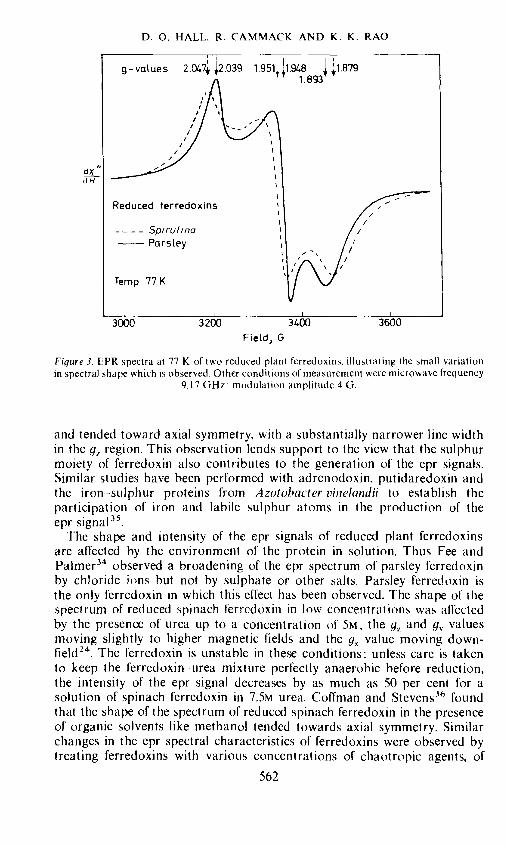
The epr spectra of reduced plant ferredoxins exhibit orthorhombicsymmetry. Typical spectra of two reduced ferredoxins are shown in Figure 3and the apparent g-values of a range of plant ferredoxins are given in Table 3.
Table 3. Principal apparent g-values of plant ferrodoxins at 77 K
Pure specimens of oxidized ferredoxin exhibit no epr signal since it is non-magnetic. The similarity in the shape of the epr spectra from two suchdiverse plant species as parsley and Spirulina (Figure 3), and the nearnessof the g-value parameters for so many ferredoxins is again indicative of theidentity of the nature of the active centre of all these ferredoxins. The roleof the iron and sulphur atoms at the active centre in the generation of theepr signal has been confirmed by a number of independent studies. A smallbroadening was observed in the epr spectrum of reduced 57Fe substitutedplant ferredoxins probably due to nuclear hyperfine interactions 12, 32,Fee and Palmer34 replaced the labile sulphur of parsley ferredoxin withselenium isotopes and found that the shape of the epr spectrum of theselenium-substituted protein was different from that of the native protein
561
dXdH
D. 0. HALL. R. CAMMACK AND K. K. RAO
Figure 3. EPR spectra at 77 K of two reduced plant ferredoxins. illustrating the small variationin spectral shape which is observed. Other conditions of measurement were microwave frequency
9.17 GHz: modulation amplitude 4 G.
and tended toward axial symmetry, with a substantially narrower line widthin the g1 region. This observation lends support to the view that the sulphurmoiety of ferredoxin also contributes to the generation of the epr signals.Similar studies have been performed with adrenodoxin, putidaredoxin andthe iron—sulphur proteins from Azotohacter vinelandii to establish theparticipation of iron and labile sulphur atoms in the production of theepr signal35.
The shape and intensity of the epr signals of reduced plant ferredoxinsare affected by the environment of the protein in solution. Thus Fee andPalmer34 observed a broadening of the epr spectrum of parsley ferredoxinby chloride ions hut not by sulphate or other salts. Parsley ferredoxin isthe only ferredoxin in which this effect has been observed. The shape of thespectrum of reduced spinach ferredoxin in low concentrations was affectedby the presence of urea up to a concentration of 5M, the g7 and g valuesmoving slightly to higher magnetic fields and the g value moving down-field24. The ferredoxin is unstable in these conditions: unless care is takento keep the ferredoxin urea mixture perfectly anaerobic before reduction,the intensity of the epr signal decreases by as much as 50 per cent for asolution of spinach ferredoxin in 7.5M urea. Coffman and Stevcns3 foundthat the shape of the spectrum of reduced spinach ferredoxin in the presenceof organic solvents like methanol tended towards axial symmetry. Similarchanges in the epr spectral characteristics of ferredoxins were observed bytreating ferredoxins with various concentrations of chaotropic agents, of
562
Field, G
EVOLUTION OF PLANT FERREDOXINS
which urea is one example25. On adding low concentrations of the agents,e.g. O.04M trichloroacetate, to reduced Spirulina ferredoxin there were de-creases in the linewidth and small shifts in the apparent g-values until itresembled more closely that of parsley ferredoxin (Figure 4). The effect isreversed on adding 2M NaCL. It therefore seems that there is a balance between
3000 3200 3400 3600
the 'sharpening' effect of chaotropic agents, and the 'broadening' effect ofchloride. Since ferredoxins are more stable in the presence of chloride it istempting to speculate that those ferredoxins which have a broader eprspectrum in the reduced state are more stable than those with narrower linewidths under equivalent conditions of salt concentration. Thus a comparisonof the epr spectra of reduced parsely and Spirulina ferredoxins clearly showsthat parsley ferredoxin has narrower linewidths (Figure 3) and thereforeshould be more sensitive to denaturing agents than the blue-green algalferredoxin. Comparative stability studies37 have shown that Spirulina ferre-doxin is much more stable at room temperature than parsley ferredoxin(Table 4). Since the chromophoric groups in the two ferredoxins are identical,the reason for the difference in stabilities should be found in the differencesin the amino acid compositions of these proteins.
The real cause of the induced changes in the epr spectral characteristics
563
//'V /
dy'dH
Reduced Spirulina Ferredoxin
Ferredoxin alone
+O•04M TrichLorocicetate
0.04M Trichloroatatef2M NcCI
Field1 G
Figure 4. EPR spectra of Spirulina ferredoxin illustrating the effect of low concentrations of achaotropic agent, and the reverse effect of NaCI. Conditions of measurement as in Figure 3.
D. 0. HALL. R. CAMMACK AND K. K. RAO
Table 4, Relative activities of plant ferredoxins after storage fortwo months37
of original activitySpecies -—----
Liquid N2 Room temperature (21 C)
Spirulinu 93 35Scenedesmus 79 5
Spinach 81 3
Alfalfa 78 8
Parsley 74 3Zea mars 81 15
of reduced ferredoxins by chloride ions, organic solvents and chaotropicagents is not yet definitely known. No corresponding changes have beenobserved in the CD or pmr spectra of the reduced ferredoxins in solution.One possible explanation is that the spectra were measured in frozensamples and the reagents might have induced small changes in the spectraby alteration of the structure of the ice around the ferredoxin molecule.On adding higher concentrations of chaotropic agents such as guanidinehydrochloride the epr spectrum undergoes a more dramatic change, whichagain is reversible25. This may be related to the changes in protein conforma-tion which are seen in the CD.
As early as 1966 Gibson et aL38 and Thornley et a!.39 proposed a modelfor the active centre of plant ferredoxins. based on epr studies and the mag-netic susceptibility data then available. According to their postulate the twoiron atoms in oxidized ferredoxin are high spin ferric antiferromagneticallycoupled to give a net spin of zero and no epr signal; on reduction one
Figure5. Model for the iron sulphur group of oxidized and reduced plant ferredoxins'2.
of the iron atoms accepts an electron and becomes high spin ferrous to givea net spin of S = and gives the characteristic epr signal (Figure 5).
Mössbauer spectraThe Mössbauer effect is a very useful tool to study the nature of the iron
atoms in proteins4041. Since it is not a magnetic resonance technique it
564
EVOLUTION OF PLANT FERREDOXINS
can be observed equally well in paramagnetie (e.g. reduced ferredoxin)and non-magnetic (e.g. oxidized ferredoxin) materials. The Mossbauer effectcan be used to measure the effective magnetic field produced at the nucleiby the electrons. Two parameters measured by Mössbauer spectroscopyare the chemical isomer shift (ö) which gives an idea of the valence and spinstate of the iron atom and its degree of covaleney, and the quadrupolesplitting (AE) which is a probe of the local stereochemistry of the iron atom.Considerably more information regarding the valence and spin state of theiron can be obtained from the magnetic hyperfine spectra observed at lowtemperatures when the Mössbauer effect is studied in the presence and absenceof an external magnetic field42.
Since the Mossbauer effect in thc case of iron is specific to 57Fe nucleiand since the natural abundance of "Fe nuclei in iron is very low (about2.2 per cent) it is advantageous to enrich the ferredoxins with 57Fe beforeMössbauer effect measurements. This can be easily achieved by chemicalsubstitution in the case of ferredoxins without changing the conformationor biological activity of the protein. Enrichment with "Fe has also beenachieved by growing Euglena on "Fe and then extracting the enrichedferredoxin44. The results were very similar to those from the chemicallysubstituted ferredoxin.
The Mössbauer spectra of three plant ferredoxins in the reduced state attwo different temperatures are shown in Figure 6. The spectrum of the
199K 4.2K HII I I I I I I
Microcystis Microcystis
o .Wa$'-,., ,1nxrr 0'-J%/%i !V I'1 - 1
- : Scenedesmus Scenedesmus
¶ 0 t"e.s;ii- t/ . 1 .: . .s
Spinach Spinach
o **4<q4.,,j , ct14S 0 k4 -1— -! 1 ,,'
2- 2
____________________________ I I I
—4 —2 0 2 4 —4 —2 0 2 4
VeLocity, mm VeLocity, mm
Figure 6. Mossbauer spectra of reduced ferredoxins from two algal and one plant species, at198 K (left), and at 4.2 K (right) in a small field perpendicular to the y-rays, illustrating the
magnetic hyperfine splitting92.
565
D. 0. HALL, R. CAMMACK AND K. K. RAO
oxidized ferredoxins (not shown in the diagram) has two lines of almostequal width with a chemical shift of about + 0.20 mm s -' relative to
6000 5000 I000 3000Hz
Hz
Figure 7. Low-field region of the pmr spectra of Scenedesmus ferredoxin, D20, 22C. in thereduced form, the resonances due to six protons are visible and the peaks arc numbered, (Un-
published results obtained in collaboration with W. D. Phillips and C. C. McDonald).
metallic iron and a quadrupole splitting of 0.60 mm s .The spectrum is con—sistent with the high-spin Fe3 + atoms in almost equivalent states. The spectraof the reduced ferredoxins at high temperature (Figure 6) show four linesof almost equal width and depth. These are a superposition of two quadrupolesplit doublets; one doublet similar to that of the oxidized ferredoxm (Fe3)and the second with a larger chemical shift and quadrupole splitting whichis characteristic of the high-spin Fe2 atom.
The effect of a small magnetic field on the Mässbauer spectrum of reducedferredoxin at very low temperatures showing magnetic hyperfine splittingis shown on the right hand side of Figure 6. The MOssbauer spectrum issplit by the application of large magnetic fields, the effective field of the Fe3 +ions decreasing and that of the Fe2' ions increasing in the appJied field.The details of the interpretation of the Mössbauer spectra are given else-where45: suffice it to say that the resemblance of the spectra of ferredoxinsfrom spinach, Scenedesmus and Microcystis again confirms the view that theactive centres of all plant ferredoxins are identical in structure. Mössbauerstudies have also been carried out on parsley, spinach and other ferredoxins byDunham et al.46 All these studies confirm the original model of Gibson38,Thornley39 and associates proposed for the active centre of ferredoxins(Figure 5).
566
Reduced
8000 7000 6000 5000 4000 3000
. 1
Spi
rulin
a fe
rred
oxin
4
3000
L
5 6—
I I
I I
I
2000
1 0
5 10
15
20
25
30
35
Tem
pera
ture
. °C
- S
cene
desm
uS
0 5
10
15
20
25
30
30
Tem
pera
ture
. °C
- S
pina
ch
0 5
10
15
20
25
30
Tem
pera
ture
1°
C)
— S
piru
1ira
(b
)
I 35
Figu
re 8
. T
empe
ratu
re de
pend
ence
of
the
low
-fie
ld c
onta
ct-s
hift
ed
prot
ons
from
red
uced
ferr
edox
ins
of (
a) s
pina
ch a
nd S
cene
desm
us a
nd (
b) S
piru
lina
and
Mic
rocy
stis
fer
redo
xins
. T
he te
mpe
ratu
re sc
ales
hav
e be
en of
fset
to s
how
the
sim
ilari
ty o
f the
patte
rns.
Pro
ton
peak
s are
num
bere
d as
in F
igur
e 7.
(Unp
ublis
hed
resu
lts o
btai
ned
in c
olla
bora
tion w
ith W
. D. P
hilli
ps an
d C
. C. M
cDon
ald)
.
ScenedesmuS ferredoxin — —
—
Spi
nach
fer
redo
xin
—
—
—
,—
—--
- —
3
—2
7000
6000
5000
4000
3000
Mic
rocy
stis
fer
redo
xin
—
N I
2
cr1
—4
6000
5000
4000
N z
-J 45
6
0 5
10
15
20
25
30
Tem
pera
ture
1°C
) -M
icro
cyst
is
rn C
—
C
C
-n z ni
ru
35
cm
2000
-
D. 0. HALL, R. CAMMACK AND K. K. RAO
Proton magnetic resonance spectraPhillips and his associates have studied the proton magnetic resonance
(pmr) spectra of spinach, parsley, alfalfa, and soyabean ferredoxins47' 48In order to measure the pmr spectra, the proteins were lyophilized, anddissolved under a nitrogen atmosphere in 0.2M Tris—DCI in 99.77 per centD20, d 7.8. Contact shifted resonances were detected to low field in thespectra of both oxidized and reduced ferredoxins (for example, see Figure7).The resonances are assigned to the -CH2 protons of the four cysteineresidues that are thought to bind the iron--sulphur redox centre to the poly-peptide chain. The temperature dependence of the contact shifts supportedthe proposal that the two iron atoms are antiferromagnetically coupled inboth oxidized and reduced forms and the assignment for the valence statesof high-spin Fe3 + — Fe3 and Fe2 — Fe3+ for the iron pairs in the oxidizedand reduced ferredoxins. respectively, It seems that the temperature depend-ence of these contact shifts is very sensitive to protein structure. The wholepattern seems to be shifted at different temperatures in spinach with Scene-desmus (Figure 8a). The temperature dependent contact shifts of two blue-green algal ferredoxins are compared in Figure 8b. Once again these indicatethat while there is variation between the algal and higher plant ferredoxinsthe blue-green algal ferredoxins are very similar. Interestingly in the contactshift spectrum of reduced soyabean ferredoxin preparations it was possibleto detect the presence of two distinct genetic variants in approximatelyequal concentration48. Thus pmr studies, in addition to being a valuable toolfor the study of the nature of the paramagnetic species, can also be used todetect minor variations in the amino acid sequences of ferredoxins preparedfrom the same species.
AMINO ACID SEQUENCES OF PLANT FERREDOXINS
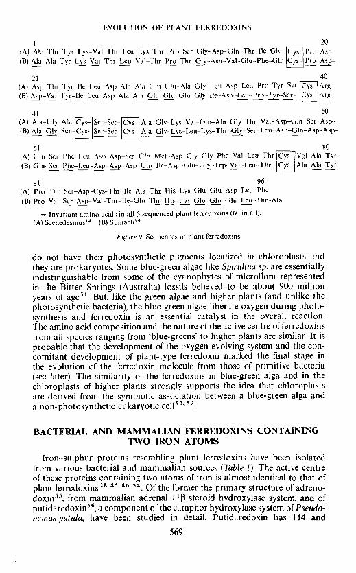
The primary structures of ferredoxins from five species of plants areknown49. There is remarkable similarity in the structures from that ofScenedesmus (green alga) to Leuceana glauca (free). These proteins consistof 96 or 97 amino acid residues and all of them have alaninc at their aminoterminal end. About 60 per cent of the residues are invariant in the fivesequences. Other prominent similarities are, (1) that five cysteine residuesare found in identical positions in all the ferredoxins sequenced; there is nocysteine in position 18 in Equiseturn ferredoxin5° thus indicating that theother four invariant cysteines are involved m the binding of the two ironatoms in the active centre (Figure 5); (2) segments containing residues 18 to26, and 35 to 50 and 62 to 66 are invariant except for a substitution of Alain residue 42 of Scenedesmus ferredoxin instead of Ser in all other ferredoxins.Though the sequences of other ferredoxins are not known, from their aminoacid compositions and spectral properties we may assume close similaritiesin the primary structures of ferredoxins from the blue-green algae to higherplants. The primary structure of' Scenedesmus ferredoxin is compared withthat of spinach ferredoxin in Figure 9.
Blue-green algae are considered to be the most primitive algae, ecologicallyrelated. and evolutionarily close, to the photosynthetic bacteria since they
(A) Asp Thr Tyr lIe 1eu--Asp--Ala- Ala Glu Glu-- Ala Gly leu -Asp-Leu--Pro--Tyr Ser Cys— Arg—(B) p-Vai Fyr-fle---Lcu -Asp-Ala--Ala Glu--Glu-Glu-çy--fle--Asp-Leu--Pro---Ser-- Cys- Arg
41 60
(A) Ala—Gly- Ala Cys— Ser-Scr-- Cys-- Aia-Giy--Lys--Val-Glu—Aia—Giy—Thr -Vai--Asp—Gin Ser -Asp--(B) Ala-y--Ser-- Cys- Ser--Ser- Cys- Ala_Giy_Lys_Leu—Lys—Thr--Giy—Scr--Leu--Asn—Gin—AsP—ASP--
= Invariant amino acids in all 5 sequenced plant ferredoxins (60 in all).(A) Scenedesmus'4 (B) Spinach94
Figure 9. Sequences of plant ferrcdoxins.
do not have their photosynthetic pigments localized in chioroplasts andthey are prokaryotes. Some blue-green algae like Spirulina sp. are essentiallyindistinguishable from some of the cyanophytes of microflora representedin the Bitter Springs (Australia) fossils believed to be about 900 millionyears of age51. But, like the green algae and higher plants (and unlike thephotosynthetic bacteria), the blue-green algae liberate oxygen during photo-synthesis and ferredoxin is an essential catalyst in the overall reaction.The amino acid composition and the nature of the active centre of ferredoxinsfrom all species ranging from 'blue-greens' to higher plants are similar. It isprobable that the development of the oxygen-evolving system and the con-comitant development of plant-type ferredoxin marked the final stage inthe evolution of the ferredoxin molecule from those of primitive bacteria(see later). The similarity of the ferredoxins in blue-green alga and in thechioroplasts of higher plants strongly supports the idea that chloroplastsare derived from the symbiotic association between a blue-green alga anda non-photosynthetic eukaryotic cell52 .
BACTERIAL AND MAMMALIAN FERREDOXINS CONTAININGTWO IRON ATOMS
Iron—sulphur proteins resembling plant ferredoxins have been isolatedfrom various bacterial and mammalian sources (Table 1). The active centreof these proteins containing two atoms of iron is almost identical to that ofplant ferredoxins28'45'46' Of the former the primary structure of adreno-doxin55, from mammalian adrenal 113 steroid hydroxylase system, and ofputidaredoxin56, a component of the camphor hydroxylase system of Pseudo-monas putida, have been studied in detail. Putidaredoxin has 114 and
569
1 7
(A)
A1a
—A
1a—
Tyr
-Lys
—V
aI—
Thr
-Leu
—
C
(B)
Ser—
Ser-
-Ser
—G
n-A
sp--
Lys
-He-
Thr
-Val
--H
is—
Phe-
fle—
Asn
—A
rg--
Asp
8
37
(A)
(B)
Giy
—G
!u--
Thr
—L
eu—
Thr
—T
hr—
Lys
—G
Iy-L
ys—
IIe—
Giy
—A
sp—
Ser—
Leu
—L
eu —
Asp
—V
al—
VaI
—V
aI--
-Gln
—A
sn—
Asn
—L
eu—
Asp
—IJ
e—A
sp--
-GIy
—Ph
e—G
ly—
Ala
—
67
(A)
Ser—
f--A
rg—
A1a
—G
1y—
Ser-
Cys
Se
r—Se
r—
ys A
Ia—
GIy
--L
ys—
Leu
—L
ys—
Thr
—G
Iy—
Ser—
Leu
--A
sn—
GIn
—A
sp—
Asp
---G
In—
Ser—
Phe—
Leu
—A
sp--
Asp
--A
sp—
(B)-
G1u
-Gly
—T
hr—
Leu—
AIa
C
ys S
er—
Thr
Cys
His
—Le
u—IIe
—P
he—
GIu
--G
In—
His
--Ile
Phe
—G
lu—
Lys—
Leu-
-Glu
--A
la—
I!e--
Thr
--A
sn—
GIu
—G
lu—
Asp
-
(A)
GIn
—II
e—A
sp—
Glu
—G
Iy—
Trp
—V
aI—
Leu
—T
hr ys
A1a
—A
1a—
Tyr
—Pr
o—V
a1--
--Se
r—A
sp--
Va1
—T
hr—
I1e—
GIu
—T
hr-H
is--
-Lys
—G
tu—
G1u
—G
1u--
Leu
—T
hr—
A1a
--
>
(B)
Asn
—M
et—
Leu
—A
sp—
Leu
—A
la—
Tyr
—G
Iy li
e ys
Leu
—T
hr—
Lys—
A1a
—M
et—
Asp
--A
sn—
Met
—A
sp—
Thr
—V
aI—
Arg
—V
al—
Pro
-Asp
—A
1a—
Va1
—S
er—
Asp
—A
1a
/ \
Thr
/ /
Cys
A
sp
Gly
A
rg
/ Leu
\ /
Ser
—A
rg
Figu
re 1
0. C
ompa
riso
n of s
pina
ch f
erre
doxi
n an
d ad
reno
doxi
n seq
uenc
es.
(A)
Spin
ach
ferr
edox
in94
, (B
) B
ovin
e ad
reno
doxi
n55
1 28
(A)
Ala
—Ph
e—V
al—
lie—
A
su
Asp
—Se
r--
Cys
V
ai—
Ser—
C
s G
ly—
Ala
C
ys A
ia—
Giy
—G
iu ', P
ro—
Vai
—S
cr--
Aia
—iie
—T
hr—
Gin
--G
ly—
Asp
--T
hr—
(B
) A
la—
His
—lie
—lie
—
Thr
—A
sp--
Gin
—
Cys
Il
e—Se
r—
Cys
G
iy—
Ala
C
ys A
ia—
Ala
—G
iu
Cys
4-Pr
o—V
ai—
Giu
—A
ia—
Iie—
His
—G
in—
Giy
—T
hr—
Gly
—
(C)
Aia
—L
eu—
Met
—Ii
e—
Thr
—A
sp—
Gin
—
Cys
T
le—
Asn
— C
ys
Asn
—V
al
Cys
Gin
—Pr
o—G
lu
Cys
Pr
o—A
sn—
Giy
—A
ia—
Iie—
Ser—
Gln
—G
iy—
Asp
—G
iu
1 28
29
55
(A)
Gin
—Ph
e—V
ai—
Ile—
Asp
—A
ia—
Asp
—T
hr C
ys
Tie
—A
sp—
Cys
G
iy—
Asn
ys
Aia
—A
sn—
Vai
C
ys
Pro—
Vai
—G
iy—
Aia
—Pr
o—A
sn—
Gin
—G
iu
(B)
Lys
—T
yr—
Gin
—V
ai—
Asp
--A
Ia—
Asp
—T
hr C
ys
Tie
—A
sp—
Cys
G
ly—
Ala
C
ys G
in—
Ala
—V
al
Cys
Pro
—T
hr—
Gly
—A
ia—
Vai
—L
ys—
Aia
—G
iu
(C)
Thr
—T
yr—
Vai
—Ii
e—G
iu—
Pro-
-Ser
—L
eu—
Cys
T
hr—
Giu
Cys
V
a! A
sp
Cys
Val
—G
lu—
Val
C
ys P
ro—
Iie—
Lys
—A
sp—
Pro—
Ser—
His
—G
iu—
Giu
—T
hr
Gly
V
a!
C
His
C
ys
C
Tyr
G
in
/ G
b Se
r
Thr
67
81
(C)
Giu
—A
sp—
GIu
—L
eu—
Arg
—A
ia—
Lys
—T
yr—
Giu
—A
rg—
TIe
—T
hr—
Gly
—G
iU—
G1Y
rn
C
(A)
C. b
ut yr
icur
n:10
7 (B
) C
. tar
tariv
oruv
n:61
(C) C
hrom
atiu
m:6
2 z
Fig
ure
11.
Com
pari
son o
f se
quen
ces
of C
lost
ridi
um a
nd C
hrom
atiu
m f
erre
doxi
ns
D. 0. HALL. R, CAMMACK AND K. K. RAO
adrenodoxin 115 amino acid residues. In common with plant ferredoxinsthey contain at least one —Cys—x—x--Cys- grouping in their sequence. Tsai eta!.56 have identified a number of homologous segments in the primarystructures of putidaredoxin and adrenodoxin and the sequences of thesetwo ferredoxins exhibit a fair degree of similarity to the sequences of plantferredoxins. We have aligned the sequences of adrenodoxin and spinachferredoxin in Figure 10 to point out some of the similarities. A statisticalanalysis of various ferredoxin sequences by Barker et a!.57 confirms therelationship between adrenodoxin and plant ferredoxins which suggests thatall these ferredoxins could have evolved from a common ancestral gene.However, in spite of the identity of their active centre and similarity in theiramino acid sequence, the two-iron ferredoxins of one type are not completelyinterchangeable with the two-iron ferredoxins of another type in theirbiological function. This could possibly be due to differences in their tertiarystructures or due to differences in the structures of various reductases withwhich they combine during physiological electron transfer processes.
FERREDOXINS CONTAINING EIGHT IRON ATOMS
Ferredoxins cortaining eight atoms of iron have so far been obtained onlyfrom bacteria, both photosynthetic and non-photosynthetic. The opticalabsorption of these ferredoxins arc characterized by a maximum at 390 nmand they differ in their biological action from the plant ferredoxins in thatthey transfer two electrons at a time. All these ferredoxins, thus far analyzed,except Chromatium ferredoxin, consist of about 55 amino acid residues with amolecular weight of approximately 6000 daltons. As in the plant ferredoxinsthe iron and 'labile sulphur' of these proteins can be removed by the action ofreagents like trichloracetic acid and mercurials and the original ferredoxincan be reconstituted from the apoprotein by the addition of iron salt andsuiphide in a reducing medium58. The structure of the iron—sulphur complexof this type of ferredoxin is not as well established as the plant ferredoxins.Magnetic susceptibility measurements have indicated strong antiferromag-netic exchange coupling between the component iron atoms in C. pasteuri-anum ferredoxin59. An x-ray investigation of crystalline M. aerogenesferredoxin has shown that the eight iron and sulphur atoms exist as twoidentical clusters of 4Fe plus 4S atoms60. However, we still do not have enoughinformation to correlate the magnetic resonance properties of these proteinsand their electron transferring capacity.
The amino acid sequences of five ferredoxins from obligate fermentativebacteria are known61. In addition to the invariant positions of eight cysteineresidues in the sequences of all these five ferredoxins there are many identicalsegments in their structures confirming the homologous nature of theseproteins. The primary structure of Chromatium ferredoxin contains an extra26 amino acid residues62. However, the similarity of this molecule to theother bacterial ferredoxins in this group is borne out by the invariant posi-tions of many amino acid residues when its primary structure is realigned,as was done by Matsubara et at.63, and then compared with the clostridialferredoxins. A similar comparison of Chromatium ferredoxin with twoclostridial ferredoxins is depicted in Figure 11.
572
(A)
Aia
—Ph
e-V
a1—
Ile-
Asn
—A
sSer
Cys
V
a1-S
er
ys G
ly—
Ala
C
ys
Ala
—G
ly--
Glu
C
ys
Pro-
Val
--Se
r-A
la- I
1e—
Thr
—G
in—
G1y
—A
s T
br
29
(B) P
ro—
Ile—
Gln
—V
al—
Asp
--A
sn C
ys
Met
—A
la
ys G
in—
Ala
C
ys
lle—
Asn
—G
lu--
Cys
Pr
o—V
ai—
Asp
--V
aI--
Phe
—G
in--
Met
—A
sp—
Giu
—G
ln—
GIy
—A
sp—
C
(C
) Glu
Cys
Pr
o—A
sp—
Asp
—V
al-T
yr—
Ile—
Leu
—A
sp--
Ala
—
Ala
—G
iu—
Glu
—
17
30
r—
30
56
(B)
Lys
—A
la—
Val
---A
sn—
Ile—
Pr
o—A
sn—
Ser—
Asn
—L
eu—
Asp
—A
sp--
-Giu
Cys
Val
—G
lu—
AIa
—II
e—G
ln—
Ser—
Cys
—Pr
o--A
la--
Ala
—Il
e--A
rg—
Ser
(C)
Glu
—G
Iy—
Ile—
Asp
--L
eu—
Pro—
Tyr
—Se
r—C
ys—
Arg
—A
la—
G
iy—
Ser—
Cys
Se
r—
31
45
Figu
re 1
2. S
eque
nce
of D
. gi
gas f
erre
doxi
n66
(B) c
ompa
red w
ith a
nalo
gous
se
gmen
ts o
f the
ferr
edox
ins
of C
. but
yric
um'°
7 (A
) an
d sp
inac
h94
(C).
Ii 0. HALL, R. CAMMACK AND K. K. RAO
THE FERREDOXINS CONTAINING FOUR IRON ATOMSThree ferredoxins containing four atoms each of iron and sulphur per
molecule are fairly well characterized (Table 1). One of these, Chromatiumhigh potential iron protein (HIPIP) is quite distinct in its properties fromother ferredoxins64' . No biological function has yet been found for thisprotein. The ferredoxin from the sulphate reducer Desulphovihrio qiqas'6 jçinteresting due to the fact that the primary structure of this protein is homo-logous in parts to segments of Clostridia ferredoxins and is similar in otherparts to segments of plant ferredoxins (T. H. Jukes, personal communication).Their primary structures are compared in Figure 12. Bacillus polymyxaferredoxin67 has a molecular weight of 9000 and on reduction accepts oneelectron per molecule68. These four-iron ferredoxins may be evolutionarylinks between the eight- and the two-iron ferredoxins and so are being activelystudied by various groups.
ARE FERREDOXINS PRIMITIVE PROTEINS?Ever since Oparin enunciated his theory about the abiogenic origin of
life on earth attempts have been made to synthesize in the laboratorycompounds like amino acids, purines, pyrimidines etc. under conditionswhich simulated prebiotic environments69. There is also a vigorous searchto detect the presence of precursors of biological molecules like sugars,proteins and nucleic acids in extra-terrestrial matter70-72. The presence ofthese precursors in meteorites and other planets would indirectly provetheir existence in the prebiotic earth's atmosphere. Amino acids like glycine,alanine, valine, glutamic acid, aspartic acid, proline, serine, cysteine andisoleucine have been synthesized under simulated primitive earth condi-tions49. Analysis of the Murchison and Murray meteorites and moon samplescollected in the Apollo missions have shown the occurrence of the first sixof the above-mentioned amino acids in these bodies. It is interesting that64 per cent of C. butyricum ferredoxin is made up of these six amino acids.The clostridial ferredoxins are relatively small proteins and the iron andsulphur which form the active centre of these proteins can be added to theapoprotein by a non-enzymic process. They have a very low redox potentialclose to that of hydrogen gas. All these properties suggest the possibilitythat a ferredoxin-like protein would have functioned even in very primitiveorganisms.
FERREDOXINS AS A TOOL IN PHYLOGENETICCLASSIFICATION
It is now a generally accepted thesis that any mutation in the nucleotidecodons of the gene directing the synthesis of a particular protein will beexpressed by an amino acid substitution in the sequence of the protein, i.e.the primary structures of homologous proteins are reflections of the nucleo-tide sequences of their genetic material. The ferredoxins are found in allspecies of organisms. The amino acid sequences of ferredoxins from fivespecies of anaerobic bacteria are known and they show a high degree ofhomology. Also, the positions of certain key amino acids like the aminoterminal alanine residue and the eight cysteine residues, which are thoughtto chelate the iron atoms, are identical in all these five ferredoxins. Thus these
574
EVOLUTION OF PLANT FERREDOXINS
[erredoxins would probably have evolved from a common ancestral gene.The plant ferredoxins are about double the length of clostridial ferredoxinsbut the two groups show certain similarities in their properties and aminoacid sequences. A quantitative comparison of the amino acid sequences ofplant and Clostridia ferredoxins has indicated evolutionary relationshipsbetween these two groups but the evolutionary distances may be large5 7 63, The[erredoxin from the red photosynthetic bacterium Chrotnatium is inter-mediate in length between Clostridia and plant ferredoxins but in its sequenceand function it is closer to the Clostridia type than to the plant ferredoxins.We do not yet know the complete sequence of a ferredoxin from a greenphotosynthetic bacteria. However, from the known amino acid compositionand terminal residues of ferredoxins from this latter group we could predictthat they are intermediate between the non-photosynthetic anaerobes and:he red Chromatium7375. The position of the sulphate reducer D. gigas isbased on a comparison of the amino acid sequence of its ferredoxin withbacterial and plant ferredoxins (Figure 12) and on the suggestion of Schlegel75[rom metabolic considerations that sulphate reducing bacteria can be placedbetween the anaerobic photolithotrophs (e.g. Chromatium) and the blue-greenalgae in the evolutionary ladder. Thus from a comparative study of the primarystructure and function of ferredoxins from various species we could tenta-tively propose a phylogenic classification as outlined in Figure 13.
\ naerobic bacteria —* Green photosynthetic bacteria — Red (sulphur) photosynthetic bacteria —
Figure 13. Evolutionary development of ferredoxins49.
AKNOWLEDGEMENTSWe thank the Science Research Council for grant support and Misses A. J.
Cox and J. Zantovska for assistance.
REFERENCESL. E. Mortenson, R. C. Valentine and J. E. Carnahan, Biochem. Biophys. Res. Comm. 7, 488(1962).2 K. Tagawa and D. 1. Arnon, Nature 195, 537 (1962).D. 1. Arnon, Experientia 22, 273 (1966).C. Nolan and F. Margoliash, Ann. Rev. Biochem. 37, 727 (1968).J. E. Haber and D. E. Koshland, Jr., J. Mo!. Biol. 50, 617 (1970).
o P. Jolles and J. Jolles, Progr. Biophys. Mo!. B!. 22, 97 (1971).R. E. Dickerson, J. Mo!. Biol. 57, 1(1971).D. Boulter and J. A. M. Ramshaw, Phytochemistry 11, 553 (1972).R. Malkin and J. C. Rabinowitz, Ann. Rev. Biochem. 36, 113 (1967).
o D. 0. Hall and M. C. W. Evans, Nature 223, 1342 (1969).B. B. Buchanan and D. I. Arnon, Advn. Enzymo!. 33, 119 (1970).
2 K. K. Rao, R. Cammack, D. 0. Hall and C. F. Johnson, Biochem. J. 122, 257 (1971).B. B. Buchanan and D. I. Arnon, Methods in Enzymo!ogy 23, 413 (1971).K. Sugeno and H. Matsubara, J. Biol. Chem. 244, 2979 (1969).F. R. Whatley, K. Tagawa and D. I. Arnon, Proc. Nat!. Acad. Sci. U.S. 49, 266 (1963).
o M. C. W. Evans, D. 0. Hall, H. Bothe and F. R. Whatley, Biochem. J. 110, 485 (1968).M. Losada, A. Paneque, J. M. Raminez and F. F. Del Campo. Biochem. Biophys. Res. Comm.10, 298 (1963).
575
D. 0. HALL, R. CAMMACK AND K. K. RAO
I 8 A. San Pietro, Meth. Enz. 6, 439 (1963).9 E. Bayer, J. D. Krauss. P. l-lagenmaier. H. Roder and A. Trehst. Biochim. Biophvs. i4cta
143. 435 (1967).° S. Keresztes-Nagy, F. Perini and E. Margoliash. J. Biol. Chem. 244, 981 (1969).21 D. Petering. J. A. Fee and G. Palmer, J. Biol. Chem. 246, 643 (1971).22 K. R. Garhett, R. D. Gillard. P. K. Knowles and J. E. Stangroom. Nature 215, 824 (1967).23 R. Padmanahhan and T. Kirnura. J. Biol. C/win. 245, 2469 (1970).24 D. H. Petering and CI. Palmer. Arch. Biochein. Biophys. 141, 456 (1970).25 R. Cammack. K. K. Rao and D. 0. Hall, Biochem. Biophys, Res. Comm. 44, (1971).26 N. Nelson and J. Neumann. Biochem. Biophys. Res. Comm. 30, 142 (1968).27 R. Cammack. J. Neumann, N. Nelson and D. 0. Hall. Biochem. Biophys. Res. Comm. 42,
292 (1971).28 W. A. Eaton. G. Palmer, J. A. Fee. T. Kimura and W. Lovenberg. Proc. Nati. Acad. Sci. U.S.
68, 3015 (197!).29 G. Palmer and R. H. Sands. J. Biol. Chem. 241, 253 (1966).° D. 0. Hall, J, F. Gibson and F. R. Whatley, Biochem. Biophys. Res. Comm. 23, 81(1966).31 W. 11. Orme-Johnson and H. Beinert. J. Biol. C/win. 244, 6143 (1969).32 G. Palmer. Biochem. Biophys. Res. Comm. 27, 315 (1967).
C. E. Johnson, R. Cammack. K. K. Rao and D. 0. Hall. Biochem. Biophys. Res. Comm. 43,564(197 1).J. A. Fee and G. Palmer, Biochim. Biophys. Acta 245, 175 (1971).J. C. M. Tsibris and R. W. Woody. Coord. Chem. Rev. 5, 417 (1970).
36 R. E. Coffman and B. W. Stevens. Biochem. Biophys. Res. Comm. 41, (1970).' D. 0. Hall, K. K. Rao and R. Cammack, Biochein. Biophys. Res. Comm. 47, 798 (1972),38 J. F. Gibson. D. 0. Hall, J. H. M. Thornley and F. R. Whatley, Proc. NatI. Acad. Sci. U.S.
56. 987 (1966).39 j H. M. Thornlcy. J. F. Gibson, F. R. Whatley and D. 0. Hall, Biochem. Biophys. Res.
Comm. 24, 877 (1966).° W. D. Phillips. E. Knight and D.C. Blomstrom in Non-heme iron proteins,(A. San Pietro, ed),p. 69. Antioch Press, Ohio (1965).
4! G. Lang. Quart. Rev. Biophys. 3. 1(1970).42 C. F. Johnson in Magnetic Resonance in Biological Systems, (A. Ehrenberg, B. G. Malmström
and T. Vhngard, eds.). p. 405. Pergamon Press, Oxford (1967).C. E. Johnson. J. Appl. Phys. 42, 1325 (1971).C. E. Johnson, F. Elstner, J. F. Gibson, G. Benfield, M. C. W. Evans and D. 0. Hall, Nature220, 1291 (1968).R. Cammack. D. 0. Hall, K. K. Rao and C. E. Johnson, Biochem. J. 125, 849 (1971).
46 W, R. Dunham. A. J. Bearden, 1. T. Salmeen. R. H. Sands, W. H. Orme-Johnson and H.Beinert, Biochim. Biophys. Acta 253, 134 (1971)." M. Poe, W. D. Phillips, J. D. Glick son, C. C. McDonald and A. San Pietro, Proc. Nat!. A cad.Sci. U.S. 68, 68 (1971).
48 J. D. Glickson. W. D. Phillips, C. C, McDonald and M. Poe, Biochem. Biophys. Res. Comm.42,271 (1971).D. 0. Hall. R. Cammack and K. K. Rao in Theory and Experiment in Exohiologv, Vol. 2.(A. Schwartz, ed.) p67, Wolters-Noordhof, Netherlands (1972).° S. J. Aggarwal, K. K. Rao and H. Matsuhara, J. Biochem.(Tokyo),69, 601 (1971)
SI J W. Schopf, Biol. Rev. 45, 319 (1970).52 L. Sagan, J. Theoret. Biol. 14, 225 (1967).
D. 0. Hall, R. Cammack and K. K. Rao, Nature 233, 136 (1971).E. Munck, P. G. Debrunner, J. C. M. Tsibris and 1. C. Gunsalus, Biochemistry 11, 855(1972)." M. Tanaka, M. Haniu and K. T. Yasunobu. Biochem. Biophys. Res. Comm. 39, 1182 (1970).R. L. Tsai, I. C. Gunsalus and K. Dus. Biochem. Biophes. Res. Comm. 45, 1300.1306(1971).W. C. Barker, P. J. McLaughlin and M. 0. Dayhofl Fed. Proc. 31, 837 Abs (1972).
58 R. Malkin and J. C. Rabinowitz, Biochem. Biophys. Re.s. Comm. 32, 822 (1966).M. Poe, W. D. Phillips, C. C. McDonald and W. Lovenberg, Proc. Nat!. A cad. Sci. U.S. 65,798 .804 (1970).
60 L. C. Sieker, E. Adman and L. H. Jensen, Nature 235,40 (1972).SI M. Tanaka, M. Haniu, 0. Matsueda, K. T. Yasunohu. R. H. Himes, J. K. Akagi, E. M.
Barnes and T. Devanathan, J. Biol. ('hem. 246, 3953 (1971).
576
EVOLUTION OF PLANT FERREDOXINS
62 H. Matsubara, R. K. Sasaki, D. K. Tsuchiya and M. C. W. Evans, J. Biol. Chem. 245, 2121(1970).
63 H. Matsuhara, T. H. .Iukes and C. R. Cantor, Brookhaven Symp. Biol. 21, 201 (1968).64 K. Dus, H. De Klerk, K. Sletten and R. G. Bartsch, Biochim. Biophys. Acta. 140, 291 (1967).65 K. Dus, S. Tedro, R. G. Bartsch and M. 0. Kamen, Biochem. Biophys. Comm. 43, 1239
(1971).66 j Travis, D. J. Newman, J. Le Gall and H. D. Peck, Biochem. Biophys. Res. Comm. 45, 452(1971).
67 y I. Shcthna, N. A. Stombaugh and R. H. Burns, Bloc/tern. Biophys. Res. Comm. 42, 1108(1971).
68 W. H. Orme-Johnson, N. A. Stombaugh and R. H. Burns. Fed. Proc. 31, 448 Abs. (1972).69 0. H. Kenyon and 0. Steinman, Biochemical Predestination. McGraw—Hill, N.Y. (1969).70 K. A. Kvcnvolden, J. G. Lawless, K. Pening, E. Peterson, J. Flores, C. Ponnamperuma, I. R.
Kaplan and C. Moore, Nature 228, 923 (1970).J. G. Lawless, C. E. Folsome and K. A. Kvenvolden, Sci. Amer., 226(b), 38 (1972).
72 K. Harada, P. G. Hare, C. R. Windsor and S. W. Fox, Science 173, 433 (1971).B. B. Buchanan, 11. Matsubara and M. C. W. Evans. Biochim. Biophys. Acta. 189, 46 (1969).K. K. Rao, H. Matsubara, B. B. Buchanan and M. C. W. Evans, J. Bacteriology 100, 1411(1969).H. G. Schlegel, Intl. Symp. Origin of Life and Evolutionary Biochemistry, Varna, Bulgaria(1971).J. S. Hong and J. C. Rahinowiti, J. Biol. Chem. 245, 4982 (1970).M. Tanaka, T. Nakashima, A. M. Benson, H. F. Mower and K. T. Yasunobu, Biochemistry5, 1666(1966).
78 G. Palmer, R. H. Sands and L. E. Mortenson, Biochem. Biophys. Res. Comm. 23, 357 (1966).K. T. Shanmugam, B. B. Buchanan and D. I. Arnon, Biochim. Biophys. Acta. 256, 477 (1972).
80 R. Bachofen and D. I. Arnon, Biochim. Biophys. Acta. 120, 259 (1966).81 D. C. Yoch, J. R. Benemann, R. C. Valentine and D. 1. Arnon, Proc. Natl. Acad. Sci. U.S.
64, 1404(1969).82 D. V. Dervartanian, Y. I. Shethna and H. Beinert, Biochim. Biophys. Acta. 194, 548 (1969).83 Y. 1. Shethna, Biochim. Biophys. Acta 205, 58 (1970).84 K. A. Davis and Y. Hatefi, Biochemistry 10, 2509 (1971).85 5. 0. Mayhew, 0. Petering, G. Palmer and 0. P. Foust, J. Biol. Chem. 244, 2830 (1969).86 R. W. F. Hardy, E. Knight, C. C. McDonald and A. J. D'Eustachio in Non-heme iron
proteins. (A. San Pietro, ed), p. 275. Antioch Press, Ohio (1965).87 Nakos and L. E. Mortenson, Biochemistry 10, 455 (1971).
0. Nakos and 1.. E. Mortenson. Biochemistry 10. 2442 (1971).89 j Multani and L. E. Mortcnson, Biochim. Biophys. Acta 256, 66 (1972).90 Y. I. Shethna, D. V. Dervartanian and H. Beinert, Bloc/tern. Biophys. Res. Comm. 31, 862
(1968).91 H. Vetter and J. Knappe, Hoppe-Seyler's Z. Physiol. Chem. 352, 433 (1971).92 K. K. Rao, R. V. Smith, R. Cammack, M. C. W. Evans, D. 0. Hall and C. E. Johnson,
Biochem J. 129, 1159 (1972).A. Mitsui and 0. I. Arnon, Physiol. Plant. 25, 135 (1971).H. Matsubara and R. M. Sasaki, J. Biol. Chem. 243, 1732 (1968).T. Kimura, Struct. and Bond. 5, 1(1968).
96 5 Rieske in Non-heme iron proteins. (A. San Pietro, ed), p. 461. Antioch Press, Ohio(1965).
" T. Yamanaka, S. Takenami, K. Wada and K. Okunuki, Biochim. Biophys. Acta 180, 196 (1969).98 P. BOger, Planta 92, 105 (1970).
A. Mitsui, Biochim. Biophys. Acta 243, 447 (1971).too A. A. Mutuskin, K. V. Psheneva, 0. P. Kaverina and P. A. Kolesnikov, Doklady Akad. Nauk.
SSSR 183, 715 (1968).tOt P. Schjirmann, B. B. Buchanan and H. Matsubara, Biochim. Biophys. Acta 223, 450 (1970).02 C. 0. Crawford and R. G. Jensen, Plant Physiology 47, 447 (1971).03 S. S. Lee, J. Travis and C. C. Black, Jr. Arch. Biochem. Biophys. 141, 676 (1970).04 K. K. Rao and H. Matsubara, Biochem. Biophys. Res. Comm. 38, 500 (1970).05 0. J. Newman, J. N. IhIe and L. Dure, Biochem. Biophys. Res. Comm. 36, 947(1969).06 A. M. Benson and K. T. Yasunobu, J. Biol. Chem. 244, 955 (1969).107 A. M. Benson, H. F. Mower and K. T. Yasunobu, Arch. Biochem. Biophys. 121, 563 (1967).


![1 A Novel Three-Component Rieske Non-Heme …aem.asm.org/content/early/2014/06/09/AEM.00659-14.full.pdf15 a transposable element that is highly conserved ... [2Fe-2S] ferredoxins and](https://static.documents.pub/doc/80x56/5ac076c57f8b9a213f8bfe3b/1-a-novel-three-component-rieske-non-heme-aemasmorgcontentearly20140609aem00659-14fullpdf15.jpg)

