THE PREGNANEDIOL EXCRETION IN NORMAL AND ABNORMAL PREGNANCY THESIS PRESENTED IN PULEILMENT OE THE REQUIREMENTS EOR THE DECREE OE DOCTOR OE MEDICINE OE THE trNIYERSITY OE CLASCOW BY MARY G. COYLE, M.B.CH.B., M.R.C.O.
Transcript
THE PREGNANEDIOL EXCRETION INNORMAL AND ABNORMAL PREGNANCY
THESISPRESENTED IN PULEILMENT OE THE REQUIREMENTS EOR THE
DECREE OE
DOCTOR OE MEDICINE OE
THE trNIYERSITY OE CLASCOW BY
MARY G. COYLE, M.B.CH.B., M.R.C.O.
ProQuest Number: 13848955
All rights reserved
INFORMATION TO ALL USERS The quality of this reproduction is dependent upon the quality of the copy submitted.
In the unlikely event that the author did not send a com p le te manuscript and there are missing pages, these will be noted. Also, if material had to be removed,
a note will indicate the deletion.
uestProQuest 13848955
Published by ProQuest LLC(2019). Copyright of the Dissertation is held by the Author.
All rights reserved.This work is protected against unauthorized copying under Title 17, United States C ode
Methods of determining the urinary pregnanediol excretion.A comparison of the method of Sommerville, Marrian and Gough (1948) and ofMitchell (1954).A critical review of the method of Klopper, Michie and Browne (1955)•The relationship between the Blood progestational activity” and the urinary pregnanediol excretion in normal pregnancy.
Page. 1 .
6 .
8.
29.
SECTION II. Normal pregnancy.SECTION III. Abnormal early pregnancy. SECTION IV. Abnormal late pregnancy.
Toxaemia of pregnancyPart A. Part B . Conditions associated with
foetal death in late pregnancy.
PINAL SUMMARY AND CONCLUSIONS. ACKNOWLEDGEMENTS.PUBLICATIONS*.REFERENCES.
45.50.67.96.98.
154.202.207.209.210.
GENERAL INTRODUCTION.
Eor many years it had been, clear that a reliable means of assessing placental function would provide clinicians with a valuable tool, in controlling treatment and guiding the prognosis, in abnormal pregnancy conditions. The placenta is known to produce an enormously wide variety of substances, directly or indirectly influencing foetal development. Some of these are incapable of assessment and are not produced continuously during pregnancy. Others are in constant production and because they give rise to well recognised excretion products lend themselves to a study of placental function.Oestrogens and progesterone are examples of such products, but whereas the chemical nature of the various end products of oestrogen metabolism has not been fully investigated, it has long been recognised that sodium pregnanediol glycuronide forms a major part of the katabolites derived from progesterone.
Various methods have been formulated for the urinary pregnanediol estimation. It may be estimated in conjugated form, Sodium Pregnanediol Glycuronide ^/Na.P.G^ (Venning, 1937, Allan &
Viergiver, 1941; Kaufman & Westphal, 1947) or as free pregnanediol (Davis & Fugo, 1947; G-uterman 1947; Sommerville, Gough & Marrian, 1948; de Watteville, 1952, Klopper, Michie & Browne, 1955)* Before this work was undertaken considerable controversy existed regarding the normal range of values and the clinical significances of pregnanediol excretion. Thus the values given for the peak excretion in the last months of pregnancy vary according to different workers:- Venning (1937)72 + 22, Davis & Fugo (1947) 73 + 12, Michie (1953)62 + 21, de Watteville (1951) 49+ 12 and Kaufman, Westphal & Zander (1951) 2 2 + 2 2 .
Pregnanediol is one of the excretory end products of progesterone metabolism produced in the secretory phase of the normal menstrual cycle and throughout pregnancy. Progesterone is essential for the embedding and growth of the early ovum. Many observers have thought that lack of progesterone was related to imperfect embedding and abortion, the pregnanediol assay has been advocated as a guide to prognosis and therapy in threatened and recurrent abortions (Cope, 1940; Hain, 1941 and Bender, 1948). Others have found it of limited value in these
conditions Swyer & Daly (1953) and de Watteville (1951).
The placenta takes over the function of the corpus luteum after the 3rd month of pregnancy where a rise in the level of pregnanediol excretion is noted. Late pregnancy disorders may be associated with a drop in progesterone production and hence a fall in the pregnanediol excretion. Smith & Smith (1954) showed that when regular assays were performed on 104 normal pregnant women the excretion dropped or failed to rise 3 to 5 days before the patients showed signs of toxaemia. Venning (1938); Hain (1941); and de Watteville (1951) failed to find any correlation between the pregnanediol excretion and the degree of toxaemia, but they found that in certain cases where there was danger of intrauterine foetal death there was also a low pregnanediol excretion. Trolle (1955) suggested that the presence of albumen in the urine in these cases might give rise to fallacious results, The albumen inducing, during toluene extraction, the formation of a firm emulsion, making recovery of pregnanediol difficult.
In chronic nephritis associated with essential hypertension, Pigeaud, Burthiault & Bertoux (1954) found that the pregnanediol excretion was low if intrauterine foetal death occurred. This was not so when chronic nephritis alone was present.
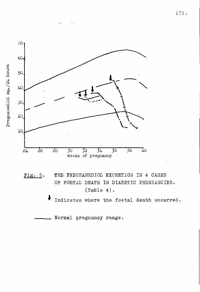
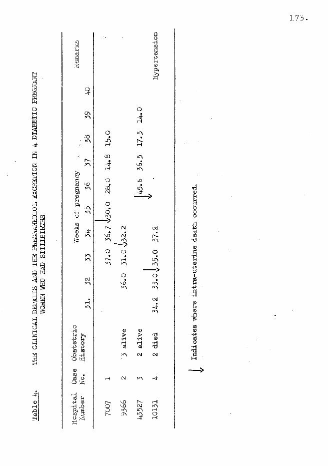
White & Hunt (194-0) , White (1949) and Smith & Smith (1947) showed that in the diabetic patient low pregnanediol values were associated with intrauterine foetal death. Peel (1955) stated that a fall in excretion of pregnanediol preceeded the foetal death in diabetes, but Gray (1955) was unable to show any significant difference between the results obtained in diabetic and in normal pregnant women.
The conflicting views in the literature cited showed that no clear picture of the normal range of pregnanediol excretion in pregnancy or of the variations which occur in early or late abnormal pregnancy^ could be obtained, and it was decided therefore to make a more comprehensive study of the problem. The first part of the work comprises an investigation into the variations which occur
when different methods are used to extract and purify pregnanediol. A review of the literature showed that by the method used by Sommerville et al (1948) a purer extract was obtained than by any of the shorter methods. This method was chosen for comparison with a modification of the method of Henderson et al (1949) which had been in current use at the Jessop Hospital for Women, Sheffield for the previous five years. Since neither of these methods proved to be satisfactory, an analysis was made of a new chromatographic method by Klopper, f'Michie & Brown (1955)• This method was found to give a purer extract and it was adopted subsequently for all the work on the pregnanediol excretion in normal and abnormal pregnancy described below.
The investigaticnfell into 4 divisions:-
SECTION I. METHODS.
Part A . Methods of Determining the UrinaryPregnanediol. This consists of a comparison between the method of Sommerville, Marrian and Gough (1948), and of Mitchell (1954).
Part B . Critical review of the method of Klopper,: Michie and Brown (1955)•
Part C . The relationship between the Blood"progestational activity", estimated by the Hooker and Forbes (1947) biological assay and the urinary pregnanediol excretion in normal pregnancy.
SECTION II. NORMAL PREGNANCY.
The range of pregnanediol excretion during normal pregnancy, before the onset of labour and in the puerperium was investigated.
SECTION III. ABNORMAL EARLY PREGNANCY.
The pregnanediol excretion in cases of threatened abortion, recurrent abortion and in Hydatidiform mole was investigated.
7.
SECTION IV. ABNORMAL LATE PREGNANCY.
Part A . The pregnanediol excretion in the Toxaemiasof Pregnancy. The following conditions were investigatedA) Pre-eclamptic toxaemiaB) Essential HypertensionC) Stillbirths and neonatal deaths
associated with ToxaemiaD) Chronic renal disease
Part B . The pregnanediol excretion in conditionsassociated with foetal death in late pregnancy. The following conditions were investigated:-A) Antepartum HaemorrhageB) Diabetes mellitusC) Rhesus immunisation including cases
of unexplained foetal deathD) Bad obstetric history.
Each section begins with a short introduction which includes references to the literature and it is concluded by a general discussion. A summary of the work reported and the conclusion drawn from it completes the thesis.
SECTION I .Part A . ERRORS IN THE DETERMINATION OP THE
URINARY PREG-NANEDIOL EXCRETION.
This section presents the results of a study of the variations found in the pregnanediol excretion in late pregnancy when a detailed examination of two methods of assay was made, together with a brief review of previous clinical experiences with these methods in the light of the results obtained.
INTRODUCTION.
Marrian isolated pregnanediol from the urine of pregnant women in 1929 and the chemical constitution was determined by Butenandt (1930 & 1931) who named the substance pregnanediol. Progesterone was isolated as a chemically pure substance in 1934.There is a striking relationship between these steroids and Venning & Browne (1936) demonstrated that sodium pregnanediol glycuronide could be recovered from the urine in the second half of the menstrual cycle and in pregnancy. Venning, Henry & Browne (1937) showed that when progesterone was injected into post-menopausal women and men pregnanediol was
9.
recovered from the urine.
Pregnanediol can either he estimated as "free pregnanediol” or in the conjugated form, sodium pregnanediol glycuronide /la P.G/7. In the method used by Venning (1937) the /la P . G ^ is recovered from the urine by extraction with butyl alcohol and subsequent precipitation from acetone. This method was later modified by Allan & Viergiver (1941) and Kaufman & Westphal (1947)• It requires large quantities of urine and the final extract contains only 80 per cent of the /la P.GjJ7 extracted, (Marrian & G-ough, 1946)* Moreover, bacterial hydrolysis of the /la P s o m e t i m e s occurs and this causes misleading low recoveries, (Bucher & Geschicter,1940).
Astwood & Jones (1941) hydrolysed the urine by boiling with acid and estimated the "free pregnanediol" gravimetrically. "Pree pregnanediol" when treated with concentrated sulphuric acid gives a yellow colour, and Talbot, Berman, Maclaglan &Wolfe (1941) used this to devise a colorimetric method for estimating the pregnanediol.
Sommerville, G-ough & Marrian (194-8) increased the purity of the final extract by the introduction of fractional precipitation of the pregnanediol extract. Modified precipitation techniques are used in the shorter methods of G-uterman (1944) and of Sommerville, Marrian & Kellar (1948) but both these modifications reduce the accuracy and the specificity of the extraction process.
Other methods purify the pregnanediol by chromatography, Stimel, Randolf & Conn (1952),Huber (1947) and de Watteville (1951), but impurities are still present in their final extracts. These may be removed by washing with cold solvents, but this causes a considerable loss of the free pregnanediol. (Bradshaw & Jessop, 1953? and Klopper, Michie & Browne 1955)•
When "free pregnanediol" is estimated, the final measurement is made either by a non-specific colour reaction obtained by adding concentrated sulphuric acid to the dried extract or by weighing it. The accuracy of the gravimetric method is dependent upon the purity of the residue and this may be determined by comparison of the melting
11.
point and the absorption in ultraviolet light of the final extract with those of pure pregnanediol. These considerations led to the selection of the method of Sommerville, G-ough & Marrian (1948) and to that of Henderson, Maclaghan, Wheatley &Wilkinson (1949), because they gave a white crystalline extract, and no loss was entailed by leaching the extract with cold solvents to purify.
DESIGN OF EXPERIMENT.
Three consecutive 24 hour specimens of urine were collected from 10 normal primigravid patients in the 34th - 35th week of pregnancy. Every care was taken to ensure a complete 24 hour specimen in each case; also the volume and creatinine content of each specimen of urine were noted and where these showed a marked daily variation the specimens were discarded and others obtained: thiswas only necessary on 2 occasions. Purified extracts were prepared in duplicate by myself and a technician. One of us used the method of Sommerville et al (1948) and the other used that of Henderson et al (1949)•The pregnanediol content was estimated gravimetrically
and colorimetrically on each specimen of urine by both methods. The gravimetric extracts were also examined for purity by melting point determinations and absorption in ultraviolet light. Each patient was kept under observation until delivery when the weight of the baby, and the weight, macroscopic appearance and histology of the placenta were noted.
METHOD I.
The method of Sommerville et al (1948) was used without major modification. The urine (500 ml.) after acid hydrolysis, was extracted with toluene, and any emulsion obtained was broken down by the addition of a few drops of Teepol (Shell Chemicals, Ltd., London) instead of the tedious filtration process used by Sommerville and his colleagues; this modification was shown not to interfere with the extraction. The toluene extract was washed with N-NaOH and water and evaporated to dryness; the residue was then dissolved in ethanol and precipitated under carefully controlled conditions of time and temperature,onc@ with 0.1 N-NaOH and twice with water, the precipitate in each case being controlled
by centrifugation with the aid of Hyflo Super-Cell (Johns Manville Co.,Ltd., London). After final decolonization with charcoal in ethanol, concentrated sulphuric acid was added to a portion of the dried extract which was than placed for 24 minutes in a water bath at 25°C ; "the absorption of the solution was then measured in the Spekker Photoelectric Absorptiometer using an Ilford Spectrum Violet 601 Pilter (transmission max. 403 atpc) . The remainder of the dried extract was weighed to give an alternative method of estimation.
METHOD II.
Henderson et al (1949) used a precipitation from acetone because this solvent appeared,from preliminary work,to give the best results. They showed that the time and temperature of precipitation from acetone were not determining factors in the recovery of added pregnanediol, and, as pregnanediol is less soluble in acetone than in ethanol, recovery from acetone should compare favourably with that obtained by the method of Sommerville et al (1948).
In the original method Henderson et al (1949) used only one precipitation; for Method II, the two
techniques have been combined, in that three precipitations from acetone instead of ethanol were used.
The dried toluene residue obtained as for Method I was taken up in 2.5 ml. acetone in a 50 ml centrifuge and 50 ml. IN-NaOH was added. The mixture was stirred and placed in a boiling water bath for 10 minutes and subsequently transferred to a refrigerator at 4^ for 2 hours. Hyflo Super- Cel 10 mg. was then added, with stirring, and the tube was centrifuged at 2,500 r.p.m. for 20 minutes The supernatant liquid was decanted and the precipitation twice repeated on the residue using distilled water instead of 0.1 N-NaOH. The remainder of the procedure was as for Method I.
Recovery experiments were carried out for comparison with Method I. One precipitation using pure pregnanediol, pure solvents and 0.1 1-laOH or distilled water, gave similar results for both methods; but when pure pregnanediol was added to male urine (0.5 - 2.0 mg. added to 500 ml. of urine) the recoveries using each technique in full
I1 ab
le
I.
i^C
jdil!
lGi\i
La
VEIB
OF
±Jra
Gl''tA
]NilD
IQL
IN m
gtns
/24
ho
urs
.
a<HO
i—I rHH
,Q
1—|
%
Pd«]•H03=> 5pq
oHOo
PQ
bO•H03=!>jPQ
orHOO
>»pq
oC•H03
lbpq
orHOO
lbpq
wps•H-P<1
VO A LA IA
rH A A CM
d
3VOA
ACM
VO A'A .—1 O.-)
• . !hr—1 CM -V)
0d d ao o dd d Hp p o0 0 H
d Oo
CM H A r'~ Hi—1 p- O oA vo A p- -d
O l"-VQ CM
fHC\J
A -4 00 A
VO CM A A
A -T VO -T
fv. A CM CM
-d" r>-A A
A A A CM
A 00 r- i—i
O A A rH
A V-Q A A
LA VO -d- A
rH CM
d d o o d d p p0 0
i—i
00 A A A
■4* OvO CM
A- O A CM
-d*-OCMA
15-
3
H O00 A
A A A CM
rH CM VO CM
CM f"- A rH
A O 00 P-
O CMr- h
r - aA CM
CM 00 r- vo
co AVO A
32*rH
rH CM VO A
A -d* VO CM
-d" H A CM
VO A
CM O f - rH
A O A CM 3 ^
CM
vo
A Pr VO -T
O A O -d-I—i
A d" A A
AOQ A -T
A A A CM
O -d vo vo A
O d 00 A 3
rHA
CM d
I
vq a d CM
CM i—I A CM
VOrHvO
A gr>-
d h vo. A
AVOA
CMCMCOrH
OQ VO d CM
-d- CMVO CM
A d A A
r> cm4- CM
CM H CM H CM H CM H CM H CM H CM H CM
d d d d d d d d d d d d d d do o o o o o o o o o o o o o od d d d d d d d d d d d d d dp p p p p P p p p p p p p p p0 0 0 0 0 0 0 0 0 0 0 0 0 0 0:-rT 53 a d >2 d V-| A V d 3;
A VO r -
VO vo ov A H Ap— vo d cm p- a
o Op OQ A H A00 d -d CM VO A
H CM
d d o odp p 0 0
orH
COA
r -HoCM
olH0j
Po;'H
00 A
16.-p•H!H•H■atP4
H■3
H•
w (0
O& & &a> 0 <0t> > >
CMCO S &
oVOfnoinLf\CMOJ
8m•»VO
3
CM ON VO
3
rlCMn-3
•»ON
oininCMCM
8in•svo
von -o 3m
•Oin
H
■S-p &1
vu0<qorHA o.rH8 tJ Hg -pd 3 00 1
■p•nI0PQ
■a-poEh
were approximately 10$ less for Method II than for Method I: this discrepancy was greater whenpregnancy urine was examined. As the final product from pregnancy urine using either method was not pure pregnanediol, this discrepancy could be due to a difference in the amount of impurity present.
RESULTS Of THE EXPERIMENT.
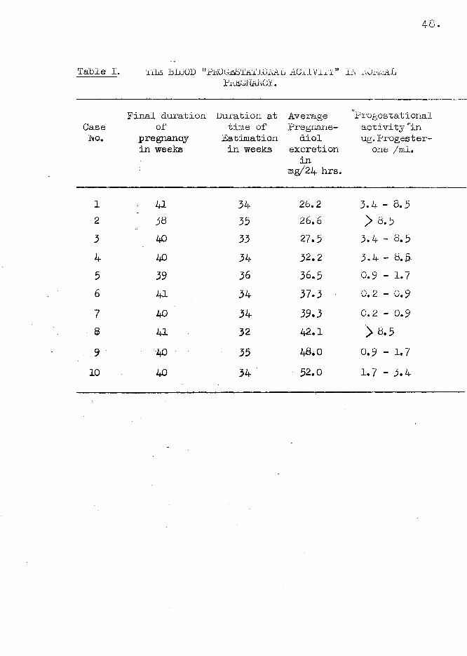
The duplicate readings were first examined and the differences between pairs of readings analysed.The mean difference of duplicate readings in Methods I and II was 3-9 + 9*57 (standard error of mean of 30 observations) and 2.5 + 0.38 (30): showing that the technical accuracy of the two methods was similar. The duplicate readings were therefore averaged leaving 4- estimations of pregnanediol for each of the 10 patients on each of 3 hays, making 120 estimations in all (Table I).
The marginal totals shown in Table I indicate the main results of the experiment. There is a large difference between individual patients and a small difference (not significant) between days, which may be ignored in view of the other and more
TABLE
II MELTING- POIN
TS AND ULT
RA VIOLET LIG
HT ABSORPTION
OP PURIFIED EXTRACTS
18.
rHO3
3CMH•
o
orn•o
8ft
No.
of
Estimations
3om 3
0
2nd
Product
Meth
od
2.
Standard
Error CM
•CM
+1
ON•in
+1
ON•m
+1
Mean ON
HCM
VO* o•CMn
CQg
O -P
n ,2CMrn
oin 3
0
u *n
w
-pop ••O H
2nd
Prc
Method
Standaj
Error lA
r-•H+i
r -•CM
+i + 2.2
Mean
207
CM
iON•n-CM
Melt
ing
points
fnhoCM+»cd
£
m
onCM
8
£» am
important findings. There are marked differences between the results for Method I and for Method II and between the gravimetric and colorimetric estimations, differences which are far greater than could possibly be the result of chance variation. These observations, together with the high mean square error, suggested that all four estimates were at fault, and this was shown to be the case when the final products were tested for purity by measuring their melting point and ultraviolet absorption spectra (Table II).
The average melting point for the product of Method I was 207°+ 1.8° (standard error of the mean of 32 observations), and for Method II 219° + 2.2°(4l), levels which were clearly different from those of pure pregnanediol (243°)> all melting points being measured on a G-allencamp (London) stage. Though Method I gave estimations for pregnanediol twice those for Method II (see totals Table I), the higher melting point of the final extract of Method II indicated that this method gave the purer products.
20.
Case ■ Duration Babies' Placental Macroscopic appear-No. of Weight Weight and or Placenta
39* H 6 lb. 3 oz. 1 lb. 3oz. Unhealthy "giitty" apx earance. Long cord. No infarcts.
3 u it 8 lb. 1 oz. 1 lb. 3oz. Pew small infarcts present.
4 43 it 8 lb. 1 oz. 1 lb. 8oz. " Gri t ty11 app eararic e Pew small infarcxs.p; k0 it 4 lb. 9 oz. 1 lb. 2oz. Small but healthy.
6 40 it 8 lb. 6 oz. 1 lb. 8oz. Healthy.-? 3E 14 it 9 lb. 7 oz. 1 lb. 12oz. Unhealthy “gritty1'
appearance.8 T m i it . 6 lb. 13 oz. 1 lb. lloz. Healthy. Pew
small infarcts.9 41 it 6 lb. 12oz. 1 lb. 3oz. Healthy.10 40 H 7 lb. 5oz. 1 lb. 3oz. Healthy with few
small infarcts.
* In case 2 there was marked foetal distress But a living child was obtained.
3t In case 7 the infant was stillborn.
21
The ultraviolet absortion spectra were measured in ethanolic solution and peaks or inflections were obtained in all cases at approximately 204 and 230 mu. The average values for
E i om for Methods I and II at 204 m}i. were 4 1 + 2 . 7 (30) and 5 7 + 5 - 9 (30) and at 203 mg|* the corresponding values were 2 8 + 2 . 2 (30) and 32 + 3*9 (30). As pregnanediol at the concentration and within the range investigated has a negligable absorption, these peaks must have been due to impurities and the absorption measured at these wavelengths is an indication of some of the impurity present.
The large difference between colorimetric and gravimetric estimation (see totals Table I) stresses the impurity of the product and the non-specificity of the method of assay.
STUDY 0E PLACENTAE.
The relationship between the duration of pregnancy and Baby's weights are shown on Table III. There was no correlation found between the pregnanediol excretion, by either method and these results.
Nor was there any correlation between the histological appearances of the placentae and the pregnanediol assays.REVIEW OE PREVIOUS CLINICAL EXPERIENCE WHEN THE
METHOD OF HEHDERSOU ET AL (1949) WAS USED.
The records of 24 gynaecological cases were available for study; infrequent or irregular menstruction or infertility were the main complaints In all cases pregnanediol was estimated by Method II Nine of these patients had levels ranging from 0.7 - 4.5 mg./ 24 hours from the 14th to 28th day of the menstrual cycle, and in all cases the endometrium showed histological evidence of progesterone activity. In 2 other cases, although pregnanediol was detected in the urine, the endometrium was of the hypoplastic non-secretory type.In the remaining patients, evidence of ovarian or pituitary dysfunction was detected by other endocrine assays, and no pregnanediol was present in the urine.
Only 2 abnormally high levels of pregnanediolexcretion were found among 700 patients investigated both occurring in hermaphrodite children aged 4
Table 4,THE KESULTS OF THE ENDOCHIHE IW E S T1 G iiTlQ E
years. The final pregnanediol fraction consisted in each case of a brown oily substance instead of white crystalline solid. The results are shown in Table IV. The ultraviolet absorption spectra showed a marked peak with a very high extinction value in both cases at 236 mp., thus indicating the possible presence of a large quantity of unsaturated ketone. Cortisone therapy (50 mg./day intramuscularly) reduced the level of excretion of this material to zero within 4 days, indicating that the substance was probably of adrenal origin. Thus false positive pregnanediol excretions were found in these cases.
The individual records of 20 cases of habitual or threatened abortions were also available for close study. Not all cases where low levels of pregnanediol were detected, miscarried, and in some cases with normal levels the outcome from pregnancy was unsatisfactory. Weekly determinations were carried out in one patient with a history of 2 previous miscarriages. She had stilboestrol and progesterone therapy during the investigation and delivered a living child at term;
25.
the pregnanediol excretion in this case varied markedly from week to week.
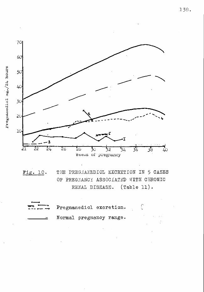
In a few cases of pre-eclamptic toxaemia wheie an attempt had been made to use the estimation as a measure of placental function, marked variation in the results obtained rendered this impossible. Thus in 2 cases where the patient delivered living babies at 34/35 weeks the urinary pregnanediol excretion levels were 5*6 and 6.5 mg. respectively.
DISCUSSION.
The work reported here shows clearly that the estimation of urinary pregnanediol by the above techniques, which were chosen as being the most satisfactory available, must be of very doubtful clinical value. Considerable variation has been shown in the level of excretion of pregnanediol in normal pregnancy between individuals and possible a small day to day variation in the same individuals. A marked variation was apparent when comparisons were made between the
two methods of obtaining the purified extract and between the two methods of estimating the pregnanediol content. There was also a high error variation.The melting points and absorption spectra of the end products together with the difference in values obtained by colorimetric and gravimetric estimation indicate that much of this variation is due to the presence of impurity. This has been proved in the past to give fictitiously high results for most of the shorter methods for estimating pregnanediol based on the procedures of Astwood & Jones (1941) and Talbot, Berman, Maclaghan & Wolfe (1941).
In the light of these errors a careful re-assessment of. previous experience with these methods of assay was undertaken lest any useful clinical correlation be overlooked. None was found. The variation in the results in cases of habitual and threatened abortion and pre-eelamptic toxaemia made it impossible for the test to be used as a diagnostic aid in cases cited.
Palse positive results for pregnanediol were obtained in 2 gynaecological cases in which the
27.
endometrium was of the hypoplastic non-secretory type and in 2 cases of hermaphroditism in children. These observations are in line with that of Swyer (1949) who stated MThe total excretion of pregnanediol in the luteal phase of the menstrual cycle was so variable that one cannot hope to give indisputable evidence of eorpus luteal activity'’.
SUMMARY AED CONCLUSIONS.
The levels of urinary pregnanediol excretion in late pregnancy were studied by two methods of assay.
In a planned study of the variations in these levels, estimations were made on the 24-hour urine specimen of each of 10 normal 34-36 week primigravid women oh each of three consecutive days. Purified extracts from each urine sample were prepared by each method and the pregnanediol content estimated both gravimetrieally and colorimetrically.
The 4 methods of assay gave widely different results which were analysed by the analysis of variance.
The difference between the results obtained by the two methods and by the gravimetric and colorimetric assay were highly significant.
The melting points and the ultraviolet light absorption of the purified extracts were also investigated and were found not to be those of pure pregnanediol.
It was concluded that the impurities present in the purified extracts accounted for much of the variation in the results obtained and for the high level of uncontrollable error.
A review, to determine any clinical value of these assays, carried out over several years, did not reveal any clear evidence of usefulness which might be set against the errors above described.
It was thus concluded that these methods were not sufficiently specific, sensitive or accurate for the estimation of urinary pregnanediol, and that further research upon the method of extraction would be necessary before any study of the pregnanediol excretion in abnormal pregnancy could be contemplated.
Part B .ANALYSIS OP THE METHOD FINALLY CHOSENFOR ESTIMATING- THE URINARY PREG-NANBIIOL.
INTRODUCTION.
A new method of estimating pregnanediol was reported by Klopper, 'Mtchie & Brown (1955).Through the courtesy of Dr. Klopper, the details of this method were made available to me before publication and an experiment, similar in design to that previously employed, was undertaken to evaluate this new method.
DESIG-N OF EXPERIMENT.
Three consecutive carefully collected 24 hour specimens of urine from each of 10 normal patients in the 32 - 36th week of pregnancy were examined.The pregnanediol content was estimated independently by myself and a technician, duplicate estimations being carried out on the same specimen of urine.
METHOD.
The only major modification to the method of Klopper et al (1955) was the omission of the
permanganate wash. Recent work hy Klopper (1955), completed after we had begun this investigation, indicated that this step is necessary in the case of non-pregnant urines and although desirable in pregnancy urines, is not essential in the majority of cases. (Klopper .1955) .
In 5 cases ( 1 - 5 Table I) duplicate samples of urine were taken, each being one twentieth of the 24 hour volume; these provided sufficient material for the tests for purity on the final extract in addition to the normal assay. In the other 5 cases (6 - 10 Table I) pregnanediol assays only were carried out and a smaller quantity of urine, (l/250th) was taken. After acid hydrolysis the urine was extracted with toluene and this extract washqd first with N-l\TaOH and then with distilled water, and evaporated to approximately 10 ml.This was applied, after cooling to room temperature, to the surface of a chromatography column previously prepared from 3 g alumina in a tube of 1 cm. diameter.
The column was washed with 2 5 ml. 0.87° ethanol in benzene,' and the pregnanediol fraction then eluted
31.
with 13 ml. 3% ethanol in benzene. (The volumes of solvents used in chromatography have to be slightly modified with different batches of alumina). The second eluate was evaporated to dryness and the residues acetylated with acetyl chloride in benzene,2 ml. of each. Light petroleum, (b.p. 40° - 60°) was then added, and the solution washed with 25 ml.80% strength aqueous bicarbonate, and twice with 25 ml. distilled water and chromatography was again carried out. The column was similar to that used previously but it was prepared in light petroleum instead of benzene. The pregnanediol diacetate was eluted with 15 ml. of benzene. This was evaporated to dryness and approximately 10 mg. sodium sulphite was added. The colour was developed by adding 10 ml. of concentrated sulphuric acid and allowing the solution to stand in a water bath at 25° for 1 hour, the absorption of the solution was measured in the Spekker Photoelectric Absorptiometer using an Ilford Spectrum Violet 601 filter, (transmission maximum 403 m\x.) .
When the twentieth of the 24-hour specimen was
Tab
le
I.
MilO
TIN
G
POIN
TS
AND
UIT
RA
VIO
L1T
ABSO
RPT
ION
01
PU
RIP
InD
EX
TRAC
TS
32.
n m
EHShH2ci)s9
oo
4) .03 O d O
CM
CO
OJ
m
(M
rn
CM
O COrH H
CM
i—I
3
CM MOCT\ Orn CM
CM i—I CM
00 rHLP\ VDH H
p- cmLP\ vO rH rH
rv. mLTV VO I—I I—I
CM
3 vO CM rH
Pm —1-1 coI—I I—I
lT\CX)
VOLT\
ONLf\rH
p--ini—i
m3
-1 A -1 vOCM OV VQ inrn CM -T rn
O co3 3
i—I H
-1 i—I CM
o in VO p - inp- r n r n p - CPrH rH CM 1— 1 1— 1
nm
op~-CM
= JCM
in CM CM CM i— 1• • • • •
CM CM rH rH CMrH rH CM i— 1 rH
n rn CM O -1• • • » •
rH i— 1 in CP vorH i— 1 CM H i— I
'-X* r n O CO n* • « • .
CM 1— 1 r n CM CPi— 1 1— 1 CM CM
p- inin mH rH
o vo vo in H H
CT\ vo in ni—i i—i
-dh n
nnrH
rnCT\CM
cr\rn
oOV -H P- 03 ri|I f
r n *Hvo PQi—li—Io•H 0)
St ' H P 0 erf d P erf 0 d O PO erfP 9 » |ij d H Cm ‘O
0
03 .erf 0Pderf •H03 d03 derf o0d po oT) -H0 d03 perf 0O £j03 'u•H o
1—1d oo o•H-Perf od cnp CMd cd0o 0d ho po
n0 Anoerf i—i-p orw ■H0 do 0H c0 coft a0 0XI dcM ft* Si
Produced by
acetylation
of pregnanediol (Ivi.P.
243°)
followed
oy chromatography and repeated re
crystalisation fro
m di-ethyl fcthex.
extracted, the final products were examined for purity by measuring their melting points on a Grallencamp (London) stage and their ultraviolet absorption spectra both in ethanol and sulphuric acid in a Unicam (Cambridge) Quartz Spectrophotometer,S.P.500.
Recovery experiments were carried out by adding 0.2 mg. pure pregnanediol in 0.2 ml ethanol to 150 ml. male urine or 10 ml. late pregnancy urine, followed by hydrolysis and extraction, as previously described. Pour male urines and two late pregnancy were studied.
RESULTS.
The melting points of the final acetylated products in the case of patients 1 - 5 are shown in Table I. The melting points were bi-phasic, but only the lower temperatures are shown. The ultraviolet absorption was measured in ethanolic solution, and after 1 hour in concentrated sulphuric acid at 25° C.The peaks in all cases were in the same positions as those for pure pregnanediol di-acetate but the extinction values of these wavelengths were
extinction34.
4
3
2
.1
200 210 2£0220 230Wavelength
240 260
Graph I a. Tffiti UIERAVIOLET ABSORPTION CURVE FOR PUREFREGMInIM)IO L DI-ACETATE IN ETHANOL IN
1 mg. /ml. ETHANOL.
E. 210 » .300 E 390Ion.
Extinction
35.
200 210 220 230 270230 Wavelength
250
Graph l.h. UlATtAVIOLBT ADSORPTION CURVE FOR PREGNANDIOL DI-ACETATE UM ETHANOL (EXTRACT) FROM
CASE No. 1.
E1E. 210 mp./ml. Ian.
_____ . Day I • 434 12-6 __ Day II . 500 11.3
Day III .456 12.5
Ex
tin
ct±
o
n
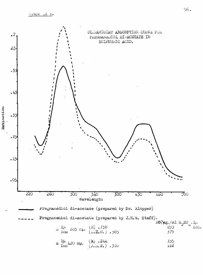
Vj'X'cXOi'I II cl*36.
ULiidLVIOLET ABSORPTION CUWE FOR PPEODAIIEDIO L DI-ACETATE ID
S uLtfiU rvIC AC ID .
23-
2 0220 380300 340Wavelength
430 4b0
Pregnanediol di-acetate (prepared by Dr. hlopper)
Pregnanediol di-acetate (prepared by J.H.W. Staff).
The final extract was thus purer than that which was obtained by the methods which had been previously tested.
The 24-hour pregnanediol excretiohs wereanalysed statistically. After averaging theduplicate readings, the results were set out intable form, (Table II), where the marginal totalsreveal the main features of the experiment. Usingthe analysis of variance the patients differedsignificantly, as was expected. The interactionPatients X days is significant, indicating thateven when allowance is made for the testing error(deduced from comparison of the readings of the twoobservers) the estimated excretion in any particularpatient does not vary from day to day. This"occasion" variation has an estimated standarddeviation oj 8.5 - 2.1 i.e. 1.8 appears to be random,
y 2the lack of significance between day mean square suggesting the absence of any appreciable daily pattern of variation common to all patients. The testing error of a single sample has a standard
Table III.APALTSIS OP VARIANCE
Source of Variation.
Degreesof.
FreedomSum of Squares
MeanSquare
Between women 9 4280XXX
475.6Between days 2 8 4» 8
nsInteraction or Random error 18 153
XXX8.5
Residual or Testing error 30 62 2.10
Total 39 4503 -
The mean squared difference between women is significant on the 0.1% level. The difference between days is not significant when compared with the interaction mean square which foims the appropriate basis for such a test. The interaction mean square itself is significant on 0.1°/q level when tested against the testing
error mean square.
41.
deviation of approximately 1.45* Hence if any attempt is made to measure the general level of output at any particular phase of pregnancy within the period covered by the experiment using only one test by one person the error will be the sum of the occasion variation and the testing error and will
phave an estimated standard deviation (1.45 ) +(1.8 )2 i.e. 2 .30.
DISCUSSION.
In the critical evaluation of this new method of assay, some uncertainties need to be discussed.
The specificity of this method, as with the older techniques previously studied, depends upon the purity of the final extract. Using the melting points and ultraviolet absorptions as criteria of purity, it has been shown that a final product is obtained which is only slightly less pure than a specimen of pregnanediol-di-acetate, produced by acetylation of a pure sample of pregnanediol followed by chromatography and repeated recrystalisations from ether.
Recovery experiments when pure pregnanediol was added to the urine show that 80-96/ of the pregnanediol originally present ih the urine is estimated in the final extract. These findings are similar to those reported by Klopper et al (1955)•
The- laboratory error of this method was determined when the assay was performed on three consecutive 24-hour specimens of late pregnancy urine. G-ood duplication of the results by independent observers shows that the laboratory error is low; however with pregnancy urine it is necessary to use only a small portion, (1/250), of the 24-hour specimen and thus any laboratory errors are multiplied by 250 when the 24-hour output is estimated. Despite this, the standard deviation of the testing error is only 1 .49*
The random errors due to day to day variation in laboratory conditions, faulty urine collection, etc., have been reduced so far as could be contrived in the results reported here: but these known andunknown sources of variation can never be entirely eliminated. The best that can be hoped for is that
the residual error is lower than the changing levels which are sought. In this study the standard deviation for the residual error was encouragingly low 2.3 due in a large measure to co-operation of the patients selected. Five were doctors’ wives and the other five were nursing sisters, all of whom were aware of the need for great care in collection complete 24-hour specimens of urine.
The evident superiority of the method of Klopper et al (1955), as compared with the previous techniques examined, thus provided a means for routine clinical investigations. Provided that great care is taken at all stages of the assay, particularly in the accurate collection of the twenty-four hour sample, results of reasonable dependency can be expected.
SUMMARY AND CONCLUSIONS.
The method developed by Klopper et al (1955), for estimating urinary pregnanediol has been investigated in late pregnancy, and the results reported here.
44.
As a planned study of the method and of the variations in the daily excretion under known conditions, estimations were made on 24-hour specimens of each of 10 normal 34-36 week primigravid women on each of three consecutive days.
Measurement of the melting points and ultraviolet light absorption showed the final extract to have a high degree of purity.
The analysis of variance of the results showed a marked difference between the amounts of pregnanediol excreted by the different patients; but no difference between the daily outputs. The testing error of the method as estimated from the duplicate results of two independent observers was low. The residual or ''occasion" error was small enough to justify a further study of the test.
This method was thus chosen to study the application of this assay in the management of early and late pregnancy disorders.
45.
Part G .
THE RELATIONSHIP BETWEEN MPROGESTATIONAL ACTIVITY*1 OP THE BLOOD AHD URINARY PREG- NAKEDIOL EXCRETION IN NORMAL PREGNANCY.
Hooker & Porbes (1947) described a technique whereby’’progestational activity11 could be measured by observing the response of the stromal cells of the endometrium of mice to local applications of progesterone or extracts containing substances of ’’progestational activity" Essentially the test is carried out by injecting measured microquantities (0.0006 ml. or less) of the fluid under test into the uterine horn of a previously ovariectomised mouse, leakage of the injected fluid being prevented by two ligatures one above and one below the injection mass. The characteristic response in the endometrial stromal cells, seen 48 hours after injection, consists of a change from stellate or polygonal cell forms to a smooth, slightly elongated oval outline; the chromatin threads become fine and evenly distributed, and the nucleolus is conspicuous.
The minimal dose of progesterone to produce this effect in the strain of mice employed by these workers was 0.0002 p. g. The authors showed that other steroids failed to produce this change and concluded that the test was reasonably specific in denoting; progestational activity Forbes (1950) also produced evidence to show that progestational activity could be demonstrated in plasma extracts of as little as 1 ml. of blood taken from women during the secretory phase of the menstrual cycle.
Hitherto the biological test for progestational activity was carried out in rabbits and the amount of progesterone necessary to produce positive responses precluded the possibility of applying this test to the routine study of blood plasma levels of progesterone in women. With the Hooker & Forbes’ Test available, it was decided to determine whether the progestational activity level, measured in terms of progesterone, bore any relationship to the level of pregnanediol in the urine. Preliminary tests, using solutions of progesterone in sesame oil, showed that with the
47.
strain of mice available the sensitivity of the test was strictly comparable with that described by Hooker & Forbes (1947).
METHOD.
Three consecutive 24 hour specimens of urine were collected during the 34th to 36th week from each of two patients who subsequently were delivered of live babies. Pregnanediol estimations were carried out on each of these three specimens in each case, and the average daily excretion determined.On the second day of collection, a specimen of citrated blood was obtained. The citrated plasma was extracted twice with 10 ml. ether, and then 2 ml. sesame oil was shaken up with the ether extract.The ether was removed by distillation under reduced pressure, and 0.0006 ml. of the final solution or dilutions thereof were injected into the uterine horns of ovariectomised mice. The greatest dilution giving a positive response was considered to show progestational activity equivalent to 0.0002 u g. progesterone, the activity of the plasma then being expressed in terms ofu g. progesterone per ml.
No attempt was made to hydrolyse the plasma extract and therefore the recorded values are an expression of free progestational activity, and dLO not include any combined or "bound” progestins.
The results in the ten cases are set out in Table I. -
The results show that whereas positive responses were obtained by the Hooker Forbes (1947) technique in all ten cases examined, there was no correlation between the level of response and the level of pregnanediol excretion. It seemed improbable that this technique would give an index of placental activity more reliable than the pregnanediol assay, and no further work using this technique was carried out.
50.
SECTION II.
THE PREGNANEDIOL EXCRETION IN NORMAL PREGNANCY AND IN THE PUERPERIUM.
The excretion of pregnanediol during pregnancy has been studied by many workers and the results show a general trend of high level excretion in the last three months of pregnancy with a fall to zero levels soom after delivery. Thus Browne, Henry & Venning (1937) estimating sodium pregnanediol glycuronide, obtained values of 40 mg./24 hours at the 21st week, peak values of 73 to 80 mg./24 hours at the 32nd week and showed that there was a fall just before delivery. Estimating ’'free pregnanediol” by precipitation techniques values of between 36 and 120 mg./24 hours were obtained in the later weeks of pregnancy by Davis & Eugo (1947), Guterman (1944) (1945), Jones, Delfs & Straun (1944) and Michie (1953) • Lower values were obtained by de Watteville (1951) and Trolle (1955). The pattern of excretion immediately preceding parturition is not clear. Stover & Pratt (1939) and Hain (1941) found that the excretion was
51.
maintained until the onset of labour. In contrast, a fall in excretion rate was demonstrated by several workers (Pratt, Simmonet & Robson, 1940, Lyon, 1940 and Maunsey, 1950).
Other workers were unable to demonstrate a characteristic excretion pattern for the period immediately preceding delivery (Venning, 1948, Bradshaw & Jessop, 1953> and Trolle, 1955)*There was general agreement that pregnanediol falls rapidly after delivery and reaches low levels in 3 to 6 days.
Variations in the techniques of assay are inpart clearly the cause of the lack of uniformity inthe results reported by the above workers and accordingly it was necessary to study the pattern of pregnanediol excretion throughout normal pregnancy and the early puerperium by the method of Klopper et al (1954).
PLAh OP INVESTIGATION.
The following aspects of the pregnanediolexcretion in normal pregnancy were studied:-
Pregnanediol
Pregnanediol mg./24 hrs.
rng./24
hrs.
52.
70 1
60 ■
50-
30 •
20*
10*
Weeks before deliveiy
B.
IQ« *»15 20 25
Weeks before deliveiy10
Fig. I.A. MEAN ESTIMATES OP URINARY PREGNANEDIOLEXCRETION FOR EACH WEEK OP PREGNANCY.
Fig. I.B. STANDARD DEVIATIONS CALCULATED FOREACH WEEK OF PREGNANCY.
______ Range of excretion * 2 x S.D...._______ Mean estimates...
l) The pregnanediol excretion from the 10th week of pregnancy till the onset of labour.
Pregnanediol estimations were made on the urines of 24 women at intervals varying from 1 to 3 weeks throughout the course of pregnancy. They were a highly selected group of patients, being either the wives of doctors or former nursing sisters of the hospital. They all delivered living babies at the 38th to 42nd week of pregnancy and in each case the birth weight of the baby and the weight of the placentae was noted.
2) The pregnanediol excretion after parturition. Four hourly estimations were made in five cases during the 24 hours after delivery. All specimens were collected by catheter to prevent contamination with blood. In 9 cases pregnanediol determinations were made at Hintervals” until the. 9th or 10th day after delivery.
RESULTS.The mean estimated of the urinary pregnanediol
excretion during each week of pregnancy are shown on Fig.I. The range of excretion throughout normal
Pregnanediol mg./24 hrs.
54.
701 ’«s
10• •
0 10 15 20 25 :Weeks before delivery
Pig. II. THE PREGNANEDIOL EXCRETION INnormal p r e g n a n c y calculatedPROM THE DURATION OP TIME
BEPORE DELIVERY.
Range of pregnanediol excretion.------ Average excretion.
Pregnanediol mg./24 hrs.
55.
Delivery
10
Weeks before delivery
Pig. III. THE PREGNANEDIOL EXCRETION, IN 5 NORMAL PREGNANT WOMEN, DURING THE 6 WEEKS BEPORE DELIVERY.
In these cases there was a fall in excretion before
delivery.
56.
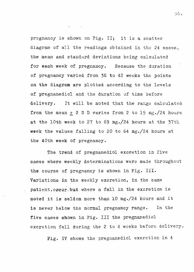
pregnancy is shown on Pig. II; it is a scatter diagram of all the readings obtained in the 24 cases, the mean and standard deviations being calculated for each week of pregnancy. Because the duration of pregnancy varied from 38 to 42 weeks the points on the diagram are plotted according to the levels of pregnanediol and the duration of time before delivery. It will be noted that the range calculated from the mean + 2 S D varies from 2 to 15 mg./24 hours at the.10th week to 27 to 69 mg./24 hours at the 37th week the values falling to 20 to 64 mg./24 hours at the 40th week of pregnancy.
The trend of pregnanediol excretion in five eases where weekly determinations were made throughout the course of pregnancy is shown in Pig. III. Variations in the weekly excretion, in the same patient, occur .‘but where a fall in the excretion is noted it is seldom more than 10 mg./24 hours and it is never below the normal pregnancy range. In the five cases shown in Pig. Ill the pregnanediol excretion fell during the 2 to 4 weeks before delivery.
Pig. IV shows the pregnanediol excretion in 4
Pregnanediol mg./21+
hrs.
Delivery
20 .
Weeks before delivery
ffig. IV. THE PREGNANEDIOL EXCRETION, IE 5 NORMAL PREGNANT WOMEN, DURING THE 6 WEEKS BEEORE DELITERY.
In these cases a fall before delivery was not
detected.
Pregnanediol mg./24
hrs.
58.
Deliveiy
40 ■
10 .
.3-11 9 7Days before delivery
.gig. V . THE DAILY PREG-NANEDIOL EXCRETION IN A NORMAL PREGNANT WOMEN.
Table
VI. SHE
PKEGMUNEDIQL
KKGHUIION
EEFOMi
i AND
AFT
ER Ds&DtSm
IN9 NOR
MAL
PREGNANT M3MKN,
59.
3
ON
CO&0) fs-
3nrf v£>u.<0-P
~1 CM
m n CM CM
n n -t
m -d* n~VH4 n r"- «n CM
~r n
3*.*ftn
CM
r~i&0)£-a'
<D•5H CM(DncJ nOQJ03&
•Os;Q)3o
VO r- n
CO mH CO □V
VOH r-f-*
•(V- CMH CO o\ nrHnrH CMCM nrH ONCM CM fv.CM nCM 3 nn
cases where a fall before the onset of labour was not detected. In one of these cases a rise in excretion was actually detected during the 2 weeks before delivery.
When daily estimations were made in 4 cases a constant excretion was maintained for 10 days in 3 cases and in one patient a rise occurred five days before delivery, Fig. V.
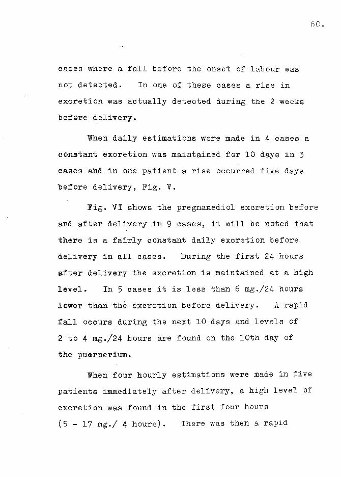
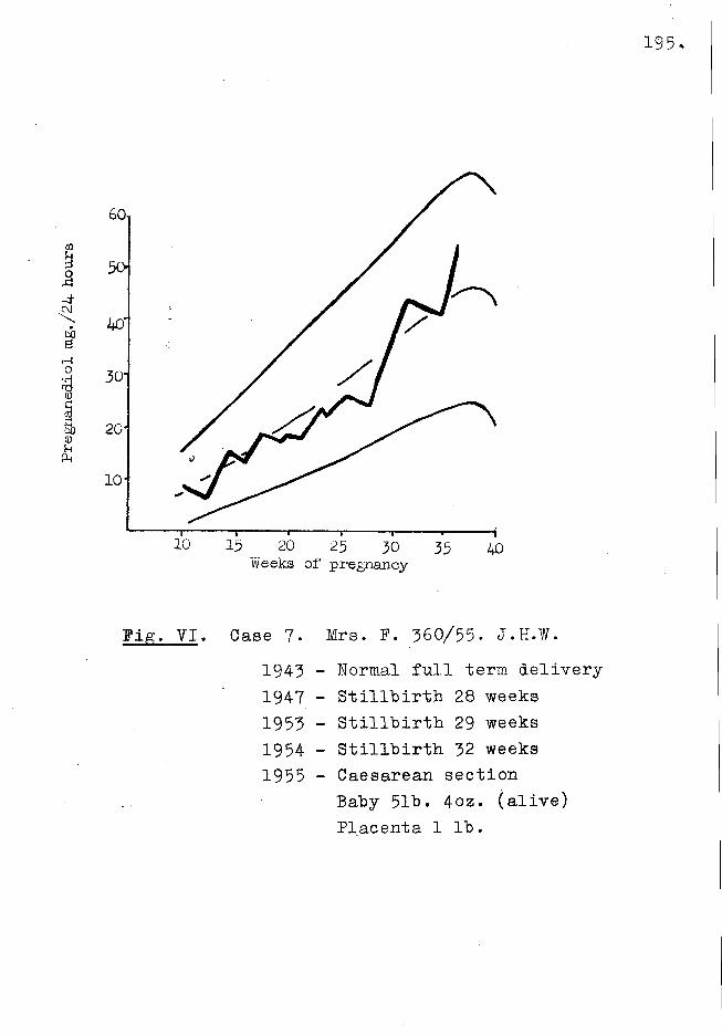
Fig. VI shows the pregnanediol excretion before and after delivery in 9 cases, it will be noted that there is a fairly constant daily excretion before delivery in all cases. During the first 24 hours after delivery the excretion is maintained at a high level. In 5 cases it is less than 6 mg./24 hours lower than the excretion before delivery. A rapid fall occurs during the next 10 days and levels of 2 to 4 mg./24 hours are found on the 10th day of the puerperium.
When four hourly estimations were made in five patients immediately after delivery, a high level of excretion was found in the first four hours ( 5 - 1 7 mg./ 4 hours). There was then a rapid
Pre
gnan
edio
l m
g. f/
Ur
hrs
.
61.
Deliveiy
20
T T8 i16 T20hours a f t e r d e liv e ry
Fig. YII. THE 4 HOURLY PREGNANEDIOLEXCRETION IN 5 NORMAL PREGNANT WOMEN FOR 24 HOURS AFTER
DELIVERY.
fall to levels of 2 to 4 mg./ 4 hours and in the last 12 hours the excretion remained fairly constant (Fig. VII).
DISCUSSION.
The range of pregnanediol excretion in normal pregnaney for the method of Klopper et'al (1954) was studied by making regular estimations at regular "intervals” throughout the course of pregnancy in 24 intelligent women. The range is lower than that obtained, by other workers, using precipitation techniques, G-uterman (1954), Davis & Fugo (1947) and JMtchie (1953) • It is higher than that obtained by other chromatographic techniques, de Watteville (1951) and Trolle (1955)• This is understandable as it has already been shown that the formertechniques estimate an impure form of pregnanedioland in the latter technique pregnanediol is purified by leaching with cold solvents and this may remove some of the extracted pregnanediol (Klopper et al (1954)).
The pregnanediol excretion in pregnancy reaches apeak level at the 37th week and there is usually a fallin the test two or three weeks preceding delivery
(Fig. II); but the results obtained in the study of individual cases show that, of the 17 patients who were under observation until the onset of labour 13 had a fall in the excretion during the 36 - 38th week of pregnancy', excretion was maintained at a high level in the other 4 cases (Fig. IV).
When daily determinations were made in 9 cases the excretion level did not fall before delivery (Fig. V & VI). These results confirm the observations of Stover & Pratt (1939) and Hain (1941).
The pregnanediol excretion during the 24 hours following delivery was found to be high. All .the patients were catheterised immediately after the placenta was expelled thus the pregnanediol excreted during that period may have been present in the blood at the time of delivery. In 5 of the 9 cases the excretion was less than 6 mg./24 hours lower than the level before the onset of labour. In case I (Fig. VI) , although the patient was catheterised after completion of the third stage of labour the 4 hourly collection of urine was not organised for 5i hours during which time the patient had passed
urine. This may account for the low value obtained in this case in the 24 hours after delivery (15 mg./24 hours). Bradshaw & Jessop (1953) obtained values ranging from 5 to 28 mg./24 hours during the first 24 hours after delivery and 1 - 2.7.mg./24 hours on the 33rd day. Trolle (1955) found that the excretion during the first 24 hours in 4 cases was the same as that before delivery (15 - 30 mg./24 hours) but he obtained a fall to nil in the course of the next 3 - 6 days.
A gradual fall in excretion was obtained during the puerperium by the method of Klopper et al (1954), levels of 2 - 4 mg. being found on the 10th day after parturition. When four hourly excretion rates were studied, excretion immediately after delivery was high ( 5 - 1 7 mg./24 hours) and then there was a gradual drop, the level being maintained for the 12 to 24 hours after delivery (Pig. VII
SUMMARY AND CONCLUSIONS.
The range of pregnanediol excretion throughout normal pregnancy was studied using the method of Klopper et al (1954). The range is wide. It varies
from 2 - 1 5 mg./24 hours at the 10th week to 27 - 68 mg./24 hours at the 37th week. In most cases there is a fall in excretion in the weeks preceeding labour but in 4 of the 17 patients studied here the excretion was maintained.
When daily determinations were made for 10 days preceeding the onset of labour little variation was found in 3 patients but one patient had a 10 mg./24 hours rise in excretion.
After delivery the pregnanediol excretion was maintained at a high level for 24 hours and then there was a gradual fall to a level of 2 - 4 mg./24 hours on the 10th day after parturition. More than -§• of the pregnanediol excreted during the 24 hours after delivery appears in the urine in the first 4 hours.
CONCLUSION
The pregnanediol excretion in normal pregnancy for the method of Klopper et al (1954) is now established. There is no specific variation characteristic of the approach of labour in the daily excretion but in the majority of cases the excretion
reaches a peak level at about the 36 to 38th week of pregnancy and then there is a gradual fall before delivery.
Luring the first 24 hours after delivery the excretion is only slightly lower than the excretion before delivery, the major part of this excretion occuring in the first four hours after delivery.
SECTION III.THE PREGNANEDIOL EXCRETION IN THREATENED
AND IN RECURRENT ABORTION. AND IN HYDATIDIEORM MOLE.
Among the many factors which are alleged to play a part in the aetiology of spontaneous abortion hormonal imbalance occupies an important place;There is as yet little knowledge and few measures of what constitutes hormonal balance. This section is concerned with only one aspect of the problem, the excretion of pregnanediol. Opinion as to the value of this assay differ; there are some who have found the pregnanediol excretion to be a reliable guide by demonstrating subnormal values preceding abortion (Venning 1937, Cope 1940, Guterman 194 7, Bishop 1948, Guterman & Tulsky 1949, & Borglin 1956); whereas others maintain that normal values do not exclude the possibility of impending abortion, and doubt the significance of low values, (Hamblen, Cuyler &Baptist 1942, Swyer 1949, Plotz & Darup 1950,Zander 1951 and Swyer & Daly 1953)*
Despite these uncertainties, progesterone has been used in the prevention of abortion (Bender 1948, Guterman 1953, Bishop, Richards & Doll 1950) and Smith & Smith (1948), considering that the metabolism of oestrogen and progesterone were interdependent, advised _the administration of oestrogen and claimed an increase in the pregnanediol excretion when stilboestrol was administered during pregnancy.Similar findings were reported by Davis & Rugo (1947 and Sommerville, Marrian & Clayton (1949)•
Guterman (1953) maintained that when progesterone was injected intramuscularly in cases of threatened abortion, abortion ensued if under 20$ of the progesterone was converted into pregnanediol but if the conversion rate was about 20$, the pregnancy continued undisturbed.
Most workers who have assessed the progesterone metabolism in early abnormal pregnancy have used the methods for extracting and purifying pregnanediol which according to Marrian (1954) are not sufficiently sensitive, specific or accurate for the small amounts of pregnanediol found in early pregnancy urine.
69.
The investigation detailed here is a reassessment of the value of the pre-gnanediol assay in the management of cases of threatened and recurrent abortion using the more reliable method of Klopper et al (1954)•It is divided into 4 sections1) Tire pregnanediol excretion in cases of threatened and recurrent abortion where no specific form of therapy was used;2) The pregnanediol excretion in cases of recurrentabortion when stilboestrol therapy was used:3) The conversion rate of exogenous progesteronein 15 cases of threatened abortion; and4) The pregnanediol excretion in 5 cases ofHydatidiform mole.
TEE PREG-NAKED101 EXCRETION IN THREATENED ABORTION.
Patients with a history of vaginal bleeding and abdominal pain in early pregnancy had a twenty four hour specimen of urine collected on admission to hospital. Cases in which abortion occurred within 3 days of admission were not included in the series, as they were assumed to have been inevitable dr incomplete when first seen. In a series of 70 primary
Pregnanediol mg./24
hrs.
70.
A . h .
30 „
20
10* *
2012 16 Weeks of pregnancy
20. 12 16 Weeks of pregnancy
Fig. I. THE PREGNANEDIOL EXCRETION IN 61 CASES OE PRIMARY THREATENED ABORTION
A. *** Live births (29 cases)B. • •• Abortions (32 Eases)
Normal pregnancy range.Average readings for normal pregnancy.
threatened abortions, single pregnanediol estimations were made in 61 cases the remaining 9 cases having estimations carried out at weekly intervals. The duration of pregnancy at the time when the patients threatened to abort varied from 8 to 20 weeks. The results are shown on Eig. I. Of the 61 patients, 29 delivered live babies and 32 aborted. Before the 12th week of pregnancy there was no difference between the results obtained in the live birth and the aborted group. After that period 7 (30$) of the results obtained in the live birth group were above the average values obtained in normal pregnancy, whereas in the aborted group only 2 (10$) were above it; moreover in the latter group 5 (26$) of the readings were below the limits of the normal pregnancy range as compared with the live birth group where all the values were within the normal pregnancy range.
Pregnanediol estimations were made at weekly intervals in 9 cases; of these 5 delivered live babies (29 observations)and 4 aborted (17 observations) The results for each week of pregnancy are shown on Eig. Ila and lib. which are scatter diagrams of all
Pre
gnan
edio
l m
g./2
4. h
rs.
72.
A.30 ,
t—
16 2012weeks o f pregnancy
10
12 16 Weeks of pregnancy20
Pig. II. THE PREGNANEDIOL EXCRETION IN 9 CASES OE PRIMARY THREATENED ABORTION.
A.xxx Live Births (5 cases)B .•••Abortions (4 cases)
Normal Pregnancy Range Average readings for Normal Pregnancy
Average readings for cases of Threatened abortions.
Table J .TKS BhiGNaNEulOL aICHETION IN 9 GASES OF FNIMAHr. THREATENED ABOHTION.
the readings obtained in these 2 groups of patients.The values obtained for the pregnanediol excretion are similar to those obtained when only single estimations were made. When, however the trend in individual cases is studied there is some evidence that declining results indicate that abortion is likely, and rising levels that the pregnancy will continue.These changes ’. are more marked after the 12th week by which time careful clinical examination should give as much information as the hormonal assay. Nevertheless, the trend after the 12th week might on occasion be of value in adding weight to a clinical judgement.
THE PREGNANEDIOL EXCRETION IN RECURRENT ABORTION.
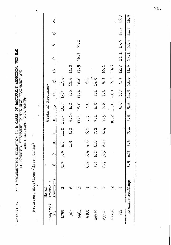
This group of patients received no specific form of treatment during this pregnancy. They had a' history of 2 or more abortions in previous pregnancies. There were 13 patients in the series, 8 delivered live babies and 5 aborted. They were kept under observation throughout the course of pregnancy and urine was collected at daily or weekly intervals.
The results of the weekly pregnanediol estimations are shown on Big. Ill a and b, which are scatter
75.
13.A .
20
10„ — *" x*
2012 16 Weeks of pregnancy
20Weeks of pregnancy
Fig. III. THE PREGNANEDIOL EXCRETION Iff 13 CASES OE RECURRENT ABORTION WHEN NO SPECIFIC
. FORM OF THERAPY WAS USED IN THE NEXT PREGNANCY.
A. xxx Live births (8 cases)B. * • * Abortions (5 cases)
■—.. Normal pregnancy range.-------Average readings for normal pregnancy.------ Average readings for recurrent
abortions.
Table
II a.
THE PREGNANEDIOL EXCRETION
IE 8 CAS
ES OF
RECURRENT
ABORTION,
WHO HAD
NO SPECIFIC TREATMENT
IN THE NEX
T PREGNANCY
AND
WHO DELEVERED
LIVE
BABIES
76.
00
H
VO
LP\H
rn
5t,•H crsft
0>
•H 00rH
09do•H 0913 3 §O O -H
f t -H -P o > U0 o4“J O U f t30 ft ft -ftHHft i— !o ft0 -Pft •H •ft O c/9 — Oft
CM VO
in rHm vo r- crsft
OJ\CMr-COrH
o m o3 •
r"- oi—i rH H
ft vo O CM : o rn•r -
•rH
•vo GO ; -J-
•ON
H rH rH rH
ft o ft CM vO• • • • •
r-- 00 CJ\ r -H i— 1
r-- O VO O o 00•J- CO
•r"- CO
•r -
H rHm
CM CM ft rn ft m•VO
»n- in •r-- •n-rH rH
CM o o CM ft• • • • •rH VD VD VOrH
f t o\ CO 00 O•vo 00
•vo
m ft 1—1 rn• • • •m vo vo r-
r"- CM CM r->-• « • •m CO m so
m m
mvo
oCOvo
rn
ovoLft3
3rnnCM
VOoCMCMr-rHOoCM
O3CM•Oi—I
ft- CM
i—IOn-r-CM
OVVOH
ftrHin
mrH
Hn-CMHinCO
COVO
LALPn
r-CM
cr\VO i—ICM-J-rH
K-\CMCM
LCvrH
ONrH
00•
CMrH
rn•
CMrH
VO•CT\00cAftn-voVO
mvoLf\vo
09pf'i0U0bOao
Table
II b.
TEE HUa&NANEDIOL EXCRETION
IN 5 CAS
ES OP
RECURRENT
ABORTION
WHO HAD
. ANOTHER
UNSUCCESSFUL EREGNANCI.
77.
m0XQ
oA
rd0o
o•3§0UO0H
A Aln • •rH A A
o O-4H
O UN AA • • •rH rA - CM CM
!>>O§ rH O O COerf C\Ja H
♦CM CM
•CMuu
0)A.t-H m UN O h-
of 11
•(—1 ON
ao0 rH CO CM O Ow 10 00
•CO •A
•co
CM A 4" <S» AON * • • • •-T i—1 r - A COrH
-t o CMCO • • •
CM 00 COrH
IQw hOfl do ■H
^ 'H •o° * A •4 -4 CM A
cd0• o UO rQS <S 0bO
oj•a
U0-p i—1 o CO 00 CO >•H • CO CO Cl UN A 4o O ON -4 r - ONw ;z; co rH r-- rHO HH
78
diagrams of the results obtained in the 8 patients who delivered live babies and in the 5 who aborted. Table II shows the individual results for each patient and the average readings for each group. The
results are similar to those obtained in the live birth groups and aborted group of threatened abortions All the readings in the live birth group are within the normal pregnancy range and the average readings rise after the 12th week of pregnancy to above the normal pregnancy average. In the aborted group 7 results lie outside the lower limit of the normal range and the average readings show a fall after the 12th week of pregnancy.
The results in this group are similar to those in the group of primary abortions. Values below the normal range were associated with abortions, values below the average but still within the normal range did not appear to have any clear prognostic value, and values within the normal range but above the average were usually associated with continuing pregnancy.
Pregnanediol mg./24- hours
79.
30.
20-
10-
* 12 16 Weeks of pregnancy
20
B.
20-
10-
16b 12 20Weeks of pregnancy
Fig. IV
A.B.
THE PREGNANEDIOL EXCRETION.IN CASES OE RECURRENT ABORTION WHO DELIVERED LIVE BABIES IN THE NEXT PREGNANCY.No specific therapy (8 cases)Stilboestrol therapy (6 cases)
The shaded areas show the limits of the lowest and highest
readings obtained.Normal Pregnancy RangeAverage readings for normal pregnancy
80
a
EH
EH
<d
MHH
0)rHftcd
on
X)i—i
ft
vOH
AH
0UPi
A ft H O
JS00 CM,:3r H
i—I H
O
CTn
CO
VIft cJ o o•HU ft
0 MA O £ ,-Q 3 3
i—Iaj Hft 0•H ft ft g m 3O ftft!
O CM '-0•
CM•
ftH H H
o -4 4" CM•
'A o•
ft nfH H H fH
O
CMCM
CM
H
CMe
VO
X) O ft X) LA o • • • • » •
.0 H O X) A A H CM CM H
O 4 cm CM K>0• • • • • o
A vo ON 00 ON 4" i— I i— I i— I rH
X> -4- f t cm ft o• • • • • o
-4 CM A VO ft AH H H i—I
co o a 4- 4*• • • • •
CO CM A 'ft CMsH H
X) O 4" A• • •
00 COrH
•CMH
•Oi—1
-d“ -d- co--- 7
O i—1• • •
00 CM H H H
•CMH
•HH
CM 4" 00n MrH rH
•CMH
vo O 4" ft• • •
00 A O H rH 3
— > -4
03 CM A CM• • •
CO A A•
rH
CM CM A A A A
-4 CM vo A O O A O ft CO A p-— ft- i—I A O V-D f t A vo A A
ftAH
-4•
AH
ftAH
VOHH
O
A
0hi)cdu0
aH
EH
-4CM,
ACM
CMCM
HCM
OCM
a
COH
0U ft ft HftO
a00vo
AH
-4H
AH
CMH
H
ft M O pj
O•H1!O3 rQ
4 cd
I—Icd Hft 0 •H ft Ph g w 3o ft
X)AI—IA•
H
O
00H
ft•
voH
A
ft i—ICM•
ACM
i—I ON
A
voH
A
di—1-4i—I i—ICM
CM
3i—I A
ft0O
3oo
i>scd*H0ft!ftr—IoH-ft100oft I—I•H
0ft!ft§iM0ftcdo•Hft!H
— >
Table 4 THE DAI IX PiiECkANEDIGL EXCNETION IK 6 CASTS ON xtilJUilTlNT XBOitTION VViiO i)E1 .T vTk pi ) LIVE BABIES
No Specific I‘herapy Stilboestrol TherapyHospitalNumber 127 4755 961 6465 5080 65Numberof
indicates when the stilboestrol therapy was commenced
82.
2) THE PREGNANEDIOL EXCRETION WHEN STILBOESTROL THERAPY WAS USEE IN THE TREATMENT OP 7 PATIENTS WITH A HISTORY OP RECURRENT ABORTION.
Stilboestrol in increasing doses was administered to 7 patients with a history of 2 or more abortions in previous pregnancies. The initial dose was 30 mg. daily until the 16th week of pregnancy when it was increased by 5 mg. daily at weekly intervals until the 36th week. Six patients delivered living babies at or.near term and one patient aborted at the 24th week of pregnancy.
The daily pregnanediol excretion in 2 patients who were treated with stilboestrol and in 4 of the untreated group are shown on Table IV. The daily excretion in both cases was constant and the stilboestrol treatment neither increased nor decreased the pregnanediol excretion. The results of the weekly determinations are shown on Table III where this is again demohstrated.In Pig. IV. a and b the mean values and the range of excretion are plotted against the mean range of normal pregnancy. The range in the recurrent abortion group is represented by the highest and lowest values obtained
83*
for each week of pregnancy. The range in "both groups lies between the normal pregnancy range and that obtained in the stilboestrol-treated group is neither higher nor lower than in the untreated group.
The pregnanediol excretion in the patient who aborted at the 24th week of pregnancy is shown on Table III. There was a rise till the 20th week when a slight fall occurred, the excretion rate being 15*5 mg./24 hours when the patient aborted.
3) THE CONVERSION OF EXOGENOUS PROGESTERONE IN THREATENED ABORTION.
Fifteen patients between the 9th and 20th week of pregnancy were admitted with a history of vaginal bleeding and abdominal pain and had pregnanediol assays made on 3 consecutive days after admission. On the 4th day 90 - 100 mg. of accurately weighed progesterone in 2 ml. sesame oil was injected intramuscularly.The percentage of the progesterone excreted as pregnanediol was then calculated from the pregnanediol excretion during the next 3 "to 4 days.
The progesterone conversion in non-pregnant states
Table 5. THE PROGESTERONE RECOVERY' IN 15 CASES 01- THREATENED ABORTION.
Duration LL'VE BIRTHS Duration ABORTIONof
Pregnancy Recovery Pregnanediol
Excretionbefore
injection
Recovery Pregnancy%
Pregnanediol
Excretionbeforeinjection
12 weeks 2.4 3.5 13 Nil 10.7
11 7.0 10.8 16 5.8 9.0
t14 7.0 17.0 20 6.3 10.9
16 11.6 14.4 11 6.4 7.3
18 26.0 7.0 16 8.6 5.0
17 27.0 9.0 13 10.9 3.3
-13 30.0 7.5 10 17 2.5
- 9 29 4.0
Average 15 o 1<$
85.
was found by injecting the same amount in the proliferative and in the secretory phase of a menstrual cycle. The recovery was 9$ in the proliferative phase and 11$ in the secretory phase. These results agreed with those of Xlopper & Mitchie (1955) and the experiment was not repeated.
In the threatened abortion group of 15 patients 8 aborted and 7 retained the pregnancy (Table V). The conversion rate in the aborted group varied from zero $ to 29$ and in the live birth group from 2.4$ to 30$.The amount of progesterone converted bore no relationship to the urinary pregnanediol level nor the duration of pregnancy at the time of the injection. The conversion rate was not lower in the aborted group than in the other. Thus the progesterone conversion test was of no value in determining the outcome from pregnancy in this small series of cases.
4) THE URINARY PREGNANEDIOL EXCRETION IN 5 CASES OP HYP ATIEIE OHM MOLE.
These patients came under observation in the course of the investigation into the pregnanediol excretion in cases of threatened abortion. They were
86.
Table 6. TiiE HMJMINQiiDIOL M.CEETION IK 3 CASKS OPfflDATIDIFOKM MOLS.
Before 3 days 7 days 28 days 6 weeks 10 weeks operation after after after after after
operation operation operation operation operation
1 26 23 17.0 ‘ 12.8 3.2 0.7
Before 1 day 2 days 7 days 3 weeks operation after after after after
operation operation operation operation
2 6.6 2.8 2.8 2.0 1.3
3 8.8 7.8 3.4 3.2 1.6
4 16.2 10.8 7.8 0.73
5 3.2 2.2 0.9 0.73
87.
all from 14 to 18 weeks pregnant and in all cases the Ascheim-Zondek test was positive in high dilution.The results of the pregnanediol excretion are shown on Table VI. Case I is the most interesting; she was 14 weeks pregnant and the size of the uterus corresponded to a pregnancy of 20 weeks. The Ascheim-Zondek test was positive in a 1 in 1000 dilution. The ovaries were enlarged to 4 times the normal size by large theca-lutein cysts. The pregnanediol excretion was 26 mg./24 hours before evacuation of the mole and 23 mg./24 hours the next day, 17 mg. 7 days later, 12.8 mg. after 28 days,3.2 mg. after 6 weeks and 0.7 mg. after 10 weeks.The gradual wall in the pregnanediol excretion was roughly parallel to the reduction in the size of the ovaries which were palpably enlarged to twice normal size on vaginal examination 28 days after the operation, and were only slightly enlarged 4 weeks later.
In Case 4 the ovaries were enlarged to about 5 times normal size by theca-lutein cysts and the pregnanediol excretion was 17 mg./24 hours. The mole was removed by abdominal hysterectomy and about 75$ of the ovaries, which were enlarged by theca-lutein cysts,
were removed at the same time. Thus the regression of the pregnanediol excretion was not so marked in this case.
In the other 3 cases no palpable enlargement of the ovaries was noted. The pregnanediol excretion before evacuation of the uterus varied from 8.8 - 2.4- mg./24 hours and after evacuation it gradually fell to normal levels.
DISCUSSION.
The pregnanediol excretion in 70 cases of primary threatened abortion was studied. Before the 12th week of pregnancy the results obtained did not give any indication of the final outcome of the pregnancy in the majority of the cases. After the 12th week significantly lower results were obtained in the group where abortion occurred. When the results obtained in the individual cases were studied it was found that in cases where excretion fell, the patient was more likely to abort than in cases where a normal level of excretion was maintained. However in 3 cases where abortion occurred the excretion level was maintained (Cables I and II) .
The bleeding in threatened abortion |s due to haemorrhage into the chorio-decidual space. This may- upset the nutrition of the trophoblast and cause foetal death but the progesterone production of the trophoblast will only cease when the damage is sufficient to cause degeneration of large areas of placenta. The cases investigated here show that it is difficult to diagnose pi acental degeneration, before the 12th week of pregnancy, by the pregnanediol excretion because the range of excretion in normal pregnancy is so wide that it is difficult to detect abnormal variations. Moreover plaeental degeneration is not the only cause of abortion in early pregnancy. The foetus may die from some other cause, e.g. congenital defect and the placenta may still survive.
When the daily or the weekly pregnanediol excretion was determined in 6 patients treated with stilboestrol who had a history of abortions in previous pregnancies, neither an increase nor a fall in excretion was noted. The range of excretion compared favourably with that found in normal pregnancy (fig* III) The dosage of stilboestrol used was similar to that
90.
recommended by Smith & Smith (1948) who demonstrated that stilboestrol increased the pregnanediol excretion during pregnancy. Sommerville, Marrian & Kellar (1948) found that there was a fall in excretion during the administration of 50 mg. of stilboestrol orally.The findings in the :small series of cases followed here confirm the work of Michie (1955) who could not demonstrate a rise nor a fall after the oral administration of stilboestrol.
The management of cases of recurrent, abortion have long aroused controversy. In 8 patients who received no specific form of therapy and where the outcome of the pregnancy was successful, wide variation in the pregnanediol excretion, between patients, occurred. These were more marked after the 12th week of pregnancy (Fig. lib).
- Patients with low levels of excretion had as successful an outcome from pregnancy as those with high levels and Bevis (1951) and Swyer & Daly (1953) stressed the part played by psychotherapy in the management of these cases. King (1953) summarised the results obtained by 14 authors who used a variety of treatments
91.
and came to the conclusion that the success rate was not altered hy hormone replacement therapy.
When large doses of progesterone (90 to 100 mg.) were injected into patients threatening to abort, the amount recovered did not depend onthe outcome of the pregnancy. Venning & Browne (1940) and Sommerville & Marrian (1950) observed that there was a higher conversion of progesterone to pregnanediol in pregnancy than in the non-pregnant state. G-uterman (1953);showed that in cases of threatened abortion the conversion was lowere where abortion occurred than were the pregnancy continued. Borth (1954) confirm this but Hobson & G-ornall (1951) found this test to be of little value. Klopper & Michie (1955) obtained a conversion rate of about 16$ in men, pregnant and non-pregnant women when a single injection of 50 mg. of progesterone was given. The conversion rate in the present series was 15$ in the live birth group and 10$ in the group which aborted. The range of values obtained was 2.4$ to 30$ in the latter group and zero $ to 29$ in the former. Thus the conversion rate in this series varied considerably; it was not influenced
by the outcome of the pregnancy, the initial pregnanediol excretion nor the duration of pregnancy.
The high level of pregnanediol excretion over a six weekly period after evacuation of a hydatidiform mole deserves special mention. The large theca-lutein cysts of the ovaries must have been responsible for the production of progesterone in this case. In the other cases where the ovaries were not enlarged the pregnanediol excretion before evacuation of the uterus varied from 2.8 to 7*8 mg./24 hours and there was a rapid return to normal levels after evacuation. Plotz &Darup (1950) reported the continued excretion of pregnanediol in large quantities for 20 days after evacuation of a hydatidiform mole, but they did not associate it with theca-lutein cysts of the ovaries.
SUMMARY.This investigation was undertaken to assess the
value of the urinary pregnanediol assay in the management of cases of threatened and recurrent abortion. The chromatographic method of Klopper et al (1954) was used in the extraction and purification of the pregnanediol.
The assay was found to he of very little value in determining the outcome of pregnancy in cases of threatened abortion before the 12th week of pregnancy. After the 12th week a lower level of excretion was found in the cases where abortion occurred than in the cases where the pregnancy continued.
In 8 patients with a history of recurrent abortions in previous pregnancies who received no specific form of therapy in the next pregnancy and who delivered live babies, the pregnanediol excretion varied from 5 to 15 mg./24 hours at the 12th week of pregnancy. The range of excretion was within the normal pregnancy range.
When stilboestrol was used in the treatment of 7 patients with a history of recurrent abortion the range of the pregnanediol excretion was again within the normal pregnancy range and neither an increase nor a decrease in the pregnanediol excretion was noted.One of the patients aborted at the 24th week of pregnancy and a falling excretion was noted before abortion occurred.
94.
When one injection of (90 - 100 mg.) progesterone was given to 15 patients with a history of threatened abortion the amount recovered as pregnanediol was not related to the outcome of the pregnancy, the duration of pregnancy or the initial pregnanediol excretion.
In one case of hydatidiform mole where large theca-lutein cysts of the ovaries were present the pregnanediol excretion was abnormally high (26 mg./24 hours) for the duration of pregnancy and it remained high after evacuation of the mole, returning to normal limits when the ovarian enlargement had subsided 6 weeks later. In 3 cases where there was no ovarian enlargement the pregnanediol excretion was lower and normal values were found 2 to 4 weeks after evacuation of the moles.
CONCLUSION.
It may be concluded that the estimation of the urinary pregnanediol excretion in the early months of abnormal pregnancy is seldom helpful because in normal pregnancy the excretion varies over such wide limits. There is a trend however which suggests that after the 12th week of pregnancy an excretion level below the normal average for the period of gestation
carries a somewhat worse prognosis than one above the normal average.
In the small series of cases of recurrent abortion, detailed here, hormone replacement therapy with stilboestrol did not appear to increase or decrease the pregnanediol excretion.
The conversion rate of progesterone to pregnanediol in 15 cases of threatened abortion was not influenced by the duration of pregnancy, the initial pregnanediol excretion nor the outcome of the pregnancy.
An abnormally high level of excretion in early pregnancy may indicate the presence of a hydatidiform mole and theca-lutein cysts of the ovaries.
SECTION IV.
THE PREGNANEDIOL EXCRETION IN TOXAEMIA OE PREGNANCY AND ESSENTIAL HYPERTENSION.
INTRODUCTION.
The stillbirth and neonatal mortality from toxaemia of pregnancy ranges from 5$ to 30$ and it is higher in the severe than in the mild and moderate cases (Dieckman 1952) . Induction of labour or Caesarean section, often performed before term in the severe cases of toxaemia, for the benefit of the mother results in the birth of premature infants. According to Cross (1952) 18$ of the premature babies which die are delivered by toxaemic mothers. It is likely that the death of some of these babies is due to placental insufficiency causing malnutrition in utero.
The pregnanediol excretion has been used as an idex of placental efficiency, and according to de Watteville (1951) and Richli (1953) the foetal prognosis is poor if the pregnanediol levels are low. On the other hand Trolle (1955) showed that a low pregnanediol excretion was not always indicative of poor foetal prognosis. Other workers were unable to establish a
97.
relationship between low pregnanediol excretion and the severity of the toxaemia (Yenning, Henry and Browne, 1938; Hain, 1940 and Ghandhuri et al 1953).
This investigation was undertaken to establish the value of the pregnanediol assay in the management of cases of toxaemia and essential hypertension. Regular pregnanediol estimations were performed on a number of these cases and they were kept under observation until delivery, when the type of delivery and the duration and character of the labour were noted. The weight and condition of the baby and the weight, macroscopic and histological appearance of the placenta were also recorded.
Patients who came under observation during this investigation were referred for pregnanediol assays by the hospital staff on account of mild, moderate or severe toxaemic signs, or essential hypertension in early pregnancy. In some cases the investigation was undertaken because the uterine growth was less than was expected for the duration of ammenorrhoea, although the signs and symptons of toxaemia were very mild and improved after admission
98.
to hospital for treatment.
The cases followed during the course of this investigation have heen subdivided into:-
A. Toxaemia of pregnancy.B. Essential Hypertension in pregnancy.C. Stillbirths and Neonatal deaths associated
with toxaemia or Essential Hypertension.D. Pregnancy associated with chronic renal
disease.
RESULTS.
A. THE PREGNANEDIOL EXCRETION IN TOXAEMIA OPPREGNANCY.
The cases in this group all had living babies which survived. A subdivision is made into:-1) The less severe cases which responded to treatment and were not delivered until after the 37th week of pregnancy.2) The severe and fulminating cases which did not respond to treatment and which were all delivered between the 34th and 36th week of pregnancy, except one case which was delivered at the 38fb week by Caesarean section.
rQ A A A 0 ,-Q rQ rQ -Q 1— 1 « CM A A P A A ,-Q1—1 1—1 1—1 P 1— 1 1— 1 P 1— 1 P Ar—1 P 1—1 1— 11—1 1— i
1—1 1—1 1—1 P P 1—1 1—1 1— 1Pi 1CM 1—1 1—1 1—1 P I—1
C M S M f c M C M C M e M C M t M C M t M C MO O O O O O O O O O Op n - t O on n O 3 3 o o
P 1—1 P p• • • • » • • • • • •_q p> n ,o a n 32 n a n a p p p p p p p p p p p
« n tq cq cq n 0 0 0 0 0 0 on o n n cm p 1—1 p 1—1 1—1cm cm cm 0 0 0 •n n o1—i
A A n a ,n a 1—I 1—1 1—I 1—I a rQP 1—I A
u n u n u n v o n vo r-- n vo -o u n vo n n n vo vo v o n v o
P C M n - 3 nvor^-ooaNO!—(KNVO NNC\I iAinCM NONINO m o !NP3 - n c o p- n o n eg P o n vo v o 3 - n o n o 3 * cm .on ON
P P P -n
1— 1 CM cn 3" n ••-0!-1 1— 1 1—( 1— 1 1— 1 p
00 n n o 3" COCM ON 3-cck n m'ON O .:q .0 p pP- P- -3,— 100 p-1—I
00p- 1—1 1—1>n 3--3 cm O P P
|CMcoNN
100.
1). The less severe cases of toxaemia.
In this group of 18 cases, all were delivered of live babies which survived; there were 2 cases of twin pregnancy. The duration of toxaemia varied from 3 to 7 weeks before delivery and after admission to hospital with the institution of treatment (rest and sedatives) there was a clinical improvement in all the cases, and the pregnancies were continued until after the 37th week. The clinical findings are shown on Table 1, the highest blood pressure recorded was 160/100 mm.Hg. and the albuminuria varied from a trace to 2.5 g* per 1000 ml. but in one case albumen was not present. In 5 cases oedema was not detected.
Living babies were delivered by Caesarean section in two cases, and vaginally in sixteen cases. Surgical induction by artificial rupture of the membranes was performed in 11 cases. Foetal distress was not detected in any case. The average weight of the babies was 61b. loz. with a range of 51b.loz. to 71b. lOoz. The average weight of the placentae was 1 lb. 4oz. with a range of 1 lb. to 1 lb. lOoz.
Pregnaneeliol
mg./2
4- hrs.
101.
XX
20
10.
Weeks of pregnancy
Fig. 1. . A SCATTER DIAGRAM 0E THE PREGNAEEDIOLEXCRETION LEVELS, IE 18 CASES OE LESS SEVERE TOXAEMIA, WHO DELIVERED LIVE
BABIES (Takle 2)
Normal pregnaney range.------- Average readings for normal pregnancy.■------ Average raadings for Toxaemia Group.
Sable
2. THE PREGKANEDIGL EXCHETIOH
IN 18
CASES
OP LES
S SEV
ERE
TOXAEMIA WHO
DELIVERED
LEVE
BABIES
102.
^ 0 4UN CO CM CM CM A
on rH
4 4 O 4 O• • • f • •O O N -4 O CO
A A A CV] A kn
A CM O O1-1 A A . CM A r"’ A
CMCOA VO
CMCOA
4 ON CO H CO O O O
CM On 00 ON VO A A Q A CM A CM CM A OM CM
VOCOCM ON
CM
COCM
ONCM CM
A
CO 00CM
H CM A 4 A VO r .00 ON O H CM A 4 A VO r-CO
A
'3334
r--r--A
103.
the twin pregnancies being excluded. Thus both the babies and placentae in this group were of average for an average duration of pregnancy of 38-g- weeks except 4 which were under lb.
The pregnanediol excretion (Fig.l, Table 2) was on the average low for the period of pregnancy.Case 5 showed a subnormal excretion (17-5 mg. at the 39th week) with the low baby weight of 51b. 9oz. at 39 weeks' maturity. Case 10 showed a subnormal excretion at the 3Sth week but a slight rise at the 39th week brought it into the normal range. Case 17 showed a fall in excretion after the 33rd week, but it was not until the 37th and 38th weeks that the excretion became subnormal, surgical induction at the 38th week resulted in the spontaneous delivery of a live infant which weighed only 51b. 3oz.Thus, of the 3 patients who showed subnormal excretion, only two were delivered of babies whose weight was low for the relevant gestation period. In cases 1, 2 and 8 unduly small babies for the respective gestation period were also delivered, the pregnanediol excretion was within the normal range, but below the normal average
Tabl
e 3-
TH
E C
LIN
ICA
L D
ETA
IIS
OP 9
PATI
ENTS
WH
O HA
D 13S
S&
SEVE
RE
TOXA
EMIA
AN
D W
HODELIVERED
LLVE
BABIES.
u00 >Ph ch -H A O rHEH 0
P
ft0§•H-P1p
O P rQ O
erf53 0 erf 0
£0 O1 O •3 O '—ID rl g
q0 H 0
rHcdft30Oerfi—IPM
'S)•H05*
B oBSOf•H 0 :Q :3
0CQerfoI—Ierf-p•Hft g 0 3 o •c
r'* Ho O o O OCQ •rH CQ ■H (Q H ■H ■H3 P 3 ft 3 -CJ P ■ftOo o O O o O O0- CD 0 i) CD 0 0 0
-j CQ § CQ S CQ CQ CQGj Crf 3f t
3-p£ s B «i O0 § SO 0 o 0 o 0 B 0 0
Oh H ft H ft H co B Hcq erf CQ erf CQ -p erf OTCQ . CQ . erf B CQ 02IH 0 H 0 H 0 o 0 <D• erf • erf « erf ft erfC/3 O CQ O 03 c3 03 O o
VO A 00 A vO - Ia a a a a a a a a
oo i co i vo it rH
CVJ NVO IACVJVO m o O rH rH
+ H* + + ++ + + + + + + + +4- + +
O O A O O O A O O. * • • • • • • •A i— I CO CM vO CM O CM A
o O 0 0,000A O O O O H O O Q ON rH rH ON rH jH H rHo ' o ' O ' O O O O O-I A CO -3" UN UN VO UT\ ON
rl i—I rl H H H rl rl H
tq NCnJ is! P P H H ^ N No o i —I i—I • . o o o_rf- q -O -T A Ai— | i— | — I i— I rH I— I I— I I— I I— I
rH rH
0 0 0 0 0 0 0 0 0 00 CM A A A A 00 vo A
H r H r H
P p P P _Q P P P PH H r j H r l H H r J H1 CM -1 A -T -nr A Pr -T
H CM A -3" A vO I"-- CO A
O COCO A vOA ON rH H Q H CM GO AQ N O CM r- CM H 03 r--•Ct cm a cm h n h A voUN H H ,.0 C- rH H .Q
voA
oqrH
No3
4A
3O•H-O0
1H i—IerfOu
02II•H03
0oO20■5
104.
105.
Considering all the 18 cases of toxaemia in which the pregnancy was terminated after the 37th week, the babies were all over 5 lb. in weight, and the placental weight over one pound. The average pregnanediol excretion, although within the range for normal pregnancy was below the mean excretion for the relevant duration of pregnancy.
2). Cases of severe and fulminating toxaemia.
Of nine patients considered in this group, some improved slightly with bed rest and sedatives, but none completely recovered, and the pregnancy was terminated between the 34th and the 36th week in 8 cases and at the 38th week in the remaining cases. The clinical details are shown on Table 3* The blood pressure range was from 140/90 to 190/100 mm.Hg. and the albuminuria from 0.5 g./lOOO ml. to 8.5 g./lOOO ml., all the patients had oedema. Five cases were delivered by caesarean section, 3 vaginally after artificial rupture of the membranes, and one spontaneously.They all had living babies which survived. The average weight of the babies was 31h• lOoz., the range being 21b. 12oz. to 41b. 15oz. There was thus
Pregnanediol mg./24 hours.
106.
60 -
50 -
xx10 . x—
i 30 32 34Yveeks of pregnancy
gig. 2.’ .A SCATTER DIAGRAM OP THE PRE GEARED IOL EXCRETION LEVELS, IN 9 CASES OP SEVERE TOXAEMIA, WHO DELIVERED LIVE
BABIES.
- Normal pregnancy range._______ Average readings for normal pregnancy._______ Average readings for the Toxaemia Group.
107.
T a b le 4 . Tim) H m )G N iud !i)I0L NXONbTION XlM B ) C iiS iiS OH SWiSRH TOXAEMIA WHO D H IiV iH M )
U V A B A B IiiS
Case Weeks of pregnancy.No. 29 30 31 32 33 34 33 36 37
Average Headings11.2 10.7 11.3 16.0 16.3 18.9 18.9 16.3 16.5Normalpregnancy Average Headings38.4 37.2 26.9 37.8 39.3 40.7 43.4 46.0 57.7
108.
a low average birth weight for an average duration of pregnancy of 36 weeks. One of the patients (Case 6) who was delivered by Caesarean section at the 38th week, had a 41b. 13oz. baby which was asphyxiated at birth and had convulsions with cyanotic attacks for 2 to 3 days after delivery; it responded to treatment and survived.
The average weight of the placenta for this group was 15oz. ranging from 13 to 16oz. The pregnanediol excretion is shown on Pig. 2, and Table 4. It will be noted that the excretion in all but 2 cases was below the lower limit of the normal pregnancy range and. that the average excretion was well below that range. The excretion levels for cases 2, 7 and 8 are shown in Pigs. 3b and 4a and 4b., a fall in excretion was noted before delivery in each of these cases. The average excretion of pregnanediol in normal pregnancy falls at the 37th week, but in this group of cases the fall occurred between the 31st and 35th week and was associated with subnormal birth weights.
The pregnanediol excretion in case 6 is shown in Pig. 3a. It will be noted that a low level was
Pre
gnan
edio
l m
g./2
4 ho
urs
Pre
gnan
edio
l m
g./’
24 ho
urs
109.
401
30
20
10J
30 32 34 36 38 43Weeks of pregnancy
3 a.
30-
20.
Weeks of pregnancy
Case 6. (Table 4) .Baby 41b. 13oz.(alive)S.I. Surgical Induction
37 weeks.
Case 2. (Table 4)Baby 21b. 12oz. (alive)L.S.C.S. lower Segment
Caesarean Section 35 weeks.
Big. 3 b.The pregnanediol excretion in 2 eases of severe
Toxaemia.-— - - Pregnanediol excretion. - __ Normal pregnancy range (lower limit)______ Average readings for normal pregnancy.
Pregnanediol mg./'24 .ho
urs
Pregnanediol mg./24 hours
110
30 •
10 •
34 36 , 3830 32
Case 8 (Table 4)Baby 41b. 6oz.(alive)L.S.C.S. (Caesarean
Section) 36 weeks.
Weeks of pregnancy
Fig. 4a,
20
10
>2 34 36 3<Weeks of pregnancy
Case 7 (Table 4)*Baby 31b. 8oz.(alive)Spontaneous Delivery
(35 weeks).
Fig. 4b. THE PREGNANEDIOL EXCRETION IN 2 CASES OF SEVERE TOXAEMIA.
Pregnanediol excretion.Normal pregnancy range.Average readings for normal pregnancy.
111.
maintained till delivery at the 38th week. This is in agreement with the small size of the baby (41b.l3oz.)
Thus in this group there is good correlation between baby weight and pregnanediol excretion. An association between pregnanediol excretion and foetal prognosis cannot be posulated since all these babies survived.
Discussion.
In the two groups of toxaemia presented here, the only clinical difference was that the cases in the less severe group responded to rest and those in the severe group did not. The difference in pregnanediol excretion between the 2 groups was marked; the excretion was almost entirely subnormal in the severe group and almost entirely normal in the less severe group. The conclusion to be drawn is that subnormal pregnanediol values are associated with the types of toxaemia which are resistant to treatment and consequently low pregnanediol levels may indicate that the toxaemia is of this severe type. Live babies resulted from all these cases, so it cannot be concluded that low pregnanediol excretion is always associated with poor foetal prognosis; this
Tabl
e 3
. TH
E C
LERI
CAL
DET
AILS
OF
13 PA
TIEN
TS
WHO
HAD
ESSE
NTI
AL
HYPE
RTEN
SIO
N NO
TCO
MPL
ICAT
ED
BY TO
XAEM
IA.
112.
&0.arH0A
&ft o
§ csj 0ft S) ft -H a) 0 cj rl 3 3 0«
0f t .0 3 0 o
0•H pq 3 IE ft
cdft -p1 'So -H 3 0ft :3=
11 •H 0PQ «=
0 .0 O
I—Icd 0
-p 0 •H cdft O 0 O
3 o£ 0 Xj 0 \
S3o
•H-PO00£ OCO00 d3 3cd -P0 530 ocd ftO 03
3H3
0 3 o o .3 3 •H H f t i—I 3o o ft d cd 3 o0 0 O
0f t0 A ' N
0O•H •H
f td 0 0 0 0 o . 3 0 aOO■p 3 -p 3 3
S-0 3 3 3 3
3 o 3 o g o 0 o o 3 ndo 0 o 0 0 0 0 -3 0 03 3ft f t 3 s 0 3 Hm d 03 d 3 0 f t f t - ± d II
ft ft <d ft 0 0 -p9 3 • 3 0 3 o •H 3 .
H o H o 0 o 3 H o H• ft • ft cd ft O f t •
C/3 m m 03 o 03 ft 03 03
M 3 Q Q N N"V -T ~T CO CT\0O
o ^ q o o o H O O H H H O OON ON rH rH H . O N . HO ' Q ' O O i A O O O vo -A r- oo r- vo la oo i— | H H i— [i— I i— I H H
O,CT\
O O i—I O H I— I 0"\ rHO' o o'oLT\ VO LT\ crs i— i i— i i— i i— i
fti—itq0 M 0 N 0 0 0 0 0 N 0o o o o o o o o o o o o 00 i—I CO CO CvJ rH CO i—I l's'- CT\ i I -d" •^ ^SftftftftftftftftftftftMD H 1—I I—I i I I—I i I i I r—I i—I !—I rH 1 I
p. r- m vo vo r-'-oovo crv -r vo
H CVJ i^v-liftvo P'-CO crv
OO-tCQiON^ON HiOvCvJ H VO VO OV CMOV OV -T Q -ft r \ 00 OVO ' — I rH O r - CJV o coVO i— I
O rH CVJ rH rH rH
3
OONO'vorH
• . . • . * • , • • . •0 0 0 0 0 0 0 0 0 0 C3o O O O o O O o • o O o O
v o CMH
H crv vo H CQ H 0o
H N-^ - 4
• • • • • • • . -A « • • •,Q f t f t f t f t <Q f t .jQ i—I f t f t f t rOH H H i—i H rH i—i i—1 H 1—1 H rH
H H i—1 i—i i—1 i—1 i—i CM H 1—1 H —1
-TCAHCMH
tsjoVO
av o
0taO&05
113.
conclusion is in agreement with that of Trolle (1955) but in disagreement with that of de Watteville (1951) and Richli (1953).
B. THE PREGNAHEDIOL EXCRETION IN PREGNANCY COMPLICATE!)- BY ESSENTIAL
HYPERTENSION.
The patients in this group were all delivered of live babies which survived. A subdivision of cases is made into:-1) Essential hypertension not complicated by toxaemia.2) Essential hypertension complicated by toxaemia.
l). Essential hypertension not complicated by toxaemia.
Table 5 shows the clinical details of 12 patients with single pregnancies and one with twin pregnancy. Caesarean section was performed at the 36th week in 2 cases and the remainder were delivered vaginally between the 37th and 41st week. In one case (Case 12, Table 5) foetal distress was noted after a 13 hour labour, but forceps delivery resulted in the birth of an asphyxiated baby which responded to treatment and survived.
Pregnanediol mg./24 hours
114.
60
20
10
24 26 3-4 36 3b28 30 32Weeks of pregnancy
Pig. 5. A SCATTER DIAGRAM OE THE PREGNANEDIOL EXCRETION LEVELS, IN 13 CASES OE HYPERTENSION WITHOUT TOXAEMIA, WHO
DELIVERED LIVE BABIES.
Normal pregnancy range. Average readings for normal pregnancy. Average readings for hypertensive group.
Table
6. THE
HM4NANEDI0 L
EXCRETION
IN 13
CASES, OE
ESSENTIAL
HYPERTENSION NOT CGi^LECAIED
BY TO
XAEM
IA,
Y/HO
LEII
VEHE
D LEVS
BABI
ES.
11
8
3
ONrr>
OOf*'*
r-
vo
o
0uft
ur\N~\
«H 4 o ^00{3: foro*
C\JrO
Hfo>
O
CTSCM
H 0 0 01 rQ cd gO .3
o o o CM CM CO 4 crv4
. .i— 1 ’MO CM
• «Q as -4•ov 4 i
o O O CM O 4 f''- vo4
• •4oo 3 3 .rH
•00 •CM -4 rH CM 4
CM O • •
ON 00 CM
O O CO VO» • • t00 00 Q iO CM -3" 4
O O O O O• • • • m +as cm co 4 a- .CM CO n">i-Ov lT\ 43*5 34 H
O£VOCM
-1 VO • •CM Q CM 4
cT*vQ cm • « •
O ^ rH CM
oI—I CM4•
vorH
CO1
O
OCM
COGOrH
CM O -4 • • •O 'MO VO i—I i—I i—IO 4 4 • • •O '—I i—Ii—I r*"\ i—1
VOCT\rH
VO o'MO fv- CM i—I
CO
ro*CM
CM
COI—I
O•vo
CM
H CM -4 LPv VO lN- GO CT\ O r-j CMI ^ r-i r-i r—i r-i
CMO-OCMH
Oo
CM
CM
'MOOVCM
4•COCM
01bQ3
•H
0:40oQS0■5
r.
04-4
3I"*-£
t*Scr\
a\
ov•
vo,
010 bo bO d cd -H H d 0 cd > 0 cd u5so£>0uftHcdcjfiO
VJ1
116.
The lowest birth weights were found in the 2 cases delivered by Caesarean section, 41b. loz., and 41b. 8oz. The other babies weighed more than 51b. 8oz. The placenta of the 41b. loz. baby weighed 14 oz. and the others weighed over 1 lb. Thus both the birth and the placental1 weights corresponded with the appropriate gestation period except in the g cases delivered by Caesarean section. In one of these the baby and the placental weight was low and in the other the baby weight was low.
The pregnanediol excretion is shown in Fig. 5 and Table 6. It can be seen that although some of the results lie below the level limit of the normal pregnancy range, the mean values lie within the range yet at a lower level than the mean values for normal pregnancy. Four patients had subnormal values,Case 4 excreted 18.5 mg. at the 36th and 22 mg. at the 37th week when surgical induction resulted in the delivery of a normal baby weighing 51b. 8oz.Case 10 has also a low pregnanediol excretion and Caesarean section at the 38th week resulted in the delivery of a live infant weighing only 41b. loz.
CVJ mm crv v o cvj VO H N - tm cvi m cvj -d- rH m rH
Som
with a placenta of 14-oz., these weights are low for this period of pregnancy. A low level of excretion was present in Case 11 but in spite of this a normal infant weighing 61b. 4oz. was delivered. Foetal distress after a 13 hour labour developed in the fourth case (Case 12) where low pregnanediol excretion was detected. Thus of four patients with low pregnanediol excretion, two had an uneventful delivery, in one there was poor foetel growth and one had foetal distress in labour. In the nine patients who had normal levels no signs of placental insufficiency in the form of foetal distress or poor growth of the baby was detected.
It may be concluded that in this type of case when the pregnanediol excretion is low, placental insufficiency is more likely to be present than when the excretion level is normal.
2). Essential hypertension complicated by toxaemia.
Table 7 shows the clinical details of the 10 patients in this group. Six cases had toxaemia which responded to treatment and four had severe and fulminating toxaemia resistant to treatment. The
119.
former cases were delivered at the 37th to 40th week of pregnancy, either after artificial rupture of the membranes or spontaneous labour, the remainder had their pregnancies terminated at the 35th or 36th week by Caesarean section (3 cases) or artificial rupture of the membranes (1 ease) .
Foetal distress occurred in Case 8 where an accidental haemorrhage at the 36th week precipitated a decision for immediate delivery by Caesarean section, the infant weighed 31b. 6oz. at birth but cyanotic attacks occurred during the first four days of extra uterine death. Case 2 was delivered of a healthy infant weighing 51b. 7oz. although infarcts were present over 50^ of the placental surface. Macroscopic and microscopic examination showed the remainder of the placenta to be healthy.
The birth weights of the less severely toxaemic group ranged from 51b. 2oz. to 81b. 2oz. with an average of 61b. 2oz. The placental weights were also within the normal range. In the severely toxaemic group, the birth weights varied from 31b. 6oz. to 51b. 2oz. (average 41b. 8oz.) yet the placental weights
Preg
nane
diol
mg./24
hour
s.
120.
20
30 32 34Weeks of pregnancy
■Fig. 6. THE PREGNANEDIOL EXCRETION, IN 10 CASESOE SEVERE PRE-ECLAMPTIC TOXAEMIA? WHO
Pregnanediol excretion. _ Lower limit of the normal pregnancy
range.
122.
were still within the normal range. The average duration of pregnancy in this group was 36 weeks so it is evident that the hahies* weights were subnormal for the gestation period.
The levels of pregnanediol excretions are shown in Pig. 6, individual graphs have been made for each patient. In three patients excretion was within the normal range and all three belonged to the less severe group of toxaemia. Two patients initially excreted subnormal amounts, but after the institution of treatment the excretion increased until normality was reached. These two patients belonged to the less severe group.
Pour patients had a consistently low excretion, and of these, 3 belonged to the severe group whilst the fourth patient (Case 2) had of the placentalsurface replaced by infarcts (Pig. 7b). Case 8 had a normal excretion but a gradual fall occurred which preceded and accompanied an accidental haemorrhage. The subnormal birth weights in this group correspond with subnormal levels of pregnanediol excretion.
123.
In this group of patients it has heen shown that the pregnanediol excretion varied with the severity of the toxaemia. In those eases where the toxaemia improved hut did not disappear altogether, the pregnanediol excretion rose into the normal pregnancy range. In one of these cases the placenta was grossly infarcted hut the functioning part was normal on histological examination and the pregnanediol excretion improved with rest and sedation; the hahy was of normal weight when delivered.
In cases where the toxaemic symptons were severe and did not respond to treatment, the pregnanediol excretion was lower or a fall in excretion was noted. The hahies survived although they were small for the period of gestation.
Discussion.
Ih: pregnancies associated with essential hypertension the pregnanediol excretion may give some indication of the rate of growth of the foetus and the functioning capacity of the placenta. In the majority of cases the excretion is within the normal pregnancy range but below the normal average, and the
124.
foetal development and prognosis is good. In some cases the excretion is subnormal and in these foetal development may be slow.
When pre-eclampsia is superimposed on the hypertension, the picture of pregnanediol excretion is slightly changed. If the toxaemia is responsive to treatment, the foetal growth and the pregnanediol excretion levels remain or become normal. If however the toxaemia does not improve, the foetal growth is retarded and the pregnanediol excretion is subnormal or becomes so.
It seems that the pregnanediol assay may be of use to the clinician in the management of this type of case. The general policy in the treatment of the hypertensive pregnant woman is rest, sedation of the patient and possibly the administration of hypotensive drugs. These measures are specially enforced when toxaemia becomes superimposed on the hypertension. Normal pregnanediol excretion may serve to reassure the obstetrician, whereas subnormal or falling levels may serve as a warning that either foetal growth is slow or the placental reserve is poor, particularly in
0H031
Ph
lI034O
oehagappps§
3ooCM
Poa
3
aH3ao
COoHIdeh
d>1a)0 >ti) 4 *H !>> O r—I H 0d
d0o
om3O■—I3 rH p 0 ■H d Pn'gC/3 3o ,:C
3 ;*hO O0 •H 03-Hp p 3 -O ~o O o O "0 OJ 0 03 :.Q 3 03■p£ 3 A 3o 0 o 0Ph Ph uC/3 3 m 3 s03 • 03H 0 H 03 « 3C/3 O C/3 O
4 4/H CM
-P : P O Ph 03
•jO•Hc-H~p Hh P m3 3 3CD ci u od 3 0p o 5H 0 rQ 33 di 3 -P
P d i P 30O _pP 0
CJP
Tv o 4 4 a p u r p o 4 o o c o c > v o c M v o 4 4 4 u p c o 4 Kp Np r/p CM i^i^i^^i^\-T^\i^C\|i^N^rr\K>cM Np
A"8 = 0di—i3-P
CO CO 00 CM rr\ VO CM VO NP CM Np 4 4 4 npnpCMnpnpnpnpcm vp
+ + + + + + + +
fv. o o o• • • frl CM K> 4UPCMO
+ + +
0osEH
+ + + + + ++
up o o o• * t •O CUN 4 CM
I I I + + +
I I O O UP • • •CM CO CM
O O O O O O O O O O Q OO ^ O O O O O / H H O O C M C T n H H O O - v t H 00 .ON i—I i—I ON ON ON H r-1 ON ON .H H rH H H rH rH rHo ' O' O' O 'O 'Q ' Q' Q' o ' o ' O' O 'O ' o ' o ' o 'o 'o 'o 'r?\ 00 VO 00 N 4 d 4 D LfN-d-00 UP up VO r^-p-ONCOi—| i—I i— I i—I i—I i—I i—I rH i I rH P i—1 i I i—I i I i—I i I rH i 1EMO • .C\i EM tsj • •O O EM EM . CM UP O
, . EM CMCM • EM O O. O EM O H HtM UP O NPO . 4• . EM . EM EM . •t q E M O N O O E M C M
O O 4" O O EO O. VMLi. w j • • . W v —-i H ‘ xo H .P-apUP-QHXJUD-QCOCOHEDPrHHvoaP 4 rQ rH rH rH rHH
I-1
H i—I rH rH rH rH
J3 &P H -4 np
C M » E M » « » » » » « » » » » * ?O E M O E M C M E M E M t M E M E M t M l M I M t M i M E M O O H O O O O O O O O O O O O O H UP H 4 GO CO VO CM 4 H H CO vO CM 4 P, t l—I • :H •CO • -O • • • • * • ,-0 • • • • fUD •H rl J3 rl D -Q ,Q ,Q D d H d d d d H drH i—j i—i i—I I I i—I i—I rp i I i—I i I rHrH H Np H 4 UP up up LCP cm H cm CM p p r l M
EMorHA
i—| CM Hp 4 UP VO P~- 00 OP O r-j CM vp 4 -£N VO P- CO OP i—I i—I i—I i—IrH1—I i—i‘ • 1 I fH
O on 4UP Pj_ VO NPUP H OP
CO
M p 4 v M O -p OP )P UP p ..0 d VO CO 4 OP rH 4 d gp d CO CM '-p o. O OP CM o VO O H UP P H f> o O VOO rH P- CJP OP OP CM OP DP 4 rH CJP O r'- oH rH I—I rH UP rH,—| i—I VO H CO
I—I
o$
CMNP
CMNP
oup
oCMoCO
EMoCO
EM5-I—I
■CM
OCM
4
EM O i—fai—I NP
0Oil3H0%
126.
those cases where the toxaemia is resistant to treatment. The pregnanediol level may act as a guide for premature delivery in the difficult borderline cases.
C. TEE PREG-NANEDIOL EXCRETION IN 25 GASES OPPRE-ECL1MPTIC TOXAEMIA OR ESSENTIAL HYPERTENSIONIN WHICH INTRA-UTERINE OR NEONATAL DEATH OCCURRED.
Pregnanediol estimations were made on 25 patients who had an intra-uterine or neonatal foetal death associated with pre-eclamptic toxaemia or essential hypertension. In 5 of these cases the additional complication of accidental haemorrhage occurred, the 'two groups have been studied under a separate heading.
Gases not complicated by accidental haemorrhage.
Table 8 shows the clinical details of the 20 patients in this group; all had hypertension, the blood pressure ranging from 190/140 mm.Hg. to 154/90 mm.Hg.: 14 had albuminuria with from 0.25 to8.0 of albumen per 1000 ml. of urine and 14 had oedema.
Neonatal death occurred in 5 cases (1 to 5), the babies being delivered by Caesarean section in 3 cases and after a 14 and 24 hour surgically induced labour in 2. The birth weights at delivery were
Pregnanediol mg./24
hours
127.
60.
20
10
27 33 35 37weeps of pregnancy
Eig. 8 . A SCATTER DIAGRAM OE THE PREGNANEDIOL EXCRETION IN 20 CASES OE TOXAEMIA,WHERE THE EOETUS WAS EITHER STILLBORN
OR DIED IN THE NEONATAL PERIOD.
*** Eoetus still alive when estimation made
••• Eoetal death occurred before estimation made.
Normal pregnancy range.Average readings for toxaemia group.
TABLE
9. THE
ER
EGNA
NEDI
OL
EXCRETION
IN 5
CASES
OF NEONATAL
HEATH
AND
15 CASES
OP IN
TRA-
UTER
INE
.DEATH
PROM
TOXA
EMIA
onN-\OO
MDN"\in
-4•A
S CM PM m
05 »*N
Is o•AOnCMCOCM
h-CMVOCM
inCM-d*CM
CMCM•COH
VO
4 0 H • • •A-CO OO Hn-rH
mrH
rHCM
H«CMCM
-d-$
in*VO
s
s
t • • • *H CM -T m
HCM
CM O • •cm asCM CM
rHCM
inH
mCO
•§
■3
<D <D O
V O Is - C O O N ^ H C\j
128
m o cm -d- • • • •"ino o oa'asisf
CT&
■ s~o o- c f CM CM n rl rl
vo•rHH
OCM
O CO4vO#CM
CM•HrH
ON
COON
CO
-d"
-4-*nvoOOmmrHVO•rH
Om
-4 mvo co on orl n rl rl H H H CM
129.
subnormal for a gestation period of 29 to 36 weeks.In the remaining 15 cases the foetus died within 1 and 4 weeks before delivery, the birth weights varied from 1 lb. loz. to 51i>» 8oz. with an average of 31b. “koz. This is a low for the average duration of pregnancy which was 34 weeks.
The pregnanediol excretion in this group of patients is shown on Table 9 and Pig. 8 . In 7 of the cases studied (cases 6 to 12) it was not possible to obtain specimens before the foetal death but specimens were collected 1 to 14 days after the foetal death. It is assumed that these readings reflect the pre-death levels because in 5 cases (cases 13 to 17) where the pregnanediol excretion was determined before and 7 days after the foetal death, no appreciable fall was noted, and in the other 3 cases (18, 19 and 20) only a slight fall ranging from2.0 to 4.4 mg./24 hours was noted.
The mean readings for the 20 patients were well below the lower limit of the normal pregnancy range.In 2 cases (18 and 19) the levels were initially normal but became subnormal before intra-uterine death
130.
Foetal death
§a<ohft 10 -
10 15 20 25 30Weeks of pregnancy
ffig- 9. Case 18. Table 9-Baby 1 lb. 4-oz. (Foetal Death
at 26 weeks)
Normal pregnancy range.
131.
occurred. Case 18 developed severe toxaemia at the 26th week of pregnancy and the baby died a few days later. The pregnanediol excretion had fallen in the 2 weeks preceding the foetal death (Fig. 9) and there was a slow steady fall to 3.0 mg./24 hours during the remaining 4 weeks before delivery. The placenta was fibrotic and histologically no evidence of functioning placental tissue could be found. The foetal death was associated with an improvement in the patient’s general condition, the albuminuria improved and the blood pressure fell from-190/140 to 140/90 mm.Hg.In the 7 cases where pregnanediol determinations were made after intra-uterine death a subnormal level was found in 5, and a normal level in 2 cases. It is unfortunate that specimens were not obtained in these cases before the death because there may well have been a downward trend in the excretion.
Thus in these cases where the pregnancy was complicated by hypertension or toxaemia, and intra-uterine or neonatal death occurred, a significantly low level of pregnanediol excretion was found. This was associated with subnormal foetal development and again demonstrates
that low levels of pregnanediol excretion may indicate that the foetus is developing slowly and that the placental reserve is already reduced. A further reduction may occur if the maternal toxaemia does not respond to treatment or if a further deterioration in the maternal condition occurs.
Cases not complicated by accidental ante-partumhaemorrhage.
Of the 5 patients in this group the clinical details are shown in Table 10; all patients were severely toxaemic and three had eclamptic fits preceding the haemorrhage. One was delivered by Caesarean section, 2 had labour induced and 2 delivered spontaneously. Three of the infants were stillborn and 2 died in the neonatal period. The average birth weight was 21b. 15oz. This is low for an average duration of pregnancy of 32 weeks.The placentae were examined and the diagnosis of Accidental Ante-partum Haemorrhage was confirmed.
The pregnanediol excretion before and after the haemorrhage is shown in Table 10. In all cases the level was normal before the haemorrhage and in 4 cases
134.
a subnormal level was found on the following day.In the 5th case the level fell significantly but not to a subnormal level. These results accord confirmation of the fact that the pregnanediol levels are related to the amount of functioning placental tissue remaining after the haemorrhage and that a fall in pregnanediol excretion indicates a decrease in placental function and consequently reflects incipient danger to the foetus.
DISCUSSION.
It is a curious fact that there is no change of pregnanediol excretion preceding eclampsia or accidental haemorrhage in this small series. This seems to be at variance with a statement made in an earlier section, namely that the more severe type of toxaemia which is resistant to treatment is associated with low pregnanediol levels.However in these cases the onset of the toxaemia was sudden and the duration of the symptons before the accidental haemorrhage ranged from 1 - 3 weeks and of the factors which contribute most to the reduction in placental function the duration of the toxaemia is one of the more important.
135.
D . ■ THE PREGNANEDIOL EXCRETION IN PREGNANCYASSOCIATED WITH CHRONIC NEPHRITIS.
INTRODUCTION.
In pregnancy associated with chronic nephritis the prognosis for the foetus is poor. The babies are small for the duration of pregnancy and the placentae small and insufficient. Pigeaud and Bureiul (194-7) showed that in these cases there was a relationship between the placental weight and the pregnanediol excretion in that when the placenta weighed under 1 lb., low levels of excretion were found and when it was between 1.5 and 1.9 lb. a higher excretion was present. They suggested that the foetal prognosis was worse when the impaired renal function was associated with hypertension. Trolle (1955) maintained that the presence of albumen in the urine in these cases, could give rise to fallacious results the albumen inducing a solid emulsion and making recovery of the pregnanediol difficult.
Pregnancy complicated by chronic renal disease is a rare condition but 5 patients were admitted to
TABLE
11.
THE CLINICAL DETAILS
AND PEEQTANEDIOL EXCRETION
IN 5 PATIENTS
WITH
CHRONIC REN
AL DISEASE
P
•P H •« rd P &0o o « w'Sjh S >a w s g
0) 0CO 01 P h
-d’UN oun -4-CM
OVO
-4H
o nCM
00
CM
o O Oo o UNH rH rH\ \o O Oo V O r * -
CM H rH
• • •63 63 63O O O
IQ O N
•-QrH
CM
V O
• • •63 63 63a O
rHO
CM• •a’ a a
UN rH
rH CM U N
V OVO
8in- 4
&H
IQin
c3 c3 a0 o 0 oH •H M •Hcd +» CO -Pm O 0 00 0 0 0aJ CO OS 01O o
ONun
I# • • •Hh Tj h h s0 0 rd 0 A H•H -rl *H dp vo p cm p m co
o & 5« l 1 I^ t
ONUN
oUN
OHVDrH
ao
aam
CMrHr-i"-
o
.3o'H63c8
63OmaCM
m
u\O NVD3
r-CM
inCM
- 4CM
unCM
Si
in
un
unCM
VO
inCM
m o • 0m HON O • •oo m
CM
U N
4
UN*in
o•COrH
O•rHCM
rH
CMoCOrH
UNrH
00rH
VO
UN.VO
voH
OSI
VO
00UNO
CM
00
OH
H CM un -4 UN
L36.
137.
hospital during the time when this work was in progress, 4 had chronic nephritis and one chronic pyelonephritis with marked impairment of renal function.
RESULTS.
The clinical findings are shown in Table U.The "blood urea varied from 29 to §0 mg. per 100 ml, albumen from 1.4 to 8 gm. per 1000 ml. and granular casts were found in all cases. The pregnancies were complicated by hypertension in all cases, the blood pressures varying from 160/100 to 200/100 mm.Hg.
The pregnancy in case 3 was terminated by hysterectomy at the 24th week because the patient's general condition was deteriorating: the bloodpressure was rising and the albuminuria increasing.The baby was 1 lb. 2oz. in weight and lived for 5 hours. The placenta weighed 6oz., it was unhealthy; there"was evidence of an accidental haemorrhage and histological evidence of insufficiency. The pregnanediol excretion was abnormally low (1.0 mg./24 hours at the 21st week of pregnancy) and it remained low during the 3 weeks before delivery. In the first
Preg
nane
diol
iag./24
hour
s138.
6(7
20
10.
Weens of pregnancy
Pig. 10. THE PREGNANEDIOL EXCRETION IN 5 CASES OP PREGNANCY ASSOCIATED WITH CHRONIC
RENAL DISEASE. (Table 11).
Pregnanediol excretion. Normal pregnancy range.
r
139.
24 hours following delivery, the patient excreted 1.5 mg./24 hours (Pig. 10) and the excretion was only 0.5 mg* on the 4th day of the puerperium. It is difficult to believe that a baby could grow to 1 lb. 2oz. with a blood progesterone level indicated by a pregnanediol excretion which was at a similar level to that found in the proliferative phase of the menstrual cycle, yet a functioning placenta must have been present.
Oase I was somewhat similar to the above. The patient was delivered by Caesarean section at the 34th week of pregnancy, of a baby weighing 41b. 4oz.,which lived for 6 hours. The placenta was 13oz. in weight, and there was an area of accidental haemorrhage and histological evidence of insufficiency. The pregnanediol excretion (Pig.10) was again abnormally low (3 to 8 mg.) throughout the pregnancy, the level being .3*5 mg. before delivery. In the 14 days after delivery the level varied from 1.2 to 0.4 mg./24 hours.
In case 5, Caesarean section was performed at the 33rd week of pregnancy and the baby which was only 21b. 5oz. in weight lived for 24 hours. The
140.
placenta weighed only 8oz. and about half of its functioning area was replaced by an infarct. The pregnanediol excretion in this case was low ( 6 - 7 mg.) which was in agreement with the low birth weight of the baby and the poor functioning capacity of the placenta.
The duration of pregnancy at delivery in case 4 was 39 weeks. The baby which was 51b. 12oz. in weight survived, after delivery by Caesarean section. The pl¢a weighed 16oz. and there was histological evidence of insufficiency. The pregnanediol excretion was maintained at a level of from 16 to 21 mg./24 hours from the 27th to the 39th week of pregnancy. This is below the normal pregnancy range. In this case the low birth weight, the placental histology and the pregnanediol excretion were in agreement.
"Rhesus immunisation as well as chronic nephritis was present in case 4» The baby, a hydrops, died within 2 hours of birth, and the placenta, which was erythroblastatic, weighed 21b. 9oz. The pregnanediol excretion was within the
141.
normal pregnancy range at the 29th week of pregnancy hut if fell to 17*0 mg./24 hours before delivery at the 30th week. It was the highest pregnanediol excretion noted in this series and corresponded with an enlarged and erythroblastatic placenta.
DISCUSSION.
Low levels of pregnanediol excretion were found in 4 patients whose pregnancy was complicated byimpaired renal function.
. .*
In one of these cases the excretion was maintained at a level just below the lower limit of the normal pregnancy range and the baby was small for a pregnancy of 39 weeks gestation (case 4). Histological evidence of placental insufficiency was found. This is in agreement with the results already reported in the section on toxaemia of pregnancy.The findings in case 5 were similar; the pregnanediol excretion was 6*3 mg./24 hours and the baby weighed only 21b. 5oz. at the 33rd week of pregnancy. The placenta was grossly infarcted.
In the other 2 cases abnormally low levels of
142.
excretion were detected, and although placental insufficiency was probably present, it is difficult to believe that live babies can be produced with a functioning placental capacity inferred by a pregnanediol excretion of only 1.0 or mg./24 hours. It is possible that in these cases the pregnanediol excretion was not a true index of placental function and that the abnormally low levels were caused by failure of the kidneys to excrete the total amount of pregnancy formed by the placenta. If however the pregnanediol had been accumulating in the blood, then it should have been excreted in the puerperium when the renal function improved. This did not occur in the 2 cases studied. These cases were however only studied for periods of 4 and 14 days and the kidney function may not have recovered sufficiently during that time for the accumulated pregnanediol to be excreted. Pregnanediol before it is excreted by the kidneys is conjugated either in the liver or the kidney to pregnanediol glycuronide, which is water soluble. It is possible that if the kidney is the site of conjugation then in cases where the kidney function is impaired the conjugation is
143.
incomplete, and the kidneys may be unable to excrete pregnanediol which is insoluble in water. It may then be excreted by the alimentary tract or further metabolised.
In the last case the pregnanediol excretion was normal initially and then a slight fall occurred. The placenta was erythroblastotic and the cause of foetal death was Rhesus immunisation and not placental insufficiency.
These patients all had albuminuria and it was suggested by Trolle (1955) that this could cause an incomplete recovery of pregnanediol because solid emulsions, which are difficult to disperse, form in the toluene extracts of urines containing albumen. In the work reported here, the emulsions were broken down by adding 2 drops of "Teepol” (Shell Chemicals, London) to the extracts followed by washing with 25 ml..N/NaOH containing 25$ NaCl.
This technique was used on 12 urines containing from 0.05 to 3 g. per 1000 ml. of albumen, to which 0.4 mg. of pure pregnanediol had been added to 150 ml. of urine. The recovery rates varied from
144.
T a b le 1 2 . T in i iu iC Q V jhtl OP r iit ta P A J ik i^ iis D Iu L lAON Uj.u.i\iiib) CONTAINING- AIAIMNN.
80io to 100c/o (Table 12.) , these are satisfactory recoveries for this method. This experiment only shows that free pregnanediol can be recovered from urines containing albumen and if the pregnanediol glycuronide is bound to the protein molecule some may be lost during extraction.
DISCUSSION AND PROPOSALS FOR FUTURE WORK.
This investigation was undertaken to establish the value of the urinary pregnanediol assay in the management of cases of toxaemia of pregnancy. The results obtained in the 75 eases have been grouped according to the patients' symptons, their response to treatment and the outcome of the pregnancy.
In the cases where the toxaemia was of the less severe type of where there was a good response to treatment (rest and sedatives) the- pregnanediol excret'ion was either within the normal pregnancy range or a subnormal level was found which became normal when the patient's toxaemia or hypertension improved. These patients were delivered between the
146.
57th and 40th week of pregnancy and the babies which were of normal weight survived.
Subnormal or falling levels of pregnanediol excretion were found in the patients who had a severe type of toxaemia, but who were delivered of live babies. The excretion levels in these cases appeared to give some indication of the foetal prognosis, because although duration of pregnancy at delivery was earlier than in the less severe group, the babies were of subnormal weight for the gestation period.In the group who had stillbirths or neonatal deaths, in some cases very low levels of pregnanediol excretion were found (6 to 10 mg./24 hours) and the average excretion of the whole group was below the lower limit of the normal pregnancy range.
The results in these cases showed that, in cases of hypertension or toxaemia, the pregnanediol excretion gave some indication of the response to treatment, and of the final outcome of the pregnancy.In the treatment of severe toxaemia the pregnancies are often not terminated before the 36th week because of the high foetal mortality from prematurity. In
147.
some of the cases presented here, intra-uterine foetal death occurred "before the 36th week of pregnancy and subnormal levels of pregnanediol excretion were found before the foetus died. The cause of the foetal death was probably anoxia caused by a gradual reduction in the functioning capacity of the placenta. Potter & Adair (1939) performed post mortem examinations on a large number of stillbirths and neonatal deaths and found that anoxia was one of the factors in which contributed to the high foetal mortality from toxaemia of pregnancy.In cases, where a subnormal level of pregnanediol excretion or a significant fall in excretion is found, and clinically the baby does not appear to be growing, earlier delivery may prevent the foetus from being subjected to a prolonged period of anoxia and the foetal prognosis may be improved.
In this type of case it would be useful to correlate the cord blood oxygen saturation and the level of pregnanediol excretion. Clemetson & Churchman (1953)9 Walker & Turnbell (1953) and Walker (1954) showed that in toxaemia of pregnancy
148.
the cord blood oxygen saturation is reduced. Thus, if a relationship between the level of oxygen saturation, the pregnanediol excretion, and the fate of the foetus was found in cases of severe toxaemia it might be possible to establish the true pathological levels of pregnanediol excretion which indicate that the placental function is insufficient for the foetal requirements.
The factors which appear to have most influence on the pregnanediol excretion are the severity and the duration of the toxaemia and the maternal response to treatment. In 5 cases of fulminating toxaemia of short duration (l to 2 weeks) where the babies died from the effects of accidental haemorrhage the pregnanediol excretion was normal before, but subnormal after, the haemorrhage.
It has been assumed that the low pregnanediol levels“found in some cases of toxaemia may reflect the functioning capacity of the placenta and it is possible to correlate the pregnanediol excretion levels with low birth weights and with the foetal mortality.There are however 2 other possible explanations for
149.
the low levels found, namely, that the kidney function was disturbed; or that when albumen is present in the urine, there is an incomplete recovery of pregnanediol. In 4 patients who had chronic renal disease, 2 excreted pregnanediol at the rate of 6.3 and 21 mg./2-4 hours and 2 at 1.5 and 3.5 mg./24 hours, the babies’ weights were subnormal in all 4 cases and there was histological evidence of placental insufficiency. In these cases both a diminution in the progesterone production and the disturbed kidney function may have been responsible for the subnormal pregnanediol excretion. This could be proved if blood and urinary pregnanediol levels could be compared but there is as yet no satisfactory method of estimating the blood pregnanediol (Klopper 1956,Personal communication). It was decided that if pregnanediol clearance tests were performed on patients with low levels of pregnanediol excretion, and the rate of clearance compared with the normal pregnancy rate, the results obtained might show whether the injected pregnanediol was being retained. It is difficult to obtain pure pregnanediol commercially but 2 gms. of pure pregnanediol was prepared from
150.
200 litres of late pregnancy urine. A fine suspension of pregnanediol (50 mg.) in 2 ml. of eihyl oleate was injected into 3 members of the staff but no pregnanediol was recovered in the urine during the 7 days following the injection. Guinea pigs were then injected with a similar preparation of pregnanediol and 3 days later 75$ of the pregnanediol was recovered from the site of the injection.The pregnanediol was probably not absorbed, because
*.it is insoluble in water but if sufficient quantities of pure pregnanediol glycuronide were extracted andpurified, this being water soluble would probablybe more suitable for the clearance test. Theresults of this test might help to prove whatfactors are responsible for the low pregnanediol excretion levels found in the toxaemias of pregnancy.
When pregnanediol was added to 12 urines containing albumen, satisfactory recoveries of 80$ to 100$ were obtained., It was thus assumed that the albumen present in the urine was not responsible for the subnormal levels of pregnanediol excretion found in toxaemia of pregnancy. Moreover in some cases of
151.
essential hypertension where albuminuria was not present low levels of pregnanediol excretion were found.
In some cases of toxaemia or hypertension where subnormal levels of pregnanediol excretion are initially found, a rise to normal levels may occur in response to treatment. In toxaemia and hypertension the blood supply to the uterus is reduced by spasm of the spiral arteries and this reduces the placental blood supply and may cause placental insufficiency. In those eases where a response to treatment is noted, the placental blood supply and function is probably improved, and this appears to be reflected by an increase in the pregnanediol excretion. This may be useful in determining the value of new forms of treatment in these conditions.
Summary and Conclusions.
The urinary pregnanediol assay appears to be of value in the management of cases of toxaemia of pregnancy, Normal levels have been found in cases where the foetal prognosis is good, and subnormal
152.
levels in cases where the toxaemia is complicated by intra-uterine or neonatal foetal death. A falling level below the normal pregnancy range, appears to be significant if the fall is maintained over a period of 2 or more weeks or if the fall is associated with a deterioration in the maternal condition.
The low levels of pregnanediol excretion found in toxaemia of pregnancy may not be caused by a fall in progesterone production. They may be due to either failure of the kidneys to excrete the pregnanediol produced or the incomplete recovery of pregnanediol from urines containing albumen. In 2 cases of chronic renal disease abnormally low levels of pregnanediol excretion were found, but in the cases of toxaemia good correlation was found between the pregnanediol excretion level, the birth weight and the pl_acental histology. Satisfactory recoveries were obtained when pregnanediol was added to urines containing albumen and in cases of hypertension where albuminuria was not present low levels of pregnanediol excretion were found.
153.
It may thus be concluded that the pregnanediol excretion can reflect placental function in cases of toxaemia and that it may be used to assist in the management of cases where there is doubt about the foetal prognosis. This assay may also be of value in assessing the effects of treatment, i.e. by hypotensive drugs, in cases of severe toxaemia and hypertension in pregnancy.
154.
PART B .THE PREGNANEDIOL EXCRETION IN CONDITIONS
ASSOCIATED WITH POETAE DEATH IN LATE PREGNANCY.
It has been shown that in toxaemia of pregnancy subnormal levels of pregnanediol excretion may be found in those cases complicated by foetal death.It was decided to investigate patients who had foetal deaths due to other late pregnancy disorders. The investigation was extended to include patients with a history of an unexplained stillbirth or neonatal death in a previous pregnancy and who were again pregnant.
The investigation is divided into 4 parts according to the associated condition :
A) Antepartum haemorrhage.
B) Diabetes mellitus.
--£) Rhesus immunisation, including eases of unexplained foetal death.
%•H •H 3 0 •ft 3 •H •H ftf t 3 ft o ft ft ft o ft ft ft f t o HO cd O ft js 3 o 0 3 O 3 O 53 •rl 30 0 0 0 0 0 ft 0 05 0 0 f t 0in cd ft Cd 3 ft ft 0 3 ft 0 ft 0 o O ftd cd - p —✓ cd f t 3 3 •H 3 3
Cd Cd ft 0 ft 0 0 OQ rft 00 0 o 0 o 0 0 ft ft 0cd cd ft 3 ft 3 3 3cd H 3O O C/2 O 02 O O O
I—I ro\O00CM
ONm
«OLTV5
i—IOp -UN
O00CM
VO
p-CM
CM
3
00NN
VO
3
o .o
CM
mn
o•nn
n oo
o in corn n n
oom
oCMn
ONn p>-rn vo oo in m
CM
cdrn-4-
rn
nn
vo
-4- vo• •m nin -d-
CM
3
p -n
•0 • •N N •0 •EM CM 0 •CMo o o o O O O Ooo r— 4 CM VO nrft P- nrft• • • • • • • •O E E E E E ■53 H En vo rft
VOi—1vo 3 i—in H00
CM n -4 n vo p- CO
CO CM o -4 % -4 inn ON CO CM i—i mn i—i 0 r> ON m CMJO oo Ei—1 -4 p-CM oi—i o 1—1
ooCM
onvo
o3
0
1
n ooom-4oinco•ftin
in VOm m
•CM N 0O Q <0n -4 CME• • «E XI EE E Evo vo VOON 10
0VO ON J0CM CM 3i—1 vo ftvo i—1 0■5'4
nftrn
00-J’rn
0
3
n1coft-4"p-.
p -rn
0
1
156.
A. THE PREGNANDIOL EXCRETION IN ANTEPARTUM HAEMORRHAGE.
INTRODUCTION.
Placenta previa and accidental Haemorrhage not associated with toxaemia or hypertension are two of the principal causes of vaginal bleeding in late pregnancy. In both these conditions there is partial separation of the placenta from the uterine wall and this may reduce the area of functioning placental tissue.
Patients admitted with a history of vaginal bleeding in the later weeks of pregnancy had pregnanediol estimations made at regular intervals until delivery. There were 20 cases investigated, the final diagnosis was placenta previa in 10 and in the other 10 cases a diagnosis of accidental haemorrhage was made.
RESULTS.
Placenta Previa G-roup.
The clinical details are shown on Table I In one patient twin babies were born at the 39th
Pregnanediol mg./24 hours
60
50
40
50
20
10
24 26 34 36 3826 30 32Weeks of pregnancy
• Big. I. A SCATTER DIAGRAM OP THE PREG-NANEDIOLEXCRETION LEVELS, IN 10 CASES OPPLACENTA PREVIA (Table I).
j/;-- Normal pregnancy range. .... Average readings for tiie Placenta
Previa group.
158.
week of pregnancy, in the other cases living babies were delivered between the 35th and 39th week. Caesarean sections were performed in 7 cases and 3 cases had spontaneous vaginal deliveries. The birth weights varied from 41b. 8oz. to 81b. 15oz., the average being 61b. 12 oz. which is normal for an average duration of pregnancy of 37 weeks although 2 babies were small for the relevant gestation period (cases.2 and 6) and one of these babies died in the neonatal period (case 2).
The pregnanediol excretion Pig. 1., Table 1 was within the normal pregnancy range but the mean values were below the mean values for normal pregnancy. It is of interest to note that in case 2 a significant and sustained fall within the normal pregnancy range occurred at the 36th week of pregnancy. This has been observed in normal pregnancy but in this case the baby's weight was subnormal and neonatal death occurred. The excretion level in the case of twin pregnancy was not higher than in normal pregnancy in spite of the excessive weight of the placenta (2-g-lb.) and the combined weights of the babies (11 lb. lloz.).
Table
2. THE CLE
IMICAL
DETAILS
AND THE PiT
EGilAH
EDIOL
EXCLETION
ID 10
CASES
OE ACCIDENTAL
ANTEPARTUM HAE
Ij/IOAb
HAGE.
o -0
&a)0 >Cd A!>} O H A 0
ONA
COA
p-A
'd>A
AA
-1A
AA
CMA
i—I A
oAONCM
COCM
CM
A 3
d b u)-P cd cdHP h
•p A _d-P 3) U Aa 0 ;h fs
0 .0 Ocd .Zj O
I rHCd cd 03 -P O ACP
1?H 03 03 M > 0 •H 0> H |5 0 xd
£A d din H H HO O o oA ..Q A n
vi) iH 0) 0 rH 1---1 0 0 i—l:> -H !> [> r—i H > > A• i—i ■H •H 1H — i •H •H ■H ■ H- i o :H H p , r> rH A -O<4 in 3 CO 01 h =4 01
0 d 3 0 d d 3 qOd o CO d 3 o d
aM
o H3 •H A C/3 o A 0 d •H d0 P d d 0 d P 3
0A 0 CQr-l 0 O o O £ = d o O 0 d3 H 0 X 0 3 Cd 0 U 0 <3 = II
A cd 0 01 A 01 3 0d 0 d a 0 d .o 0 .—1 o iH 0 o IHCd cd Z - Cd . 3 Cd •01 O cq 01 CQ O Ol CQ
VO
A
AI—I
o£
A*8-AJUL.
■ ^ o "•rH
ALA ON CO
lt\ q -4-
O A ON A CM A CMTTTr
AA
-Q JLONp-^h
ooAX
1—I O P~- A CM i— I— o—
vOATrv
CPCM
onCM o nA
•A
PCXp~CO_l\Lo
A_C=LA
•CMA —
TTT6
ACM■vTT
O£
00
—*ACM
p-A LO•
ACMTsT
t tAOH-
A
ONCMtrrvO i—I oCM
p-A
00 CO ON A A A A A
0 CM0 O VO CMi—I
• •rQ Q 1-1 I-1A CM
i— I CM
A ON i— I CM-A
0«CM 0
O O Oi— 1 . A • CMi— 1 rP i— 1 a i— 1. 1— 1 a rH aA ■JO ,£3 A A3 i— 1
CMAA
A -3 A VO r-
r- VO CO P-A A A A
t •0 0 0O O OCM a 1— 1 AA ,-P H r— 1. I— 1 • •
vo ,o ,Q3 3 A
-I
00 A Oi— 1
-3 A H ON CM M A O H H H 0A VO VO VO
OvO5
O CMo p~-A i—I O A — I rH
O -p A O
159.
J, Indicates
when
the 1‘o
etus
died
Pregnanediol mg./24 hours
160.
60‘
/A
20.
10-
3826 28 34 36 433230Weeks of pregnancy
Pig.2 . THE PREGNANEDIOL EXCRETION IN 10CASES OE ACCIDENTAL ANTEPARTUM
HAEMORRHAGE (Table 2).
------ Live birth Caesarean section.u— __ « Live birth Spontaneous delivery.•_____• Foetal death. ^ Time of foetal death.
-- Normal pregnancy range.
161.
Accidental Haemorrhage Group.
The clinical details of the 10 patients are shown on Table 2. Living babies were delivered vaginally in 4 patients, by Oaesarean section in 2 patients and 4 patients had stillbirths. The average birth weight for this group was 41b. 15oz. which is low for an average gestation period of 37 weeks. The diagnosis of concealed and revealed accidental haemorrhage was confirmed in all cases by examination of the placenta.
The pregnanediol excretion levels are shown on Table 2 and Fig. 2. The levels were normalin 2 cases (4 and 9); both delivered live babies which were normally developed for the relevant gestation period. In 3 cases (2, 5 and 8) the pregnanediol excretion started to fall at the 27th (case 2) and the 33rd weeks (cases 5 and 8) and it had dropped below the normal pregnancy range before delivery. The normal pregnancy fall occurs between the 36th and 40th week. Live babies were delivered in these cases, 2 by Ceasarean section (cases 2 and 8) and one spontaneously after a short labour (case 5).
Pregnanediol mg./24 hours
162.
-p
-pHeavy vaginal bleeding.
20
10
Weeks of pregnancy
.Fig. 3 » Case 6. Table II (Accidental Haemorrhage) Baby 51b. (died 24 hours before delivery)
Pregnanediol excretion.
Lower limit of the normal pregnancy range
163.
The babies survived although their birth weights were subnormal.
In the other 4 oases (3> 6, 7 and 10) stillborn babies of low weight for the gestation period were delivered and the pregnanediol excretion was again subnormal.
Thus, low pregnanediol levels can be correlated with slow foetal growth in the majority of these cases and persistantly subnormal levels with a poor foetal prognosis. fn view of these findings it is suggested that in cases of accidental antepartum haemorrhage a fallipg pregnanediol excretion, before the 36th week of pregnancy, may be used as an indication for early delivery, either vaginally or by Caesarean section and in cases where subnormal levels persist labour should be induced at an early date to avoid the incidence of intra-uterine foetal death.
DISCUSSION.
Haemorrhage in the later weeks of pregnancy from a low implantation of the placenta does not affect the pregnanediol excretion. The excretion
164.
in 10 cases of placenta previa was within the normal pregnancy range and the average birth weight of the babies, 61b. 5oz., was normal for an average duration of pregnancy of 37 weeks.
In the accidental haemorrhage group the pregnanediol excretion was either inititally below or fell below the normal pregnancy range in 7 cases and in 4 of these cases the babies were stillborn.The average weight of the babies was low, 41b. 13oz.,
*for an average duration of pregnancy of 37 weeks.Thus in this group the pregnanediol excretion was lower than in the placenta previa group and the babies were smaller although the duration of pregnancy was the same in both groups. There were more stillbirths in the accidental haemorrhage group.Thus, in placenta previa, although the placenta becomes partly separated from the uterine wall the separation is not due to an inherent fault in the placental vascularity or function and the pregnanediol excretion is often maintained at a normal level after the separation has occurred. In accidental haemorrhage the placental function is reduced, because the
165.
placenta is separated from the uterine wall by a retroplacental clot and the pregnanediol excretion may fall to subnormal levels after the haemorrhage occurs. In 3 of the cases presented here the fall in excretion occurred between 27th and 33rd weeks and placental insufficiency may have been present before the final haemorrhage at the 31st, 36th and 37th weeks of pregnancy.
CONCLUSIONS."The pregnanediol excretion may give some guide
£o placental function in cases of antepartum haemorrhage. A fall or a sustained low level of excretion may indicate that the foetus is in danger and if the pregnancy is sufficiently advanced for the baby to lead an independent existance delivery should be precipitated.
In accidental haemorrhage there is a reduction in the area of functioning placental tissue, and the baby may die from malnutrition. In this type of ease the pregnanediol excretion is subnormal and the birth weights are low and the incidence of intra-uterine foetal death is increased.
B. THE PREG-NANEDIQL EXCRETION IN DIABETES AND PREaNANCY.
INTRODUCTION.
The management of the pregnant diabetic is a difficult problem because in about 10$ to 30$ of cases the baby is stillborn or dies in the neonatal period. It has been shown that there is a fall in the pregnanediol excretion preceding intrauterine death in a number of cases (White & Hunt, 1940, and Smith & Smith 1947)• These workers presupposed that the low pregnanediol excretion was due to insufficient progesterone production. Working on these results White (1949) and Smith & Smith (1953) showed that the foetal survival was increased if replacement hormone therapy in the form of stilboestrol and progesterone was administered throughout pregnancy. Cray (1954) and Holland (1954) however found that in diabetic pregnancy the pregnanediol excretion was within the normal range and that the assay could not be used as a guide to foetal prognosis.
167.
PLAN OP INVESTIGATION.
Nineteen diabetic patients, all having insulin therapy, were kept under observation throughout pregnancy. The diabetes was diagnosed during pregnancy in 2 cases and between 1-g- and 10 years before the pregnancy in the remaining seventeen.The duration of pregnancy at delivery varied from 34 to 40 weeks and the babies’ weights from 5 to 7 pounds, there were 4 stillbirths. Three patients had hypertension in early pregnancy and intrauterine foetal death occurred in one of them. Two patients developed pre-eclamptic toxaemia during pregnancy but both delivered live babies. The other 3 stillbirths, all the result of antepartum foetal death, occurred in patients who had no complications other than diabetes. In 15 cases live babies were delivered either by Caesarean Section (9 cases) or spontaneously after surgical rupture of the membranes (6 cases). Pregnanediol estimations were made at regular intervals after the 28th week of pregnancy in all cases.
Pregnanediol mg./24 hours.
168.
50-
X*
20-
10
30 32 34 :Weeks of pregnancy
ffig. 4 . A SCATTER DIAGRAM OF THE PREGHANEDIOL EXCRETION LEVELS, IN IS PATIENTS WHO HAD DIABETES AND DELIVERED LIVE
BABIES.
xx Uncomplicated diabetes.•• Diabetes complicated by hypertension
q •Oho o fHO . p ■H 0P P -id P P;j . b 1 O JJ r ~ ~ 1O yfl 0 0 O0 0 0 00 o 0 oHo 53ffl O cj d)0 H -H ■ k 3 S aJ P 0 Cj 0 O
H C0 53 H 0in 0 hat P a. 0 H 0 .cd a) 0 0 0 =
00 cd
H cd ft cd d ft O Q S o cd—i t_i i-th r'iO \Q ft O
00«ACM
a o o . • •lpv p*. o
A A CMCM O O • • •
A A vo A A CMO O CMQ ^ A 4 A CMOOiA-I ft AA A CMO O O • • •cm a a
-4- CM CMOaCM
CMh-A
voAA
S oo ~a
0 fit H> rl Hft ft ft H -H Pcd 0 0CM i—I CM
rl CM
■ON VO r"-avo avo
O•
ACMvoCOCMVO•r--ft
AaPIs-4
-4
AAA
p H p 0 o o
tt . p : 0 p a ft . os> CO Ph:h cq
o•
vovoO
A
. . oP P ftoial o
0
0H000cdo
O4
i JCM AA
OCMA
00 -4 vo • # •a g o r> 4 cm 4
CM O A O t • • •-4 Q N a A -4 CM A
OA
4 CM O-j1 -IA A 4
A•
CMAAAA
OvoA
O
A
gOrH
I I0t>•Hftcd
P i—I
A V O r«-a0 A
■CM A A -O A CM 0 VQ vO CM O 4 CM P P O A A >—I i—I'JO3
O CMr .A AO vo • •o r-*.
a
o 4A CM A V O
o M0 4J A A A A A^ , 0 0 00 • .• -4 Q 00 A 4 A .4 CM
g o o
ovoA
AI"-CMA
O voA G O 4 A
4•
PA
A A
4 CM • •vo r-A A
MO VO O CO00CM
O•
PA
a a f"-~CM A A
O
%00 o• ±A 4 A A
0 0 > > P P ft ftcd cd
0 03 8 3 8 8 8
4 i— I P i— I CM
O p CM A 4 Ai—I i—I i—I i—I i—I i—I
cm 4 cm r - a a4 oo co 0 4 4i—I i—I 4 A A AO CM O CM CM CMi—I i—I i—i ft i—I i—I
169
S. I.
= Surgical Induction.
170.
RESULTS.
The pregnanediol excretion in the live birth group is shown in Table 3 and Eig. 4. The excretion was within the normal pregnancy range except in cases 3 and 4. Case 3 was one of the cases of essential hypertension (180/110) mm.Hg.) and case 4 had toxaemia with albuminuria (| to 2 gm./lOOO) and hypertension (150/100 mm.Hg.). Live babies which survived were delivered by Caesarean section in both these cases.
Table 4 and Eig. 5 show the pregnanediol excretion in 4 cases where intra-uterine foetal death occurred. In all cases the pregnanediol excretion level was maintained for 7 to 14 days after foetal death had occurred. Eig. 6 shows the pregnanediol excretion in one of these cases who had daily estimations made for 9 days after the foetal death and then every third day until delivery. The excretion was 32 mg./24 hours 16 days after the foetal death and there was a gradual fall during the next 3 weeks until a level of 15 mg./24 hours was found before delivery. The birth weights of the babies in this group varied from 6 to 7i lbs. although foetal
Pregnanediol mg./24
hours
171.
701
60.
50
40
30
20
10
24 26 28 30 32 54 56 38 2o"Weeks of pregnancy
Pig. 5. THE PREGHAHEHIOL EXCEETIOH IH 4 CASESOP POETAL HEATH IH HIABETIC PREG-HAHCIES.
(Table 4) .* Indicates where the foetal death occurred.
Hormal pregnancy range.
Preg
nane
diol
mg
./24
hour
s
172.
601
40..
30-Foetal death Delivery20-
10
Weeks of pregnancy
Fig. 6. Case 1. Table 4.
THE PREGNANEDIOL EXCRETION IN A CASE OF FOETAL DEATH FROM DIABETES.
Nineteen diabetic pregnant patients had regular pregnanediol estimations made after the 28th week of pregnancy. Fifteen of these had live births and 4 had stillbirths.
The pregnanediol excretion in the group who delivered living babies was within the normal pregnancy range except in 2 cases, one complicated by hypertension and the* other by toxaemia. It seems likely that the cause of the low pregnanediol in these cases was not the diabetes but the complication, namely the hypertension and the toxaemia.
The pregnanediol excretion in the 4 cases where intrauterine foetal death occurred was maintained at a normal level for a period of 7 to 14 days after the foetal death. These results suggest that the placenta continues to live after the foetus dies. They are in agreement with those of Rolland (1954), who, using
175.
the method of Marrian, Sommerville & Kellar (1948) showed that the overall picture for the pregnanediol excretion in 44 diabetic pregnant patients, 12 of whom had stillbirths, differed little from that obtained in normal pregnancy. They are however at variance with those of Smith & Smith (1947) who, using the method of Venning (1938) showed that there was a low pregnanediol excretion in pregnancy diabetes. White (1949) used the findings of Smith & Smith (1947) as a basis for hormone therapy and showed an increase in the foetal survival when progesterone and oestrogen were administered in pregnancy diabetes. Peel (1955) was unable to show any significant increase in the foetal survival in diabetic patients treated with stilboestrol therapy. The results reported here indicate that progesterone production is normal in the diabetic pregnancy and consequently there is no rationale for replacement hormone therapy.
CONCLUSIONS.
The diabetic pregnant woman has a normal pregnanediol excretion.
176.
Subnormal levels were found in two cases where live babies were born. One of these cases was complicated by hypertension and the other by toxaemia and thus the low values of pregnanediol excretion could have been due to the complications and not to the diabetes.
In 4 cases where intrauterine foetal death occurred the pregnanediol excretion was maintained at a normal level for 7 to 14 days after the foetus had died. It is postulated that placental insufficiency was not the cause of the foetal death in these cases.
It may thus be concluded from this small series that the pregnanediol assay is of no value in determining the outcome of the pregnancy in uncomplicated diabetes since a normal level of excretion can be maintained before and for sometime after intrauterine foetal death.
Pregnanediol mg./24 hours
177.
60-
30-
10.'
24Weeks of pregnancy
Fig. 7 . THE PEEG-NANEDIOL EXCRETION IE 5 GASES OF FOETAL DEATH ASSOGIATED WITH RHESUS IMMUNISATION (Table l).
V Indicates when the foetal death occurred. Normal pregnancy range.
Table
3. THE CMIMICAL DETAIIS
AND THE HM3NANEDI0L
EXCRETION
IN 5 CAS
ES OP
FOETAL DEATH
ASSOCIATED WIT
H RHE
SUS IMMUNISATION.
CO['O*
C\J rH<+-1(M(M
CT\COCM
COCM
ON
-P
-P CM-Q•H
i—I
CT\Or-CM
•H O Pi ,o K"\CM
179.
C. THE PREGNANEDIOL EXCRETION IN CASES OP POETAL DEATH PROM RHESUS IMMUNISATION
AND PROM CAUSES UNKNOWN.
It Has been shown that in cases of foetal death in the diabetic patient normal levels of pregnanediol excretion may be found for 7 to 14 days after the foetal death. Cases of intra-uterine death from Rhesus immunisation and from unknown causes were next investigated.
The pregnanediol excretion in 5 cases of foetal death associated with Rhesus immunisation.
Pregnanediol estimations were made at regular intervals throughout pregnancy in 5 patients who were sensitised to the Rhesus antibody. Intra-uterine death occurred in all cases between the 27th and 36th week of pregnancy, the birth weights varying from 1 lb. loz. to 61b. 7oz. with an average of 31b. 12oz. which is considered normal for the average gestation period of 31 weeks.
The results of the pregnanediol estimations are shown on Figure 7 and Table 5* H will be notedthat except in case 1 , the excretion was within the
180.
normal pregnancy range before and after the foetal death. In case 1 a significant drop (9 mg./24 hours) was noted before the foetal death and a level just below the lower limit of the normal pregnancy range was maintained for 19 days afterwards. This is the only case in which a subnormal birth weight was present ( 1 lb. loz. at the 28th week of pregnancy).In case 2 the placenta weighed 41b., the pregnanediol excretion was within the normal pregnancy range but the baby, a hydrops, was heavy (61b. 7oz.) for a 32 weeks' gestation.
It has been shown that in some cases of foetal death associated with Rhesus incompatibility, normal levels of pregnanediol excretion are found before and after the foetal death. The cause of the foetal death in Rhesus immunisation is often erythroblastosis, not placental insufficiency. Thus the placenta can continue to function and produce hormones after the foetus has died. The placental function can be demonstrated by the level of pregnanediol excretion.
LAdLa
6. THE CIAHICAL DETAILS
At® THE EKEGMKEDIOL
EXCEE1TOH
IISl 5
CASES
OFFOETAL DEA
TH OF
UNKNOW
N AETTOIOGY. .
181.
~T.-ICM
ONtN CO•
NOCM
CO
L CO Pi n n<hOCD
. CDr-.N >
o00LCN
o*
r .H
—> NO •UNNN
ONN i—I rH
4H I—I .M
H
E 5•H <DPQ «£
ONN
tsOCO
srH
P"- CONN NN
NOUN.Q
NN
NONN
X2rHsNO
oCMrH
*
r-
nrf6Io
E"8CD
■3 '-pfl)o<P0)XIp
g■gCOCDPerfO
$H
0) .02 O3*3 H P CD •H Q02 3O Sw
I—I
COnnNO
CM
'h1NNCMrH
n n ' -I UN
NO NO NNrH r-- OCO CJN rHON LfN UN
182.
The pregnanediol excretion in 5 gases of foetal death of unknown aetiology.
The clinical findings of this group are shown in Table 6. Of the 5 patients, 3 had foetal deaths between the 30th and 37th week of•pregnancy and 2 between the 40th and 41st week. The cause of the foetal death was not known in any of these cases.In cases 1 and 2 the babies’ weights were subnormal for the gestation period at the time of foetal death, but normal in the remainder.
The pregnanediol excretion is shown on Table 6. In cases 1 and 2, subnormal levels were found 2 and 4 days after the foetal death; in case 3, the level was within the normal pregnancy range 4 days after the foetal death. In cases 4 and 5, normal levels were found 2 and 4 days before and 1 and 2 days after the death of the foetus. The birth weights were subnormal in the cases where a subnormal excretion was found, and normal when the excretion was normal. Thus it may be assumed that in cases 1 and 2 placental insufficiency, judged by the subnormal weights of the babies and the low pregnanediol levels, was present,
183.
and that if the foetal death in cases 3> 4 and 5 was caused by placental insufficiency, this was not demonstrated by the level of pregnanediol excretion or by the birth weights of the babies.
Summary and conclusions.
In 3 cases of foetal death of unknown aetiologyand in 4 cases from Rhesus immunisation normal levelsof pregnanediol excretion were found after the foetal death. The birth weights were normal for the gestation period except in one case associated with Rhesus immunisation where the baby, a hydrops, was 61b. 7oz. at the 32nd week of pregnancy.
In 2 cases where the aetiology of the foetaldeath was uncertain, the pregnanediol excretion levels after the foetal death were subnormal for the gestation period and the birth weights were subnormal. Thus the cause of the foetal death was probably placental insufficiency. a fall in pregnanediol excretion to a subnormal level was found before the foetal death in one of the eases of Rhesus immunisation, but the baby's weight was subnormal.
184.
It may be concluded that in some cases of foetal death of unknown aetiology or from Rhesus immunisation, the placenta can continue to function after the foetus has died and pregnanediol is excreted at a normal level. The cause of the foetal death in other cases may be placental insufficiency and in these cases subnormal or falling pregnanediol excretions are associated with subnormal baby weights.
185.
D. THE PREGNAIEDIOL EXCRETION IN GASES WHERE THE OBSTETRIC HISTORY WAS POOR.
Patients who have had an unexplained stillbirth or neonatal death are difficult to manage in a future pregnancy. There is always a possibility that the baby will again be stillborn but at present there is no'- test which will detect incipient f,foetal distress". This investigation was undertaken to establish the value of the pregnanediol excretion in these cases, both to detect placental insufficiency and to determine when the baby should be delivered.
The patients in this group had a history of one or more stillbirths or neonatal deaths in a previous pregnancy. Details of the obstetric history and the method of delivery are given in all cases.
Case I. (Mrs. C.) age 30 years, had had a history of 3 abortions and a neonatal death of a baby weighing 51bs. 9ozs. at the 36th week of pregnancy. In the next pregnancy she was admitted to hospital at the 27th week on account of premature
Pregnanediol mg./24
hours
186.
50
40*
20
10
10 15 20 25 35 40Weeks of pregnancy
-Fig. I. Case I. 6586/55- J-H.W.1949 - Neonatal death 5-1 lb-161951 - Abortion.1952 - Abortion.1954 - Abortion.1955- Baby 3-1 lb. (alive)
Placenta l l lb. Spontaneous delivery.
Pregnanediol mg./24- hours
187.
50
20 -
10.
34Weeks of pregnancy
ne- II. Case 2. Mrs. L. 1428/55 J.H.W.1953 - Neonatal death. Pull term
31b.1954 - Abortion1955 - Caesarean section.
Baby 41b. 14oz. (alive). Placenta 1 lb. 2oz.
Lower limit of the normal pregnancy range.
188.
rupture of the membranes and at the 35th week she had a spontaneous vaginal delivery, the baby weighing 31b. loz. The size of the baby was small for a pregnancy of 35 weeks maturity, but it survived.The placenta weighed 1 lb. 3oz. and was normal.The pregnanediol excretion is shown on Pig. 1.There was a fall below the normal pregnancy range at the 23rd week and then a low level was maintained until delivery. The correlation between the low birth weight and the low pregnanediol excretion is to be noted. Delivery in this type of case is often witheld until the foetus is approximately 51b. in weight. Such a decision was not to be made as in this case spontaneous labour ensued. However it is suggested that the maturity rather than the size of the foetus should be taken into account in the decision to induce since the growth of the baby appears to be slow in cases where a low pregnanediol excretion is present.
Case II. (Mrs. L.), age 2 5 years, had a history of an abortion following a stillbirth weighing only 3 lb. at the 40th week of pregnancy.
Pregnanediol mg./24 hours
189-
50
20-
10.
10 20 25 50Weeks of pregnancy
■Fig. III. Case 3. Mrs. W. 7198/56. J.H.W.1953 - Neonatal death 28 weeks1954 - Neonatal death 28 weeks 1956 - Live birth 81b. 8oz.
Placenta - 1 lb.
190.
She remained well throughout the next pregnancy but the growth of the uterus was poor in comparison with the duration of amenorrhoea and Caesarean Section was performed at the 36th week. The baby weighed 41bs. 14oz., which was small for a 36th week pregnancy but it survived. The placenta, 1 lb. 2oz. in weight, showed a few small areas of infarct and on histological examination signs of ageing and insufficiency was found. The pregnanediol excretion (Pig. II) was at the lower limit of the normal pregnancy range but there was a fall just before delivery. This is in agreement with the slow foetal growth and the placental histology and in association with the history could have been used as an indication for delivering this patient at the 36th week of pregnancy.
Case III. (Mrs. W.) had had stillbirths at the 26th week of her 2 previous pregnancies. In the next pregnancy the uterine size increased slowly and at the 37th week it was decided to induce labour surgically.A living 51b. 8oz. baby, which survived, was delivered vaginally after a 6 hour labour. In this case the pregnanediol excretion (Pig. Ill) was maintained at
Pregnanediol
mg.
/24
hours
191.
20..
10
36 37Weeks of pregnancy
Fig. IV. Case 5- 33692/56. C.G.H.1954 - Stillbirth 36 weeks
31b. 3oz.1955 - Stillbirth 34 weeks
31b. 4oz.1956 - Surgical induction 38 weeks.
Baby 6 lb. (alive)Placenta 1 lb. loz.
192.
a level below the lower limit of the normal pregnancy range from the 31st to the 37th week of pregnancy.The significance of this observation is uncertain as the baby was of reasonable weight and the placenta 1 lb. in weight was normal.
Case IY. (Mrs. H.) had a history of 2 intrauterine foetal deaths, one following an accidental haemorrhage. She was well throughout the next pregnancy and the baby continued to grow normally.She delivered a living 81bs. Og-oz. baby at the 39th week. The duration of labour was 3 hours and the placenta appeared to he healthy and weighed 1 lb. 8ozs. The pregnanediol excretion at the 37th week was 58 mg./24 hours and at the 39th week it was 49 mg./24 hours. Thus the normal excretion levels in this case agreed with the good placental function and normal growth of the haby.
In her previous pregnancies, Case V., (Mrs. R.) had had 2 stillborn babies weighing 31b. 3oz. and 31bs. 4oz. and at the 36th and 34th weeks. In the next pregnancy the uterus continued to grow normally
and labour was induced surgically at the 37th weeks.
Pregnanediol mg.
/24 hours
193.
20
10
24 26Weeks of pi'egnancy
Fig. T . Case 6 . 58189/56 C.G.H.101944 _ Neonatal death. Pull term 4^5 lb*
A living 6lb. baby was delivered and the placenta weighed 1 lb. loz. and was healthy. The pregnanediol excretion maintained at a normal level until delivery (Pig. IV) tallied with the successful outcome.
Case VI. (Mrs. A.) age 35 years, had been married 10 years and her 3 previous pregnancies had resulted in the delivery at the 40th week of a 41b. lOoz. baby which died during labour, and 2 stillbirths at the 32nd week of pregnancy. Her next pregnancy was uncomplicated and a living 51b. 12oz. baby was delivered by Caesarean section at the 35th week. The placenta weighed 1 lb. and on histological examination evidence of ageing and insufficiency was found. The pregnanediol excretion (Pig. V) was normal but started to fall at the 32nd week until at the 35th week it was below the lower limit of the normal pregnancy range. This fall could have been related to the senile changes which had occurred in the placenta and have been used as an indication for terminating the pregnancy.
Case VII. (Mrs. P.) had had one normal delivery followed by 3 stillbirths and an abortion. In her next pregnancy vaginal bleeding occurred at irregular
Pregnanediol mg.
/24- hours
40'
20'
10-
10 15 20 30 35 40Weeks of pregnancy
Fig. VI. Case 7- Mrs. P. 360/55. J.H.W.1943 - Normal full term delivery 1947 - Stillbirth 28 weeks1953 - Stillbirth 29 weeks1954 - Stillbirth 32 weeks1955 - Caesarean section
Baby 51b. 4oz. (alive) Placenta 1 lb.
196.
intervals and she was kept in hospital from the 8th to. the 56th week when the membranes ruptured spontaneously and the foetal heart became irregular.A living 51b. 4oz. baby was delivered by Caesarean Section. The placenta weighed 1 lb. 4oz. and it appeared to be healthy on macroscopic and histological examination. Pregnanediol estimations (Pig. VI) madb at regular intervals of 3 to 5 days throughout pregnancy, remained within the normal pregnancy range and reached a level of 54 mg./24 hours before delivery. In this case the pregnanediol excretion was in agreement with the placental histology. The cause of the continued vaginal bleeding may have been placental separation but it had no effect upon the pregnanediol excretion, the growth of the placenta nor of the baby.It is possible that the irregularity of the foetal heart was due to a cause other than placental insufficiency, since the pregnanediol excretion was high; the most likely cause was rapid engagement of the foetal head when the membranes ruptured in this muMparous patient.
Case VIII. (Mrs. B.) had a history of a neonatal
Pregnanediol mg./24
hours
197.
60,
20
10.
10 L5 20 25 3<Weeks of pregnancy
Big. VII. Case 8 . Mrs. B. 4755/55- J.H.W.1952 - Neonatal death1953 - Abortion1954 - Abortion1955 - Spontaneous delivery
(Poetal distress)Baby 81b. lOoz. (alive) Placenta 1 lb. 14 oz.
198.
death, and 2 abortions in previous pregnancies. The next pregnancy was prolonged for 6 days after the expected date of delivery when labour occurred spontaneously and lasted 10 hours. There was "foetal distress" in the second stage of labour and the baby was asphyxiated at birth but it responded to ..resuscitation and survived. It weighed 81b. lOoz. and the placenta weighed 1 lb. 14-oz. Histologically the placenta showed evidence of poor function and postmaturity. The pregnanediol excretion (Pig.VI) was within the normal range till the 40th week when it dropped to 17 mg./24 hours. In view of the placental histology it seems probable that the cause of the foetal distress was placental insufficiency. It is reasonable to deduce that the fall in pregnanediol excretion was due to the reduced placental function and it is tempting to suggest that drop in excretion could have been used as the indication for immediate delivery in this case.
DISCUSSION.
The cause of the stillbirth and neonatal deaths in previous pregnancies in this group of patients was
199.
not known but it is possible that placental insufficiency may have been present in some of the cases where small babies, born at term, did not survive. This complication could have recurred in subsequent pregnancies. It is difficult to assess the growth of the foetus clinically but it may-be possible to demonstrate placental function by the pregnanediol assay.
A normal level of pregnanediol excretion was found in Cases IV, V, and VII who delivered live babies of average weight for the gestation period.In Case VII irregularity of the foetal heart was detected immediately the membranes ruptured but this may have been caused by the sudden engagement of the foetal head and not by placental insufficiency, the placental histology was in agreement with the pregnanediol excretion.
The fall in excretion noted in Case II, VI and VII was associated with placental senility detected on histological examination. living babies were delivered by Caesarean section at the 35th week in Cases II and VI and spontaneously after a 10 hour labour in Case VIII, foetal distress was noted before
200.
delivery in this case.
Cases I and III had low normal levels followed by a maintenance of excretion instead of a rise.The excretion in Case I remained at the low level of 14 mg./24 hours till delivery at the 35th week of pregnancy. The clinical association in this case was the low weight of the baby which was only 31bs.loz. at the 35th week. The other foetus which was of low weight in relation to the maturity was Case II where a falling pregnanediol Ibvel was detected. A reasonable explanation cannot be found for the subnormal values detected in Case IHand no clinical association was found.
CONCLUSIONS.
The results of the pregnanediol assay in this small series of cases where the obstetric history was poor lead to the following conclusions. Normal pregnanediol values suggest that the foetus is growing normally and that a successful outcome of the pregnancy can be anticipated.
Values which suddenly fall may indicate that
201 .
the placenta is showing signs of ageing and insufficiency and that delivery should not he postponed.
Consistently low values are of doubtful significance but in these cases the growth of the foetus may be slow and the maturity rather than the foetal size should be taken into account when the decision for induction of labour is weighed.
202.
GENERAL SUMMARY AND CONCLUSIONS.
Some of the results presented in this section confirmed the opinions already expressed in the section on toxaemia of pregnancy in that a relationship was found between the subnormal levels of pregnanediol excretion and subnormal birth weights in cases of accidental antepartum haemorrhage, foetal death of unknown aetiology and in patients with a poor obstetric history. In some of these cases histological evidence of placental insufficiency was also found.
A normal level of pregnanediol excretion was found in 17 cases of uncomplicated diabetes and in 4 of these cases the level was maintained for 7 to 14 days after the foetal death had occurred. Similar results were obtained in 4 cases of intra-uterine death from Rhesus immunisation and 3 cases of foetal death of unknown aetiology.
Good correlation was found between pregnanediol excretion and the clinical findings in the group of patients who had a poor obstetric history and it was decided that the assay was of value in the
203-
management of these cases.
It may be concluded that the pregnanediol assay may be of value in the management of some cases of accidental haemorrhage, diabetic pregnancy complicated by toxaemia and hypertension, and in cases where the obstetric history is poor.
When a small series of cases of foetal death from Rhesus immunisation and foetal death of unknown aetiology were investigated the results showed that placental function was maintained at a normal level before, and for some time after the foetal death had occurred.
204.
PINAL SUMMARY AND CONCLUSIONS.
This work was undertaken to establish whether or not the urinary pregnanediol assay could be used as an indication of placental function in abnormal pregnancy.
A biochemical survey of two precipitation methods and of the chromatographic method of Klopper, ■‘Hichie & Brown (1955) proved that the chromatographic method was the most accurate.
The blood "progestation activity" and the urinary pregnanediol excretion were compared in 10 normal women at the 34 to 36th week of normal pregnancy. No correlation was found between the two.
The daily variation on 3 consecutive days, in the pregnanediol excretion in these ten women, at the same period of pregnancy was found to be insignificant.
Regular estimations were made during pregnancy on 24 patients and the normal pregnancy range was established. A peak level of excretion was found between the 36th and 38th week, and there was a fall just before delivery in the majority of cases. The
205.
excretion before and after delivery was studied in detail and only a slight fall in excretion was found on the day following delivery.
The range of excretion in early normal pregnancy is very wide, and for this reason the assay proved to be of limited value in the management of cases, of recurrent and threatened abortion.
In 6 cases of recurrent abortion stilboestrol therapy neither increased nor decreased the pregnanee diol excretion, and using the progesterone recovery test of Gruterman (1953) it was impossible to foretell the outcome of a pregnancy in cases of threatened ab or t i on.
Evidence was presented to show that the pregnanediol excretion may reflect placental function in cases of severe toxaemia. In many cases a relationship between the pregnanediol excretion, the birth weight and the placental histology was shown. Other factors which might be responsible for altering this relationship have been discussed.
In some cases of non-toxic accidental antepartum haemorrhage the same relationship between the
pregnanediol excretion, the hirth weight and the placental histology was noted.
A small series of cases of diabetes in pregnancy was investigated, and the pregnanediol excretion in uncomplicated diabetes was found to be normal. In cases of intra-uterine death, normal levels were found in diabetes, Rhesus immunisation and in foetal death of unknown aetiology.
It may be concluded that the urinary pregnanediol assay can be used to indicate placental function in some abnormal conditions of late pregnancy. The results obtained may give considerable assistance to the clinician, in assessing cases where a reduction in the functioning capacity of the placenta is suspected.
207.
ACKNOWLEDGEMENTS.
I have pleasure in acknowledging my indebtedness to the Consultant Staff of the Jessop Hospital,in particular Mr. Patrick and Professor C.S.Russell for allowing me to investigate their patients and for their advice and interest in this work; to Dr. C.G. Paine for his guidance and help at all stages of the investigation and for permission to use the histological reports on the placentae.and on the Hooker Eorbes technique. I am indebted to Dr. E.L. Mitchell for advice, assistance and encouragement in the biochemical part of this investigation; to Dr. A. Klopper of the Medical Research Council Unit, Edinburgh for giving me details of his method before publication and for help and encouragement; to Mr. R. Hall, E.I.M.L.T. who kindly carried out many of the duplicate estimations of pregnanediol throughout, and to Miss M. Early, B.Sc., for technical assistance. I am grateful to the medical, nursing and labotatory staff of the hospital for assistance in many ways, to Dr. P.M. Smith for encouragement and advice and to my sister Dr. A.C. Doyle for translating the German literature.
208.
My grateful acknowledgements are also due to the Board of Governors of the United Sheffieldi
Hospitals for generously financing the work.
Oh&.tfct. lyuae ? Ur
209.
PUBLICATIONS.
Part of the work presented in this thesis has been previously published, the references to these publications being:-
1. "Errors in the Determination of the Urinary Pregnanediol", Coyle M.G., Mitchell E.L., Russell C.S., and Paine C.G. (1955)J. Obstet.Gynae. Brit.Emp. 2, 291*
2 . VA report of the chromatographic assay of Urinary Pregnanediol in Pregnancy", Coyle M.G., Mitchell E.L., and Russell C.S. (1956)J. Ohstet.Gynae, Brit.Emp. 4, 560.
210.
Allen, W.M
Astwood, E
Bender, S Bevis, R., Bishop, P. Bishop, P.
Borglin, N Borth, R.,
Bradshaw,
Brpwne J.S
Bucher, M.:
Butenandt, Butenandt, Cope, C.L. Chondhuri,
REFERENCES.
and Viergiver, E., (1941) • J. "biol.Chem., 141, 837*
.B., and Jones, G.E.S., (1941)* J. "biol. Chem., 127, 397.