77
The Radiology of Tuberculosis Ronald J. Karpick, M.D. Fairfax County Health Department 9/15/09 New England TB Intensive

The Radiology of Tuberculosis
Ronald J. Karpick, M.D.
Fairfax County Health Department
9/15/09
New England TB Intensive


Fairfax County, VA
Population 10.5 Million
400 square miles

The Nations Capitol is 9.6 miles from my office

Today’s Objectives
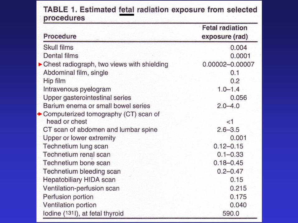
• Learn how Radiation is produced and how
much an individual receives during a CXR
• Learn the major landmarks of the CXR
• Learn to distinguish Primary and Post
primary Pulmonary TB on the CXR
• Review the appearance of TB infection in
other organ systems

Lead Shield X-ray tubeFilm
Posterior-Anterior (PA) Chest X-ray


Grant’s Atlas of Anatomy 11th Ed

F.J. Netter, Respiratory System, Ciba
Collection, 1980

F.J. Netter, Respiratory System, Ciba Collection, 1980
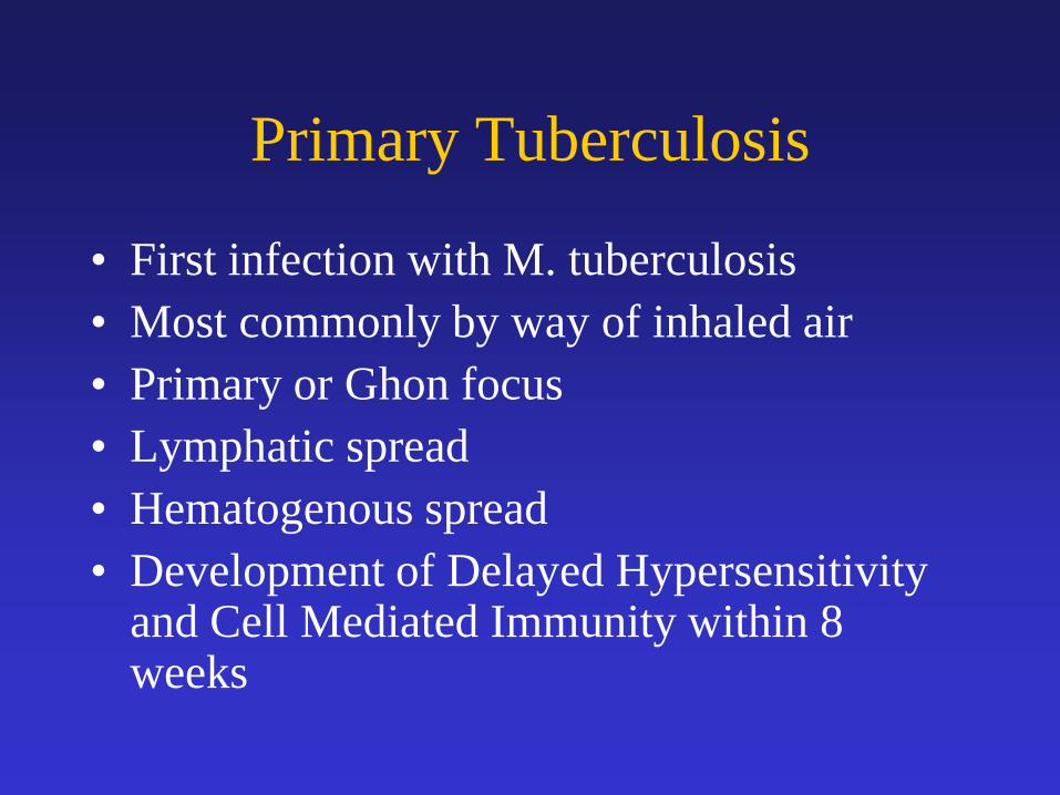
Primary Tuberculosis
• First infection with M. tuberculosis
• Most commonly by way of inhaled air
• Primary or Ghon focus
• Lymphatic spread
• Hematogenous spread
• Development of Delayed Hypersensitivity and Cell Mediated Immunity within 8 weeks

Ghon
focus
a calcified
granuloma
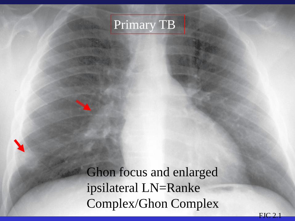
Ghon focus and enlarged
ipsilateral LN=Ranke
Complex/Ghon Complex
Primary TB
FJC 2.1

FJC 2.9a
PRIMARY TB
MEDIASTINAL AND HILAR LN

Right Hilar
LN
Subcarinal
LN
CAT Scan w Contrast
L
post

PET Scan
Positive
Emission
Tomography
Radiolabeled
Glucose

FJC 2.11PRIMARY TB, LN & INFILTRATE

FJC 2.4
PRIMARY TB
CONSOLIDATED RIGHT UPPER
LOBE, NO AIR BRONCHOGRAMS

FJC 2.12
PRIMARY TB-PLEURAL EFFUSION
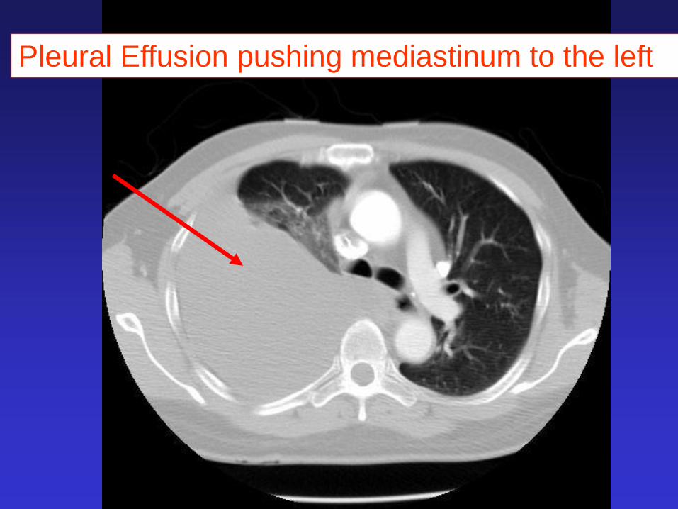
Pleural Effusion pushing mediastinum to the left

Primary Tuberculosis
in adolescents and
young adults

Post-Primary or Reactivation TB
• Reactivation of a previously dormant primary infection, though in a small number of cases, it is the extension of the primary infection if it occurs within a year of the initial infection.
• It is a disease of adolescents and adults
• It affects primarily the upper lobes with a propensity to cavitate,
• Extra pulmonary sites include the LN, Pleura, bone, CNS, GU, Peritoneum

FJC 2.13a
POST PRIMARY or REACTIVATION
TUBERCULOSIS
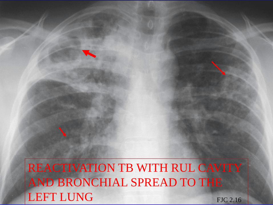
FJC 2.16
REACTIVATION TB WITH RUL CAVITY
AND BRONCHIAL SPREAD TO THE
LEFT LUNG
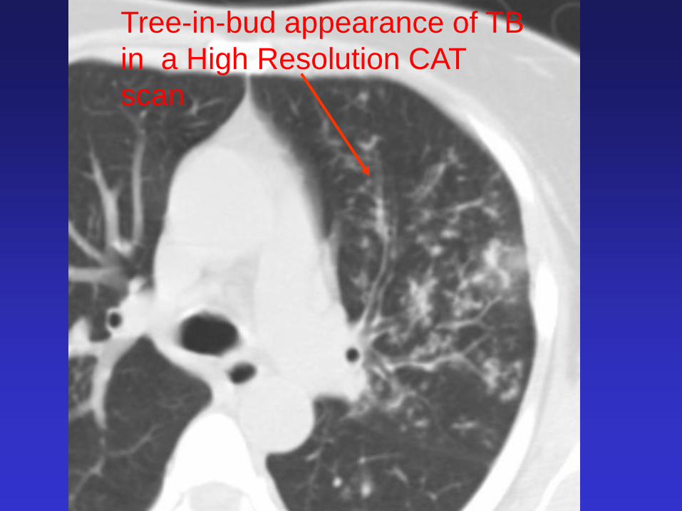
Tree-in-Bud
appearance
Tree-in-bud appearance of TB
in a High Resolution CAT
scan

Respiratory
bronchiole
Alveolar duct
Eisenhuber, Radiology 2002;222:771-72
Tree-in-Bud

Dense calcification
in Right Upper
Lobe


Post Primary Tuberculosis
Reactivation TB

Miliary Tuberculosis


Miliary Tuberculosis
CAT Scan

Millet seeds

CENTRAL NERVOUS SYSTEM

Infiltrating process
MRI

Infiltrating process with pressure
compressing the brain
edema
Tuberculosis
mass
CAT Scan

Tuberculomas in the Brain
MRI

TB Meningitis on cortex and Sylvian
fissure, MRI with contrast
R

Skeletonal TB


Anterior

Collapsed
Vertebrae
Spinal Cord
MRI
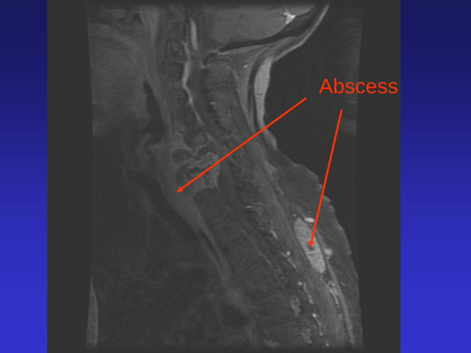
Abscess

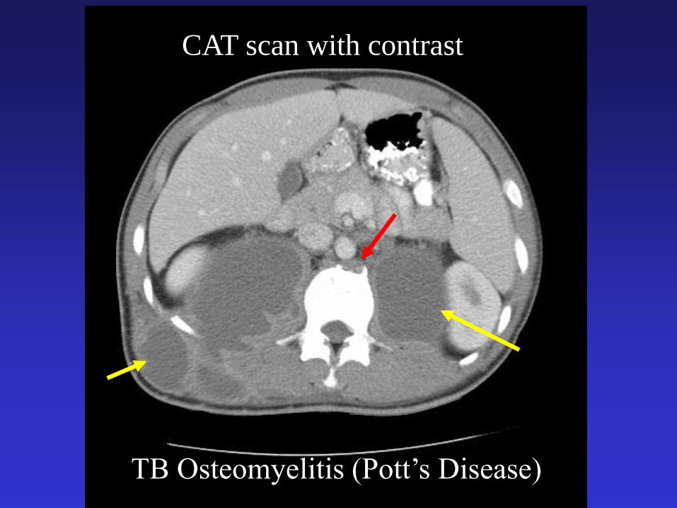
TB Osteomyelitis (Pott’s Disease)
CAT scan with contrast
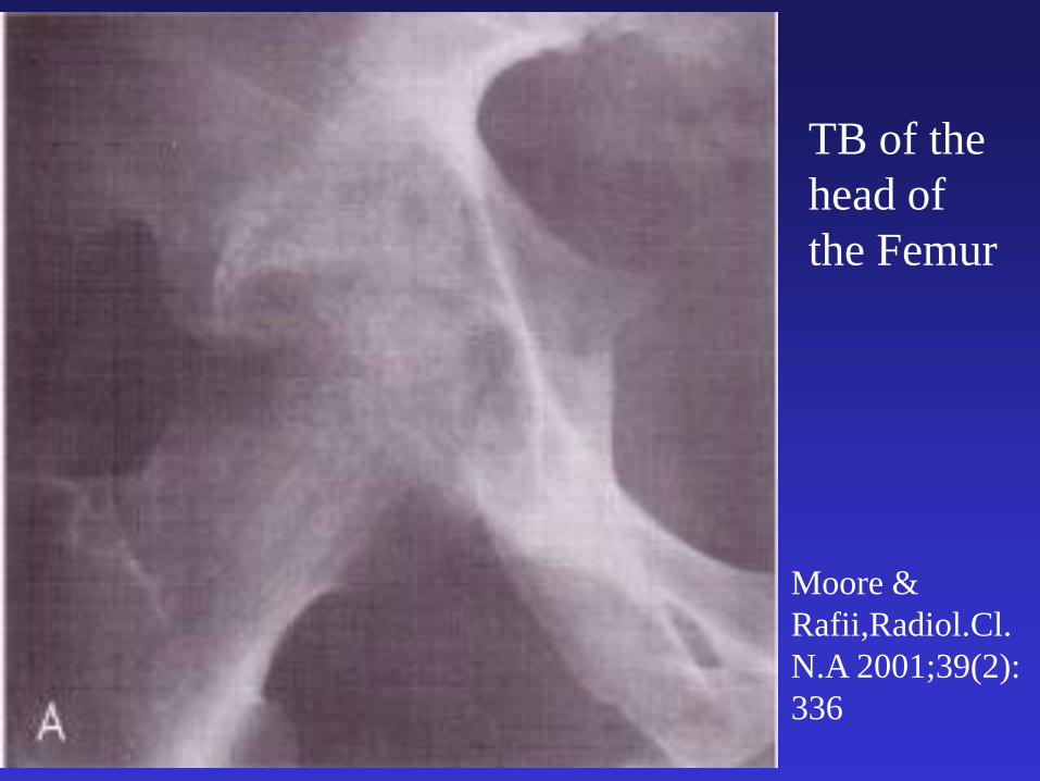
Moore &
Rafii,Radiol.Cl.
N.A 2001;39(2):
336
TB of the
head of
the Femur

TB of the
Head and
Neck of
the
Femur

Tuberculosis Arthritis of
the knee

Moore &
Rafii,Radiol.Cl
.N.A
2001;39(2):
336
TB of the
Elbow

Gastrointestinal TB
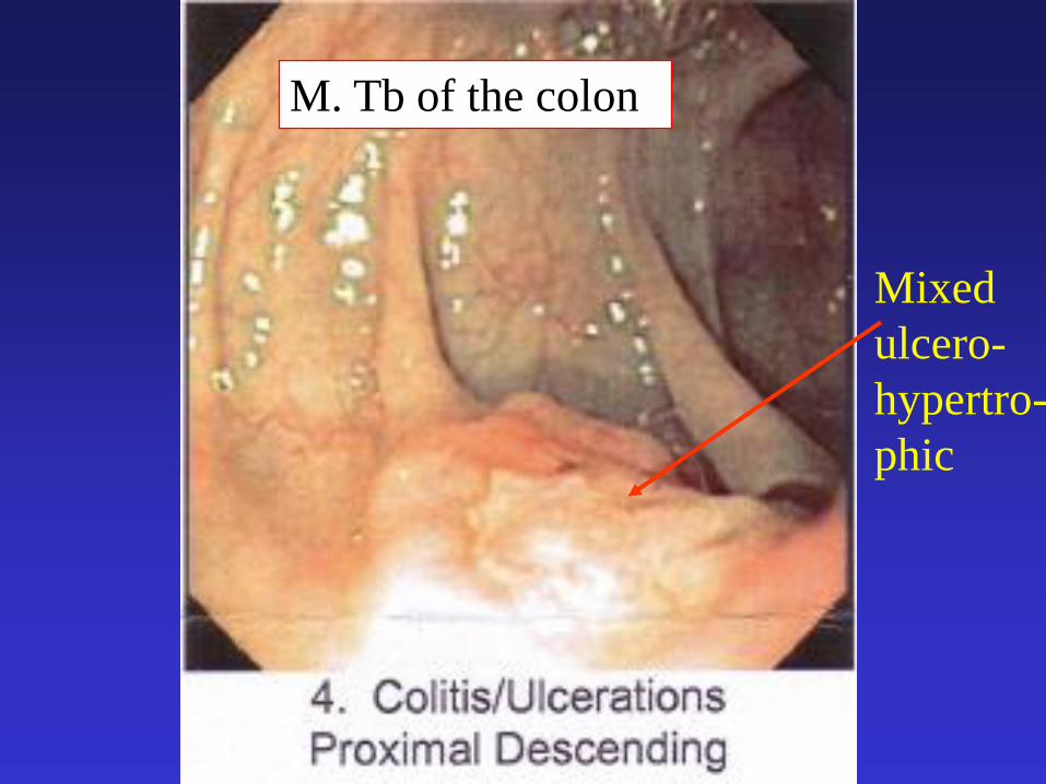
Mixed
ulcero-
hypertro-
phic
M. Tb of the colon


12-7-07
CAT Scan with contrast
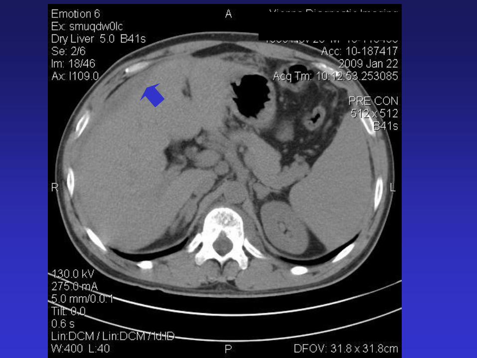
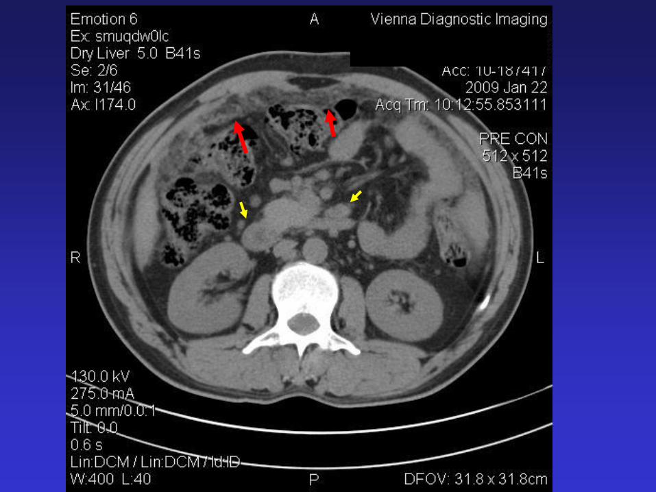

HIV +, liver and SpleenTW
CAT scan with Contrast

Peripheral Lymph Node
Tuberculosis


3-27-09
Lymph
Node

Genital-Urinary TB

Renal TB
TW
IVP

Pericardial TB

Water Bottle Heart

CAT Scan

Today’s Objectives
• Learned how Radiation is produced and how much an individual receives during a CXR
• Learned the major landmarks of the CXR
• Learned to distinguish Primary and Post primary Pulmonary TB on the CXR
• Reviewed the appearance of TB infection in other organ systems

References
• Radiographic Manifestations of Tuberculosis, 2nd
Ed., Daley et al., 2006, Francis J. Curry National Tuberculosis Center
• Diagnosis of Diseases of the Chest, Fraser & Pare’ WB Saunders, 1970
• Tuberculosis from Head to Toe, Harisinghani et al., RadioGraphics 2000; 20: 449-470
• Introduction to Chest Radiology; SB Gay et al., UVA Health Science Center, Department of Radiology, 2003
www.med-ed.virginia.edu/courses/rad/

References
• Atlas Radiologic Anatomy; L.Wicke, 7th English
Edition, Icon Learning Systems, 2004
• Blueprints Radiology; A. Uzelac and RW Davis,
Lippincott Williams & Wilkins, 2006
• Introduction to Diagnostic Imaging; G. Stimac,
WB Saunders 1992
• Getting Started in Clinical Radiology; G. Eastman,
Wald and Crossin, Thieme, 2006

Questions ???
![Pediatric TB radiographsnid]/05a...1 Pediatric TB radiographs Ann M. Loeffler, MD Curry International Tuberculosis Center Radiology Best quality frontal and lateral views of the chest](https://static.documents.pub/doc/80x56/609633be3922801af21c02d2/pediatric-tb-radiographs-nid05a-1-pediatric-tb-radiographs-ann-m-loeffler.jpg)


