Page 1
Edith Cowan University Edith Cowan University
Research Online Research Online
Theses: Doctorates and Masters Theses
2015
The regulation of human iron metabolism in hypoxia The regulation of human iron metabolism in hypoxia
Andrew Govus Edith Cowan University
Follow this and additional works at: https://ro.ecu.edu.au/theses
Part of the Sports Sciences Commons
Recommended Citation Recommended Citation Govus, A. (2015). The regulation of human iron metabolism in hypoxia. https://ro.ecu.edu.au/theses/1719
This Thesis is posted at Research Online. https://ro.ecu.edu.au/theses/1719
Page 2
Edith Cowan UniversityResearch Online
Theses: Doctorates and Masters Theses
2015
The regulation of human iron metabolism inhypoxiaAndrew GovusEdith Cowan University
This Thesis is posted at Research Online.http://ro.ecu.edu.au/theses/1719
Recommended CitationGovus, A. (2015). The regulation of human iron metabolism in hypoxia. Retrieved from http://ro.ecu.edu.au/theses/1719
Page 3
Edith Cowan University
Copyright Warning
You may print or download ONE copy of this document for the purpose
of your own research or study.
The University does not authorize you to copy, communicate or
otherwise make available electronically to any other person any
copyright material contained on this site.
You are reminded of the following:
Copyright owners are entitled to take legal action against persons who infringe their copyright.
A reproduction of material that is protected by copyright may be a
copyright infringement. Where the reproduction of such material is
done without attribution of authorship, with false attribution of
authorship or the authorship is treated in a derogatory manner,
this may be a breach of the author’s moral rights contained in Part
IX of the Copyright Act 1968 (Cth).
Courts have the power to impose a wide range of civil and criminal
sanctions for infringement of copyright, infringement of moral
rights and other offences under the Copyright Act 1968 (Cth).
Higher penalties may apply, and higher damages may be awarded,
for offences and infringements involving the conversion of material
into digital or electronic form.
Page 4
EDITH COWAN UNIVERSITY
School of Exercise & Health Science
The Regulation of Human Iron Metabolism in Hypoxia
Andrew Govus
BSc. (Hons), Grad. Dip. Ed
This thesis is submitted for the award of Doctor of Philosophy (Sports Science) from
the School of Exercise & Health Science, Faculty of Engineering & Health Science
Principal Supervisor:
Assoc/Prof. Chris Abbiss (Edith Cowan University)
Co-Supervisors:
Dr. Peter Peeling (University of Western Australia)
Dr. Laura Garvican-Lewis (University of Canberra & Australian Institute of Sport)
Prof. Chris Gore (Australian Institute of Sport)
Date of Submission:
28/08/2015
Page 5
The declaration page
is not included in this version of the thesis
Page 6
USE OF THESIS
The Use of Thesis statement is not included in this version of the thesis.
Page 7
i
ACKNOWLEDGEMENTS
On the Process...
“Philosophy starts with wonder...
Indeed it does. Wonder, not merely in the academic sense, but also in life’s bigger
questions. What do you want? How far can you go? What are you willing to do to get
there? In this respect, the PhD process has taught me many things, patience, persistence
and determination. Quite often, it takes you to your limit, or beyond, but that is the very
thing that keeps us progressing. In doing so, we learn much about our character. In
essence, the most important teaching of the PhD is thus not academic at all. The most
important lesson is perhaps not what we get, but who we become from our endeavours,
that make them worthwhile.
The acknowledgements section was the most enjoyable part of the PhD to write. Where
else can you be (almost) excused from writing in non-academic English? Consequently,
I’ll take this rare opportunity to be slightly rebellious and use the passive voice, split the
infinitive, abbreviate words using apostrophes, start sentences with the word “but” and
use the Oxford comma. More importantly, where else can you truly thank those who’ve
helped you develop both academically and interpersonally? Through this process, we
realise how far we’ve progressed in our life, the friendship we’ve made or strengthened
and what is truly important to us. Therefore, as this journey nears its end, it fills me with
nothing but gratitude for those who’ve walked this road with me.
...and, at the end, when philosophic thought has done its best, the wonder remains”
Alfred North Whitehead, Mathematician (1861-1847)
Yes, I think it does. On the other hand, perhaps a better state of confusion?
Page 8
On those who have Shared the Journey...
Supervisory Panel
A/Prof Chris Abbiss: Thank you for your supervision, patience, understanding and
guidance throughout the process. A fellow coffee lover, although I would have been
hospitalised with heart palpitations if I consumed half as much as you did! I hope we
can continue to do good things together.
Dr. Peter Peeling: You have been there from the very beginning. Thank you for
believing in me and mentoring me every step of the way, both in sport and in life.
Whether it was me falling off a treadmill at 18 km.h-1 during your own PhD, or me
trying to beat your half marathon time (that took a while), or debating why 03:00 in the
morning is definitely not the best time to go to the beach – it has been good fun. I am
truly grateful for your commitment and efforts on my behalf.
Dr. Laura Garvican-Lewis: Laura, Laura, Laura. Thank you for keeping me on track
throughout the process. You have gone from strength to strength in the four years I have
known you. When we first met, you were almost at exactly this stage, ready to submit
your PhD. Since then, you have got a PhD, run under 3:00 for a marathon, got married,
had a daughter and continued to publish extensively. I’m not sure how you fit it all in. I
think we have learnt a lot from each other during this thesis and I hope we continue to
do so.
Prof. Chris Gore: A wholehearted thank you for giving me an opportunity at the
Australian Institute of Sport as a physiology Occupational Trainee in 2011. Your
guidance has been invaluable. You set a great example everyday with your integrity,
Page 9
work ethic and attention to detail. I only hope I can emulate these qualities throughout
my career.
Edith Cowan University
Dr. Greg Haff: Doc Haff. Your guidance, friendship and mentorship have been helped
me immensely throughout the process. I know I could always count on you for a good
chat about physiology and the scientific process. You even managed to get me to lift
(albeit briefly), which is no mean feat, given my physique! I look forward to our
friendship in the years to come, we’ve work to do!
Dr. Ihsan: A trusted friend and colleague. I am very grateful for guidance and advice.
Even if you were constantly pestering me to play “wall-y” or telling why Arsenal will
win the Premier League this year. However, you were always on hand with some sage
advice or just to talking about running! I look forward to working together again in the
future.
Dr. Chris Joyce: Joyle – my fellow Englishman, even if you are from Middlesbrough.
Thanks for everything early on. Another one who has quietly achieved – PhD, job, got
married and had children...Curry and beers, mate?
Dr. Carl Woods: Carlitos, I always enjoyed your insights and friendship. You worked
very hard to be to be where you are now, so you very much deserve it. You have some
great ideas and I know you will go from strength to strength in the coming years. I hope
we can continue to share this journey.
Alan Metcalfe: Thank you for being a good friend. We’ve had a blast. So many
memories, although I think half of them are “not suitable for print”. No doubt, you’ll
retain your love of biscuits and baked beans, sometimes together in the same meal.
Page 10
Keep plugging away and moving forward. My advice to you: 1) Don’t buy any magic
beans or book any holidays without asking me first, 2) avoid German girls, 3) why are
you reading this? Don’t you have work to do? ;-).
PhD Office: So many people are part of the bigger picture here. A sincere thank you
those in the ECU PhD suite who have shared the journey with me or had to enduring my
venting (everyone) or blindly walked into my humour (Tina – every time!). You’ve
made it fun.
Australian Institute of Sport, Canberra
Lachlan Mitchell: We have already learnt so much from each other. Whenever I get an
idea, as rare as it may be these days, I always think “if only Lach was here!” Similarly,
if my R code wasn’t working (pretty much daily), I always had you to bounce some
ideas off, or to point out I’d missed a comma somewhere on line 3,567, hence why the
code won’t run. Indeed, we’ve had endless debates about everything, where of course,
we are both ALWAYS right. We still do. When all is said and done, I am happy you
could be part of this with me. Furthermore, who else would retrieve my bag from the
police station after I’ve left it in a park during a half marathon, or organise my birthday
party when I’ve managed to lock myself out of my house one hour before my birthday
party is about to start. As you know, this is normal for me, so you just sigh and say “you
idiot” but then help me out. We’ve come a long way together and I look forward to our
friendship in the years to come.
Dr. Brad Clark: My partner in crime. I don’t need to say anything to you, we’ve already
solved the world’s problems. At the end of the day, we’re not saving lives here mate.
Coffee?
Page 11
Mel Shurrey (aka Mrs. Clark): Mel, it has been an absolute pleasure getting to know
you throughout my PhD. They say behind every great man, is a great woman. That is
certainly true here. Thanks your hospitality, cooking, patience (especially when Brad
and I had taken over the lounge to play FIFA) and, most of all, your friendship.
Avish Sharma: An original A-Team member. You’ve a bright future ahead of you
Avish. I really enjoyed watching you grow both professionally and personally during
my time at the AIS. Thanks for all your help throughout data collection. We’ve had
some great times haven’t we? There are many more to come mate! #gotitdone.
Val Chan: Confusing at the best of times, but always willing to help out. You’ve
overcome a lot yourself and I’m thankful for your listening ears and occasional piece of
salacious gossip.
Dr. Philo Saunders: Philo, thanks for all your help with data collection and your
mentorship over the years. I won’t thank you for consisting taking me to pieces on
every long run and track interval session, but keep doing what you’re doing. You seem
to be getting younger with age. I’m not sure how, but it works.
Prof. David Pyne: Pyney, I have always been thankful for your guidance and insights on
all things sport. You always had time for me and steered me in the right direction. I do
recall endless hours during my time at the AIS repairing many spreadsheets and doing
all things Rugby League – a sport I am happy to admit I know absolutely nothing about
and is the antithesis of my physique. I will never forget your pearling one-liners, mostly
at AIS staff meetings but they could crop up just about anywhere. Only you can get
away with those!
Page 12
Dr. Chris Barnes: Much like the first Turning machine, somewhat of an enigma. When
we can find you. However, a man with a big heart who was always willing to provide
some statistical support and a quirky anecdote. Upon reflection, perhaps refrain from
telling your story about when you spent time in the markets in Addis Ababa...Thank you
very much for your patience and understanding.
Dr. Graeme Allbon: Absolute legend! Thank you very much for your assistance in
running bioassays for this project and general life advice. Another who know the truly
valuable things in life.
Institut für Sports-und Präventivmedizin, Saarbrücken
Dr. Sabrina Skorski: Frau Skorski. Be it in Perth, Adelaide, Canberra, Malmö, Paris, or
Saarbrücken, I’ve always enjoyed your company and our heated debates. We’ve had
some great times and I hope there are many more to come. Danke schön!
Caroline Schneider: From when we first met, to the present moment, I’ve enjoyed every
minute. Your friendship, support and happy disposition has meant a lot to me. Merci
beaucoup et j'attends avec impatience te voir encore
Murdoch University
Nathan Lawler: Half of this is yours too Nathan. Thanks for your help during to data
collection. I couldn’t have done it without you. I have an idea...;-).
Family
Mum & Dad: To you, I owe everything. You selflessly sacrifice so much on a daily
basis to make sure I’ve always had what I needed. So many times, I’ve been so grateful
to come home to Mum’s cooking, or watch the football with Dad (Chelsea of course), or
Page 13
just someone to chat to. You keep me balanced. You invested in my education to make
sure I had a better life, so this thesis is for you. I very much hope I can make the most of
the opportunities you’ve given me.
Daniel, Matthew, Luke: Boys, thank you for keeping me grounded and motivating me
to be better. That said, we all know I’m still house FIFA champion.
Arthur: If you think you can, you might. In the end I did. Rest in peace. With a decent
pint of real ale of course and with Fulham, Chelsea and Kingstonian playing good
football. Well, maybe 1/3 ain’t bad?
To the Reader...
I recommend reading this thesis with a glass of red wine (for medicinal purposes of
course!) or another suitably strong alcoholic beverage. Whilst this thesis may not be a
literary masterpiece, I hope its outcomes inform you as much as they have informed me.
Are you sitting comfortably? Then I’ll begin...
Page 14
ABSTRACT
Athletes commonly use altitude exposure in an attempt to improve their aerobic
performance at sea level. Altitude exposure enhances erythropoiesis and iron-dependent
oxidative and glycolytic enzyme production, for this reason, athletes must maintain a
healthy iron balance at altitude. A negative iron balance at altitude may limit such
physiological adaptations, potentially reducing the performance benefits of altitude
exposure.
This thesis examined the regulation of iron metabolism during acute (~31 min, Study
One) and prolonged altitude exposure (14 days, Study Two). Finally, Study Three
examined how daily oral iron supplementation influenced haemoglobin mass (Hbmass)
and iron parameter responses to prolonged, moderate altitude exposure in a large cohort
of elite athletes. Specifically, Study One found acute (~31 min) interval exercise [5 × 4
min at 90% of the maximal aerobic running velocity (vVO2max)] increased post-exercise
interleukin-6 (IL-6) production and elevated hepcidin production 3 h thereafter in both
normoxia (fraction of inspired oxygen (FIO2) = 0.2093) and normobaric hypoxia (i.e.
3,000 m simulated altitude; FIO2 = 0.1450). These results suggest exercise performed in
acute hypoxia does not alter the post-exercise hepcidin response, relative to exercise in
normoxia, possibly owing to the short duration of the hypoxic stimulus.
Prolonged altitude exposure suppresses resting hepcidin levels in sojourning
mountaineers, but its influence on the post-exercise hepcidin response exercise has not
yet been investigated. Therefore, Study Two investigated how 14 days of live high: train
low (LHTL) (exposure to 3,000 m simulated altitude for 14 h.d-1) influenced resting
levels of hepcidin, erythropoietin (EPO) and blood iron parameters. Study Two also
examined the post-exercise hepcidin and iron parameter responses to interval exercise
Page 15
(5 × 1,000 m at 90% of the maximal aerobic running velocity) performed in normoxia
(600 m natural altitude) and normobaric hypoxia (i.e. ~3,000 m simulated altitude),
following 11 and 14 days of LHTL. The post-exercise hepcidin response was compared
with interval exercise performed at a matched exercise intensity in normoxia or hypoxia
before LHTL. Here, LHTL suppressed resting hepcidin levels after two days of
exposure, but the post-exercise hepcidin response to interval exercise was similar in
normoxia and hypoxia, both before and after LHTL. Additionally, Hbmass increased by
2.2% and plasma ferritin levels decreased following LHTL. In conclusion, prolonged,
moderate altitude exposure suppresses resting hepcidin levels, which likely ensures
more iron can be transported to the erythron to support accelerated erythropoiesis.
Prolonged altitude exposure places a large burden on body iron stores because
additional iron is required to support accelerated erythropoiesis. Accordingly, athletes
often ingest oral iron supplements during altitude exposure to ensure they maintain a
healthy iron balance. By analysing ten years of haematological data collected from well-
trained athletes who undertook two-to-four weeks of LHTL at simulated (3,000 m) or
natural (1,350-2,700 m) altitudes, Study Three established how oral iron supplement
dose moderates the Hbmass, serum ferritin and transferrin saturation response to
prolonged moderate altitude exposure. In general, athletes supplemented with 105 mg.d-
1 or 210 mg.d-1 of oral iron supplement increased their Hbmass from pre-altitude levels by
3.3% and 4.0% respectively. Serum ferritin levels decreased by 33.2% in non-iron
supplemented athletes and by 13.8% in athletes supplemented with 105 mg.d-1 of oral
iron, however, those athletes who ingested 210 mg.d-1 markedly increased their iron
storage compartment by 36.8% after moderate altitude exposure. Thus, daily oral iron
supplementation at altitude assists athletes to maintain a healthy iron balance, providing
them with sufficient iron to sustain accelerated erythropoiesis.
Page 16
In conclusion, this thesis suggests exercise in acute hypoxia does not seem to alter the
post-exercise hepcidin response relative to exercise in normoxia, but prolonged altitude
exposure suppresses resting hepcidin levels and may attenuate the magnitude of post-
exercise hepcidin response after 14 days of LHTL. Finally, daily oral iron
supplementation may support iron balance and Hbmass production in athletes
undertaking prolonged moderate altitude exposure.
Page 17
TABLE OF CONTENTS
ACKNOWLEDGEMENTS ............................................................................................ I
ABSTRACT ............................................................................................................... VIII
TABLE OF CONTENTS ............................................................................................. XI
LIST OF TABLES ................................................................................................... XVII
LIST OF FIGURES ................................................................................................... XIX
LIST OF PUBLICATIONS ..................................................................................... XXII
1 CHAPTER ONE ................................................................................................... 24
1.1 Overview ................................................................................................................ 25
1.2 Background ............................................................................................................ 25
1.3 Statement of the Problem ..................................................................................... 28
1.4 Significance of the Research ................................................................................. 30
1.5 Research Aims ....................................................................................................... 31
1.5.1 Study One ........................................................................................................ 32
1.5.2 Study Two ....................................................................................................... 32
1.5.3 Study Three ..................................................................................................... 32
1.6 Research Questions & Hypotheses ...................................................................... 32
1.6.1 Study One ........................................................................................................ 32
Page 18
1.6.2 Study Two ....................................................................................................... 33
1.6.3 Study Three ..................................................................................................... 34
1.7 Definition of Terms ............................................................................................... 36
2 CHAPTER TWO .................................................................................................. 38
2.1 Introduction ........................................................................................................... 39
2.2 Hypoxic Exposure Methods ................................................................................. 41
2.2.1 Acute Hypoxic Exposure Methods ................................................................. 41
2.2.2 Prolonged Hypoxic Exposure Methods .......................................................... 45
2.2.3 Summary ......................................................................................................... 51
2.3 The Regulation of Iron Metabolism in Normoxia .............................................. 51
2.3.1 Iron – Biological Functions & Distribution .................................................... 52
2.3.2 Regulation of Systemic Iron Balance .............................................................. 52
2.3.3 Regulation of Intracellular Iron Balance ......................................................... 56
2.3.4 Summary ......................................................................................................... 57
2.4 Daily Iron Turnover .............................................................................................. 57
2.5 Iron Deficiency in Athletes ................................................................................... 59
2.5.1 Definition ........................................................................................................ 59
2.5.2 Prevalence ....................................................................................................... 62
2.5.3 Influence on Athletic Performance ................................................................. 63
2.5.4 The Hepcidin Hypothesis ................................................................................ 65
2.5.5 Summary ......................................................................................................... 67
2.6 The Regulation of Iron Metabolism in Hypoxia ................................................ 67
Page 19
2.6.1 Acute Hypoxic Exposure ................................................................................ 70
2.6.2 Prolonged Hypoxic Exposure ......................................................................... 74
2.6.3 Summary ......................................................................................................... 81
2.7 Maintaining Iron Balance at Altitude ................................................................. 82
2.7.1 Pre-Altitude Blood Screening ......................................................................... 82
2.7.2 Dietary Considerations .................................................................................... 84
2.7.3 Iron Supplementation ...................................................................................... 85
2.7.4 Summary ......................................................................................................... 91
2.8 Conclusion .............................................................................................................. 92
3 CHAPTER THREE .............................................................................................. 94
3.1 Abstract .................................................................................................................. 95
3.2 Introduction ........................................................................................................... 96
3.3 Methods .................................................................................................................. 97
3.3.1 Participants ...................................................................................................... 97
3.3.2 Experimental Design ....................................................................................... 98
3.3.3 Experimental Procedures ................................................................................ 98
3.3.4 Statistical Analysis ........................................................................................ 101
3.4 Results .................................................................................................................. 102
3.4.1 Interval Running Session .............................................................................. 102
3.4.2 Interleukin-6 .................................................................................................. 103
3.4.3 Hepcidin ........................................................................................................ 103
3.4.4 Iron Parameters ............................................................................................. 104
Page 20
3.5 Discussion ............................................................................................................. 106
3.5.1 Iron Parameters ............................................................................................. 109
3.6 Conclusion ............................................................................................................ 110
4 CHAPTER FOUR ............................................................................................... 111
4.1 Abstract ................................................................................................................ 112
4.2 Introduction ......................................................................................................... 113
4.3 Methods ................................................................................................................ 115
4.3.1 Participants .................................................................................................... 115
4.3.2 Experimental Design ..................................................................................... 115
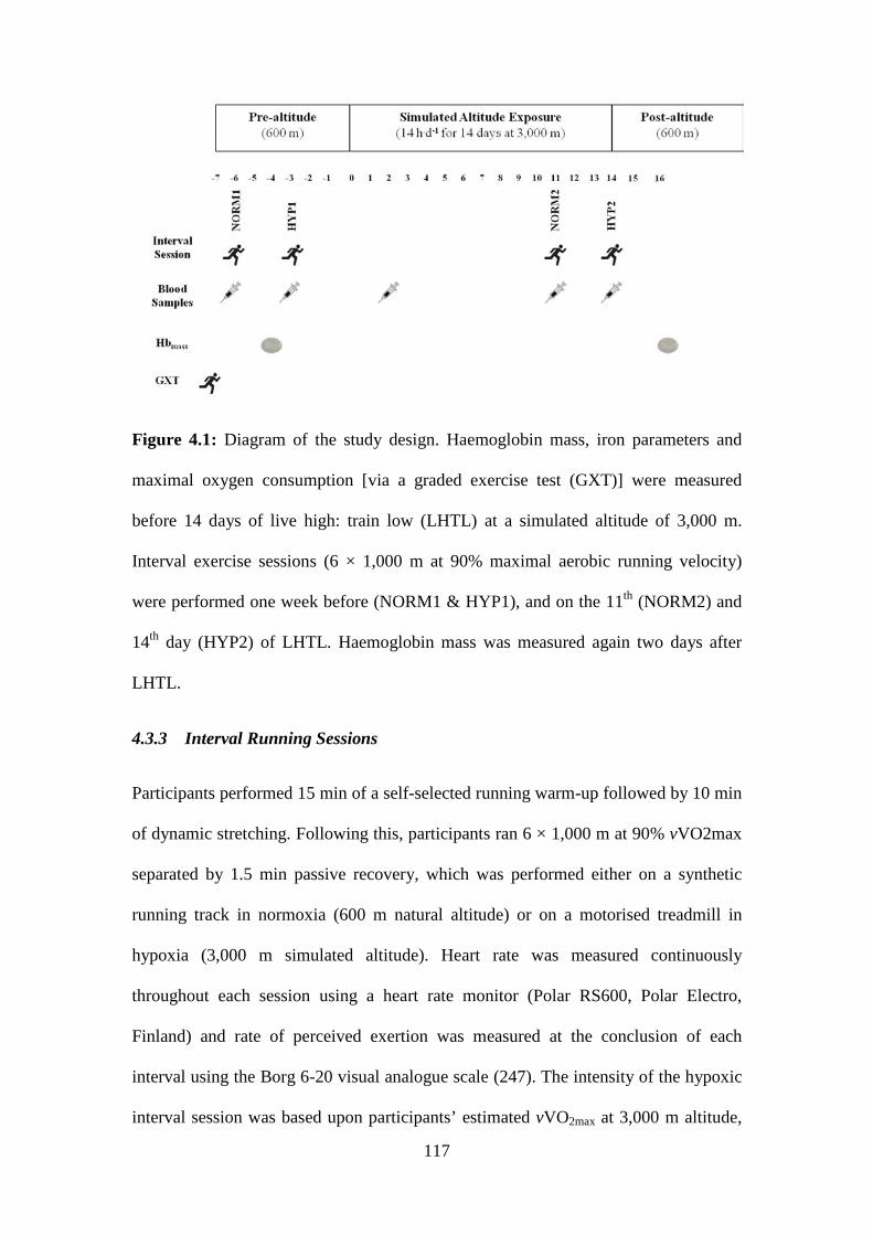
4.3.3 Interval Running Sessions ............................................................................. 117
4.3.4 Haemoglobin Mass ....................................................................................... 118
4.3.5 Venous Blood Samples ................................................................................. 118
4.3.6 Laboratory Analyses ..................................................................................... 119
4.3.7 Statistical Analysis ........................................................................................ 120
4.4 Results .................................................................................................................. 121
4.4.1 Environmental Conditions & Physiological Responses to Interval Exercise 121
4.4.2 Hepcidin Response ........................................................................................ 121
4.4.3 Iron Parameter Responses ............................................................................. 124
4.4.4 Red Cell Parameter & Haemoglobin Mass Response ................................... 126
4.5 Discussion ............................................................................................................. 127
4.5.1 Hepcidin Response ........................................................................................ 127
4.5.2 Iron Parameter Responses ............................................................................. 129
Page 21
4.5.3 Red Cell Parameter & Haemoglobin Mass Responses ................................. 130
4.6 Limitations ........................................................................................................... 131
4.7 Conclusion ............................................................................................................ 132
5 CHAPTER FIVE ................................................................................................. 134
5.1 Abstract ................................................................................................................ 135
5.2 Introduction ......................................................................................................... 136
5.3 Methods ................................................................................................................ 138
5.3.1 Ethics Statement ............................................................................................ 138
5.3.2 Participants .................................................................................................... 138
5.3.3 Altitude Exposure.......................................................................................... 139
5.3.4 Iron Supplementation .................................................................................... 139
5.3.5 Haemoglobin Mass ....................................................................................... 140
5.3.6 Iron Parameters ............................................................................................. 140
5.3.7 Total Iron Incorporation ................................................................................ 141
5.3.8 Statistics ........................................................................................................ 141
5.4 Results .................................................................................................................. 142
5.4.1 Haemoglobin Mass and Iron Parameter Responses ...................................... 142
5.4.2 Total Iron Incorporation ................................................................................ 143
5.5 Discussion ............................................................................................................. 144
5.5.1 Haemoglobin Mass Response to Moderate Altitude ..................................... 145
5.5.2 Total Iron Incorporation & Iron Parameter Responses ................................. 146
5.5.3 Serum Ferritin Threshold for Iron-Supplementation at Moderate Altitude .. 147
Page 22
5.6 Limitations ........................................................................................................... 149
5.7 Conclusion ............................................................................................................ 150
6 CHAPTER SIX ................................................................................................... 151
6.1 Thesis Summary .................................................................................................. 152
6.1.1 Iron Regulation during Acute and Prolonged Hypoxic Exposure ................ 152
6.1.2 Maintaining Iron Balance during Prolonged Hypoxic Exposure .................. 156
6.2 Practical Implications ......................................................................................... 158
6.3 Limitations ........................................................................................................... 160
6.4 Directions for Future Research.......................................................................... 161
6.4.1 Practical Directions ....................................................................................... 161
6.4.2 Theoretical Directions ................................................................................... 163
6.5 Conclusion ............................................................................................................ 165
7 REFERENCES .................................................................................................... 168
8 APPENDICES ..................................................................................................... 198
Page 23
LIST OF TABLES
Table 2.1: The three stages of iron deficiency in athletes. Adapted from Peeling et al.
(1).
Table 3.1: Response of common physiological variables to a 5 × 4 min interval running
session at 90% maximal aerobic running velocity (vVO2peak) in normoxia (FIO2 =
0.2093) and normobaric hypoxia (FIO2 = 0.1450).
Table 3.2: Interleukin-6 and hepcidin response to a 5 × 4 min interval running session
at 90% maximal aerobic running velocity performed in normoxia (FIO2 = 0.2093) and
normobaric hypoxia (FIO2 = 0.1450) conditions. Cohen’s d effect sizes are expressed
with 90% confidence limits.
Table 3.3: Response of iron parameters to a 5 × 4 min interval running session at 90%
maximal aerobic running velocity in normoxia (FIO2 = 0.2093) and normobaric hypoxia
(FIO2 = 0.1450).
Table 4.1: Environmental conditions, heart rate and perceptual response to 6 × 1,000 m
interval session at 90% maximal aerobic running velocity performed in normoxia
(NORM, n = 10) and hypoxia (simulated, normobaric altitude of 3,000 m, HYP, n = 9)
before and after 14 days of live-high: train-low.
Table 4.2: Iron parameter response to interval exercise (6 × 1,000 m at 90% maximal
aerobic running velocity) performed in normoxia (600 m natural altitude, n = 10) and
normobaric hypoxia (3,000 m simulated normobaric altitude, n = 9) both before and
during 14 days of live high: train low at 3,000 m simulated, normobaric altitude.
Table 4.3: Red blood cell parameters before and after 14 days of live high: train low at
3,000 m simulated, normobaric altitude (n = 10).
Page 24
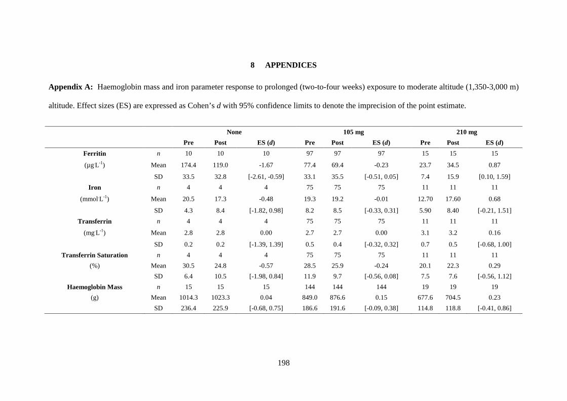
Table 5.1: Parameter estimates (Est.) with 95% confidence limits (CL) for the changes
(Δ) in Hbmass, ferritin and transferrin saturation (TSAT) during prolonged moderate
altitude exposure, when controlled for oral iron supplement dose.
Page 25
LIST OF FIGURES
Figure 2.1: Known regulators of hepcidin expression. Yellow shapes indicate known
suppressors of hepcidin expression. Blue shapes indicate known promoters of hepcidin
expression. The body’s iron stores (stores regulator) may either promote or suppress
hepcidin expression depending on the size of the iron storage compartment and is thus
shaded in both blue and yellow. Green shapes indicate emerging regulators of hepcidin
expression. IL-6: Interleukin-6, PDGF-BB: Platelet derived growth factor-BB, Ras-
RAF: Rat sarcoma-Rapidly accelerated fibrosarcoma pathway (part of the mitogen
activated protein kinase pathway), mTOR: Mammalian target of rapamycin.
Figure 2.2: Daily iron turnover and iron balance in the body. RBC: Red blood cells; RE
system: Reticuloendothelial system (or mononuclear phagocyte system).
Figure 2.3: The regulation of iron metabolism by the hypoxia inducible factor (HIF)
and erythropoietin (EPO) in response to hypoxia. The hypoxic-stabilisation of HIF-2α
stimulates increased EPO production in the kidney (REPC: renal EPO producing cells)
and liver, in turn up-regulating erythropoiesis. Iron availability increases to support
accelerated erythropoiesis in hypoxia requiring several changes in iron protein
expression. In the gut, duodenal cytochrome B (DctyB) reduces Fe3+ to Fe2+, which is
then transported to the enterocyte via divalent metal transporter-1 (DMT-1). Iron
absorbed by the gut or contained in reticuloendothelial macrophages are exported into
blood plasma via ferroportin (FPN), forms a complex with transferrin (Tf) and is then
transported to erythron and other body organs. Simultaneously, EPO acts upon erythroid
progenitor cells, stimulating the release of erythroferrone (ERFE), which suppresses
hepcidin expression. Hepcidin suppression reduces FPN degradation, increasing iron
export from reticuloendothelial macrophages, enterocytes and hepatocytes. DcytB,
Page 26
DMT-1, FPN expression are HIF-2α mediated, Tf is HIF-1α mediated. Figure adapted
from Haase (2).
Figure 4.1: Diagram of the study design. Haemoglobin mass, iron parameters and
maximal oxygen consumption [via a graded exercise test (GXT)] were measured before
14 days of LHTL at a simulated altitude of 3,000 m. Interval exercise sessions (6 ×
1,000 m at 90% maximal aerobic running velocity) were performed one week before
(NORM1 & HYP1), and on the 11th (NORM2) and 14th day (HYP2) of LHTL.
Haemoglobin mass was measured again two days after LHTL.
Figure 4.2: Pre- and 3 h post-exercise hepcidin response to a standardised 6 × 1,000 m
interval session at 90% maximal aerobic running velocity performed in (A) normoxia
(600 m natural altitude) before and on the 11th day of LHTL and, (B) hypoxia
(simulated altitude of 3,000 m) before and on the 14th day of live high: train low
(LHTL).
Figure 5.1: Iron incorporation into the iron storage (SII) and red cell compartment (HII)
for each category of iron supplement dose [0 mg (n = 15), 105 mg.d-1 (n = 144), 210
mg.d-1 (n = 19)]. Overall, iron supplemented athletes had greater total iron incorporation
(TII) compared with non-iron supplemented athletes.
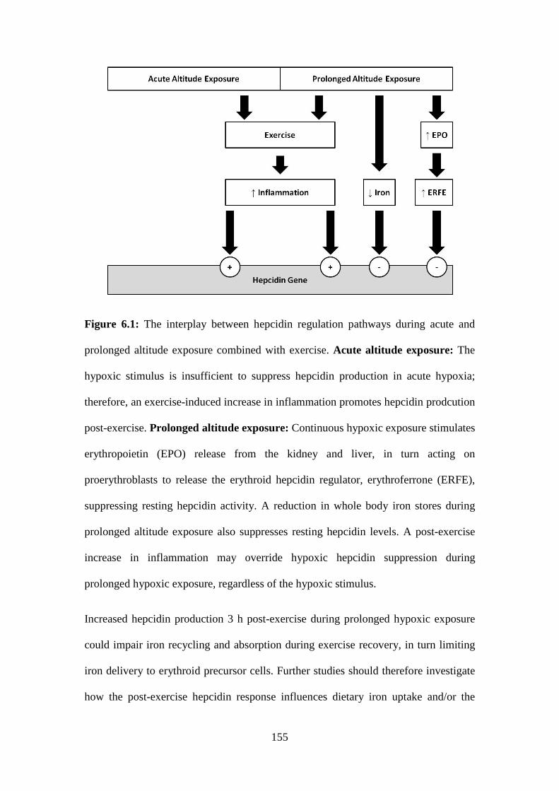
Figure 6.1: The interplay between hepcidin regulation pathways during acute and
prolonged altitude exposure combined with exercise. Acute altitude exposure: The
hypoxic stimulus is insufficient to suppress hepcidin levels in acute hypoxia; therefore,
an exercise-induced increase in inflammation promotes hepcidin production post-
exercise. Prolonged altitude exposure: Continuous hypoxic exposure stimulates
erythropoietin (EPO) release from the kidney and liver, in turn acting on
proerythroblasts to release the erythroid hepcidin regulator, erythroferrone (ERFE),
Page 27
suppressing resting hepcidin levels. A reduction in whole body iron stores during
prolonged altitude exposure also suppresses resting hepcidin production. A post-
exercise increase in inflammation may override hypoxic hepcidin suppression during
prolonged hypoxic exposure, regardless of the hypoxic stimulus.
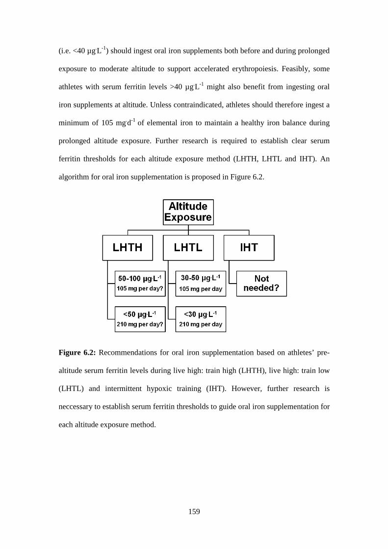
Figure 6.2: Recommendations for daily oral iron supplement dose based on athletes’
pre-altitude serum ferritin levels during live high: train high (LHTH), live high: train
low (LHTL) and intermittent hypoxic training (IHT). However, further research is
neccessary to establish serum ferritin thresholds to guide oral iron supplementation for
each altitude exposure method.
Page 28
LIST OF PUBLICATIONS
The following chapters have been published in, or submitted to, academic journals for
peer-review:
Chapter Three
Govus, A. D., Abbiss, C. R., Garvican-Lewis, L. A., Swinkels, D. W., Laarakkers, C.
M., Gore, C. J., & Peeling, P. (2014). Acute hypoxic exercise does not alter post-
exercise iron metabolism in moderately trained endurance athletes. European Journal of
Applied Physiology, 114(10): 2183-2191. Impact factor: [2.3].
Chapter Four
Govus, A. D., Peeling, P, Abbiss, C. R., Lawler, N.G., Thompson, K., Peiffer, J.J.,
Swinkels, D. W., Laarakkers, C. M., Gore, C. J., Garvican-Lewis, L. A., (2015). Live
high: train low - influence on resting and post-exercise hepcidin levels (In review:
Scandinavian Journal of Medicine and Science in Sport, Impact Factor [3.1]).
Chapter Five
Govus, A. D., Garvican-Lewis, L. A., Abbiss, C. R., Peeling, P., Gore, C.J. (2015) Pre-
altitude serum ferritin levels and daily oral iron supplement dose mediate iron parameter
and hemoglobin mass responses to altitude exposure. PLoS One. 10(8):e0135120.
Impact factor: [3.5]
Page 29
Additional Publications Related to this Thesis:
Peeling, P., Sim, M., Badenhorst, C.E., Dawson, B.T., Govus, A.D., Abbiss, C.R.,
Swinkels, D.W., Trinder, D. (2014) Iron status and the acute post-exercise hepcidin
response in athletes PLoS One 9(3): e93002. Impact factor [3.5]
Conference Presentations:
Govus, A.D., Peeling, P., Abbiss, C.R., Lawler, N., Swinkels, D.W., Laarrakkers, C.M.,
Thompson. K.G., Peiffer, J., Gore, C.J., Garvican-Lewis, L.A. (2015) Fourteen days of
live high: train low altitude exposure does not alter the post-exercise hepcidin response.
20th Annual Congress of the European College of Sports Science. Malmö, Sweden
Competitive Research Grants ($25,556):
Govus, A.D., Abbiss, C.R., Peeling, P., Garvican, L.A., Gore, C.J. (2013) Iron
regulation at altitude: and investigation into the influence of acute exposure, chronic
responses and best practice iron supplementation. ECU Higher Degrees by Research –
Mobility Grant ($5,000).
Garvican-Lewis, L.A., Govus, A.D., Abbiss, C.R., Peeling, P., Gore, C.J. (2013) The
effect of acute hypoxic training on post-exercise iron status, inflammatory cytokine and
hepcidin levels in well-trained male endurance runners. Australian Institute of Sport –
High Performance Sports Research Fund ($20,556).
Page 30
24
1 CHAPTER ONE
INTRODUCTION
Page 31
25
1.1 Overview
This doctoral thesis contains three studies investigating iron metabolism during either
acute or prolonged hypoxia. Specifically, the first two studies (Study One and Two) of
this thesis examined the regulation of iron metabolism following exercise performed
in normoxia and hypoxia during acute (~31 min, Study One) and prolonged (two
weeks, Study Two) hypoxic exposure. Finally, the third study analysed the effect of
oral iron supplement dose on the haemoglobin mass (Hbmass) and iron parameter
responses to prolonged, moderate altitude exposure in a large cohort of well-trained
athletes.
1.2 Background
Endurance athletes use two main methods of hypoxic exposure to enhance their
aerobic performance: 1) acute hypoxic exposure, which involves intermittent
exposure to, or exercise in, hypoxia several times per week, and 2) prolonged hypoxic
exposure, consisting of several weeks exposure to natural or simulated, moderate, or
high altitudes (3). Acute hypoxic exposure (<48 h) is considered too short to stimulate
erythropoiesis, but may instead improve aerobic performance by enhancing oxidative
and glycolytic enzyme concentrations (4). In contrast, given a sufficient hypoxic dose
(i.e. altitude duration × elevation) (5), moderate altitude (i.e. >2,200 m) enhances
Hbmass by ~1% per 100 h of exposure (6). In addition, two-to-four weeks of moderate
altitude exposure improves exercise economy, skeletal muscle buffer capacity,
oxidative and glycolytic enzyme concentrations, in turn enhancing aerobic and
anaerobic metabolism (7).
Haematological adaptations to prolonged altitude exposure are highly variable and are
influenced by factors such as training volume, nutrition, illness, injury and pre-
Page 32
26
altitude iron stores (8). Sufficient iron stores are required to support a three-to-five-
fold increase in erythropoiesis as well as increased iron-dependent oxidative enzyme
production at altitude (9). Athletes who are unable to maintain a healthy iron balance
at altitude may develop a functional iron deficiency and form microcytic and/or
hypochromic red cells, thus blunting the haematological benefits of altitude exposure
(10). Additionally, low iron availability may compromise the synthesis of several
iron-dependent non-haem proteins (11). Maintaining a healthy iron balance is
therefore extremely important in supporting the hypoxic-mediated haematological and
non-haematological adaptations to prolonged altitude exposure.
In addition to low pre-altitude iron stores, erythropoiesis may be compromised if iron
is not rapidly mobilised from the labile iron pool to meet erythroid iron demands
during initial altitude adaptation (12). The body’s master iron regulatory hormone,
hepcidin, regulates iron mobilisation from the labile iron pool (consisting mostly of
iron contained in reticuloendothelial macrophages) (13). Hepcidin is a 25 amino-acid
peptide hormone synthesised by liver hepatocytes that regulates systemic iron
availability by internalising and degrading ferroportin iron export channels located on
the cellular surface of iron storage cells such as reticuloendothelial macrophages and
hepatocytes (14). Elevated hepcidin levels reduce iron efflux from storage cells into
the blood plasma (15) consequently limiting iron delivery to the erythron. An increase
in the inflammatory myokine, interleukin-6 (IL-6), immediately post-exercise has
recently been shown to up-regulate hepcidin 3-6 h thereafter (16). Exercise intensity
(17), duration (18) and pre-exercise serum ferritin levels (19) also moderate the post-
exercise hepcidin response. Increased hepcidin levels post-exercise may impair
intestinal iron absorption and iron recycling from senescent red blood cells, thereby
transiently reducing plasma iron availability. In the long-term, such post-exercise
Page 33
27
elevations in hepcidin may require athletes to mobilise iron from storage cells to
maintain daily iron balance, which could reduce an athlete’s iron stores if daily iron
losses are not replenished.
Exercise during acute and prolonged hypoxic exposure presents two opposing
mechanisms of hepcidin regulation. Whilst hypoxic exposure and exercise both
increase IL-6 production (20), increased erythropoietin (EPO) production at high
altitude suppresses hepcidin levels within 24-48 h (21-23). Hepcidin suppression is a
favourable response to early altitude exposure since it enhances intestinal iron
absorption and promotes the mobilisation of iron from storage sites to the blood
plasma to support accelerated erythropoiesis. Conversely, since free (unbound) iron
catalyses reactive oxygen and nitrogen specie formation (24), increased post-exercise
hepcidin production is thought to protect the body from oxidative damage by
sequestering iron in reticuloendothelial macrophages, thereby transiently reducing
iron available to catalyse the formation of free oxygen and nitrogen radicals via the
Fenton-Haber-Weiss reaction (25). To date, hepcidin’s contribution to post-exercise
iron metabolism during acute or prolonged hypoxic exposure has not been
investigated. The dominant hepcidin regulatory pathway during either acute or
prolonged hypoxic exposure protocols is therefore unknown. Further investigation
into the hormonal control of post-exercise iron metabolism in acute and prolonged
hypoxia may thus help athletes to maintain a healthy iron balance when using hypoxia
as a complementary training method.
Oral iron supplementation may help athletes to maintain a healthy iron balance when
undertaking prolonged, moderate altitude exposure. To date, no clear oral iron
supplementation guidelines exist for athletes planning to undertake either acute or
prolonged altitude exposure. Generally, oral iron supplements are prescribed to ensure
Page 34
28
athletes have sufficient iron stores to sustain the three- to five-fold increase in
erythropoiesis associated with prolonged altitude exposure (9) and are especially
important in individuals with low pre-altitude serum ferritin levels (i.e. <35 µg.L-1).
Oral iron supplementation, however, may not be required in those athletes with
otherwise healthy pre-altitude serum ferritin levels (i.e. >35 µg.L-1), or if the hypoxic
dose is low [for example, short sojourns (2 weeks or less) to low or moderate altitude
(12)]. Furthermore, the optimal oral iron supplement dose necessary to support
erythropoiesis during moderate altitude exposure is highly individual and iron uptake
is influenced by an individual’s current iron status and/or their ferrokinetics (26). Yet,
despite the widespread use of hypoxic exposure methods, it is unknown how oral iron
supplement dose influences Hbmass production in athletes undertaking prolonged
altitude exposure.
1.3 Statement of the Problem
Low pre-altitude iron stores and/or an inability to rapidly mobilise iron from storage
sites to service erythropoiesis may blunt the haematological adaptations associated
with prolonged altitude exposure (12). Hypothetically, an exercise-induced increase in
hepcidin expression during the post-exercise recovery period could also compromise
haem synthesis at altitude by limiting the transport of iron from storage cells to the
bone marrow. Furthermore, elevated post-exercise hepcidin levels at altitude may
transiently impair intestinal iron absorption during exercise recovery, thereby
reducing plasma iron availability and iron incorporation into storage cells.
Consequently, athletes may be unable to replenish their iron stores via dietary means,
and thus become reliant on iron mobilised from the labile iron storage pool and
hepatocytes to maintain iron status, further reducing their iron stores.
Page 35
29
Exercise in hypoxia may augment post-exercise IL-6 production since exercise and
hypoxia both promote IL-6 production (27). Theoretically, increased post-exercise IL-
6 production following exercise in acute hypoxia could augment the magnitude of the
post-exercise hepcidin response compared with similar exercise in normoxia.
Practically, augmented post-exercise hepcidin production after exercise in hypoxia
could make it difficult for athletes to maintain a healthy iron balance when using
acute hypoxic exposure protocols since iron availability may be reduced.
Alternatively, acute hypoxia may alternatively attenuate the post-exercise hepcidin
response. Study One of this thesis therefore investigated how interval exercise
performed in acute hypoxia influenced IL-6, hepcidin and iron parameter responses
both immediately post-exercise and 3 h post-exercise in well-trained endurance
athletes.
In contrast to acute hypoxic exposure, prolonged altitude exposure (>1 week)
stimulates a three-to-five-fold increase in erythropoiesis (9). Insufficient iron delivery
to the bone marrow during prolonged altitude exposure may therefore limit the
maturation of erythroid precursor cells into erythrocytes. High altitude exposure
suppresses hepcidin production within 24-48 h of (21-23) and likely helps to improve
iron availability for erythropoiesis by promoting iron mobilisation from the labile iron
pool. In comparison, increased post-exercise hepcidin production at altitude may
reduce intestinal iron uptake and iron recycling from reticuloendothelial macrophages,
potentially limiting iron available for haem synthesis. The post-exercise hepcidin and
iron responses during prolonged altitude exposure, however, are currently unknown.
To address this question, Study Two investigated how live high: train low (LHTL)
influenced resting hepcidin, erythropoietin (EPO) and iron parameters levels after two
and 14 days exposure to normobaric hypoxia (14 h.d-1 at 3,000 m simulated altitude).
Page 36
30
Additionally, Study Two compared the post-exercise hepcidin and iron parameter
response to a standardised interval running session performed in normoxia and
simulated hypoxia both before and after two weeks of LHTL.
Athletes require sufficient iron stores to support accelerated erythropoiesis during
prolonged altitude exposure, but may be unable to provide sufficient iron to synthesise
haemoglobin if pre-altitude iron stores are too low (10), iron intake is inadequate, or
iron transport to the erythron is insufficient (12). Athletes are accordingly prescribed
oral iron supplements several weeks before, and during, prolonged altitude exposure
to raise their iron stores to cope with accelerated erythropoiesis. The influence of
different oral iron supplement doses on Hbmass production and iron parameter
responses, however, has not yet been investigated in a large cohort of well-trained
athletes. Furthermore, a better understanding of how different oral iron supplement
doses influence Hbmass and iron parameter response to prolonged altitude exposure
may allow sport scientists to develop altitude-specific iron supplementation guidelines
for athletes planning to undertake moderate altitude exposure, in turn ensuring
athletes can better maintain a healthy iron balance at altitude.
1.4 Significance of the Research
This thesis aimed to further the current understanding of how athletes regulate iron
metabolism following interval exercise performed during acute and prolonged
hypoxic exposure. Specifically, the findings of this thesis could enable medical
practitioners, sports physiologists and dieticians to refine the current guidelines for
dietary iron intake and oral iron supplementation relevant to acute and prolonged
altitude exposure, which may in turn enable athletes to better regulate their iron
balance when undertaking altitude exposure. Additionally, improving iron availability
Page 37
31
by providing oral iron supplements during prolonged altitude exposure could reduce
an athlete’s risk of developing a functional iron deficiency by ensuring their pre-
altitude iron stores can support accelerated erythropoiesis without depleting the
body’s iron storage pools. Finally, maintaining a healthy iron balance at altitude may
ensure sufficient iron is available to support iron-dependent, non-haematological
adaptations to altitude, potentially enhancing aerobic performance in normoxia or
hypoxia.
Occupations requiring workers to perform physical and cognitive tasks at high
altitude, such as military personnel, may also be interested in the outcomes of this
thesis. Maintaining a healthy iron balance at altitude may assist these individuals to
adapt to high altitudes more effectively since low serum ferritin levels are associated
with reduced work capacity and cognitive ability (28). Furthermore, iron
supplementation at high altitude may protect against acute altitude sickness (29).
1.5 Research Aims
This thesis aimed to investigate how acute (single session) and prolonged (two weeks)
altitude exposure (~3,000 m simulated altitude) affects the body’s master iron
regulatory hormone, hepcidin, and to determine its influence on iron metabolism in
well-trained endurance athletes undertaking different forms of altitude exposure. This
thesis also aimed to determine how oral iron supplement dose moderated the Hbmass
and iron parameter responses during two-to-four weeks of altitude exposure in well-
trained athletes.
Page 38
32
Specifically, the studies presented in this thesis aimed to investigate:
1.5.1 Study One
• The post-exercise inflammatory, hepcidin and iron parameter response to
acute exercise in hypoxia (3,000 m simulated altitude).
1.5.2 Study Two
• The influence of 14 days of LHTL on resting and post-exercise hepcidin and
iron parameter levels.
1.5.3 Study Three
• The influence of oral iron supplement dose on Hbmass and iron parameter
responses in well-trained athletes during prolonged (two-to-four weeks),
moderate altitude exposure.
1.6 Research Questions & Hypotheses
The research question (denoted “Q”) and corresponding hypotheses (denoted “H”) for
each study of this thesis are outlined below:
1.6.1 Study One
Acute hypoxic exercise does not alter post-exercise iron metabolism in well-
trained endurance athletes
Q1: How does an interval running session (5 × 4 min at 90% vVO2max, separated by
1.5 min passive recovery), performed in hypoxia [3,000 m simulated altitude (FIO2 =
0.1408)] influence iron parameter [serum iron, serum ferritin, soluble transferrin
receptor (sTfR)], inflammatory cytokine (IL-6) and hepcidin levels during exercise
Page 39
33
recovery (immediately post-exercise and 3 h post-exercise) compared with an
equivalent session performed in normoxia (FIO2 = 0.2093)?
H1: Compared with interval exercise in normoxia, interval exercise in hypoxia will
result in:
a) higher IL-6 levels immediately post-exercise;
b) suppressed hepcidin levels 3 h post-exercise, independent of a post-exercise rise in
IL-6;
1.6.2 Study Two
Live high: train low - Influence on resting and post-exercise hepcidin levels
Q1: How does two nights of LHTL at 3,000 m simulated altitude affect resting
hepcidin and erythropoietin levels in well-trained distance runners?
H1: Compared with baseline levels, two nights of LHTL will:
a) suppress resting hepcidin levels;
b) elevate resting erythropoietin levels.
Q2: How does 14 days of LHTL affect resting hepcidin, iron parameter (plasma iron,
plasma ferritin, plasma transferrin, plasma transferrin saturation) and Hbmass levels in
well-trained distance runners?
H2: Compared with baseline levels, 14 days of LHTL will:
a) suppress resting hepcidin levels;
b) decrease resting ferritin levels;
Page 40
34
c) increase Hbmass from baseline levels.
Q3: How does the 3 h post-exercise hepcidin and iron parameter response differ after
interval exercise (5 × 1,000 m at 90% vVO2max, 1.5 min passive recovery) performed
in normoxia (600 m natural altitude) and normobaric hypoxia (3,000 simulated
altitude) on the 11th and 14th day of LHTL compared with post-exercise responses to
equivalent exercise performed before LHTL?
H3: Compared with interval exercise performed before LHTL, interval exercise
performed in normoxia and hypoxia on the 11th and 14th day of LHTL will:
a) attenuate hepcidin levels 3 h post-exercise;
b) result in a comparable iron parameter response between the two exercise
conditions, 3 h post-exercise.
1.6.3 Study Three
Pre-altitude serum ferritin levels and daily oral iron supplement dose mediate
iron parameter and hemoglobin mass responses to altitude exposure
Q1: How does daily oral iron supplement dose (none, 105 mg.d-1, 210 mg.d-1) mediate
the Hbmass and iron parameter (serum ferritin and serum transferrin saturation)
response to two-to-four weeks of LHTL in well-trained athletes?
H1: Athletes who ingest a higher daily oral iron supplement dose will have a greater
increase in Hbmass and a lower reduction in serum ferritin levels following LHTL,
compared with those athletes who ingested a lower dose of oral iron supplement daily.
Q2: How do three different iron supplement doses (none, 105 mg.d-1 and 210 mg.d-1)
influence erythroid and storage iron incorporation following LHTL?
Page 41
35
H2: Athletes who ingest a higher dose of oral iron supplements daily will have greater
erythroid and storage iron incorporation compared with athletes who ingested a lower
oral iron supplement dose daily.
Page 42
36
1.7 Definition of Terms
ANOVA: Analysis of variance
CL: Confidence limit
DMT-1: Divalent metal transporter-1
DCytB: Duodenal cytochrome-B
EPO: Erythropoietin
FIO2: Fraction of inspired oxygen
GXT: Graded exercise test
HIF: Hypoxia inducible factor
HR: Heart rate
HYP: Hypoxic exercise trial
IHE: Intermittent hypoxic exposure
IHT: Intermittent hypoxic training
IDA: Iron deficiency anaemia
IL-6: Interleukin-6
INT: Interval exercise
IRE: Iron responsive element
IRP: Iron responsive protein
LHTH: Live high: train high
Page 43
37
LHTL: Live high: train low
mTOR: Mammalian target of rapamycin
NORM: Normoxic exercise trial
PDGF-BB: Platelet-derived growth factor-BB
RBC: Red blood cells
ROS: Reactive oxygen species
RPE: Rating of perceived exertion
SD: Standard deviation
sTfR: Soluble transferrin receptor
Tf: Transferrin
TfR-1: Transferrin receptor-1
TSAT: Transferrin saturation
VO2peak: Peak oxygen consumption
vVO2peak: Running velocity attained at peak oxygen consumption
VO2max: Maximal oxygen consumption
vVO2max: Running velocity attained at maximal oxygen consumption
VE (ATPS): Volume of expired gas (atmospheric temperature and pressure saturated)
VE (STPD): Volume of expired gas (standard temperature and pressure dry)
WCX-TOF MS: Weak cation-exchange, time-of-flight mass spectroscopy
Page 44
38
2 CHAPTER TWO
REVIEW OF THE LITERATURE
Page 45
39
2.1 Introduction
Hypoxia is a common state in the body characterised by a reduction in tissue oxygen
availability. Whilst an acute reduction in oxygen availability compromises energy
production via aerobic metabolism, prolonged exposure to hypoxia stimulates several
physiological adaptations necessary to maintain oxygen homeostasis. As a result,
acute (several hours) and/or prolonged (several weeks) exposure to hypoxia has
become an important supplementary training tool used by athletes to enhance their
aerobic and anaerobic performance (7). Two key adaptations to prolonged hypoxia are
an increase in blood oxygen carrying capacity, namely an increase in haemoglobin
mass (Hbmass) and an increase in energy production via aerobic and anaerobic
mechanisms as a result of enhanced oxidative and glycolytic enzyme concentrations
(30). Such adaptations to oxygen delivery and utilisation mechanisms in healthy
individuals following prolonged hypoxic exposure have been shown to enhance both
aerobic (31) and possibly anaerobic (32) exercise performance at sea level.
It is important to ensure individuals are healthy before undertaking altitude exposure
to maximise their adaptations to hypoxia. Accordingly, athletes should be free from
injury and illness, whilst also ensuring they maintain both energy and iron balance (8).
Low pre-altitude iron stores (10) and/or reduced iron availability during altitude
exposure may however jeopardise the body’s adaptive response to hypoxia (8)
because iron is essential for the synthesis of proteins involved in oxygen transport (i.e.
haemoglobin) and oxygen storage (i.e. myoglobin). Furthermore, iron plays a key role
in energy production via aerobic and anaerobic metabolism (33).
A large amount of iron is required to support an increase in the synthesis of iron-
dependent haem and non-haem proteins during hypoxic exposure. Athletes may find it
Page 46
40
difficult to maintain a healthy iron balance during hypoxic exposure owing to several
exercise-related mechanisms of iron loss such as haemolysis (i.e. the destruction of
red blood cells), haematuria, gastrointestinal bleeding and sweating (34). Daily iron
losses may be higher in female athletes depending on the volume of blood lost during
menstruation. Yet, the amount of iron lost through these mechanisms is rather
minimal (~2-3 mg.d-1) and an increase in intestinal iron absorption during hypoxic
exposure can often compensate for this (34). Recently, an exercise-induced,
inflammatory-mediated rise in the iron regulatory hormone, hepcidin, 3-6 h post-
exercise has been suggested to reduce intestinal iron absorption and the recycling of
iron from senescent erythrocytes by reticuloendothelial macrophages during the post-
exercise recovery period (16). In addition to low iron stores, an exercise-related rise in
hepcidin levels during acute and prolonged hypoxic exposure could therefore
compromise the benefits associated with hypoxic exposure modalities, since less iron
is available for the synthesis of iron-dependent haem and non-haem proteins.
The regulation of resting and post-exercise iron metabolism during acute and
prolonged altitude exposure is unclear, despite iron’s role in maintaining oxygen
homeostasis and energy production. This review will thus focus on, 1) the
haematological and non-haematological adaptations associated with different methods
of hypoxic exposure, 2) the regulation of iron metabolism in normoxia 3) the
regulation of iron metabolism in acute and prolonged hypoxia, and 4) strategies for
maintaining iron balance in hypoxia.
Page 47
41
2.2 Hypoxic Exposure Methods
Athletes use acute and prolonged hypoxic exposure methods to enhance their aerobic
performance. Acute hypoxic exposure methods include intermittent hypoxic exposure
(IHE); which involves short, episodic exposure to simulated hypoxia over several
weeks, and intermittent hypoxic training (IHT); characterised by long- or short-
duration interval training in hypoxia, interspersed by recovery periods in either
normoxia or hypoxia (sometimes termed “live low: train high”). Athletes sometimes
combine these two methods of hypoxic exposure, whereby IHE is interspersed by
periods of IHT. In comparison, prolonged hypoxic exposure methods include LHTH,
which involves athletes living and training at natural or simulated altitude, and LHTL,
where athletes live in hypoxia but instead train in normoxia.
2.2.1 Acute Hypoxic Exposure Methods
2.2.1.1 Influence on Aerobic & Anaerobic Performance
The influence of IHE and IHT on aerobic and anaerobic performance in normoxia is
equivocal [reviewed by (4, 35)]. To date, exercise in acute hypoxia has been shown to
improve several indicators of aerobic exercise performance such as maximal oxygen
uptake (VO2max) (36-40), and maximal aerobic running velocity (vVO2max), peak
power output (41) and lactate threshold power (39). Furthermore, IHE/IHT also
enhances exercise economy at submaximal workloads (40). Several authors have also
observed no improvement in VO2max following IHT/IHE (41-44). The equivocal
benefits of IHT/IHE on aerobic performance may result from differences in the
training status of participants assessed, the exercise protocol employed or the hypoxic
stimulus. Consequently, it is difficult to conclude IHE/IHT methods clearly benefit
aerobic performance in elite athletes.
Page 48
42
Few studies to date have investigated the influence of IHT/IHE on anaerobic exercise
performance. Morton & Cable (44) found intermittent hypoxic training (4 weeks of 10
× 1 min cycling at 80% of maximal power output, three times per week) did not
improve Wingate anaerobic test performance compared with a normoxic control
group. In contrast, Hendriksen & Meeuwsen (45) reported a 4.1% increase in mean
power output during a Wingate anaerobic test following a 10-day hypoxic training
protocol (2 h cycling per day at 60-70% of heart rate reserve at a simulated altitude of
2,500 m). Similarly, Hamlin et al. (46) found a 3% greater increase in mean power
output on a 30 s Wingate anaerobic test performance relative to normoxic placebo
group using the same protocol. More recently, Faiss et al. (35) demonstrated a
repeated sprint training protocol (3 sets of 5 × 10 s maximal sprints) performed at a
simulated altitude of 3,000 m (FIO2 ~14.6%) similarly improved 10 s and 30 s
Wingate power output. The hypoxic training group, however, were able to perform
more sprints before exhaustion relative to a normoxic training group. On balance,
IHT/IHE may benefit aerobic and anaerobic exercise performance, although the total
hypoxic dose (normally ~0.5-1.5 h), as well as the exercise intensity, work duration
(47) and duration of the recovery interval (48) appear to moderate the magnitude of
the benefits derived from these training methods.
2.2.1.2 Haematological Adaptations
Acute hypoxic exposure methods are insufficient to stimulate erythropoiesis.
Although 90 min of IHT up-regulates serum EPO concentration (49), at least 3-5 days
of continuous hypoxic exposure are required to stimulate the maturation of erythroid
precursor cells to reticulocytes (50). Indeed, whilst some studies report no increase in
haemoglobin concentration following acute hypoxic exposure (43, 44), others found
have found small enhancements in haemoglobin concentration (36, 51). However, the
Page 49
43
use of haemoglobin concentration to measure the erythropoietic benefits of altitude
exposure is limited since plasma volume expansion and dehydration may influence
the accuracy of this measure (52).
In contrast, measuring Hbmass via the optimised 2 min carbon monoxide (CO)
rebreathing technique instead provides a superior estimate of haemoglobin production
in response to long-term acute hypoxic exposure since it is less sensitive to the effects
of plasma volume expansion and dehydration (53). Currently, only Humberstone-
Gough et al. (54) and Robertson et al. (55) have investigated the influence of long-
term IHE on Hbmass production using the CO rebreathing technique. In a placebo-
controlled design, these authors found 17 days of IHE (60-90 min per day at a
simulated altitude of 3,500-6,000 m) did not improve Hbmass, VO2max, vVO2max or
running economy elite triathletes relative to a control group. Robertson et al. (55)
investigated the influence of combining IHT (four training sessions per week at
~2,200 m) with LHTL (3,000 m simulated altitude, 14 h.d-1) with IHT exposure alone
(four session per week at 600 m natural altitude) on 3,000 m time trial performance,
VO2max and Hbmass in 17 well-trained distance runners. Compared with the IHT group,
the LHTL + IHT group improved 3,000 m run time trial performance (-0.9 ± 1.4%),
VO2max (2.6 ± 3.2%), Hbmass (4.3 ± 3.2%). Whereas Hbmass increased by 3.6 ± 3.3% in
the LHTL+ IHT group, on average, Hbmass did not increase relative to pre-exposure
values (-0.7 ± 3.9%) in the IHT group. Collectively, the lack of change in Hbmass
suggests the possible benefits of acute hypoxic exposure do not appear to arise from
enhanced erythropoiesis.
Page 50
44
2.2.1.3 Non-haematological Adaptations
Acute hypoxic training methods may enhance aerobic and anaerobic performance
independent of a change in VO2max by up-regulating genes involved in both oxidative
and glycolytic metabolism. Indeed, combining acute hypoxic exposure with exercise
enhances HIF gene transcription beyond that observed following exercise or acute
hypoxic exposure alone (56). To date, IHT has been shown to enhance oxidative (i.e.
citrate synthase) (57, 58) and glycolytic (monocarboxylate transporter-1 & 4) enzyme
concentrations (35), which are mostly regulated by HIF-1. Additionally, IHT induces
several skeletal muscle adaptations, including enhanced capillary-to-muscle fibre ratio
(57), mitochondrial density (59, 60) and myoglobin concentration (57, 61).
Adaptations to IHT depend on training intensity, with higher intensity exercise more
likely to enhance glycolytic rather than oxidative enzyme concentration. For example,
high intensity aerobic interval training (2 × 3 × 2 min at 90% peak power output)
performed in hypoxia (3,000 m simulated altitude) did not enhance VO2max or
monocarboxylate transport-1 or 4 concentration compared with an equivalent training
in normoxia, despite improving peak power output (62). In comparison, repeated
sprint training in hypoxia (3 sets of 5 × 10 s sprints performed at 3,000 m simulated
altitude) increased glycolytic (i.e. monocarboxylate transporter-4) but not oxidative
(i.e. citrate synthase) enzyme concentrations relative to an equivalent training protocol
in normoxia (61). Furthermore, these authors found repeated sprint training in hypoxia
down-regulated genes involved in mitochondrial biogenesis such as peroxisome
proliferator-activated receptor-gamma co-activator-1α, mitochondrial transcription
factor A. Given these findings, repeated sprint training in hypoxia but not normoxia
may support a greater shift towards anaerobic (glycolytic) metabolism (61) by
enhancing by skeletal muscle glycolytic enzyme concentration.
Page 51
45
2.2.2 Prolonged Hypoxic Exposure Methods
2.2.2.1 Live High: Train High
The classical LHTH model usually involves individuals living and training at either
low-to-moderate (1,800-2,500 m) or high (>3,000 m) natural altitudes, for 3-6 weeks
(3). The continuous hypoxic stimulus of LHTH readily induces the body’s adaptive
response to hypoxia, but decreased VO2max and arterial oxygen saturation may limit
training quality (63, 64), thus blunting the benefits normally derived from high
intensity training (65).
The benefit of LHTH on endurance performance in elite athletes is difficult to
interpret since much research has been conducted on well-trained, rather than elite
athletes. In addition, blinding athletes to LHTH protocols is challenging, thus many
studies cannot rule out the possibility of a placebo (athletes believing they will
perform better because they undertook altitude exposure) or nocebo (athletes believe
they are disadvantaged because they did not undertake altitude exposure) effect. For
example, in a cross-over design, Adams et al. (66) split 12 highly-trained middle
distance runners into two group (n = 6 in both groups) who ran 19.3 km.d-1 at 75% sea
level VO2max for three weeks either at sea level or 2,300 m, switched training
conditions and then completed three more weeks of training. Two mile (3.2 km) run
time did not improve in either group after altitude exposure. Later, Levine & Stray-
Gundersen (67) compared the influence of LHTH with LHTL on 5,000 m run time
performance in 39 competitive distance runners (27 males, 12 females). After two-
weeks of familiarisation training and four weeks of supervised training, athletes were
randomised to either LHTH (living and training at 2,800 m), LHTL (living at 2,500
m, training at 1,250 m natural altitude) or a sea level training group (living and
Page 52
46
training at sea level). Following altitude exposure 5,000 m time trial performance was
13.4 ± 10.0 s faster in the LHTL group, but 3.3 ± 9.0 s and 26.7 ± 13.0 s slower in the
LHTH and sea level groups respectively. These authors proposed athletes’ ability to
maintain high intensity training explained the superior performance benefits derived
from LHTL compared with the LHTH protocol. Whilst the effect of LHTH on
distance running performance is largely unclear, when considering the within-athlete
variation in distance running performance (68), LHTH exposure may enhance
endurance performance in some, but not all, endurance athletes.
2.2.2.2 Live High: Train Low
LHTL involves athletes living at a natural or simulated moderate altitude (2,000-3,000
m) but training at lower altitudes or near sea level (3). The LHTL model allows
athletes to maintain the quality of high intensity training sessions by temporarily
reducing the hypoxic stimulus. To some extent, the ability to maintain exercise
intensity during LHTL may explain the enhancements in aerobic performance
typically associated with LHTL protocols. Although some studies to date have
demonstrated clear improvements in endurance performance following LHTL [(67,
69)], other researchers have reported variable improvements in distance running
performance after LHTL (55, 70). Meta-analytic data suggests several weeks of
LHTL improves sea level aerobic performance in elite endurance athletes by 4.0 ±
3.7% depending upon the magnitude of hypoxic stimulus applied and the training
status of the athletes (i.e. sub-elite versus elite) (31).
2.2.2.3 Haematological Adaptations
Hypoxic stabilisation of HIF-2α at altitude increases kidney and liver EPO
production, which stimulates increased erythropoiesis in the bone marrow.
Page 53
47
Erythropoietin production increases after ~90 min of hypoxic exposure (71), with
peak levels occurring within the first few days of prolonged altitude exposure (72).
Erythropoietin declines thereafter and reaches baseline levels after approximately one
week (73). Renal and hepatic EPO production increases in a dose-dependent manner
based on the altitude elevation. For example, Robach et al. (74) observed a
progressive increase in EPO in athletes who were exposed to increasingly higher
altitudes (from 5,000-8,000 m); with a maximum 33.8 fold increase in EPO
production at 8,000 m. Furthermore, Ge et al. (75) suggested a threshold altitude of
2,100-2,500 m is required to sustain EPO release. Considerable intra- (76) and inter-
individual (75, 77, 78) variation exists, however, in the magnitude of the EPO
response to prolonged hypoxic exposure.
An exercise-mediated decrease in EPO production during early altitude adaptation
may contribute towards the inter-individual variation in the EPO response. Indeed,
high intensity exercise markedly increases inflammatory cytokine and blood lactate
production (79), which impair kidney EPO release (80, 81). Athletes may therefore
benefit from performing low intensity exercise during the first few days of altitude
acclimatisation to avoid inhibiting EPO release (3).
Prolonged altitude exposure induces a three-to-five-fold increase in erythropoiesis to
maintain tissue oxygen homeostasis (82), although the magnitude of the
haematological benefits resulting from prolonged altitude exposure is a current topic
of debate. In a recent meta-analysis, Gore et al. (6) calculated moderate (i.e. 2,000-
3,000 m) altitude increases Hbmass by ~1% per 100 h of exposure, with an individual-
subject standard deviation of ~2%. These authors concluded most athletes could
expect a benefit in Hbmass following at least two weeks of LHTL at a moderate altitude
and suggested 300 h (typically ~3 weeks exposure) of LHTL likely enhances an
Page 54
48
athletes’ Hbmass by 1.1-6.0%. In contrast to Gore et al. (6), Rasmussen et al. (83) used
a Monte Carlo simulation to model the effects of altitude exposure on red cell volume
in athletes and proposed both pre-altitude red cell volume levels and altitude elevation
moderated the increase in red cell volume post-altitude. Furthermore, these authors
concluded higher altitudes produced greater benefits in red cell volume and
recommended athletes undertake more than two weeks of exposure to an altitude of at
least 4,000 m to enhance red cell volume. However, Rasmussen et al. (83) based their
conclusions on data derived from several red cell analysis methods (i.e. the optimised
CO rebreathing method, Evan’s Blue Dye, radioiodinated albumin, and brilliant red
dilution). Gore et al. (6), however, only analysed changes in Hbmass from studies using
the optimised CO rebreathing technique, which has superior reliability than the
Evan’s Blue Dye method (84). Compared with Rasmussen et al. (83) the findings of
Gore et al. (6) may provide a more accurate estimate of the increase in red cell
production athletes should expect following LHTL at moderate altitude. Hence,
prolonged moderate altitude exposure seems to enhance red blood cell production
when the hypoxic dose is sufficient.
The increase in Hbmass following altitude exposure should improve oxygen transport
to the skeletal muscle at sea level since each gram of haemoglobin carries ~1.34 mL
of oxygen (85). Specifically, a 1 g increase in Hbmass enhances oxygen transport by ~4
mL.min-1 (86) and ~2.3 mL.min-1 (8) in elite and recreational athletes, respectively.
Given the relationship between Hbmass and VO2max, increased Hbmass post-altitude
directly increases an athletes’ VO2max, which may translate into improved sea level
aerobic performance.
Saunders et al. (87) recently calculated a moderate correlation between ΔHbmass (%)
and ΔVO2max (%) (r = 0.57, 95% confidence limit [0.27, 0.87]) for athletes exposed to
Page 55
49
altitude. These authors concluded athletes could expect a ~0.6-0.7% increase in
VO2max for each 1% increase in Hbmass, although this model accounted for only ~15%
of the variance in the VO2max response to LHTL. Factors other than Hbmass, such as
daily exercise training may therefore influence the VO2max response to altitude
exposure and account for the between-athlete variability in the Hbmass response to
prolonged altitude exposure.
The influence of prolonged altitude exposure on VO2max was meta-analysed by
Bonetti & Hopkins (31). These authors estimated LHTH protocols increase VO2max by
4.3 ± 2.6% in sub-elite athletes, but decreased VO2max by 1.5 ± 2.0% in elite athletes.
A reduction in overall training volume and the time spent training at high intensities
may account for the decrease in VO2max in elite athletes during LHTH (4). In
comparison, LHTL at natural altitude increased VO2max by 6.4 ± 11.2% in sub-elite
athletes and by 6.4 ± 9.4% in elite athletes. Similarly, the superior increase in VO2max
with LHTL compared with LHTH protocols may result from athletes being able to
maintain their training volume and the intensities of interval sessions (3).
Athletes are sometimes classified as altitude “responders” or “non-responders” based
on the magnitude of their haematological response (77). The haematological response
to moderate altitude exposure, however, is not a genetically fixed trait. For example,
Robertson et al. (70) reported a moderate correlation (r = 0.47, n = 8) between the
Hbmass response of two LHTL blocks in distance runners who undertook two, three
week blocks of LHTL altitude exposure (14 h.d-1 at 3,000 m simulated altitude)
scheduled five weeks apart. Instead, ensuring athletes are in an adaptive state (i.e. well
trained, free of illness and injury, and with adequate pre-altitude iron stores) before
exposure (8) and focusing on low intensity training session during the first week of
altitude exposure (3) may help to ensure they maximise their haematological
Page 56
50
adaptations. As such, an athlete considered an altitude “non-responder” might gain a
performance benefit from altitude exposure when the training environment is
optimised (i.e. they are free of illness and injury, are training sufficiently and have
sufficient pre-altitude iron stores) (88).
2.2.2.4 Non-Haematological Adaptations
Prolonged hypoxic exposure may improve aerobic performance independent of an
increase in Hbmass (89). Indeed, successful aerobic performance requires athletes to
possess an excellent fractional utilisation of oxygen and movement economy (90).
Hence, non-haematological adaptations to hypoxia may enhance aerobic performance
in normoxia independent of an improvement in VO2max (7). Non-haematological
adaptations to prolonged hypoxia include increased blood lactate clearance (increased
monocarboxylate transporter 1 & 4 production), skeletal muscle buffer capacity (via
increased carbonic anhydrase production), aerobic (i.e. citrate synthase) and anaerobic
enzyme production (i.e. phosphofructokinase, hexokinase, lactate dehydrogenase),
and mitochondrial energy coupling [via increased nitric oxide synthase (NOS)
production] (7). Garvican et al. (89) presented compelling evidence to suggest non-
haematological factors contribute towards improved aerobic performance in
endurance athletes following LHTL. In an elegant study, 11 highly trained female
cyclists undertook 26 nights of simulated altitude exposure (16 h.d-1, 3,000 m
simulated altitude). After 14 days of LHTL, athletes were pair-matched based on their
Hbmass response and divided into a response group, who were free to adapt, and a
clamp group, where the weekly increase in Hbmass from baseline was negated via
phlebotomy. As expected, VO2peak increased by the response group (3.5 ± 2.3%) but
not in the clamp group (0.3 ± 2.6%). Interestingly, despite blocking an estimated 5%
increase in Hbmass in the clamp group, these athletes improved their 4 min maximal
Page 57
51
mean power by ~3.6% versus 5.6% in the response group, although time to
exhaustion was markedly worse in the clamp group compared with the response group
(-37%, 90% confidence limit: [-58.9, -5.0]). These findings suggest enhanced Hbmass
may not be the sole mechanism by which LHTL benefits aerobic performance post-
altitude exposure. The physiological benefits of LHTH and LHTL should be
interpreted cautiously however since changes in physiological parameters such as
Hbmass and VO2max do not imply prolonged exposure to hypoxia will enhance
endurance exercise performance.
2.2.3 Summary
Athletes use both acute and prolonged altitude exposure methods to improve their
athletic performance. The short hypoxic dose associated with acute hypoxic exposure
may not be sufficient to stimulate erythropoiesis, but may enhance oxidative and
glycolytic enzyme concentration. In comparison, prolonged hypoxic exposure induces
several haematological and non-haematological adaptations, which may translate into
improved aerobic and/or anaerobic performance. However, several factors such as a
high training volume, injury, illness and poor iron availability may compromise some
of the hypoxic-mediated physiological adaptations associated with prolonged hypoxic
exposure. Furthermore, improvements in physiological parameters such as Hbmass and
VO2max may not always translate into enhanced athletic performance upon the
removal of the hypoxic stimulus.
2.3 The Regulation of Iron Metabolism in Normoxia
A review of resting and post-exercise iron metabolism in normoxia is first warranted
before considering the regulation of iron metabolism in hypoxia.
Page 58
52
2.3.1 Iron – Biological Functions & Distribution
Iron is a dietary nutrient necessary for the synthesis of several haem and non-haem
proteins involved in oxygen transport, oxygen storage and energy production (33).
Iron forms the core of the protoporphyrin ring of the haem molecule, which is
essential for haemoglobin and myoglobin synthesis as well as the formation of several
electron transport chain cytochromes (91). Iron is also essential to several non-haem
proteins involved in oxidative metabolism, namely iron-sulphur proteins such as the
metalloflavoprotein, xanthine oxidase, and electron transport chain proteins such as
succinate dehydrogenase and nicotinamide adenine dinucleotide dehydrogenase (33).
The remaining iron-dependent proteins are specifically involved in iron absorption
[divalent metal transporter-1, (DMT-1)], storage (ferritin) and transport [transferrin,
(Tf)].
Iron is a reactive metal with excellent redox potential, which exists in two ionic states,
ferric (III) and ferrous (II). Free (unbound) iron can be toxic since iron readily
combines with oxygen and nitric oxide, catalysing the formation of a highly reactive
hydroxyl group (OH-) and peroxynitrite (ONOO-) resulting in oxidative and
nitrosative damage to proteins, lipids and nucleic acids (24). Thus, ferrous iron is
bound to glycoprotein ligands to prevent oxidative stress when stored or transported.
2.3.2 Regulation of Systemic Iron Balance
The maintenance of systemic iron balance involves three cells, 1) duodenal
enterocytes, which control iron absorption, 2) reticuloendothelial macrophages, which
regulate iron recycling and, 3) liver hepatocytes. The hepatic hormone, hepcidin, is
the body’s master regulator of systemic iron balance (14), regulates iron balance by
Page 59
53
moderating iron absorption and recycling mechanisms. The regulation of iron import,
export and recycling is reviewed below.
2.3.2.1 Iron Import
Dietary iron exists in two forms, haem iron (derived mostly from meat proteins) and
non-haem iron (derived from plants, cereals, legumes). Whereas 15-35% of haem iron
is absorbed from the diet, only 2-20% of non-haem iron is absorbed, with the amount
of iron absorbed depending on an individual’s sex, ferrokinetics, current iron status,
and rate of erythropoiesis, as well as dietary factors such as protein and calcium intake
(92).
Absorption of dietary non-haem and haem iron occurs across the apical surface of
duodenal enterocytes in the small intestine (93). To minimise tissue oxidative stress,
iron is delivered to the enterocyte bound to the glycoprotein, transferrin, in an
insoluble ferric (III) form. Thus, ferric (III) iron is first reduced to ferrous (II) iron by
the ferric reductase, duodenal cytochrome B (DcytB), assisted by the acidic
environment of the gut lumen, before it can be transported into the cell cytoplasm
(94). Ferrous (II) iron is then absorbed into the cell cytoplasm through the iron import
channel, DMT-1 (93, 95). In comparison, haem iron crosses the enterocyte brush
boarder membrane bound to an unknown haem transporter, and is then reduced by
haem-oxygenase, which extracts iron from the haem subunit (96). Iron liberated from
non-haem and haem into the cytoplasm forms a labile (or chelatable) iron pool (LIP)
that is able to supply iron to mitochondria for both haem synthesis and for the
assembly of iron-sulphur clusters involved in producing other iron-dependent proteins
(97).
Page 60
54
2.3.2.2 Iron Export
Prior to export, ferrous (II) iron in the cytoplasm is oxidised to the insoluble ferric
(III) iron by multi-copper ferroxidases, such as hephaestin (enterocyte) and
ceruloplasmin (hepatocyte) (98, 99). Thereafter, iron required for cellular functions is
exported the blood plasma by ferroportin, an iron export channel located on the
basolateral surface of iron storage cells such as enterocytes and reticuloendothelial
macrophages. During iron export, iron in the LIP migrates across the basolateral
surface of the enterocyte. Ferrous (II) iron then exits the cell via ferroportin, where it
binds to apotransferrin and is transported to various cells such as erythroid precursor
cells and hepatocytes via the blood plasma as holotransferrin. Alternatively,
cytoplasmic iron derived from both haem and non-haem sources that is not
immediately used for cellular functions combines with the glycoprotein ligand, apo-
ferritin, and is then stored as ferritin or attaches to low molecular weight ligands in the
cytosol and resides in the LIP until exported into the blood plasma (100).
2.3.2.3 Iron Recycling
Macrophages of the reticuloendothelial system (or mononuclear phagocyte system)
control iron recycling by erythrophagocytosis (i.e. the destruction of senescent
erythrocytes). Erythrophagocytosis is predominately conducted by splenic
macrophages but also occurs in the liver and bone marrow (13). In this process,
senescent red blood cells are engulfed by a phagolysosome, where they undergo
proteolysis to release the haem subunit. The haem subunit is then transported from the
lumen of the phagolysosome into the macrophage cytosol by the haem-responsive
protein gene-1 (101). Thereafter, haem-oxygenase lyses iron from the haem subunit in
the ribosome of the endoplasmic reticulum. Iron liberated from haem binds to
Page 61
55
apoferritin and is stored in the cytosol as ferritin, before being exported from the
macrophage into the blood via ferroportin and finally being delivered to erythroid
precursor cells (98).
2.3.2.4 Hepcidin – The Hormonal Regulator of Systemic Iron Balance
Hepcidin is a 25 amino-acid peptide hormone produced by liver hepatocytes and is the
master regulator of systemic iron balance. Hepcidin acts by inducing the
internalisation and degradation of ferroportin, located on the basolateral surface of
reticuloendothelial macrophages and hepatocytes, thus promoting iron sequestration
and limiting iron efflux from these cells into the blood plasma (15). Consequently,
less iron can be transported by holotransferrin to erythroid precursor cells to support
erythropoiesis. Hepcidin also acts upon the apical surface of duodenal enterocytes to
degrade DMT-1, thus reducing intestinal iron import (102). Hepcidin production is
regulated by plasma iron levels and the size of the iron storage compartment (i.e. the
stores regulator – both positive and negative regulator) (103), the rate of
erythropoiesis (i.e. the erythroid regulator – negative regulator) (104) and host
defence mechanisms (i.e. inflammatory cytokines/inflammatory regulator – positive
regulator) (105). In addition, several novel regulators of hepcidin have recently been
identified, including testosterone (negative regulator) (106, 107), estrogen (negative
regulator) (108), growth factors (such as PDGF-BB – negative regulator) (109) and
nutrient-dependent activation of the mammalian target of rapamycin (mTOR) (110)
[see Arosio (111) for an excellent review of the current hepcidin regulation pathways]
(Figure 2.1).
Page 62
56
Figure 2.1: Known regulators of hepcidin expression. Yellow shapes indicate known
suppressors of hepcidin expression. Blue shapes indicate known promoters of
hepcidin expression. The body’s iron stores (stores regulator) may either promote or
suppress hepcidin expression depending on the size of the iron storage compartment
and is thus shaded in both blue and yellow. Green shapes indicate emerging regulators
of hepcidin expression. IL-6: Interleukin-6, PDGF-BB: Platelet derived growth factor-
BB, Ras-RAF: Rat sarcoma-Rapidly accelerated fibrosarcoma pathway (part of the
mitogen-activated protein kinase pathway), mTOR: Mammalian target of rapamycin.
2.3.3 Regulation of Intracellular Iron Balance
The majority of iron contained within cells is delivered to the mitochondria for haem
biosynthesis via the Shemin pathway [see (112) for a review]. Intracellular iron
balance is regulated both transcriptionally and post-transcriptionally by the interaction
of iron responsive proteins (IRP) with iron responsive elements (IRE) [for
Page 63
57
comprehensive reviews of this process, see (30, 113, 114)]. IRPs exist in two
isoforms, IRP-1 and IRP-2, which sense the intracellular iron content and either
stabilise mRNA expression or block IRP translation by interacting with IREs located
on the 3’ untranslated regions (i.e. transferrin-receptor-1 and DMT-1) or 5’
untranslated regions (i.e. ferritin light-chain and ferritin heavy-chain). In turn,
IRP/IRE binding alters intracellular iron storage and transport (113). When the
intracellular iron pool is high, IRP-1 binds with iron-sulphur clusters, which converts
IRP-1 into a cellular aconitase, thus inhibiting IRP-1/IRE interactions and stabilising
ferritin mRNA.
2.3.4 Summary
Iron is involved in the synthesis of the oxygen transport and storage proteins,
haemoglobin and myoglobin, as well as energy production via iron-dependent
oxidative and glycolytic enzymes. The maintenance of systemic iron balance mostly
involves three cells, hepatocytes, enterocytes and reticuloendothelial macrophages.
The body’s iron master regulatory hormone, hepcidin acts to coordinate systemic iron
balance either directly or indirectly by interacting with these cells. Intracellular iron
balance is controlled predominantly by the interaction of IRPs with their IREs, which
alter intracellular iron content by moderating iron import and transport.
2.4 Daily Iron Turnover
The body must balance iron intake and absorption with daily iron losses to maintain
iron homeostasis and support physiological functions (Figure 2.2). The majority of
iron (90-95%) required for erythropoiesis is recycled from senescent erythrocytes by
reticuloendothelial macrophages by the process of erythrophagocytosis (13). In
addition, ~1-2 mg.d-1 of iron must be absorbed from the diet to replenish basal daily
Page 64
58
iron losses resulting from the desquamation of epithelial cells and faecal blood losses
of ~0.85 mg.d-1 in males and ~1.40 mg.d-1 in females (34). Daily iron losses are often
higher in active individuals (~2-3 mg.d-1), owing to iron lost through mechanism such
as foot-strike haemolysis, haematuria, gastrointestinal bleeding and, to a lesser extent,
iron lost in sweat during exercise (34). Regardless, exercise-related iron losses are
rather minimal and can often be compensated by an increase in intestinal iron
absorption (34). Failure to absorb sufficient iron to replenish daily iron losses results
in a negative iron balance and requires iron to be mobilised from storage sites to meet
the body’s iron needs. If not corrected, then a negative iron balance may deplete the
body’s iron stores leading to an iron deficiency and/or iron deficient erythropoiesis,
potentially compromising athletic performance [see (115) and (116) for
comprehensive reviews].
Page 65
59
Figure 2.2: Daily iron turnover and iron balance in the body. RBC: Red blood cells;
RE system: Reticuloendothelial system (or mononuclear phagocyte system).
2.5 Iron Deficiency in Athletes
2.5.1 Definition
Iron deficiency is a common nutrient disorder in athletes and classified by three
stages, depending on its severity (Table 2.1). Iron deficiency first manifests as a
decrease in serum ferritin levels. Mast et al. (117) calculated a serum ferritin threshold
of <30 µg.L-1 provided superior diagnostic sensitivity in identifying iron deficiency in
anaemic patients respectively compared with a serum ferritin threshold of 12 µg.L-1
(sensitivity: 92% versus 25%). The serum ferritin threshold used to diagnose iron
deficiency without anaemia in athletes, however, varies between researchers. Indeed,
several serum ferritin thresholds have been used to date: <12 µg.L-1 (118); <16 µg.L-1
Page 66
60
(119), <20 µg.L-1, <30 µg.L-1 (117, Fallon, 2004 #629, 120), <35 µg.L-1 (121). Serum
ferritin is a sensitive indicator of the body’s iron stores (117), but is falsely elevated
by inflammation (122) and may not always accurately represent the bone marrow iron
content (123).
Table 2.1: The three stages of iron deficiency in athletes. Adapted from Peeling et al.
(1).
Stage of Iron Deficiency Haemoglobin
(g.L-1) Ferritin (µg.L-1)
Transferrin Saturation (%)
Stage 1 ≥115 ≤35 >16
(Iron Deficiency)
Stage 2 ≤115 ≤20 >16
(Iron Deficient Erythropoiesis)
Stage 3 <115 ≤12 <16
(Iron Deficiency Anaemia)
The sTfR/ log ferritin index could also be useful in the diagnosis of iron deficiency in
athletes since sTfR is largely unaffected by inflammation (124). The sTfR/log ferritin
index has excellent sensitivity and specificity in diagnosing individuals with iron
deficiency (125) and is sometimes used in conjunction with reticulocyte haemoglobin
content to distinguish between individuals with anaemia of chronic disease and iron
deficient anaemia in the Thomas diagnostic plot (126). Indeed, several studies (119,
127-130) to date have assessed iron status in athletes using the sTfR/log ferritin index.
Exhaustion of the body’s iron stores reduces iron delivery to the erythron for
haemoglobin synthesis (131), resulting in iron deficiency anaemia (IDA). The World
Health Organisation define anaemia as a [Hb] <130 g.L-1 in males and <120 g.L-1 in
females (132). In the clinical population, [Hb] is commonly used as the only
Page 67
61
diagnostic measure of IDA, but since [Hb] is influenced by changes in plasma volume
[which expands as a result of endurance training and contracts during altitude
exposure (52)] athletes should not be diagnosed with IDA based on their [Hb] alone.
[Hb] is also influenced by age, sex, ethnicity and residence at altitude (133). Whilst
[Hb] thresholds can be adjusted to account for these factors, the considerable
between-subject variation in [Hb] means some athletes may not be diagnosed as iron
deficient, despite responding positively to iron supplementation. As mentioned above,
the assessment of several iron parameters should therefore accompany [Hb] when
diagnosing IDA in athletes.
Measuring the size of the red cell compartment in athletes via Hbmass rather than [Hb]
may assist the diagnosis of IDA in athletes. In contrast to [Hb], total Hbmass is not
influenced by plasma volume shifts and is a better correlated with mean cell volume
and mean cell haemoglobin than [Hb] (134) and may be a more sensitive indicator of
changes in red cell production in response to iron supplementation. For example,
Garvican et al. (135) examined the changes in Hbmass in an anaemic female endurance
athlete (Hbmass: 6.2 g.kg-1; [Hb]: 88 g.L-1; serum ferritin: 9.9 µg.L-1) following an iron
injection (Ferrum H, iron polymaltose, 100 mg of iron) combined with 15 weeks of
twice daily oral iron supplementation (325 mg ferrous sulphate, 105 mg elemental
iron). In this athlete, Hbmass continued to increase in the later stages of iron
supplementation despite [Hb] remaining stable. A similar uncoupling between [Hb]
and Hbmass in response to iron supplementation was also noted by Wachsmuth et al.
(134), who monitored the Hbmass response to 10 weeks of oral iron supplementation
(100 mg.d-1 elemental iron) in severely (serum ferritin: ≤12 µg.L-1) and moderately
iron deficient (serum ferritin: 13-25 µg.L-1) recreational athletes. These authors
reported that one iron deficient, non-anaemic athlete (serum ferritin: 14 µg.L-1; [Hb]:
Page 68
62
140 g.L-1) increased their Hbmass by 8.3% (52.7 g) over the oral iron supplementation
period, indicating this athlete was suffering anaemia despite a clinically normal [Hb].
Given the potentially superior sensitivity of Hbmass over [Hb] for assessing the red cell
compartment and its stability despite plasma volume changes associated with
exercise, the use of Hbmass may provide a better marker of iron deficiency in athletes.
2.5.2 Prevalence
The estimated prevalence of iron deficiency in the athletic population varies
depending upon the serum ferritin threshold used to define iron deficiency and the
population assessed. In 576 elite Australian athletes, Fallon (120) defined iron
deficiency as a serum ferritin level <30 µg.L-1. Iron deficiency affected ~3% of male
athletes (10 out of 303) and ~17% (52 out of 273) of female athletes respectively. In
comparison, a higher prevalence of iron deficiency was reported in a study of 71
female military recruits, where 77% met the criteria for iron depletion (serum ferritin:
<20 µg.L-1) and 15% met the criteria for iron deficiency (serum ferritin: <12 µg.L-1,
transferrin saturation: <15%) (136). Sinclair & Hinton (119) assessed the iron status
of 121 recreationally active individuals (72 females, 49 males), reporting iron
deficiency without anaemia (serum ferritin: <16 µg.L-1) in 29% of females and 4% of
males. However, the prevalence of iron deficiency without anaemia increased to 36%
in females and 6% in males when using sTfR/log ferritin index ≥4.5 as the diagnostic
criteria. Based on the series of studies above and regardless of the exact criteria used
to define iron deficiency, the prevalence of iron deficiency seems greater in athletes
than in less active individuals (137). One reason for this athlete’s lose more iron
through mechanisms such as haemolysis, haematuria, gastrointestinal bleeding and
iron lost in sweat (34). Additionally, daily iron losses may be higher in weight-bearing
sports such as running and triathlon compared with non-weight bearing sports as a
Page 69
63
result of greater iron lost through foot-strike haemolysis (138, 139) and more
prevalent in females owing to increased iron lost through menstruation (140).
2.5.3 Influence on Athletic Performance
A suboptimal iron status may reduce endurance performance in athletes. Iron
deficiency anaemia is characterised by a [Hb] <120 g.L-1 and impairs maximal aerobic
and submaximal aerobic exercise performance by decreasing oxygen transport.
Specifically, a 26 g.L-1 decrease in [Hb] is associated with an ~20% decrease in
VO2max (141). Furthermore, individuals with a [Hb] <120 g.L-1 show a clear reduction
in physical work capacity, including greater heart rates at submaximal workloads,
lower time to exhaustion and higher post-exercise blood lactate production relative to
healthy controls (142).
The relationship between iron deficiency without anaemia (i.e. [Hb] >120 g.L-1) and
exercise performance is less clear. Whereas [Hb] >120 g.L-1 is considered clinically
normal, decreased iron availability may impair athletic performance by compromising
the function of iron-dependent oxidative enzymes and respiratory chain cytochromes
(143, 144). Indeed, in addition to reducing oxygen transport via haemoglobin and
oxygen storage in myoglobin, iron deficiency also inhibits the function of electron
transport chain cytochromes a, b c oxidases and iron-sulphur containing
dehydrogenases (such as NADH and succinate dehydrogenase) (33). Consequently,
whereas iron deficient erythropoiesis (i.e. Stage 2 iron deficiency) inhibits oxygen
transport by reducing haemoglobin synthesis and thus oxygen transport; the impaired
function of iron-sulphur proteins contained in the skeletal muscle during Stage 1 iron
deficiency (33). While a low [Hb] clearly impairs oxygen transport during exercise
(141), the influence of iron deficiency without anaemia on athletic performance in
Page 70
64
well-trained athletes is less clear. Indeed, iron deficiency without anaemia has been
shown to impair submaximal (145) and maximal exercise performance (146),
although the provision of oral iron supplements may restore aerobic exercise
performance in some athletes (147, 148). More recently, Burden et al. (149) meta-
analysed 17 studies for their effect of iron supplementation on iron status and VO2max
in iron deficient, non-anaemic athletes. These authors concluded iron supplementation
had a large effect on serum ferritin (Hedges’ g: 1.10, 95% CL: [0.91, 1.26]). A
subsequent sub-group analysis determined serum ferritin levels in significantly
improved in males and females and that both oral iron supplements and intramuscular
iron injections were beneficial. Based on data extracted from 15 studies, iron
supplementation had a moderate effect on VO2max (Hedges’ g: 0.61, 95% CL [0.40,
0.82]), with a sub-analysis revealing a significant effect for oral iron supplements. The
improvement in serum ferritin levels and VO2max following oral iron supplementation
imply that iron deficiency without anaemia may impair aerobic power.
Low serum ferritin levels may not necessarily impair athletic performance, especially
in female athletes. For example, in a case study of a female, iron deficient (serum
ferritin: 24.5 ± 7.6 µg.L-1) Olympic level 1,500 m runner, Pedlar et al. (150) reported
that this athlete’s 1,500 m performance continued to improve over four years leading
into the Olympics despite an apparent iron deficiency. Furthermore, [Hb], VO2max,
running economy and blood lactate threshold remained stable despite apparent an iron
deficiency. These data suggest low iron stores may not always reduce athletic
performance in elite athletes, or that the runner examined in the study by Pedlar et al.
(150) was not iron deficient, despite her low serum ferritin levels. Theoretically, since
iron deficiency without anaemia does not always seem to impair athletic performance,
it is possible that some well-trained athletes will compensate for iron deficiency by
Page 71
65
improving the metabolic efficiency of enzymes involved in oxygen-independent
energy pathways. For example, improved lactate shuttling and generation of ATP via
the Cori cycle in the liver may provide a viable alternative energy production method
in iron deficiency, especially since the liver is less affected by iron deficiency than the
skeletal muscle. Hence, increasing the reliance on energy production via glycolytic
processes in iron deficient athletes involved in events lasting one to two minutes may
ensure these athletes are able to maintain their athletic performance.
2.5.4 The Hepcidin Hypothesis
Some athletes may be at risk of developing an iron deficiency despite ingesting
sufficient iron in their diet, owing to a transient, inflammatory-mediated increase in
the iron regulatory hormone, hepcidin, 3-6 h post-exercise (16). Such an exercise-
related increase in hepcidin during the post-exercise recovery phase may help to
explain the high prevalence of iron deficiency in athletes (16). Briefly, elevated post-
exercise hepcidin levels may transiently reduce iron availability by reducing iron
recycling from senescent erythrocytes and blocking intestinal absorption (see Section
2.3.2 for an explanation of how hepcidin regulates systemic iron balance).
Hypothetically, repeated post-exercise elevations in hepcidin throughout a
competitive season may limit iron uptake from iron rich meals and/or oral iron
supplements consumed during the post-exercise recovery phase. Consequently, the
body must instead mobilise iron from storage sites to maintain iron balance. Over the
course of a competitive season, this process may eventually exhaust an athlete’s iron
stores.
An increase in the release of the inflammatory myokine, IL-6, from the skeletal
muscle during exercise is likely the main promoter of post-exercise hepcidin
Page 72
66
expression (116). Factors influencing the magnitude of the post-exercise IL-6
response such as exercise intensity (17), duration (18), as well as the exercise
modality (i.e. weight bearing versus non-weight bearing) (17) seem to moderate the
post-exercise hepcidin response. In addition, glycogen depletion (151), and low
glycogen availability during exercise (152) may exacerbate post-exercise hepcidin
levels by augmenting skeletal muscle IL-6 production.
In addition to IL-6, elevated plasma iron levels and pre-exercise ferritin levels also
regulate the magnitude of the post-exercise hepcidin response (153). Plasma iron
levels increase post-exercise due to iron liberated from the mechanical destruction of
erythrocytes (haemolysis) via skeletal muscle contraction and ground contacts. Free
(unbound) iron is highly toxic since it catalyses the formation of reactive oxygen
species via the Fenton Haber-Weiss reaction, increasing tissue oxidative stress (154).
Thus, hepcidin production increases in response to elevated plasma iron levels
immediately post-exercise and promotes the sequestration of iron into macrophages
and reduces oxidative stress (155).
An athlete’s pre-exercise iron stores may also moderate the magnitude of the post-
exercise hepcidin response. Indeed, the post-exercise hepcidin response is often
blunted in iron deficient athletes (16, 156), which may help to improve intestinal iron
absorption and iron delivery to the bone marrow for erythropoiesis. For example,
Peeling et al. (19) demonstrated post-exercise hepcidin production following high
intensity interval exercise was greater in athletes with higher baseline serum ferritin
levels and attenuated in athletes with serum ferritin <30 µg.L-1. Interestingly, hepcidin
levels were attenuated regardless of a marked increase in IL-6 in athletes with low
serum ferritin levels (i.e. < 30 µg.L-1), perhaps to ensure sufficient iron is available to
support erythropoiesis. Hepcidin suppression in iron deficient athletes may therefore
Page 73
67
be an adaptive response in an attempt to maintain iron balance and iron availability
for erythropoiesis.
2.5.5 Summary
Iron deficiency is a common problem in athletes, owing to increased exercise-related
iron losses. Whilst anaemia reduces oxygen transport resulting in impaired aerobic
exercise performance, the relationship between low iron stores without anaemia is less
clear. In addition to exercise-related iron losses, an IL-6-mediated elevation in
hepcidin post-exercise may transiently reduce iron recycling from macrophages and
intestinal iron absorption. Repeated post-exercise elevations in hepcidin may require
athletes to mobilise iron from storage sites to maintain a healthy iron balance,
consequently decreasing athletes’ whole body iron stores throughout a competitive
season.
2.6 The Regulation of Iron Metabolism in Hypoxia
Iron links oxygen sensing, transport and energy production pathways. Maintaining a
healthy iron balance in hypoxia is therefore essential to oxygen homeostasis. The
master regulator of the body’s response to hypoxia is the hypoxia-inducible factor
(HIF), which controls the expression of over 100 target genes involved in oxygen
sensing and transport, erythropoiesis, oxidative and glycolytic enzyme production and
iron metabolism (30). In normoxia, HIF is hydroxylated by the tissue oxygen sensor,
prolpyl hydroxylase, which then binds to von Hippel-Lindau factor and targets the
entire HIF complex for rapid degradation by E3 ubiquitin-ligase pathways (157). In
comparison, exposure to hypoxia prolongs the half-life of the HIF-α subunit from <1
min to ~30 min, causing it to bind with the HIF-β subunit and enhance HIF gene
transcription (158). The HIF gene exists in three isoforms, HIF-1, HIF-2 and HIF-3.
Page 74
68
Each HIF isoform plays a distinct role in regulating the body’s response to hypoxia.
The HIF-2α isoform stimulates EPO expression from fibroblast-like cells in the renal
cortex and, in severe hypoxia, liver hepatocytes (159). HIF-2α also controls the
amount of iron available to support erythropoiesis by up-regulating the expression of
several genes involved in intestinal iron absorption (e.g.. DMT-1, DCytB), and iron
export (ferroportin) (160-162) (see Figure 2.3). In comparison, HIF-1α promotes
angiogenesis (i.e. vascular endothelial growth factor) (163) and glycolytic enzyme
production (i.e. phosphofructokinase, hexokinase, lactate dehydrogenase, glucose
transporter-1 and monocarboxylate transporter-4) (30), and increased expression of
the iron transport protein, transferrin receptor-1 (164). The functions of HIF-3α are
less well known, although it seems to inactivate HIF-1α and 2α variants (165).
Page 75
69
Figure 2.3: The regulation of iron metabolism by the hypoxia inducible factor (HIF)
and erythropoietin (EPO) in response to hypoxia. The hypoxic-stabilisation of HIF-2α
stimulates increased EPO production in the kidney (REPC: renal EPO producing
cells) and liver, in turn up-regulating erythropoiesis. Iron availability increases to
support accelerated erythropoiesis in hypoxia requiring several changes in iron protein
expression. In the gut, duodenal cytochrome B (DctyB) reduces Fe3+ to Fe2+, which is
then transported to the enterocyte via divalent metal transporter-1 (DMT-1). Iron
absorbed by the gut or contained in reticuloendothelial macrophages is exported into
blood plasma via ferroportin (FPN), forms a complex with transferrin (Tf) and is then
transported to erythron and other body organs. Simultaneously, EPO acts upon
erythroid progenitor cells, stimulating the release of erythroferrone (ERFE), which
suppresses hepcidin expression. Hepcidin suppression reduces FPN degradation,
increasing iron export from reticuloendothelial macrophages, enterocytes and
hepatocytes. DcytB, DMT-1, FPN expression are HIF-2α mediated, Tf is HIF-1α
mediated. Figure adapted from Haase (2).
Page 76
70
EPO promotes the survival, proliferation and differentiation of erythroid progenitor
cells into mature erythrocytes. Specifically, EPO acts on the bone marrow to inhibit
the apoptosis of erythroid precursor cells burst forming unit erythroid cells (BFUe),
allowing them to mature into colony forming unit erythroid cells (CFUe). Thereafter,
EPO promotes the maturation of CFUe into proerythroblasts and normoblasts within
48-72 h (12). These cells then mature into reticulocytes in the bone marrow over the
next 24-48 h. Full maturation from a BFUe to erythrocyte normally occurs within ~3-
5 days (50), but may occur in as little as one day in severe hypoxia. Furthermore, EPO
stimulates the release of the hormone, erythroferrone from proerythroblasts.
Erythroferrone is an erythroid regulator of hepcidin expression, acting to suppress
hepcidin expression in hypoxia to increase the amount of iron available for
erythropoiesis (104).
2.6.1 Acute Hypoxic Exposure
The regulation of systemic and intracellular iron metabolism during acute hypoxic
exposure is largely unknown. Short-term exposure to hypoxia increases HIF-1α and
HIF-2α expression (166), which collectively regulate genes involved in iron import
(160, 161), transport (164) and export (167), as well as several iron-dependent
oxidative enzymes (57). However, acute exercise in hypoxia presents conflicting
mechanisms for the regulation of systemic iron balance. Whereas hypoxia (153) and
accelerated erythropoiesis (104) suppress hepcidin expression, exercise in acute
hypoxia increases post-exercise IL-6 production relative to exercise in normoxia (27),
which may in turn augment the post-exercise hepcidin response.
On the one hand, an augmented post-exercise hepcidin response following acute
exercise in hypoxia could further reduce iron recycling and absorption during the
Page 77
71
post-exercise recovery period compared with equivalent exercise in normoxia, thereby
limiting iron delivery to the erythron during this time. On the other hand, low
hepcidin levels post-exercise may transiently increase iron recycling and absorption,
potentially enabling athletes to better deliver iron to bone marrow for erythropoiesis.
However, hepcidin suppression is not directly mediated by increased HIF expression
in hypoxia (168) and at least 6-15 h of continuous hypoxic exposure is required to
stimulate EPO levels sufficient to increase the expression of erythroferrone from the
proerythroblasts, which in turn acts to suppress hepcidin (104). Therefore, given the
short duration of the hypoxic stimulus applied during IHT/IHE protocols (typically
~60-120 min); these protocols are unlikely to suppress post-exercise hepcidin
expression relative to similar exercise performed in normoxia.
The regulation of systemic iron balance during the long-term (2-3 times per week for
several weeks) use of IHT/IHE protocols is largely unknown. IHT/IHE methods seem
unlikely to place a greater burden on the body’s iron stores than exercise in normoxia
since these methods do not seem to stimulate erythropoiesis. Accordingly, only a few
studies have observed reductions in resting serum ferritin levels during the long-term
use of IHT/IHE. In a randomised cross-over design, Bonetti et al. (51) compared the
responses of red blood cell and iron parameters of ten male sub-elite kayakers to a
three week IHE protocol (5 × 60 min per week: 5 min at SaO2 = 90-78%, alternating
with 5 min normoxic recovery), and normoxic exposure (placebo condition),
separated by a six week washout period. Serum ferritin levels in the IHE condition
decreased by ~19.0% more than the placebo group three days post-intervention, an
effect deemed “likely negative” using magnitude-based inferences. These authors
suggested the decrease in ferritin levels paralleled with a 4% increase in [Hb] relative
to the placebo condition provided evidence that IHE enhanced erythropoiesis.
Page 78
72
Additionally, Ventura et al. (169) found a significant decrease in serum ferritin levels
and a two-fold increase in reticulocytes (%) in seven endurance trained cyclists who
performed six weeks of 30 min cycling at a rating of perceived exertion of 15, three
times per week at a simulated altitude of 3,200 m. These authors noted a similar
decrease in serum ferritin levels and increase in reticulocytes (%) between the control
group and hypoxic groups. These authors concluded the hypoxic stimulus of the IHT
protocol was insufficient to stimulate erythropoiesis and reasoned the changes in
reticulocytes and serum ferritin instead resulted from normal exercise training.
Some authors have observed an increase in serum ferritin levels during the long-term
use of IHT/IHE. Hinckson et al. (170) reported a “clearly trivial” 10.7% greater
increase in serum ferritin levels following 14 consecutive days of IHE (36 min passive
exposure to 10-15% O2) relative to a normoxic placebo group (36 min of passive
exposure to 21% O2) in five male rugby players. Given the trivial changes in [Hb], the
increase in serum ferritin levels following IHE may have resulted from an hypoxic-
mediated increase in IL-6 production, since ferritin is a positive acute-phase protein
(122). In comparison, Humberstone-Gough et al. (54) used magnitude-based
inferences to calculate serum ferritin levels in seven national level triathletes were
31.0% higher than the placebo group by following 17 days of IHE at simulated
altitude of 3,500-6,000 m. This effect was deemed “likely higher” relative to the
“smallest worthwhile change” (0.2 × the between-subject standard deviation) of 8.2%
calculated for serum ferritin. However, since pre-altitude ferritin values were 24%
higher in the placebo group at baseline, it is also useful to express the change in serum
ferritin levels in the IHE group relative to this group’s pre-altitude values. When
doing so, serum ferritin levels increased by a “trivial” 5.9% in the IHE group, which
was lower than the smallest worthwhile change of 8.2% for serum levels. Thus, it
Page 79
73
seems unlikely that long-term use of acute hypoxic exposure methods place an
additional burden on the body’s iron stores beyond that of normal exercise training,
although no research to date has investigated the post-exercise hepcidin response
during long-term IHT/IHE.
The control of intracellular iron balance following exercise in hypoxia is not well
understood, but is thought to be coordinated by nitric oxide (NO) (171, 172). In
addition to a reduction in oxygen-dependent proyl hydoxylases, HIF-gene
transcription is also regulated by S-nitrosylation of HIF pathway components, thus
increased NO expression can induce HIF-gene transcription. The regulation of HIF by
NO will not be discussed here [see Ho et al. (173) for a review] owing to the
complexity of this pathway. Furthermore, enhanced post-exercise NO production
promotes iron redistribution and iron release from the cell cytosol following exercise
(172, 174, 175). Specifically, NO encourages iron-sulphur cluster disassembly,
resulting in increased IRP-1/IRE interactions. In turn, activation of IRP-1 down-
regulates ferritin mRNA expression and concomitantly up-regulates TfR mRNA
expression (176), thereby increasing iron uptake by erythroid cells to support
erythropoiesis. Therefore, increased in NO production following exercise in acute
hypoxia may help to regulate intracellular iron balance.
Page 80
74
2.6.2 Prolonged Hypoxic Exposure
In contrast to acute hypoxic exposure, prolonged altitude exposure places a large
demand on the body’s iron stores since iron is required not only to support accelerated
erythropoiesis (9), but is also required for the formation of iron-dependent non-haem
proteins (7). Several changes in iron metabolism occur during prolonged altitude
exposure to increase iron availability in hypoxia, including an increase in duodenal
iron absorption, iron release from hepatocytes, iron recycling from senescent
erythrocytes via reticuloendothelial macrophages and iron transport mechanisms.
Collectively, these changes in iron metabolism in hypoxia help to ensure sufficient
iron is available to facilitate the synthesis of haem and non-haem proteins (22).
Iron uptake by erythroid precursor cells approaches 100% of its maximal capacity
during the first few days of altitude acclimatisation (9). The iron demands of erythroid
precursor cells during early altitude exposure are first met by an increase in iron
recycling from senescent erythrocytes and the mobilisation of iron from the labile iron
pool (LIP) in reticuloendothelial cells (82). Indeed, iron mobilised from
reticuloendothelial macrophages can support erythropoiesis two-to-three times above
basal levels (177). Thus, if the iron storage compartment is sufficient, then ~15-40
mg.d-1 can be mobilised from the labile iron pool to meet erythroid iron requirements
over three weeks of altitude exposure (177).
The mobilisation of iron from reticuloendothelial macrophages during altitude
exposure is mediated simultaneously by the hypoxic stabilisation of HIF-2α
transcription (167) and the suppression of hepcidin within 24-48 h of altitude
exposure (22). In turn, these two mechanisms improve iron export from macrophages
either by enhancing ferroportin expression (HIF-2α) or by reducing ferroportin
Page 81
75
degradation (hepcidin). Hepcidin suppression at altitude is thought to be mediated by
the release of a circulating erythroid factor, erythroferrone, by erythroblasts in
response to increased kidney EPO production (104). In addition to erythroferrone,
Sonnweber et al. (109) demonstrated in gene knock-out mice that PDGF-BB, a
growth factor released platelets and macrophages, also plays a major role in regulating
hepcidin suppression. However, the role of each of these candidate molecules is yet to
be described in athletes undertaking prolonged altitude exposure.
Iron transport mechanisms are also up-regulated during early altitude acclimatisation
resulting in improved iron delivery to the bone marrow. Indeed, plasma transferrin
levels have been shown to increase after 16 h and 40 h of exposure to 3,400 m natural
altitude (21), and after two days of high altitude exposure (4,559 m) and remain
elevated even after four days of exposure (22). In contrast to other iron proteins,
hypoxic exposure increases transferrin expression via a HIF-1α pathway (164). In
addition, soluble transferrin receptor (sTfR) levels peak after ~5-7 days of altitude
exposure following an increase in EPO during the first few days of altitude exposure,
indicating increased iron uptake by erythroid precursor cells during this period (74,
89, 178).
As altitude exposure continues, duodenal iron absorption increases and stored iron is
mobilised from hepatocytes to maintain iron delivery to the erythron (9, 179). These
changes in iron metabolism are mediated by the hypoxic stabilisation of HIF-2α
transcription and hepcidin suppression, which results in increased DMT-1 and DCytB
expression on the enterocyte brush border after approximately 3-5 days of hypoxic
exposure (180). Consequently, dietary iron absorption increases from 10-15% in
normoxia to 20-25% at altitude to accommodate increased bone marrow requirements
(9).
Page 82
76
Similar to acute hypoxic exposure, prolonged altitude exposure increases the
formation of reactive oxygen and nitrogen species (181), which may promote iron
redistribution and turnover (182). In response to prolonged hypoxia, intracellular iron
metabolism is regulated in two distinct phases, an early and a late phase (183). In the
early phase (0-7 h), NO and H2O2 mediate the oxidation of iron-sulphur clusters,
thereby inhibiting the aconitase activity of IRP-1, increasing the affinity of IRP-1 for
its IRE (24). This is followed by an increased ferritin synthesis and TfR-1 and DMT-1
independent iron uptake. In the late phase (16-21 h), IRP-2 is the dominant regulator
of IRP/IRE binding, decreasing TfR-1 independent iron uptake and ferritin protein
synthesis. By this mechanism, iron uptake (via increased TfR-1 expression) and
import (via DMT-1) are favoured over iron sequestration in hypoxia, facilitating
increased iron availability for erythropoiesis. Consequently, an hypoxic-mediated
increase in ROS and reactive nitrogen species formation after exercise may promote
the mobilisation of iron from stores by reducing the translation of ferritin mRNA and
increasing the translation of TfR-1, mRNA and DMT-1 (113, 172). Furthermore, a
NO-mediated increase in TfR synthesis may increase TfR-Fe uptake by the bone
marrow, potentially supporting erythropoiesis at altitude (171).
The control of IRP/IRE in hypoxia is regulated predominantly by an increase in HIF-
2α transcription, which regulates erythropoiesis and intracellular iron balance in an
organ-specific fashion (184). Whereas hypoxic activation of HIF-2α in the kidneys
and liver increases EPO production, the up-regulation of HIF-2α transcription in the
intestine increases DMT-1, DCytB and ferroportin gene expression, in turn improving
intestinal iron absorption and plasma iron availability (2). However, if bone marrow
iron uptake exceeds iron delivery, then the depletion of the intracellular iron storage
pool may result in the formation of microcytic and/or hypochromic red cells. Since
Page 83
77
erythropoiesis can only successfully proceed in iron replete conditions, IRP-1 acts to
inhibit HIF-2α translation in hypoxia, thereby decreasing EPO production and
erythropoiesis to conserve limited iron stores (185).
In general, iron is not mobilised from the skeletal muscle to support erythropoiesis
under healthy conditions in normoxia (186). Although currently unclear, skeletal
muscle iron may be sacrificed to maintain erythropoiesis during severe hypoxia in an
autophagic manner (187, 188). Studies investigating the regulation of skeletal muscle
iron metabolism in hypoxia have produced conflicting results (189, 190). For
example, Robach et al. (191) reported a 35% down-regulation in myoglobin protein
expression in nine healthy males exposed to high altitude (4,559 m) for 7-9 days.
Altitude exposure also decreased skeletal muscle ferritin (35%), TfR-1 (50%) and
total iron content (37%). Collectively, these findings suggest high altitude exposure is
associated with a net loss of skeletal muscle iron content, perhaps indicating that iron
is mobilised from the skeletal muscle to support erythropoiesis. In contrast, one month
of low dose (~65 IU.kg-1) synthetic human EPO administration in eight healthy males
induced skeletal muscle iron accumulation, with no change in myoglobin content
despite the suppressed urinary hepcidin levels. Given these unexpected findings, these
authors speculated lower ceruloplasmin expression during EPO administration, which
is required for iron export via ferroportin, might explain the differences in skeletal
muscle iron content they found here compared with their earlier study at high altitude.
Finally, Robach et al. (190) compared the influence of high (~400 IU.kg-1) and low
(~63 IU.kg-1) dose EPO injection on skeletal muscle iron metabolism in eight healthy
males to elucidate a potential mechanism responsible for coordinating skeletal iron
metabolism in hypoxia. Both high and low dose EPO suppressed urinary hepcidin
expression within 24 h of administration, but putative mediators of hepcidin
Page 84
78
suppression such as TSAT, sTfR, growth differentiation factor-15 were unchanged.
Unlike altitude exposure, which stimulates EPO production secondary to the
stabilisation of HIF, synthetic EPO injections bypass HIF stabilisation (50).
Therefore, skeletal muscle iron metabolism may differ between recombinant human
EPO injections and prolonged altitude exposure given the role of HIFs in regulating
several iron-related proteins. Hence, further research is required to understand the
regulation of skeletal muscle iron metabolism during prolonged hypoxic exposure.
2.6.2.1 Influence of Pre-Altitude Iron Status on Erythropoiesis
Sufficient pre-altitude iron stores are required to sustain accelerated erythropoiesis
during prolonged altitude exposure. Iron mobilisation from hepatocytes during
prolonged altitude exposure is characterised by a progressive decrease in serum
ferritin levels, the magnitude of which is related to the hypoxic dose (74, 192), iron
intake, iron losses and an individual’s ferrokinetics (82, 179). However, the
mobilisation of iron from hepatocytes does not occur until approximately four days
after initial altitude exposure (9). Hence, if the labile iron pool is depleted before
altitude exposure, then iron may not be mobilised from hepatocytes fast enough to
support accelerated erythropoiesis during the first few days of altitude acclimatisation,
resulting in iron deficient erythropoiesis (12).
An hypoxic-mediated increase in iron transport and intestinal iron absorption
combined with the mobilisation of iron from storage sites may be sufficient to support
accelerated erythropoiesis when the hypoxic dose is low, such as during short (less
than one week) sojourns to altitudes less than 2,000 m. As the hypoxic dose increases,
bone marrow iron demands may exceed iron delivery, resulting in iron deficient
erythropoiesis and the development of a functional iron deficiency, even in athletes
Page 85
79
with otherwise healthy iron stores (i.e. serum ferritin levels >35 µg.L-1) (177). In some
athletes, low pre-altitude iron stores and/or impaired iron delivery to the erythron,
may restrict erythropoiesis thereby limiting the haematological benefits of prolonged
altitude exposure (10).
It is difficult to determine what constitutes a sufficient pre-altitude serum ferritin
threshold to adequately support erythropoiesis during prolonged moderate altitude
exposure, owing to inter-individual differences in the erythropoietic response (77, 78,
193) and iron uptake kinetics (92). For example, Stray-Gundersen et al. (10) found the
increase in red cell volume in was blunted in iron deficient (pre-altitude serum
ferritin: 15 ± 3 µg.L-1) compared with iron replete female distance runners (pre-
altitude serum ferritin: 69 ± 10 µg.L-1) exposed to 2,800 m altitude for four weeks.
Hence, the findings of Stray-Gundersen et al. (10) suggest low pre-altitude serum
ferritin levels may blunt the haematological adaptations to prolonged altitude
exposure. In contrast, Ryan et al. (194) found a weak, non-significant correlation (r =
0.34, p = 0.16) between the change in Hbmass and pre-altitude serum ferritin levels in
19 recreational athletes exposed to 5,000 m altitude for 16 days. This weak
relationship existed regardless of women with serum ferritin levels <20 µg.L-1
ingested 325 mg.d-1 ferrous sulphate, 2-3 times per day, for 2-3 weeks before altitude
departure. Notably, two female athletes both increased their Hbmass by >7% despite a
severe iron deficiency (i.e. pre-altitude serum ferritin levels <12 µg.L-1). In light of
these findings, pre-altitude serum ferritin may only weakly influence Hbmass
production during high altitude exposure, unless athletes are iron deficient before
altitude exposure.
The size of the iron storage compartment before altitude exposure may influence iron
metabolism during prolonged altitude exposure. For example, Peeling et al. (19)
Page 86
80
reported lower basal hepcidin levels and an attenuated post-exercise hepcidin
response in athletes with serum ferritin levels <30 µg.L-1 at sea-level. Since iron
deficiency induces tissue hypoxia, it is possible that the post-exercise hepcidin
response is suppressed in athletes with low iron stores during prolonged altitude
exposure, although further research is required to confirm such a hypothesis.
Furthermore, Mast et al. (195) recently demonstrated hepcidin expression was down-
regulated in response to the hypoxic-stress of haemorrhage in non-active, healthy
subjects with clinically healthy iron stores (serum ferritin: ≥26 µg.L-1). In comparison,
iron deficient subjects (serum ferritin: ≤26 µg.L-1), already had low hepcidin levels
before haemorrhage, and these subjects instead responded by markedly increasing
erythropoietin production in an attempt to maintain oxygen homeostasis in response to
hypoxia. Taken together, these two studies suggest iron metabolism during prolonged
altitude exposure could differ between iron deplete and iron replete athletes, although
further research is required to explore this hypothesis.
2.6.2.2 Influence on Post-Exercise Iron Metabolism
Performing exercise during prolonged altitude exposure places an additional demand
on an athlete’s iron stores since athletes must provide iron to support accelerated
erythropoiesis as well as replenish iron lost through mechanisms such as foot-strike
haemolysis, gastrointestinal bleeding, haematuria and sweating (116). Maintaining
iron balance during prolonged altitude exposure may be difficult since altitude
exposure presents opposing mechanisms for the regulation of systemic iron balance.
On the one hand, altitude exposure suppresses hepcidin expression to improve iron
availability and thus maintain oxygen homeostasis (21-23). Indeed, hypoxic hepcidin
suppression and an increase in DMT-1 and DCytB enhances intestinal iron absorption
(i.e. from ~10% at sea level to ~20% at altitude (179), may balance the ~2-3 mg.d-1 of
Page 87
81
exercise-related iron losses in athletes (34). On the other hand, both prolonged altitude
exposure and exercise increase systemic IL-6 production (196), which is a potent
promoter of hepcidin expression (197), consequently compromising oxygen
homeostasis by limiting iron delivery to the erythron during exercise recovery. One
possibility is prolonged altitude exposure decreases the magnitude of the post-exercise
hepcidin response relative to equivalent exercise performed at sea level. Furthermore,
pre-altitude serum ferritin levels may influence the magnitude of the post-exercise
hepcidin response (19) Thus, whilst increased erythropoiesis and reduced iron stores
may blunt the post-exercise hepcidin response during prolonged altitude exposure, at
the same time, altered iron availability exercise-induced inflammation increases the
magnitude of the post-exercise hepcidin response. However, the dominant mechanism
responsible for regulating post-exercise iron metabolism during prolonged altitude
exposure is yet to be investigated.
2.6.3 Summary
In summary, acute hypoxic exposure is insufficient to stimulate erythropoiesis, but
may enhance aerobic and anaerobic performance by stimulating oxidative and
glycolytic enzyme production. Acute hypoxic exposure methods do not seem to place
an additional burden on the body’s iron stores compared with exercise in normoxia,
although it is currently unclear whether inflammatory cytokines or EPO regulates
post-exercise hepcidin production following exercise performed in acute hypoxia.
Thus, further research is required to understand the control of systemic iron balance
during the long-term use of IHT/IHE protocols. Finally, both exercise and hypoxic
exposure may regulate intracellular iron balance by increasing post-exercise NO
bioavailability, in turn promoting iron-sulphur cluster disassembly, increasing iron
transport by Tf and enhancing the mobilisation of iron from ferritin.
Page 88
82
Prolonged altitude exposure enhances renal EPO production within the first few days
of altitude exposure and steadily declines to baseline after one week of exposure. EPO
acts on the bone marrow to stimulate erythropoiesis, but a rise in blood lactate and
inflammatory cytokines during early altitude adaptation may blunt kidney EPO
release. Iron delivery to the bone marrow increases at altitude to service accelerated
erythropoiesis, which places a large burden on the body’s iron stores. To meet
erythroid iron demands in hypoxia, the body enhances iron recycling, intestinal iron
absorption and mobilises iron from storage sites. EPO, hepcidin and reactive oxygen
and nitrogen species collectively regulate iron metabolism at altitude. Whereas an
increase in post-exercise cytokine production following acute exercise in hypoxia may
increase post-exercise hepcidin production; prolonged altitude exposure may suppress
both resting and post-exercise hepcidin expression. However, the regulation of iron
metabolism following exercise combined with acute and/or prolonged hypoxic
exposure is currently unknown.
2.7 Maintaining Iron Balance at Altitude
2.7.1 Pre-Altitude Blood Screening
Athletes planning to undertake prolonged altitude exposure require a comprehensive
blood pre-screening to ensure their iron stores are sufficient to support accelerated
erythropoiesis. The exact amount of iron required to meet erythroid iron demands at
altitude is highly individual and depends on the hypoxic dose (189), an individual’s
ferrokinetics (198) and the amount of iron lost through exercise-related processes
(34). A pre-altitude blood screening should be obtained approximately 6-8 weeks
before altitude exposure and include a full blood count and iron profile. Obtaining an
iron profile 6-8 weeks before exposure may allow sufficient time for daily oral iron
Page 89
83
supplements to restore an athlete’s iron stores, if required. Typically, a [Hb] <120 g.L-
1 in females and <130 g.L-1 in males indicates iron deficient erythropoiesis and is often
associated with the exhaustion of the body’s iron stores when serum ferritin levels are
<12 µg.L-1 (115). A comprehensive examination of an athlete’s iron status before
altitude could include an estimation of an athlete’s iron storage compartment (as
indicated by serum ferritin), iron transport capacity (via transferrin and transferrin
saturation) and the current rate of bone marrow iron uptake (via sTfR). Since ferritin
and transferrin are positive and negative acute phase proteins respectively, these iron
parameters may misrepresent the true iron storage pool in the presence of
inflammation (122). At least two or three iron parameters in addition to serum ferritin
should therefore be interpreted to determine an athlete’s iron status and bone marrow
function.
A positive haematological response to iron supplementation represents a true measure
of the adequacy of an athlete’s current iron storage pool. Recently, Hbmass measured
using the optimised 2 min CO rebreathing technique (199) has been proposed as a
more stable measure of the red cell compartment than haemoglobin concentration
since it is less sensitive to the potentially confounding effects of changes in plasma
volume at altitude (52). An increase in Hbmass may precede a rise in [Hb] in athletes
ingesting iron supplements (134, 135), therefore, measuring Hbmass under standardised
conditions is useful to determine the size of the red cell compartment before altitude
exposure. To this end, an increase in Hbmass in response to oral iron supplementation
before altitude exposure may indicate a pre-existing iron deficiency. For this reason,
measuring Hbmass before commencing oral iron supplementation and again
immediately before altitude exposure, following a period of oral iron supplementation
Page 90
84
may help to quantify whether an athlete has adequate iron stores to support
accelerated Hbmass production at altitude.
Given intestinal iron absorption approaches 20-25% in hypoxia (113), iron replete
athletes (serum ferritin: >35 µg.L-1) may be able to maintain iron balance during
short-term exposure at low to moderate altitudes by dietary sources alone, and may
not require oral iron supplements. Vegetarian athletes or athletes with a low dietary
iron intake may require oral iron supplementation to service accelerated
erythropoiesis at altitude. Since prolonged exposure to moderate and/or high altitude
exposure induces a three-to-five-fold increase in erythropoiesis (82), iron demands are
unlikely to be met by dietary sources alone. In these instances, oral or intravenous
iron supplements may be required to support erythropoiesis. Data from the clinical
population suggest oral iron supplements are able to support up to a four-fold increase
in erythropoiesis, but intravenous iron supplements are necessary to support a five-
fold increase in erythropoiesis (200). Greater increases in erythropoiesis are unlikely
in healthy subjects. Hence, Berglund (12) recommended providing oral iron
supplements to all athletes before moderate-to-high altitude exposure to compensate
for increase bone marrow iron uptake, but recommended providing IV iron
supplements to athletes with pre-altitude serum ferritin level <40 µg.L-1. No clear
serum ferritin threshold currently exists however to guide iron supplementation for
athletes planning to undertake prolonged altitude exposure.
2.7.2 Dietary Considerations
A low dietary iron intake at altitude could place athletes at risk of an iron deficiency
and limit the amount of iron available to support iron-dependent haem and non-haem
protein synthesis (201). In hypoxia, dietary iron absorption increases from ~10-15%
Page 91
85
in normoxic conditions to ~20-25% (202, 203). Despite this compensatory increase in
iron absorption at altitude, a low dietary intake of protein (204) and/or a calorie
restrictive diet may place athletes at risk of developing an iron deficiency. Non-
vegetarian athletes are recommended to increase dietary intake of iron by ingesting
food rich in haem iron (such as red meat), 2-3 weeks before altitude exposure and
continue to consume red meat during altitude exposure where possible. Vegetarian
athletes may need to increase the consumption of non-haem iron such as iron-fortified
cereals, energy bars, green leafy vegetables, tofu and legumes (205). Ingesting foods
and drinks high in vitamin C (i.e. orange juice, citrus fruits) at meal times assists
dietary iron absorption (206). Athletes should avoid coffee, tea (207) and calcium
(208) 30 min before and 60 min after meals since these food items inhibit iron
absorption.
The influence of elevated post-exercise hepcidin levels on iron absorption and
recycling at altitude is currently unknown. Altitude exposure suppresses resting
hepcidin levels (21, 22), but an increase in post-exercise hepcidin levels may inhibit
iron uptake during the post-exercise recovery period. Further research is required to
determine how elevated post-exercise hepcidin levels influence intestinal iron
absorption and bone marrow iron uptake at altitude. For example, the time course of
the post-exercise hepcidin response at altitude is yet to be established. Such research
may help to determine the optimal time to ingest iron rich compounds post-exercise at
altitude to maximise their absorption.
2.7.3 Iron Supplementation
Iron supplements may ensure athletes have adequate iron stores to sustain accelerated
erythropoiesis at altitude especially if dietary iron intake or the iron storages pool is
Page 92
86
low. An appropriate iron supplementation strategy allows athletes to maintain their
iron balance at altitude whilst also mitigating their risk of harm from oxidative stress.
Two main iron supplementation options are available, oral iron supplements and
intravenous iron preparations (including intravenous iron injections). The benefits and
limitations of oral and intravenous iron supplements are discussed below.
2.7.3.1 Oral Iron Supplements
Oral iron supplementation is the most common method used to raise an athlete’s iron
stores in preparation for altitude exposure. Several oral iron salt preparations are
available (e.g. ferrous sulphate, ferrous fumarate, ferrous gluconate) and are often
combined with ascorbic acid to enhance their absorption in the intestine (e.g.
Ferrogradumet C) (209). Ferrous sulphate, which supplies between 65-105 mg of
elemental iron per 200 mg tablet, is the most widely used preparation available on the
market (210) and is generally taken on an empty stomach to avoid interactions with
compounds that inhibit iron absorption (121). Oral iron repletion therapy involves the
supplementation of between 65-105 mg of elemental iron administered daily over 2-3
months (121) and its effectiveness is dependent on amount of iron absorbed via the
intestine. An oral iron dose of 100 mg.d-1 elemental iron may increase serum ferritin
levels by 30-50% over 8-weeks (147, 211).
Oral iron supplements are unfortunately associated with several negative side effects.
For example, a recent meta-analysis found oral iron supplements such as ferrous
sulphate were not well tolerated and were associated with nausea, blackened stools
and gastrointestinal discomfort (212). Such side effects may result in non-compliance,
therefore reducing the efficacy of this supplementation method. In the event of
gastrointestinal discomfort, athlete may be advised to progressively increase their
Page 93
87
dose of oral iron supplements, which may help athletes tolerate some of the
gastrointestinal side effects. Alternatively, ingesting oral iron supplements every 2-3
days or in combination with food may alleviate some of the negative side effects
(205).
Oral iron supplements have been shown to disrupt the bacteria of the gut mucosa,
potentially promoting the formation of pathogenic microbacteria (213). Furthermore,
unbound iron may enhance oxidative stress on the gut mucosa (24). Another potential
concern is that the provision of oral iron supplements to athletes who already have
filled iron stores before altitude exposure could place them at risk of developing an
iron overload, since altitude exposure enhances intestinal iron absorption. Considering
the short duration of iron supplementation and the increase in bone marrow iron
uptake during altitude exposure, the risk of developing an iron overload at altitude is
low unless individuals have a pre-existing iron overload disorder such as
haemochromatosis. Developing altitude-specific serum ferritin thresholds to guide the
prescription of oral iron supplements may assist to maintain iron balance during
altitude sojourns.
Several studies to date have investigated the influence of oral iron supplementation on
haematological and iron parameters in athletes planning to undertake or currently
undertaking altitude exposure. Hannon et al. (214), found iron supplementing healthy
females with 300 mg.d-1 of liquid ferrous sulphate for four weeks in preparation for
high altitude exposure (4,300 m) significantly increased their baseline haematocrit
levels, despite them having serum iron levels within the healthy physiological range.
In contrast, a randomised, double blind, placebo control trial showed 10 weeks of oral
iron supplements (105 mg.d-1 elemental iron) failed to enhance total Hbmass in 16
highly active female Air Force cadets undergoing basic combat training whilst living
Page 94
88
at an altitude of 2,200 m (215). In another study, Friedmann et al. (216) investigated
the influence of iron supplementation versus non-supplementation on total Hbmass in
non-iron deficient, national level male boxers after 18 d of moderate (1,800 m)
altitude exposure. These authors concluded oral iron supplementation (200 mg.d-1
elemental iron) did not enhance red cell production compared with a non-iron
supplemented placebo group despite signs of an increased erythrocyte iron turnover at
altitude. Since serum ferritin levels were not significantly reduced from baseline
measures in the placebo or iron supplementation group at any time during or after
altitude exposure, pre-altitude iron stores were likely sufficient to cope with the
additional iron uptake by the erythroid marrow. Moreover, the moderate altitude used
in this study induced only a small increase in Hbmass, suggesting a low erythroid iron
uptake. In comparison, the erythroid iron demand is greater at higher altitudes than
those used by Friedmann et al. (216) (i.e. 2,000-3,000 m versus 1,800 m) (74, 192),
thus exposure to higher altitudes likely places a greater burden on the body’s iron
stores. In support of this assertion, Robertson et al. (70) observed a significant
decrease in serum ferritin and increase in sTfR from pre-altitude levels in eight
distance runners exposed to 2 × 3 week LHTL camps (14 h.d-1 at 3,000 m simulated
altitude, train at 600 m natural altitude) scheduled three-weeks apart. The decrease in
serum ferritin occurred despite athletes ingesting 325 mg.d-1 of ferrous sulphate, one
week before, and for the duration, altitude exposure. In comparisons, serum ferritin
and sTfR levels were unchanged in either of the three-week training camps in the sea
level control group (n = 8). These observations suggest increased iron mobilisation
from storages cells (reduced serum ferritin) as well as increased erythroid iron uptake
(increase in sTfR). Based on the series of studies above, it is important to consider the
hypoxic dose as well as pre-altitude serum ferritin levels when deciding upon the
Page 95
89
appropriate iron supplementation strategy to ensure athletes maintain an optimal iron
balance at altitude.
In summary, oral iron supplementation may be required to sustain a three-fold
increase in erythropoiesis associated with prolonged exposure to moderate altitudes
(2,000-3,000 m). Unfortunately, oral iron supplements may be associated with several
negative side effects and are often poorly tolerated. Oral iron supplementation may
not be required for athletes with healthy pre-altitude iron stores (serum ferritin: >40
µg.L-1) or when the hypoxic dose is low, such as during short sojourns to altitudes
lower than ~2,000 m, but this has yet to be determined.
2.7.3.2 Intravenous Iron Supplements
IV iron administration may be necessary to restore the iron storage compartment in
anaemic athletes (serum ferritin <12 µg.L-1, TSAT <16%, Hb <115 g.L-1) who require
a rapid increase in serum ferritin levels before altitude exposure or those who are
unresponsive to oral iron supplementation (209). Currently, the infusion or injection
of >50 mL of fluid over 6 h is a World Anti-Doping Agency prohibited practice (217).
Moreover, one may question the ethics of providing IV iron supplements to athletes
with otherwise healthy serum ferritin levels (i.e. >35 µg.L-1), who are able to tolerate
oral iron supplements (209), on the basis that IV iron therapy may unnecessarily
increase an athlete’s risk of anaphylaxis, tissue oxidative and nitrosative damage and
infection risk (24). Yet, many of the reservations about the use of IV iron are based on
the risks associated with older, high molecular weight iron dextran formulations
(218). In contrast, newer low molecular weight IV iron formulations contained in a
carbohydrate shell (such as ferric carboxymaltose) seem to pose a smaller risk of harm
Page 96
90
than older iron dextran formulations (219) and are associated with fewer adverse drug
reactions (218).
IV iron administration is more efficacious than oral iron supplementation at restoring
the iron storage compartment and improving Hbmass in iron deficient athletes (220). IV
iron preparations such as ferric carboxymaltose are associated with fewer
gastrointestinal side effects since iron compounds are delivered directly into the blood
stream, thereby bypassing the gut mucosa (212). Furthermore, these iron compounds
can be delivered as a bolus injection of ~ 2-4 mL (delivering ~200-500 mg depending
on the athlete’s requirements), which does not breach the WADA anti-doping code
and do not require a test dose (221). Garvican et al. (220) recently compared the
efficacy of six weeks of oral (305 mg ferrous sulphate ingested daily for six weeks) or
IV iron supplementation (2 injections of ferric carboxymaltose per week for six
weeks) on iron status and Hbmass in 27 highly trained endurance athletes (14 males,
13 females) with low (serum ferritin: <35 µg.L-1, TSAT: <20% or serum ferritin: <15
µg.L-1) or suboptimal (serum ferritin: <65 µg.L-1, TSAT: >20%) iron status. Although
both iron supplementation methods improved athletes’ serum ferritin levels, IV
supplementation more effectively raised athletes’ serum ferritin levels. The change in
Hbmass was 5.2%, (90% CL: [0.8, 9.8%]) and VO2max 2.1% (90% CL: [-3.3, 7.7])
greater in IV low group compared with the oral low group, six weeks post-injection.
In another study, six weeks of IV iron administration (3 × 2 mL ferric carboxymaltose
injection) improved perceived fatigue and mood and enhanced 10 × 400 m
performance in 14 iron replete (serum ferritin: 30-100 µg.L-1), trained athletes relative
to a placebo injection group, despite no change in Hbmass, [Hb] or 3,000 m time trial
performance (222). In iron replete athletes, IV iron administration may therefore
improve exercise performance, perception of fatigue and mood, without concurrent
Page 97
91
improvements in Hbmass. These authors concluded IV iron administration increased
the amount iron available for energy production via glycolytic and oxidative
pathways. In comparison, Burden et al. (223) found no improvement in measures of
aerobic performance (VO2max, vVO2max, running economy) or total Hbmass in seven
well-trained distance runners (serum ferritin: <30 µg.L-1) 24 h or four weeks after an
IV iron injection (500 mg ferric carboxymaltose). Collectively, the series studies
discussed above suggests an IV iron injection does not seem to enhance Hbmass or
VO2max unless athletes are at least Stage 2 iron deficient (i.e. Stage 2 iron deficient,
serum ferritin < 20 µg.L-1).
IV iron administration could be a more viable option to increase the iron storage
compartment in athletes who need to replenish their iron stores rapidly before altitude
exposure, who are non-responsive to oral iron supplementation, or who are prone to
gastrointestinal side effects with oral iron supplementation. Furthermore, IV iron
supplementation may be required to sustain erythropoiesis in athletes who are living
or training at altitudes greater than 3,000 m owing to a large increase in erythroid iron
uptake (74). In future, a randomised placebo control trial is required to compare the
effectiveness of IV versus oral iron supplements in improving iron status, aerobic and
anaerobic performance in athletes undertaking prolonged altitude exposure to
establish the efficacy of iron supplementation strategies used to prepare athletes for
altitude exposure.
2.7.4 Summary
Maintaining a healthy iron balance at altitude ensures sufficient iron is available to
support an hypoxic-mediated increase in bone marrow iron uptake, as well as
increased oxidative and glycolytic enzyme production. Daily oral iron
Page 98
92
supplementation may enhance iron availability during altitude exposure and thereby
ensure athletes can sustain accelerated erythropoiesis at altitude. Unfortunately, the
gastrointestinal side effects related to oral iron supplementation mean they are often
poorly tolerated. Additionally, currently no serum ferritin thresholds exist to guide
oral iron supplementation in athletes planning to undertake altitude exposure.
Furthermore, the optimal time to ingest oral iron supplements to maximise their
absorption at altitude is also unknown. Alternatively, IV iron supplementation may be
indicated in athletes planning to undertake altitude exposure who are severely iron
deficient or cannot tolerate oral iron supplements. When used under the supervision of
a qualified sports physician, the use of low molecular weight, carbohydrate-based IV
iron supplements are safe and effective method of replenishing iron stores.
2.8 Conclusion
Acute hypoxic exposure does not stimulate erythropoiesis, but may instead enhance
anaerobic performance by increasing skeletal muscle glycolytic enzyme
concentration. In comparison, Hbmass increases by ~1% per 100 h exposure following
moderate (>2,200 m) altitude exposure, which enhance aerobic performance. Low
pre-altitude iron stores may however jeopardise both haematological and non-
haematological adaptations to prolonged altitude exposure. Hence, oral iron
supplementation may help iron deficient athletes to support accelerated erythropoiesis
at altitude, but further research is required to develop altitude-specific oral iron
supplementation guidelines to ensure athletes can maintain a healthy iron balance.
The regulation of iron metabolism during acute and prolonged hypoxic exposure is
not well understood. On the one hand, both exercise and hypoxia increase IL-6
production, which is a potent promoter hepcidin production. On the other hand,
Page 99
93
hepcidin is suppressed during prolonged hypoxic exposure (22). Hence, a better
understanding of how iron metabolism is regulated in hypoxia may enable sport
scientists to develop strategies for athletes to maintain iron balance during prolonged
altitude exposure. Indeed, the outcomes of such research may help to optimise both
the haematological and non-haematological adaptations associated with prolonged
altitude exposure.
Page 100
94
3 CHAPTER THREE
ACUTE HYPOXIC EXERCISE DOES NOT ALTER POST-EXERCISE IRON
METABOLISM IN WELL-TRAINED ENDURANCE ATHLETES
Govus, A. D., Abbiss, C. R., Garvican-Lewis, L. A., Swinkels, D. W., Laarakkers, C.
M., Gore, C. J., & Peeling, P. (2014). Acute hypoxic exercise does not alter post-
exercise iron metabolism in moderately trained endurance athletes. European Journal
of Applied Physiology, 114(10): 2183-2191. Impact factor: [2.3].
Page 101
95
3.1 Abstract
Purpose: To measure the influence of acute hypoxic exercise on Interleukin-6 (IL-6),
hepcidin and iron biomarkers in athletes. Methods: In a repeated measures design,
thirteen well-trained endurance athletes performed 5 × 4 min intervals at 90% of their
peak oxygen consumption velocity (vVO2peak) in both normoxia (NORM, fraction of
inspired oxygen (FIO2) = 0.2093, 15.3 ± 1.7 km.h-1) and normobaric hypoxia (HYP,
FIO2 = 0.1450, 13.2 ± 1.5 km.h-1). Venous blood samples were obtained pre-, post-,
and 3 h post-exercise, and analysed for serum hepcidin, IL-6, ferritin, iron, soluble
transferrin receptor (sTfR) and transferrin saturation. Results: Peak heart rate was
significantly lower in HYP compared with NORM (p = 0.01); however, the rating of
perceived exertion was similar between trials (p = 0.24). Ferritin (p = 0.02),
transferrin (p = 0.03) and IL-6 (p = 0.01) significantly increased immediately post-
exercise in both conditions, but returned to baseline 3 h later. Hepcidin increased in
both conditions 3 h post-exercise (p = 0.05), with no significant differences between
trials. A significant treatment effect was observed between trials for sTfR (p = 0.01);
but not iron and transferrin saturation. Conclusion: Acute exercise in hypoxia did not
influence post-exercise IL-6 production, hepcidin or iron parameter responses
compared with exercise at the same relative intensity in normoxia. Hence, acute
exercise performed at the same relative intensity in hypoxia poses no further risk to an
athlete’s iron status compared with exercise in normoxia.
Page 102
96
3.2 Introduction
Athletes commonly undertake acute (minutes to hours) and/or prolonged (weeks)
exposure to hypoxia to enhance normoxic exercise performance. Prolonged hypoxia
improves tissue oxygen transport and extraction by augmenting several
haematological and non-haematological biomarkers (7). Maintaining a healthy iron
status (serum ferritin >30 µg.L-1) ensures bone marrow iron delivery sufficiently
supports accelerated erythropoiesis during hypoxic exposure, because iron is involved
in haemoglobin and myoglobin synthesis whilst also playing a key role in energy
metabolism and cellular respiration (143, 144). However, low iron stores have been
shown to compromise haematological adaptations associated with prolonged hypoxic
exposure, potentially reducing the effectiveness of this training modality (10).
Exercise may transiently compromise iron metabolism in athletes 3-6 h after exercise
via an Interleukin-6 (IL-6)-mediated increase in the iron regulatory hormone, hepcidin
(16-18, 139, 152, 224). Elevated hepcidin levels reduce iron recycling from senescent
erythrocytes and intestinal iron absorption by degrading ferroportin iron export
channels, consequently blocking the efflux of iron contained on iron storage cells (15)
into the blood plasma, which in turn decreases iron delivery to the bone marrow for
erythropoiesis. Increased post-exercise hepcidin levels may therefore place athletes at
a heightened risk of iron deficient erythropoiesis, compared with the non-athletic
population.
In addition to IL-6, serum iron concentration, hypoxia, and erythropoietic activity
regulate hepcidin synthesis (153). However, the influence of exercise performed in
hypoxia on post-exercise iron metabolism is currently unclear because IL-6 and
hypoxia have opposite effects on hepcidin synthesis. Indeed, although acute hypoxic-
Page 103
97
exercise increases IL-6 production (27), continuous hypoxic exposure and an increase
in erythropoietic activity suppress hepcidin levels within 24 to 48 h (23, 225, 226).
Several other mechanisms, including an enhanced transcription of HIF-1α and HIF-2α
(159, 162), an increase in serum EPO concentration (190, 225), and an up-regulation
of growth differentiation factor-15 (227) or twisted gastrulation factor-1 (228) may
also suppress hepcidin expression.
Reduced hepcidin levels in hypoxic conditions enhances intestinal iron absorption and
iron mobilisation from storage sites in the liver, macrophages, intestine and skeletal
muscle (202, 203). Such mobilisation improves iron delivery to the bone marrow for
erythropoiesis during times of limited iron bioavailability, such as during hypoxic
exposure, or when an individual is iron deficient. With this in mind, a hypoxic-
mediated reduction in hepcidin expression could improve iron absorption from iron
rich meals consumed post-exercise, thus enabling the maintenance of iron status more
effectively. However, whether hepcidin suppression occurs after acute exercise in
hypoxia is currently unknown.
This study therefore compared the influence of high-intensity exercise performed in
normoxia and normobaric hypoxia on post-exercise IL-6, hepcidin and iron responses.
We hypothesised exercise in normobaric hypoxia would suppress post-exercise
hepcidin production, independent of increased post-exercise IL-6 levels.
3.3 Methods
3.3.1 Participants
Thirteen (seven males, six females) well-trained endurance runners (n = 9) or
triathletes (n = 4) participated in this study (mean ± standard deviation for age: 28.8 ±
Page 104
98
5.3 y, body mass: 63.9 ± 11.8 kg, peak oxygen consumption (VO2peak): 58.2 ± 6.4
mL.kg-1.min-1). Each participant provided written informed consent before testing. The
Human Research Ethics Committee at Edith Cowan University granted approval for
this study in accordance with the Declaration of Helsinki.
3.3.2 Experimental Design
Participants performed four exercise sessions, scheduled at least 7 d apart. The first
two sessions were graded exercise tests (GXT) in either normoxia (NORM, fraction of
inspired oxygen (FIO2) = 0.2093) or normobaric hypoxia (HYP, FIO2 = 0.1450). This
test determined the athlete’s VO2peak, running velocity attained at VO2peak (vVO2peak),
and peak heart rate (HRpeak) within each environment. The remaining two
experimental sessions included a high-intensity interval run (INT) on a motorised
treadmill in either NORM or HYP, in a counterbalanced order. These two interval
sessions consisted of 5 × 4 min efforts at 90% vVO2peak separated by 1.5 min of
passive recovery. Before each INT session, a standardised 5 min treadmill-based
running warm-up was conducted at 65% vVO2peak, making the total session duration
31 min. During each INT session, venous blood samples were collected from a
forearm antecubital vein before, immediately after exercise and after 3 h of passive
recovery in normoxia.
3.3.3 Experimental Procedures
3.3.3.1 Graded Exercise Test (GXT)
The GXT commenced at a speed of 10 km.h-1 (females) or 12 km.h-1 (males) in
NORM, and at 8 km.h-1 (females) or 10 km.h-1 (males) in HYP at a 1% gradient
throughout (229). Treadmill speed increased by 1 km.h-1 every 4 min, separated by 1
Page 105
99
min of passive recovery until participants reached volitional exhaustion. During the
GXT, VO2 for each workload was calculated based upon expired gas fractions and
volumes. Briefly, expired gas volumes were collected during the final 30 s of each
workload into a Collins 120 L chain compensated gasometer (Warren E Collins,
Braintree, MA, USA) as per (230). Thereafter, expired gas fractions were determined
by drawing an expirate sample out the gasometer into a 2 L Douglas bag, which were
analysed using a calibrated electrochemical oxygen analyser and an infrared carbon
dioxide analyser (Ametek Gas Analyzers, Applied Electrochemistry, SOV S-3A/1 and
COV CD-3A, Pittsburgh, PA).
3.3.3.2 High-Intensity Interval Running Session (INT)
Participants attended the laboratory having refrained from exercise and caffeine for 12
h. After collection of the pre-exercise venous blood sample, participants performed a
standardised warm-up of 5 min treadmill running at 65% vVO2peak followed by 5 min
of dynamic stretching. Thereafter, participants ran 5 × 4 min intervals on a motorised
treadmill in either NORM or HYP conditions. Additionally, during each session, heart
rate (HR) was recorded continuously and a rating of perceived exertion (RPE) was
obtained at the conclusion of each interval.
3.3.3.3 Simulated Altitude Environment
Altitude was simulated through nitrogen injection (flow rate 270 L.min-1, VPSA S325
V16, van Amerongen, Tiel, The Netherlands) in a 40 m3 environmental chamber (b-
Cat BV S879, Tiel, The Netherlands). To simulate 3,000 m altitude, the chamber was
set at a FIO2 of 0.1450 because the chamber was located at sea level (Perth, Western
Australia).
Page 106
100
3.3.3.4 Venous Blood Collection
After 10 min of supine rest to control for postural shifts in plasma volume (231), a
venous blood sample was collected from a forearm antecuitbal vein using a 22-gauge
needle, filling two 8.5 mL blood collection tubes (SST II Gel; BD Vacutainer™, New
Jersey, USA). These tubes then clotted at room temperature for 30 min before
centrifugation at 10 °C and a speed of 1.1 rcf (3,000 g) for 10 min. After
centrifugation, the serum supernatant was transferred into 4 × 1 mL aliquots and
stored at -80 °C for batch analysis.
3.3.3.5 Hepcidin
Serum hepcidin measurements were performed (Testing lab: Hepcidinanalysis.com,
Nijmegen, The Netherlands) by a combination of weak cation exchange
chromatography and time-of-flight mass spectrometry (WCX-TOF MS) using an
internal standard (synthetic hepcidin-24; custom made Peptide International Inc.) for
quantification (232, 233). Peptide spectra were generated on a Microflex LT matrix-
assisted laser desorption/ionisation TOF-MS platform (Bruker Daltonics). Serum
hepcidin-25 concentrations were expressed as nmol.L-1 [nM]. The lower limit of
detection of this method was 0.5 nM. The median reference level of serum hepcidin-
25 (Dutch population) is 4.5 nM for men, 2.0 nM for premenopausal women, and 4.9
nM for postmenopausal women. The reference levels for the WCX-TOF MS method
are derived from those of our enzyme method (234), based on the regression line
between the Enzyme Linked Immunosorbent Assay (ELISA) and WCX-TOF MS
results obtained for the same samples from patients without hepcidin isoforms. Details
on the WCX-TOF MS reference values for hepcidin in adults can be found at
www.hepcidinanalysis.com.
Page 107
101
3.3.3.6 Interleukin-6
Serum IL-6 was analysed using a commercially available ELISA kit (Quantikine, HS,
R & D Systems, Minneapolis, USA) with an assay range of 0.447–9.960 ng.L-1.
3.3.3.7 Iron Studies
Serum ferritin concentration was analysed on an Architect i4000SR analyser (Abbott
Diagnostics, Abbott Laboratories, Abbott Park, IL 60064 USA) using
Chemoluminesence Microparticle Immunoassay (CMIA) technology. Serum iron and
transferrin were measured on the Architect analyser (Abbott Diagnostics, Abbott
Laboratories, Abbott Park, IL 60064, USA). Serum iron levels were measured using
spectrophotometry after adding an iron reagent (Sentinel Diagnostics, Milan, Italy).
Serum transferrin levels were measured using immunoturbidimetry after adding
transferrin antibodies (Abbott Diagnostics, Abbott Laboratories, Abbott Park, IL
60064 USA). Additionally, transferrin saturation was calculated by dividing serum
iron by the total iron binding capacity. Soluble transferrin receptor (sTfR) was
measured by latex enhanced immunoturbidimetry via Tina Quant® (Roche
Diagnostics GmbH, Mannheim, Germany) on a Cobas Integra 800 modular system
(Roche Diagnostics GmbH) with absorbance measured at 583 nM.
3.3.4 Statistical Analysis
A 2 × 2 repeated measures analysis of variance (ANOVA) analysed the effects of
Time (2 levels: pre-exercise, post-exercise) and Treatment (2 levels: NORM, HYP) on
IL-6 and hepcidin levels. Additionally, a 3 × 2 ANOVA analysed the effects of Time
(3 levels: pre-exercise, post-exercise, 3 h post-exercise), Treatment (2 levels: NORM,
HYP) and their interaction on the abovementioned iron parameters. In the event of a
Page 108
102
significant interaction effect, a post-hoc Bonferroni correction for multiple
comparisons was applied to adjust the family-wise error rate. Significance was set at
an alpha level of p ≤ 0.05.
Data were also analysed via a contemporary statistical approach using standardised
mean differences as a Cohen’s d effect size (235). To calculate the standardised mean
difference, the mean difference between pre-exercise and post-exercise values was
divided by the standard deviation of the pre-exercise value. Effect sizes were then
interpreted using Cohen’s Scale for Effect Sizes (236) with the following qualitative
descriptors; trivial (0.0-0.2), small (0.2-0.6), moderate (0.6-1.2), large (1.2-2.0), very
large (2.0-4.0). The imprecision of the point estimate was quantified using 90%
confidence limits. Situations where the confidence interval simultaneously overlapped
positive and the negative thresholds were deemed unclear.
3.4 Results
3.4.1 Interval Running Session
There was no difference in ambient temperature (NORM: 22.2 ± 1.4 °C, HYP: 22.9 ±
1.3 °C, p = 0.12) or barometric pressure (NORM: 759.5 ± 9.41 mmHg, HYP: 758.1 ±
3.1 mmHg, p = 0.52) between the NORM or HYP sessions. The FIO2 was 0.2081 ±
0.0026 and 0.1450 ± 0.0005 for NORM and HYP, respectively. The relative running
velocity (90% vVO2peak) and HRpeak were lower during HYP as compared to NORM
(vVO2peak: p = 0.00, HRpeak: p = 0.01), but there was no difference (p = 0.24) in RPE
between trials (Table 3.1).
Page 109
103
Table 3.1: Response of common physiological variables to a 5 × 4 min interval
running session at 90% maximal aerobic running velocity (vVO2peak) in normoxia
(FIO2 = 0.2093) and normobaric hypoxia (FIO2 = 0.1450).
Normoxia Hypoxia
vVO2peak (km.h-1) 17.0 14.7**
(1.9) (1.7)
90% vVO2peak (km.h-1) 15.3 13.2**
(1.7) (1.5)
Peak Heart Rate (beats.min-1) 173 168**
(9) (11)
Rating of Perceived Exertion
15.0 15.0
(1.5) (2.0)
* Significantly different to normoxic condition (p < 0.05)
** Significantly difference different to normoxic condition (p < 0.01)
3.4.2 Interleukin-6
IL-6 increased from baseline immediately post-exercise in both conditions (p = 0.01);
however, there were treatment (p = 0.60) or interaction effects (p = 0.48) (Table 3.2).
The time effect was supported by moderate (d = 0.60) and large (d =1.49) effect sizes
for NORM and HYP conditions, respectively (Table 3.2).
3.4.3 Hepcidin
Serum hepcidin increased from baseline levels at 3 h post-exercise in both conditions
(p = 0.05). This time effect was supported by moderate effect sizes in both the NORM
and HYP conditions (Table 3.2). No treatment (p = 0.66) or interaction effect (p =
0.39) was present in NORM and HYP, respectively (Table 3.2).
Page 110
104
Table 3.2: Interleukin-6 and hepcidin response to a 5 × 4 min interval running session
at 90% maximal aerobic running velocity performed in normoxic (FIO2 = 0.2093) and
hypoxic (FIO2 = 0.1450) conditions. Cohen’s d effect sizes are expressed with 90%
confidence limits.
Interleukin-6 (ng.mL-1) Hepcidin (nmol.L-1)
Condition Pre Post Effect Size Pre 3 h Post Effect Size
Normoxia 0.50 1.11** 0.61 3.32 4.17* 0.84
(0.34) (0.43) [0.39, 0.89] (2.48) (2.94) [0.13, 1.56]
Hypoxia 0.56 1.24** 1.49 2.85 4.33* 0.74
(0.46) (0.56) [1.15, 1.82] (2.01) (3.29) [0.19, 1.29]
** Significantly different to baseline (p < 0.01)
* Significantly different to baseline (p < 0.05)
3.4.4 Iron Parameters
Iron parameters are presented in Table 3.3. No time (p = 0.27), treatment (p = 0.49) or
interaction effect (p = 0.62) was present for serum iron concentration. Serum ferritin
and transferrin concentrations increased from baseline levels immediately post-
exercise (serum ferritin: p = 0.04, serum transferrin: p = 0.08) but returned to baseline
levels by 3 h post-exercise (serum ferritin: p = 0.05, serum transferrin: p = 0.02).
Serum sTfR trivially increased (p = 0.01) in the NORM (d = 0.08) and HYP
conditions (d = 0.14) (Table 3.3), but no time (p = 0.07) or interaction effect (p =
0.06) existed. No time (serum iron: p = 0.27, transferrin saturation: p = 0.65),
treatment (serum iron: p = 0.49, transferrin saturation: p = 0.62) or interaction effect
(serum iron: p = 0.62, transferrin saturation: p = 0.72) existed for serum iron or
transferrin saturation.
Page 111
105
Table 3.3: Response of iron parameters to a 5 × 4 min interval running session at 90% of maximal aerobic running velocity in normoxia and
normobaric hypoxia. Cohen’s d effect sizes are expressed with 90% confidence limits.
Normoxia (n = 13) Hypoxia (n = 13)
Serum Iron Parameters Pre Post 3 h Effect Size Effect Size
Pre Post 3 h Post Effect Size Effect Size
(Pre vs. Post) (Pre vs. 3 h) (Pre vs. Post) (Pre vs. 3 h)
Iron 16.75 17.14 16.21 0.38 -0.55 15.48 16.24 15.59 0.22 0.03
(µmol.L-1) (3.80) 3.97 4.43 [-0.03, 0.80] [-1.66, 0.56] (3.43) (3.58) -5.23 [0.09, 0.35] [-0.49, 0.56]
Ferritin 75.67 81.72* 74.42† 0.1 -0.02 73.88 80.35* 74.20† 0.11 0.01
(µg.L-1) (61.87) 66.93 61.05 [0.04, 0.15] [-0.15, 0.10] -57.08 -60.51 -58.54 [0.04, 0.18] [-0.06, 0.07]
Transferrin 31.57 32.34* 31.13† 0.15 -0.25 30.91 31.51* 30.65† 0.01 -0.05
(mg.L-1) (5.00) (4.90) (4.68) [0.06, 0.25] [0.08, 0.17] -5.59 -5.37 -5.49 [0.04, 0.17] [-0.13, 0.04]
Transferrin Saturation 26.89 26.89 26.33 0 -0.56 25.3 26.02 25.34 0.14 0.01
(%) (6.57) -7.02 -8.04 [0.00, 0.00] [-2.55, 1.99] -5.13 -5.1 -6.5 [0.00, 0.28] [-0.58, 0.59]
Soluble Transferrin Receptor 2.94^ 2.97 2.88 0.08 -0.19 2.74^ 2.79 2.7 0.14 -0.11
(mg.L-1) (0.35) -0.37 -0.3 [-0.04, 0.20] [-0.64, 0.25] -0.37 -0.4 -0.39 [0.02, 0.25] [-0.27, 0.05]
Haemoglobin Concentration 139 141* 138 0.20 -0.02 136 140* 135 0.34 -0.04
(g.L-1) (108) (102) (95) [0.01, 0.39] [-0.31, 0.26] 141 127 113 [0.16, 0.51] [-0.25, 0.17]
Haematocrit 0.40 0.41 0.40 0.15 -0.02 0.39 0.40* 0.38 0.19 -0.19
(L.L-1) (0.00) (0.00) (0.00) [-0.02, 0.32] [-0.32, 0.27] 0.00 0.00 0.00 [0.04, 0.34] [0.41, 0.03]
^ Significant difference pre-exercise (p < 0.05)
* Significant difference post-exercise (p < 0.05)
Page 112
106
3.5 Discussion
To our knowledge, this is the first study to examine the relationship between IL-6,
hepcidin and iron metabolism after exercise conducted in acute hypoxia. We
hypothesised acute exercise in hypoxia performed at the same relative intensity as
exercise in normoxia, would attenuate hepcidin levels 3 h after passive recovery in
normoxia. Our findings did not support this hypothesis, with no differences in the
response of IL-6 and hepcidin following interval exercise in normoxic and hypoxic
conditions. Furthermore, no differences in iron biomarkers were observed between the
two conditions post-exercise, indicting a similar iron response between trials.
Exercise intensity and duration are key moderators of the IL-6 response, which has
recently been proposed as the main mechanism responsible for elevated hepcidin
levels 3-6 h after exercise in normoxia (16-18, 139, 152, 224). As such, the matched
relative exercise intensity may account for the similarity of the post-exercise
biomarker responses between the two conditions observed in the present study.
Indeed, exercise intensity, rather than hypoxic exposure itself, may be a greater
mediator of IL-6 production after an acute exercise session in hypoxia. Similar to our
findings, Lundby et al. (27) reported comparable IL-6 production immediately after
60 min of cycling in both a normoxic (FIO2 = 0.2093) and hypoxic environment (FIO2
= 0.1240) at a matched relative intensity of ~45% VO2max (27), although cycling at the
same absolute intensity (154 W, ~54% VO2max) in hypoxia augmented IL-6
production post-exercise. However, further research is required to determine whether
a similar hepcidin and iron biomarker response occurs when participants exercise at
the same absolute rather than relative intensity in hypoxia compared with normoxia.
Page 113
107
The length of hypoxic exposure in this study (~ 31 min) may have been insufficient to
up-regulate EPO, suppress hepcidin synthesis or affect iron uptake kinetics. Hepcidin
suppression occurs within 24-48 h when induced by low and high dose EPO
administration (190, 225, 226, 237), blood removal (226), or prolonged hypoxic
exposure (21, 23). Although EPO concentration was not measured in the current
study, Rodriguez et al. (49) previously observed a significant increase in EPO after 90
min hypoxic exposure at a simulated altitude of 5,500 m (540 hPa). Furthermore,
Mackenzie et al. (238) reported a significant increase in EPO levels associated with
90 min hypoxic exposure (FIO2 = 0.1480) preceding a 30 min treadmill run at 50%
VO2max, although Knaupp et al. (239) observed no increase in EPO after ~120 min of
hypoxic exposure (FIO2 = 0.1050). Considering the duration of the hypoxic exposure
used in these studies was 3 to 4 times greater than ours, but no clear or consistent
increase in EPO levels were observed, the length of hypoxic exposure in the current
study was likely insufficient to up-regulate EPO synthesis, thus having little influence
on hepcidin synthesis and iron metabolism. However, further studies are required to
determine the response of these biomarkers to prolonged, continuous or intermittent
hypoxic exposure both at resting conditions and during recovery from acute exercise.
Whilst athletes do not typically recover in hypoxic conditions after intermittent
hypoxic training (IHT) protocols (54), it is unlikely recovering in normoxia affected
the iron biomarker responses seen here. Recovering in hypoxia would have extended
the total hypoxic dose to ~3.5 h, which is still considerably less than the 24-48 h
reportedly required to suppress hepcidin (21, 23, 190, 226), and lower than
experienced by athletes undertaking prolonged hypoxic exposure (21 d of 14 h.d-1
hypoxic exposure). Nevertheless, Badenhorst et al. (240) observed ~27% and ~34%
lower hepcidin levels after 3 h and 24 h after interval exercise (8 × 3 min at 85%
Page 114
108
vVO2peak) respectively, when athletes recovered for 3 h in hypoxia (FIO2 = 0.1508)
compared with normoxia (FIO2 = 0.2093). However, a similar iron biomarker
response existed 3 h and 24 h post-exercise between these two recovery conditions.
Hence, athletes who recover in hypoxia may experience an attenuated rise in hepcidin
levels post-exercise, which may promote iron mobilisation from storage sites and
intestinal iron absorption.
Athletes may recover in hypoxia after exercise during simulated and natural live high:
train low (LHTL) and live high: train high (LHTH) protocols. The post-exercise
hepcidin response in these protocols is likely to be lower compared with acute IHT,
owing to a larger total time spent in hypoxia. Indeed, from a practical perspective,
Saunders et al. (3) recommended athletes avoid interval exercise upon initial exposure
to moderate (2,000-3,000 m) and high altitudes (> 3,500 m) to reduce the effect of an
exercise-induced increase in haemolysis or inflammatory cytokines, and an
impairment in EPO production resulting from the acidic nature of high-intensity
exercise. Thus, given high-intensity exercise is unlikely to be performed in the first
24-48 h of hypoxic exposure during LHTL and LHTH protocols, hepcidin levels
would already be suppressed if exercise were performed on the third or fourth day of
prolonged hypoxic exposure. If so, then recovering in hypoxic conditions may lower
hepcidin levels during recovery from exercise, thus improving iron absorption from
post-exercise meals and its mobilisation from storage sites, consequently providing
the bone marrow with sufficient iron to cope with accelerated erythropoiesis in
hypoxia. However, whether or not the cumulative hypoxic exposure experienced
during long-term intermittent hypoxic training protocols similarly influences post-
exercise hepcidin expression, is currently unknown.
Page 115
109
3.5.1 Iron Parameters
In the current investigation, serum ferritin and transferrin concentrations significantly
increased from baseline levels post-exercise in both trials, but returned to baseline
levels within 3 h of recovery. Serum iron, transferrin saturation and soluble transferrin
receptor concentrations however did not increase above baseline levels at any time
point in either exercise trial. The rise in IL-6 and serum ferritin immediately post-
exercise in the current study are analogous to an acute phase response (241), which
reduces iron availability to invading pathogens in a process known as iron
withholding. Serum iron, ferritin and transferrin saturation are positive acute phase
reactants (122), although an increase in serum iron and transferrin saturation may also
result from exercise-related haemolysis. Our findings are comparable to Sim et al.
(152) who also reported an increase in serum ferritin and transferrin in male
endurance athletes immediately after a 90 min treadmill run at 75% vVO2peak. Whilst
transferrin is considered a negative acute phase protein (122), a small increase (152)
or no change in this biomarker (242) has been reported after acute exercise. Indeed, as
Giminez et al. (243) proposed, haemoconcentration could explain the elevation in
transferrin after high-intensity exercise seen in the current study. In contrast to Sim et
al. (152), sTfR was not significantly elevated post-exercise in either condition. An
elevation in sTfR reflects increased bone marrow iron requirements associated with
accelerated erythropoiesis (124) such as occurs during prolonged hypoxic exposure
(178, 244). However, in this study, an exercise-induced increase in inflammatory
cytokines and/or haemolysis rather than hypoxic exposure was likely the key driver of
changes in these markers of iron status after exercise.
Page 116
110
3.6 Conclusion
Acute interval exercise performed at the same relative intensity in hypoxic and
normoxic conditions did not attenuate hepcidin production or alter iron protein
responses post-exercise, which was most likely because of the short duration of the
hypoxic exposure. These results indicate an acute hypoxic exercise session of a
similar duration and relative intensity, followed by recovery in normoxia, poses no
greater risk to athletes’ iron status than exercise performed in normoxia. However,
future research is required to determine whether exercise performed at a similar
absolute intensity elicits a similar hepcidin and iron biomarker response. Additionally,
investigating the influence of long-term (several weeks) intermittent hypoxic training
on hepcidin production and iron-related protein responses could provide a better
understanding how iron balance is maintained throughout such hypoxic training
methods.
Page 117
111
4 CHAPTER FOUR
LIVE HIGH: TRAIN LOW - INFLUENCE ON RESTING AND POST-EXERCISE HEPCIDIN LEVELS
Page 118
112
4.1 Abstract
Purpose: To investigate the influence of 14 days live high: train low (LHTL) (14 h.d-1
at 3,000 m simulated altitude) on resting and post-exercise hepcidin levels. Methods:
Ten well-trained distance runners performed 6 × 1,000 m intervals at 90% maximal
aerobic running velocity (vVO2max) in both normoxia (NORM1) and normobaric
hypoxia (3,000 m simulated altitude) (HYP1) before, and on the 11th (NORM2) and
14th day (HYP2) of LHTL. Venous blood was collected after two nights of LHTL,
before and 3 h after each interval running session; and analysed for hepcidin and
ferritin. Haemoglobin mass (Hbmass) was measured via CO rebreathing one week
before and two days after LHTL. Results: Two nights of LHTL decreased resting
hepcidin by 2.8 nM [95% confidence limit: -3.7, -5.9] from 4.0 ± 1.2 nM to 1.2 ± 0.2
nM (p < 0.01). Interval running elevated hepcidin 3 h post-exercise in NORM1
(39.3% [8.0, 79.6], p = 0.02) and HYP1 (64.5% [16.9, 131.6], p < 0.01). The 3 h post-
exercise hepcidin response was not different from equivalent exercise performed at
baseline following NORM2 (p = 0.10) and HYP2 (p = 0.37). Hepcidin was 56.6% [-
77.0, -18.1] lower 3 h post-exercise at HYP2 compared with HYPO1 (p < 0.01).
Resting ferritin decreased by 27.6% [-37.9, -15.5] and 29.9% [36.2, 22.9] following
11 and 14 days of LHTL, respectively. LHTL increased Hbmass by 2.2% [1.0, 3.4] (p <
0.01). Conclusion: 14 days of LHTL suppresses resting hepcidin levels after two
nights of LHTL, but does not alter the post-exercise hepcidin response to interval
exercise in normoxia and hypoxia, compared with equivalent exercise pre-LHTL.
Page 119
113
4.2 Introduction
Exposure to moderate (2,000-3,000 m) or high (>3,000 m) altitude enhances
erythropoiesis some three-fold during the first few days of exposure (82). Since iron is
required for haem synthesis, iron uptake by erythroid precursor cells approaches
almost 100% of their maximal capacity during early adaptation to altitude, placing a
large burden on the body’s iron stores. However, low pre-altitude iron stores (serum
ferritin levels <40 µg.L-1), or poor mobilisation of iron from storage sites during the
first few days of hypoxic exposure may compromise erythropoiesis. For example, red
cell volume was blunted in iron deplete (serum ferritin: 15 ± 3 µg.L-1) but not iron
replete (serum ferritin: 69 ± 10 µg.L-1) athletes exposed to 2,500 m natural altitude for
four weeks (10). In addition to sufficient pre-altitude iron stores, iron must be readily
available to support accelerated erythropoiesis during prolonged hypoxic exposure.
The majority of iron used for erythropoiesis is obtained from the labile iron pool,
which consists mostly of iron contained on reticuloendothelial macrophages. Iron
export from macrophages occurs via the iron export channel, ferroportin, and is
regulated by the hepatic hormone, hepcidin (15). Specifically, hepcidin controls
ferroportin degradation on the cellular surface of iron storage cells in response to
increased intracellular iron content and inflammation. As such, high hepcidin levels
promote iron sequestration in reticuloendothelial macrophages, effectively reducing
iron efflux into the blood plasma (15). Reduced plasma iron availability in response to
increased hepcidin may compromise iron delivery to erythroid precursor cells, thus
limiting their maturation into erythrocytes.
Several changes in iron metabolism occur during prolonged exposure to hypoxia to
improve iron availability. A key change in iron metabolism in hypoxia is the
Page 120
114
suppression of hepcidin within 24-48 h of exposure (21-23). Hepcidin suppression is a
favourable early response to prolonged hypoxia, which promotes intestinal iron
absorption and iron export from reticuloendothelial macrophages into the blood
plasma. Furthermore, the stabilisation of the hypoxia-inducible factor in hypoxia up-
regulate proteins involved in iron absorption (DMT-1, Dcyt B), transport (TfR),
uptake (sTfR) and export (ferroportin) (161), consequently improving iron delivery to
the erythron.
In contrast to prolonged hypoxic exposure, exercise in normoxia and acute
normobaric hypoxia (31 min at 3,000 m simulated altitude) both elevate Inteleukin-6
(IL-6) immediately post-exercise (245), increasing hepcidin levels 3 h thereafter.
Hypothetically, elevated post-exercise hepcidin production during prolonged hypoxic
exposure could inhibit iron export from reticuloendothelial macrophages and
intestinal iron absorption during exercise recovery, thus reducing iron available for
erythropoiesis and iron-dependent enzyme production. Decreased intestinal iron
absorption post-exercise may also limit iron uptake from dietary sources, which are
important to replenish daily iron losses. The post-exercise hepcidin response to
interval exercise performed during prolonged hypoxic exposure, however, is currently
unknown.
Given the conflicting effects of acute and prolonged hypoxic exposure on hepcidin
production, we investigated the post-exercise hepcidin and iron parameter responses
during prolonged hypoxic exposure. Specifically, we hypothesised two weeks of live
high: train low (LHTL) in normobaric hypoxia (3,000 m simulated altitude) would: 1)
suppress resting hepcidin levels, 2) decrease the post-exercise hepcidin production
regardless of whether exercise is conducted in normoxia or hypoxia, 3) decrease
resting plasma ferritin levels, and 4) increase haemoglobin mass (Hbmass) post-LHTL.
Page 121
115
4.3 Methods
4.3.1 Participants
Ten well-trained middle- or long-distance runners (six males, four females) were
recruited from the local running community (mean ± standard deviation; age: 28.6 ±
6.7 y; body mass: 62.6 ± 7.6 kg; maximal oxygen consumption (VO2max): 65.6 ± 8.1
mL.kg-1.min-1). Participants were pre-screened for an existing iron deficiency and
those deemed Stage 2 iron deficient (plasma ferritin <20 µg.L-1) were excluded (1).
The Human Research Ethics Committee at Edith Cowan University approved this
study in accordance with the Declaration of Helsinki. Each participant provided
written informed consent before testing.
4.3.2 Experimental Design
An overview of the study design is shown in Figure 1. Participants then undertook 14
days of continuous normobaric hypoxic exposure [simulated altitude of 3,000 m,
fraction of inspired oxygen (FIO2): ~15.5%] in a purpose-built, normobaric altitude
facility (89). Throughout the two-week intervention, participants were instructed to
accumulate a minimum of 14 h.d-1 of hypoxic exposure. With the exception of
experimental sessions, participants continued their normal training programme at
~600 m natural altitude.
One week before LHTL, athletes’ iron parameters, Hbmass, VO2max and running
velocity at maximal oxygen consumption (vVO2max) were measured. The graded
exercise protocol used has been described previously (246). Additionally, athletes
completed a standardised interval exercise trial in normoxia and, two days later, in
normobaric hypoxia (3,000 m simulated altitude). In this session, hepcidin and iron
Page 122
116
parameter responses were measured in normoxia pre- and 3 h post-exercise. An
equivalent interval running session was performed on the 11th (NORM2) and 14th
(HYP2) day of LHTL, respectively. Participants refrained from exercise for 12 h and
caffeine for 6 h before each interval exercise session.
One week before, and for the duration of LHTL, athletes with pre-altitude ferritin
levels <100 µg.L-1 (four females and one male; pre-altitude plasma ferritin: 44.2 ±
19.8 µg.L-1) ingested an oral iron supplement [one tablet of FerroGrad C, 325 mg
ferrous sulphate (105 mg elemental iron) and 1,000 mg vitamin C; Abbott Australia,
Botany, Australia] daily. Iron supplementation was provided as per the
recommendation of a qualified sports physician and in accordance with the iron
supplementation recommendations of Berglund (12).
Page 123
117
Figure 4.1: Diagram of the study design. Haemoglobin mass, iron parameters and
maximal oxygen consumption [via a graded exercise test (GXT)] were measured
before 14 days of live high: train low (LHTL) at a simulated altitude of 3,000 m.
Interval exercise sessions (6 × 1,000 m at 90% maximal aerobic running velocity)
were performed one week before (NORM1 & HYP1), and on the 11th (NORM2) and
14th day (HYP2) of LHTL. Haemoglobin mass was measured again two days after
LHTL.
4.3.3 Interval Running Sessions
Participants performed 15 min of a self-selected running warm-up followed by 10 min
of dynamic stretching. Following this, participants ran 6 × 1,000 m at 90% vVO2max
separated by 1.5 min passive recovery, which was performed either on a synthetic
running track in normoxia (600 m natural altitude) or on a motorised treadmill in
hypoxia (3,000 m simulated altitude). Heart rate was measured continuously
throughout each session using a heart rate monitor (Polar RS600, Polar Electro,
Finland) and rate of perceived exertion was measured at the conclusion of each
interval using the Borg 6-20 visual analogue scale (247). The intensity of the hypoxic
interval session was based upon participants’ estimated vVO2max at 3,000 m altitude,
Page 124
118
which we showed in Study One to be ~15-20% less than their normoxic vVO2max. The
running velocity for the normoxic and hypoxic sessions were 17.0 ± 2.0 km.h-1 and
13.3 ± 4.9 km.h-1 respectively. One athlete did not perform the second hypoxic
interval exercise session due to injury and was omitted from the data set. Barometric
pressure, relative humidity and ambient temperature were measured using a calibrated
weather meter (Kestrel 4500 Weather Meter, Kestrel, Michigan, USA) before each of
the four interval sessions. At the conclusion of each interval session, athletes
recovered in normoxia and did not engage in any recovery modalities for 3 h post-
exercise.
4.3.4 Haemoglobin Mass
A modified version of the two-minute optimised rebreathing method measures
athletes’ Hbmass (248). Briefly, the participants rebreathed a bolus of ~1.2 mL.kg-1
carbon monoxide (CO) through a closed system for 2 min. The percentage
carboxyhaemoglobin contained in capillary blood was measured at baseline and 7 min
after rebreathing using an OSM3 haemoximeter (OSM3, Radiometer, Copenhagen,
Denmark). To minimise the measurement error, at least five replicates of each blood
sample were measured. The typical error of measurement (with 90% confidence
limits) expressed as the coefficient of variation was 1.8% [1.3, 2.2] (248).
4.3.5 Venous Blood Samples
Venous blood samples were collected by a trained phlebotomist, immediately before
and 3 h after the four interval running sessions. Blood samples were collected at the
same time for each session to counteract the effects of diurnal variation on iron
parameters (249) and hepcidin (250). A venous blood sample was also collected under
resting conditions after two nights of LHTL and analysed for hepcidin and EPO.
Page 125
119
Before any blood sample collection, participants rested in a supine position for 10 min
to control for postural shifts in plasma volume (231). Venous blood samples were
collected from a forearm antecubital vein into four 2 mL lithium heparin blood
collection tubes (BD Vacutainer™, New Jersey, USA), and then centrifuged at 4 °C
and a speed of 2,200 g for 10 min. After centrifugation, the plasma supernatant was
stored at –80 °C for later batch analysis.
4.3.6 Laboratory Analyses
Plasma hepcidin-25 measurements were performed (www.hepcidinanalysis.com,
Nijmegen, The Netherlands) by a combination of weak cation exchange
chromatography and time-of-flight mass spectrometry (232). An internal standard
(synthetic hepcidin-24; custom made, Peptide International Inc.) was used for
quantification (251). Peptide spectra were generated on a Microflex LT matrix-
enhanced laser desorption/ionisation time-of-flight mass spectrometry platform
(Bruker Daltonics). Plasma hepcidin-25 concentrations were expressed as nmol.L-1
(nM). The median reference level of plasma hepcidin-25 (Dutch population) is 4.5 nM
for men and 2.0 nM for pre-menopausal women (www.hepcidinanalysis.com,
accessed April 26, 2015).
Plasma blood samples were analysed for iron, transferrin and ferritin, using a COBAS
Integra 400 automated clinical chemistry analyser (Roche Diagnostics, Switzerland).
Transferrin saturation was calculated as iron/total iron binding content (252). Whole
blood was analysed using a Sysmex XT-2000i (Sysmex Corporation, Japan) with key
markers being haemoglobin concentration [Hb] and haematocrit (HCT). Plasma EPO
was measured using an EPO immunoassay on an Immulite 1000 automated
immunoassay analyser (Siemens Diagnostic Products, Los Angeles, California, USA).
Page 126
120
4.3.7 Statistical Analysis
Differences in heart rate and perceptual responses to interval exercise performed in
normoxia and hypoxia before and after altitude exposure were analysed using a two-
way ANOVA with repeated measures [Time (2 levels): pre-exercise, 3 h post-
exercise) × Condition (2 levels): pre-LHTL, post-LHTL]. Additionally, two-tailed,
paired samples t-tests analysed the change in red cell parameters from baseline levels
after altitude exposure and the change in hepcidin levels from baseline after two
nights of LHTL altitude exposure. The change in plasma EPO from baseline were
analysed using a one-way repeated measures ANOVA [Time (3 levels): baseline, 48
h, post-altitude).
The influence of prolonged normobaric hypoxic exposure on iron metabolism interval
exercise in normoxia and hypoxia was examined using linear mixed models in the
nlme package (253) in the R statistics programme (254). Since no three-way
interaction effect existed for any iron parameter, independent models were fit for the
normoxic and hypoxic interval exercise conditions. All data were log-transformed
before analysis, then back transformed and converted to percentages to improve
interpretation. Results from each model are presented as non-standardised regression
coefficients (�̂�) as a percentage change with 95% confidence limits (CL) to denote the
imprecision of the point estimate.
Page 127
121
4.4 Results
4.4.1 Environmental Conditions & Physiological Responses to Interval Exercise
Participants accumulated 196.2 ± 25.6 h of altitude exposure over 14 days. The
environmental conditions as well as participants’ heart rate and perceptual responses
to interval exercise are presented in Table 4.1. Barometric pressure was 8.7 mmHg
higher (p < 0.01) and relative humidity was 13.2% higher (p < 0.01) in NORM2
compared to NORM1. Barometric pressure was 22.5 mmHg higher (p < 0.01) and
temperature was 1.8 °C lower (p = 0.03) in HYP1 compared with HYP2. Heart rate
was lower in HYP compared with NORM (p = 0.05). Additionally, RPE was similar
between the HYP and NORM trials (p = 0.26).
Table 4.1: Environmental conditions, heart rate and perceptual response to 6 × 1,000
m interval session at 90% maximal aerobic running velocity performed in normoxia
(NORM, n = 10) and hypoxia (simulated, normobaric altitude of 3,000 m, HYP, n =
9) before and after 14 days of live-high: train-low.
NORM1 NORM2 HYP1 HYP2
Barometric Pressure (mmHg) 706.7 ± 1.0 715.4 ± 0.7* 694.9 ± 2.3 717.5 ± 0.9*
Temperature (°C) 15.5 ± 3.3 13.1 ± 3.7 20.3 ± 1.4 18.5 ± 1.5*
Relative Humidity (%) 58.2 ± 8.5 71.4 ± 14.3* 45.6 ± 13.6 39.4 ± 3.3
Heart Rate (beats.min-1) 174 ± 7 173 ± 7 168 ± 6 165 ± 5^
Rate of Perceived Exertion 14 ± 2 14 ± 2 14 ± 2 13 ± 2
* Significantly different from pre-altitude (p < 0.05)
^ Significantly different between conditions (p < 0.05)
4.4.2 Hepcidin Response
Page 128
122
Two nights of LHTL decreased resting hepcidin levels from 4.0 ± 1.3 nM to 1.2 ± 0.2
nM (p < 0.01). Resting hepcidin levels were 58.6% [-77.0, -25.6] lower at NORM2
(day 11) compared with NORM1 and 63.1% [-77.5, -39.6] lower at HYP2 (day 14)
compared with HYP1 (both p < 0.01).
Figure 4.2 highlights the hepcidin response to interval exercise performed in
normoxia and normobaric hypoxia respectively during LHTL. Interval exercise in
normoxia elevated hepcidin from baseline, 3 h post-exercise by 39.3% [2.5, 89.4] and
80.5% [32.8, 145.4] at NORM1 and NORM2 (day 11) respectively (both p < 0.01).
The magnitude of post-exercise hepcidin response was not different at NORM1
compared with NORM2 (p = 0.10). In comparison, interval exercise in normobaric
hypoxia elevated hepcidin from baseline, 3 h post-exercise by 64.5% [8.5, 149.5] and
93.6% [27.6, 193.7] at HYP1 and HYP2 (day 14), respectively (both p < 0.01). The
magnitude of the post-exercise hepcidin response was not different between HYP1
and HYP2 (p = 0.37).
Page 129
123
* Significantly different from pre-exercise (p < 0.05), ^ Significantly different from pre-LHTL (p <
0.01), † Significantly different from pre-LHTL (p < 0.01).
Figure 4.2: Pre- and 3 h post-exercise hepcidin response to a standardised 6 × 1,000
m interval session at 90% maximal aerobic running velocity performed in (A)
normoxia (600 m natural altitude) before and on the 11th day of LHTL and, (B)
hypoxia (simulated altitude of 3,000 m) before and on the 14th day of live high: train
low (LHTL).
Page 130
124
4.4.3 Iron Parameter Responses
The iron parameter responses to LHTL and interval exercise are presented in Table
4.2. Hypoxic exposure decreased resting ferritin levels by 27.6% [-37.9, -15.5] and
29.9% [-36.2, -22.9] at NORM2 (day 11) and HYP2 (day 14) compared with NORM1
and HYP1 respectively (all p < 0.01). Transferrin levels were 5.1% [-8.6, -1.4] lower
at NORM1 than NORM2 (p = 0.01).
In the normoxic trial, iron and TSAT levels decreased by 33.7% [-44.7, -20.6] and
31.3% [-42.0, -18.5] from baseline levels, 3 h post-exercise at NORM1 (both p <
0.01), but these parameters were not different from baseline levels 3 h post-exercise at
NORM2 (both p = 1.00). Interval exercise in normoxia did not alter any other iron
parameters, 3 h post-exercise relative to baseline. The magnitude of the post-exercise
iron and TSAT responses were 51.4% [17.8, 94.6] and 42.9% [12.8, 81.0] higher at
NORM2 (day 11) and compared with NORM1 (both p < 0.01). No iron parameters
were altered from baseline levels 3 h post-exercise in normobaric hypoxia. The
magnitude of the post-exercise iron and TSAT responses were 43.6% [3.2, 99.9] and
44.8% [5.1, 99.4] higher at HYP2 (day 14) compared with HYP1 (iron: p = 0.04;
TSAT: p = 0.03).
Page 131
125
Table 4.2: Iron parameters response to interval exercise (6 × 1,000 m at 90% maximal aerobic running velocity) performed in normoxia (600 m
natural altitude, n = 10) and normobaric hypoxia (3,000 m simulated normobaric altitude, n = 9) both before and during 14 days of live high:
train low at 3,000 m simulated, normobaric altitude.
NORM1 NORM2 HYP1 HYP2
Iron Parameters Pre 3 h Post Pre 3 h Post Pre 3 h Post Pre 3 h Post
Iron (µmol.L-1) 21.5 ± 4.9 14.5 ± 4.1* 20.1 ± 5.6 20.8 ± 8.2 19.8 ± 5.2 18.0 ± 4.9 17.1 ± 4.7 22.2 ± 6.5
Ferritin (µg.L-1) 97.3 ± 66.0 100.2 ± 65.6 66.0 ± 41.0^ 65.5 ± 41.3 105.2 ± 69.2 98.7 ± 61.8 72.1 ± 44.0^ 67.5 ± 10.3
Transferrin (mg.L-1) 2.7 ± 0.2 2.6 ± 0.2 2.6 ± 0.2^ 2.6 ± 0.3 2.5 ± 0.2 2.6 ± 0.2 2.4 ± 0.2 2.5 ± 0.3
Transferrin Saturation (%) 35.3 ± 8.6 24.5 ± 6.9* 34.8 ± 10 35.2 ± 14.9 34.4 ± 9.1 30.7 ± 10.2 31.5 ± 9.1 39.2 ± 10.9
† Significantly interaction effect (p < 0.05)
* Significantly different post-exercise (p < 0.05)
^ Significantly different post-hypoxic exposure (p < 0.01)
Page 132
126
4.4.4 Red Cell Parameter & Haemoglobin Mass Response
Table 4.3 presents the red cell parameter responses to 14 days of LHTL.
Erythropoietin levels did not increase from baseline after two nights (18.4% [-4.7,
47.3]) or 14 days of LHTL (17.9% [-31.3, 102.2]) (p = 0.61). Fourteen days of LHTL
increased [Hb] by 2.7% [1.5, 3.9] and HCT by 3.2% [0.9, 5.7] from baseline values
(both p < 0.01). Hbmass increased by 2.2% [0.8, 3.6] (p = 0.01) from baseline
following 14 days of LHTL.
Table 4.3: Red blood cell parameters before and after 14 days of live high: train low
at 3,000 m simulated, normobaric altitude (n = 10).
Pre-Altitude Post-Altitude
Haemoglobin Mass (g) 783.7 ± 200.4 801.6 ± 198.1*
Haemoglobin Concentration (g.L-1) 144 ± 13.2 148 ± 14.5*
Haematocrit (L.L-1) 39.9 ± 3.6 41.2 ± 3.9*
Mean Cell Volume (fL) 84.7 ± 3.9 85.2 ± 3.7
Mean Cell Haemoglobin (pg) 30.5 ± 1.3 30.5 ± 1.2
Mean Cell Haemoglobin Concentration (g.L-1) 36.0 ± 1.3 35.8 ± 1.2
Erythropoietin (mU.mL-1) 13.6 ± 5.2 17.4 ± 9.3
*Significantly different post-altitude (p < 0.01)
Page 133
127
4.5 Discussion
The main findings of this study are 14 days of LHTL at 3,000 m simulated
normobaric altitude suppressed resting hepcidin levels after two nights of LHTL, but
did not attenuate post-exercise hepcidin production after interval exercise performed
in normoxia and simulated normobaric hypoxia, compared with an equivalent session
performed before LHTL. However, 3 h post-exercise hepcidin levels were lower in
the HYP condition after LHTL compared with before LHTL. Post-exercise iron and
TSAT levels were higher 3 h after interval exercise in both NORM and HYP
conditions following LHTL compared with equivalent exercise performed before
LHTL. Finally, Hbmass increased by 2.2%, in association with a decrease in resting
plasma ferritin levels post-LHTL.
4.5.1 Hepcidin Response
Resting hepcidin levels were suppressed after two nights, 11 and 14 days of LHTL.
Our findings are in line with previous studies that have observed hepcidin suppression
within 48 h of short-term high (> 3,000 m) natural altitude exposure (21-23). Such a
reduction in resting hepcidin expression during the first few days of altitude exposure
is proposed to improve intestinal iron absorption and iron export from
reticuloendothelial macrophages, thereby increasing plasma iron availability to
support accelerated erythropoiesis (22). Hepcidin suppression in hypoxia is thought to
be mediated by the erythroid regulator of hepcidin, erythroferrone, a hormone
released by erythroblasts in response to EPO (104). Alternatively, Platelet Derived
Growth Factor-BB, released from platelets, macrophages and endothelial cells has
also been shown to suppress hepcidin during early exposure to hypoxia (109).
Page 134
128
However, the extent to which these mechanisms are affected by combining altitude
exposure with exercise during the first few days of altitude exposure is unknown.
To our knowledge, this is the first study to report the influence of LHTL on the post-
exercise hepcidin response in well-trained endurance athletes. Here, the interval
exercise performed in both normoxia and simulated hypoxia during LHTL increased
hepcidin levels 3 h post-exercise. Furthermore, hepcidin levels increased post-exercise
despite suppressed resting hepcidin levels. Our decision to measure hepcidin level 3 h
post-exercise was based on the findings of Peeling et al. (16) who demonstrated
hepcidin peaks 3-6 h post-exercise in normoxia. We acknowledge hepcidin levels
could have continued to rise beyond the 3 h time point and prolonged hypoxic
exposure may indeed alter the time course of post-exercise hepcidin production.
Indeed, further research to establish the time course of hepcidin production post-
exercise during prolonged hypoxia is warranted. Such research may assist sport
scientists to determine the optimal time to ingest iron rich compounds during
prolonged hypoxic exposure to avoid peak post-exercise hepcidin levels and optimise
intestinal iron absorption.
Contrary to our hypothesis, LHTL did not attenuate post-exercise hepcidin levels
relative to pre-LHTL values in either NORM or HYP. In the absence of other known
promoters of hepcidin post-exercise, the most likely mechanism responsible for the
post-exercise rise in hepcidin levels observed here is an exercise-induced increase in
IL-6. Although we did not measure IL-6 in the current study, Lundby & Steensberg
(27) showed IL-6 increased after cycling exercise in hypoxia. Hence, an exercise-
induced increase in IL-6 seems the most likely mechanism responsible for increased
hepcidin levels 3 h post-exercise during prolonged hypoxic exposure, regardless of
Page 135
129
the strong inhibitory stimulus of hypoxia and increased erythropoiesis on hepcidin
expression.
Given the post-exercise hepcidin response during prolonged hypoxia exposure we
observed here, we suggest athletes avoid ingesting iron rich meals and/or oral iron
supplements after exercise when hepcidin levels are elevated. Indeed, iron ingested
during the post-exercise recovery phase is unlikely to be absorbed by the intestine,
and is therefore unlikely to be delivered to the erythron for erythropoiesis. Future
research should therefore focus on modelling the ferrokinetics of iron ingested during
the post-exercise recovery phase at altitude to discern the optimal time after exercise
to ingest an oral iron supplement and/or to consume iron rich meals to maximise its
absorption.
4.5.2 Iron Parameter Responses
Resting ferritin levels decreased after 11 and 14 days of LHTL, which is a common
response to prolonged hypoxic exposure. For example, Robertson et al. (70) observed
a decrease in serum ferritin levels in athletes undertaking three weeks of LHTL (3,000
m simulated, normobaric hypoxia). The decrease in ferritin levels during prolonged
hypoxic exposure likely results from an increase in iron export from
reticuloendothelial macrophages to support accelerated erythropoiesis at altitude
(255), since iron uptake by erythroid precursor cells reaches almost 100% of its
maximal capacity during the first few days of high altitude exposure (9). In further
support of this assertion, the reduction in ferritin we observed in the current study was
accompanied by an ~2% increase in Hbmass post-LHTL.
Page 136
130
4.5.3 Red Cell Parameter & Haemoglobin Mass Responses
The 2.2% increase in Hbmass we observed after 14 days of LHTL (for a total of 196 h)
is line in with the recent meta-analysis by Gore et al. (6), who calculated Hbmass
increases by ~1% per 100 h of moderate altitude exposure (>2,000 m). However, the
Hbmass response to hypoxia is subject to substantial inter-individual variability and is
affected by factors such as illness, injury, caloric deficit and iron deficiency with
some athletes classified as altitude non-responders (8). Indeed, five (2 males, 3
females) of our ten participants did not increase their Hbmass following LHTL beyond
the coefficient of variation for the CO rebreathing method within our laboratory
(1.8%) (248).
Erythropoietin production normally increases after ~90 min of hypoxic exposure,
reaching peak levels during the first week of altitude exposure, and declines towards
baseline levels thereafter (72). However, the EPO response to prolonged, moderate
altitude exposure varies somewhat between athletes (76, 78). In the current study,
EPO did not increase significantly from baseline after two nights or 14 days of LHTL.
Furthermore, five participants (3 Hbmass responders, 2 Hbmass non-responders)
increased their serum EPO by ~72% from baseline values after 14 days of LHTL.
Previously, Chapman et al. (77) found athletes who increased their red cell volume
following 30 h of moderate altitude exposure (i.e. responders) had a significantly
greater 3% increase in serum EPO during the first 30 h of moderate altitude exposure
compared with altitude non-responders. Since kidney EPO production depends on the
severity of the hypoxic stimulus applied (74), some of the athletes we studied may
have required a greater hypoxic dose (i.e. either a higher altitude or longer exposure
duration) to elicit a sufficient EPO levels to enhance their Hbmass production. Thus, we
believe the failure of EPO to increase despite an increase in Hbmass results from the
Page 137
131
high variation in our small sample population, rather than a true physiological
response.
Impaired kidney EPO production during early altitude exposure may also explain the
inter-individual differences in the Hbmass response we observed here in some
individuals following LHTL. One possible cause of impaired kidney EPO production
at altitude is an exercise-induced rise in IL-6 and/or blood lactate during early
exposure (80). Furthermore, elevated post-exercise IL-6 levels promote hepatic
hepcidin production 3-6 h post-exercise (16). Consequently, we speculate a large rise
in IL-6 during early altitude exposure may limit the differentiation of erythroid
precursor cells by blunting kidney EPO production, indirectly compromising their
maturation into erythrocytes by reducing iron delivery to the erythron. An
investigation of the relationship between the intensity of exercise performed during
the first few days of continuous hypoxic exposure and the post-exercise IL-6, hepcidin
and EPO response may help establish what factors limit red cell maturation in athletes
undertaking prolonged hypoxic exposure.
4.6 Limitations
Athletes were instructed to maintain their normal diet throughout the hypoxic
exposure, therefore differences in total dietary iron intake may account for some of
the between-athlete variation we observed in hepcidin, Hbmass and iron parameter
responses. Furthermore, we provided oral iron supplements to athletes with serum
ferritin levels <100 µg.L-1 based on the current medical guidelines at the Australian
Institute of Sport. The provision of iron supplements to some athletes may have
therefore confounded the post-exercise hepcidin response we observed following
altitude exposure. Future studies should directly examine the influence of oral iron
Page 138
132
supplementation on the post-exercise hepcidin response in athletes undertaking
prolonged hypoxic exposure compared with a non-iron supplemented matched for
pre-exposure serum ferritin levels.
Whilst we did not measure post-exercise IL-6 levels in the current study, alterations in
resting IL-6 levels during prolonged altitude exposure are reported elsewhere (21, 22,
196). Furthermore, Lundby & Steensberg reported cycling exercise during prolonged
altitude exposure elevated post-exercise IL-6 production.
For logistical reasons, the interval exercise trials were performed on a synthetic
running track (NORM) and treadmill (HYP) respectively. Such differences in the
running surface could have altered the amount of iron lost through foot-strike
haemolysis during running potentially altering the post-exercise hepcidin response.
Nevertheless, Peeling et al. (224) found a similar post-exercise hepcidin and iron
parameter response to an interval running protocol (10 × 1,000 m at 90% vVO2max)
performed on grass and road respectively. Thus, training surface is unlikely to have
influenced the post-exercise hepcidin response in the current study.
4.7 Conclusion
The novel finding of this study is that 14 days of LHTL at 3,000 m suppressed resting
hepcidin levels but did not attenuate the post-exercise hepcidin response to interval
exercise performed in normoxia and normobaric hypoxia, compared with an
equivalent running session performed before LHTL. Hepcidin levels were however
lower 3 h after interval exercise performed in normobaric hypoxia post-LHTL
compared with pre-LHTL. Given hepcidin production increases post-exercise during
prolonged hypoxic exposure, athletes may therefore benefit from ingesting iron rich
meals or oral iron supplements away from exercise during LHTL to ensure optimal
Page 139
133
iron absorption. The optimal timing of such oral iron consumption post-exercise,
however, remains elusive.
Page 140
134
5 CHAPTER FIVE
PRE-ALTITUDE SERUM FERRITIN LEVELS AND DAILY ORAL IRON
SUPPLEMENT DOSE MEDIATE IRON PARAMETER AND HEMOGLOBIN
MASS RESPONSES TO ALTITUDE EXPOSURE
Govus, A. D., Garvican-Lewis, L. A., Abbiss, C. R., Peeling, P., Gore, C.J. (2015)
Pre-altitude serum ferritin levels and daily oral iron supplement dose mediate iron
parameter and hemoglobin mass responses to altitude exposure. PLoS One.
10(8):e0135120. Impact factor: [3.5]
Page 141
135
5.1 Abstract
Purpose: To investigate the influence of daily oral iron supplementation on changes
in haemoglobin mass (Hbmass) and iron parameters after 2-4 weeks of moderate
altitude exposure. Methods: Haematological data collected from 178 athletes (98
males, 80 females) exposed to moderate altitude (1,350-3,000 m) were analysed using
linear regression to determine how altitude exposure combined with oral iron
supplementation influenced Hbmass, total iron incorporation (TII) and blood iron
parameters [ferritin and transferrin saturation (TSAT)]. Results: Altitude exposure
(mean ± s: 21 ± 3 days) increased Hbmass by 1.1% [-0.4, 2.6], 3.3% [1.7, 4.8], and
4.0% [2.0, 6.1] from pre-altitude levels in athletes who ingested nil, 105 mg.d-1 and
210 mg.d-1 respectively, of oral iron supplement. Serum ferritin levels decreased by -
33.2% [-46.9, -15.9] and 13.8% [-32.2, 9.7] from pre-altitude levels in athletes who
supplemented with nil and 105 mg.d-1 of oral iron, but increased by 36.8% [1.3, 84.8]
in athletes supplemented with 210 mg.d-1 of oral iron. Finally, athletes who ingested
either 105 mg.d-1 or 210 mg.d-1 of oral iron supplement had a greater TII compared
with non-supplemented athletes (0 versus 105 mg.d-1: effect size (d) = -1.88 [-2.56, -
1.17]; 0 versus 210 mg.d-1: effect size (d) = -2.87 [-3.88, -1.66]). Conclusion: Oral
iron supplementation during 2-4 weeks of moderate altitude exposure may enhance
Hbmass production and assist the maintenance of iron balance in some athletes with
low pre-altitude iron stores.
Page 142
136
5.2 Introduction
Prolonged moderate altitude exposure (i.e. several weeks at >2,000 m) enhances
oxygen transport and utilization by stimulating haematological [i.e. increased
haemoglobin mass (Hbmass)] and non-haematological (i.e. increased skeletal muscle
buffer capacity, mitochondrial density, glycolytic and oxidative enzyme
concentration) adaptations (7). Substantial inter- and intra-individual variability exists
in the magnitude of the Hbmass response to prolonged moderate altitude exposure
(193), owing to several factors including the hypoxic dose (5), variations in kidney
erythropoietin production during early adaptation (78), as well as injury and illness
(256).
Sufficient iron stores are required to support an hypoxic-mediated increase in haem
synthesis and iron-dependent enzyme production during prolonged altitude exposure
(143, 144). Indeed, altitude exposure is associated with a three- to five-fold increase
in erythropoiesis, with erythroid iron uptake approaching 100% of its maximal
capacity during the first few days of adaptation (179). However, low pre-altitude iron
stores, or an inability to rapidly mobilise iron from reticuloendothelial macrophages
may reduce the amount of iron available to support haematological and non-
haematological adaptations during initial altitude exposure. For example, red cell
volume did not improve following four weeks of moderate altitude (2,500 m)
exposure in nine, non-iron supplemented, iron deficient runners (pre-altitude serum
ferritin: 15 ± 3 µg.L-1), suggesting athletes require sufficient pre-altitude serum ferritin
levels to support accelerated erythropoiesis at altitude (10). In contrast, Ryan et al.
(194) reported 7 of 9 non-iron supplemented female subjects exposed to 5,000 m for
16 days increased their Hbmass despite low pre-altitude serum ferritin levels (28.9 ±
15.5 µg.L-1). Moreover, a weak correlation (r = 0.33, p = 0.16) existed between pre-
Page 143
137
altitude serum ferritin levels and the percentage change in Hbmass after 16 days of high
altitude exposure. The influence of pre-altitude ferritin levels on the Hbmass response
to prolonged altitude exposure, however, is currently unclear.
No clear iron supplementation guidelines currently exist for athletes planning to
undertake moderate altitude exposure. Raising an athlete’s pre-altitude iron stores via
oral iron supplementation in the weeks before altitude exposure may assist them to
maintain a healthy iron balance when training at altitude (12). Furthermore, although
oral iron supplementation guidelines at sea level recommend providing oral iron
supplements to athletes with serum ferritin levels <35 µg.L-1 (121), athletes with
otherwise healthy serum ferritin levels (i.e. >35 µg.L-1) planning to undertake a period
of altitude exposure may need to ingest oral iron supplements to provide sufficient
iron for erythropoiesis and iron-dependent enzyme production. Additionally, iron
supplementation may alleviate the symptoms of altitude-related illnesses such as acute
mountain sickness (29), and help to maintain cognitive function in hypoxia (28).
Few studies have investigated the influence of oral iron supplementation on Hbmass
production in elite athletes undertaking moderate altitude exposure. In one study, oral
iron supplementation (200 mg.d-1 elemental iron) did not enhance the Hbmass response
relative to a placebo in 17 National level boxers exposed to moderate altitude (1,800
m) for 18 days (216). Oral iron supplementation may have been unnecessary in this
group since their pre-altitude serum ferritin levels were clinically normal (serum
ferritin: 70 ± 23 µg.L-1) and the overall hypoxic dose was lower than typically used
during LHTL protocols (i.e. 21 days exposure to 3,000 m). However, male and female
endurance athletes with serum ferritin levels <40 µg.L-1, or who are training for long
periods at moderate (2,000-3,000 m) or high altitudes (>3,000 m) may require oral
iron supplementation to sustain accelerated erythropoiesis (12).
Page 144
138
To this end, we modelled the Hbmass and blood iron parameters [serum ferritin and
TSAT] response to prolonged, moderate altitude exposure in a large sample of well-
trained athletes. We also investigated how different oral iron supplement doses
moderated the Hbmass and iron parameter responses in well-trained athletes to
understand the importance of iron supplementation to Hbmass production during
moderate altitude exposure.
5.3 Methods
5.3.1 Ethics Statement
The Human Ethics Committee at the Australian Institute of Sport granted ethical
approval for each altitude exposure. The Human Research Ethics Committee at Edith
Cowan University granted ethical approval for data analysis.
5.3.2 Participants
De-identified haematological data (iron parameters and Hbmass) from 178 athletes
(males = 98, females = 80) who undertook altitude exposure at the Australian Institute
of Sport (AIS) from 2006 to 2014 were extracted from the AIS medical record
database. This sample represents the entire population of endurance athletes who
undertook altitude exposure at the AIS during this time. Athletes were from one of
seven sports, including; cycling (n = 60), rowing (n = 9), swimming (n = 2), triathlon
(n = 5), distance running (n = 43), race walking (n = 39) and water polo (n = 20).
Athletes were AIS scholarship holders and/or national team members who undertook
altitude exposure as part of a training camp, or who attended the AIS for research
purposes. Hence, some aspects of the combined data set presented here have been
published previously (54, 55, 70, 178, 244, 257).
Page 145
139
Three females were diagnosed by a sports physician as having hereditary
hemochromatosis and were removed at the data extraction phase and not included in
the final data analysis. Athletes’ iron status was classified based on their pre-altitude
exposure serum ferritin levels. Iron deficiency was categorised into three stages: Stage
1 (iron deficiency): serum ferritin <35 µg.L-1, TSAT >16%; Stage 2 (iron deficient
erythropoiesis): serum ferritin <20 µg.L-1, TSAT >16%; Stage 3: serum ferritin <12
µg.L-1, TSAT <16%. Stage 3 iron deficiency is normally associated with anemia (1).
Prior to altitude exposure, 21 athletes (5 males, serum ferritin: 30.1 ± 5.3 µg.L-1; 16
females, serum ferritin: 29.7 ± 3.4 µg.L-1) met the criteria for Stage 1 iron deficiency.
Six athletes met the criteria for Stage 2 iron deficiency (2 males, serum ferritin: 17.4 ±
0.7 µg.L-1; 4 females, serum ferritin: 15.2 ± 3.7 µg.L-1) and one female athlete met the
criteria for Stage 3 iron deficiency (serum ferritin: 11.8 µg.L-1).
5.3.3 Altitude Exposure
Four altitude training camps employed a classic (LHTH) training approach and were
conducted at either Stelvio Pass, Italy [~2,700 m, n = 5; (244)], St. Moritz,
Switzerland (~1,850 m, n = 5), Perisher Valley, Australia (~1,720 m, n = 8) or
Thredbo, Australia (~1,350 m, n = 13). The remaining altitude camps used a LHTL
approach (n = 147) and were conducted in normobaric, altitude-training facility at the
AIS (257), designed to simulate 3,000 m altitude. During LHTL camps, athletes were
required to spend a minimum of 14 h.d-1 at altitude, whilst they trained near sea level
(~600 m, Canberra, Australia). One LHTL group lived at 3,000 m but performed three
training sessions per week at 2,200 m (55).
5.3.4 Iron Supplementation
Page 146
140
Iron supplements (Ferro Grad C, 325 mg ferrous sulphate & 1,000 mg ascorbic acid,
delivering 105 mg elemental iron, Abbott Laboratories, Botany Bay, Australia) were
administered to athletes one week prior to, and for the duration of, altitude exposure.
The iron supplement dose administered during altitude exposure was based on
athletes’ pre-altitude ferritin levels. Fifteen athletes (13 males, 2 females) were not
iron supplemented because of high (i.e. >100 µg.L-1) pre-altitude serum ferritin levels
[serum ferritin: 164 ± 35 µg.L-1]. Additionally, 144 athletes (82 males, 62 females)
ingested one iron tablet per day (105 mg.d-1 elemental iron; serum ferritin: 76 ± 32
µg.L-1). Finally, 19 athletes (3 males, 16 females) ingested two iron tablets per day
(210 mg.d-1 elemental iron; serum ferritin: 25 ± 7 µg.L-1).
5.3.5 Haemoglobin Mass
Haemoglobin mass was measured before and after altitude exposure via the optimised
2 min carbon monoxide (CO) rebreathing technique (199). Briefly, this technique
involved the rebreathing of a bolus of ~1.2 mL.kg-1 CO for 2 min. Capillary blood
samples were taken before and 7 min after rebreathing CO, and measured five times
using an OSM3 Hemoximeter (Radiometer, Copenhagen, Denmark) to determine the
percentage change in carboxyhemoglobin (HbCO). Haemoglobin mass was then
calculated from the change in %HbCO before and after CO rebreathing. The typical
error of measurement for Hbmass in our laboratory is ~1.8%, 90% CI [1.3, 2.2] (248).
5.3.6 Iron Parameters
Blood samples were collected at rest from a forearm antecubital vein by a trained
phlebotomist. Pre- and post-altitude iron parameters (serum ferritin and TSAT) were
measured at the AIS using either a Hitachi 911 (Boehringer, GmbH, Mannaheim,
Ingelheim, Germany) or a COBAS Integra 400 (Roche Diagnostics, Switzerland) via
Page 147
141
immunoturbidimetry (258), with the same analyser always used for each pre-post
comparison.
5.3.7 Total Iron Incorporation
Total Iron Incorporation (TII) was calculated to determine total erythrocyte iron
uptake (HII) and iron stored as ferritin during altitude exposure (SII) (134). Here, TII
= [(Δ Hbmass (g) × 3.38 mg) + (Δ Ferritin (µg.L-1) × 8 mg)], where, 3.38 mg represents
the typical iron content of Hb (259) and 8 mg represents the iron content of ferritin
(260).
5.3.8 Statistics
5.3.8.1 Linear Regression
The influence of altitude exposure on Hbmass and iron parameters was modelled using
a multiple regression in the R statistics programme (254). Hbmass, serum ferritin,
TSAT and were log-transformed before analysis, back transformed, and then
expressed as a percentage difference from the specified reference level to improve
interpretability. Data were first modelled to investigate the influence of the following
covariates on the change in Hbmass in response to prolonged moderate altitude
exposure: altitude category (2 levels: natural, simulated), altitude duration (<2 weeks,
2-3 weeks, >3 weeks), altitude elevation (3 levels: 1,350-1,850 m, 2.600-2,800 m,
3,000 m) and sport (7 levels: race walking, distance running, road cycling, triathlon,
rowing, swimming and waterpolo). There were no significant differences between the
changes in Hbmass or iron parameters following altitude exposure for sex and altitude
category, thus these data were pooled during the final analysis. Furthermore, since the
Page 148
142
interaction between sport and oral iron supplement yielded a small sample size, we
chose to omit these covariates from the final model.
The percentage change in Hbmass and serum iron parameters during altitude exposure
was analysed via linear regression with daily oral iron supplement dose (3 levels:
none, 105 mg.d-1, 210 mg.d-1) as the only model covariate. Results are reported as the
percentage change from pre-altitude values, with 95% confidence limits to denote the
imprecision of the point-estimate.
5.3.8.2 Effect Sizes
The magnitudes of the change in pre-altitude versus post-altitude values for TII, HII
and SII are expressed as a Cohen’s d effect size (236). Effect sizes were interpreted
using Cohen’s Scale for Effect Sizes (236) with the following qualitative descriptors:
trivial (0.0-0.2), small (0.2-0.6), moderate (0.6-1.2), large (1.2-2.0), very large (2.0-
4.0). The imprecision of the point estimates were quantified using 95% confidence
limits. Since TII and SII included negative values, non-normality was addressed by
adding 1,000 to these values before applying a Box-Cox (power) transformation
(261).
5.4 Results
5.4.1 Haemoglobin Mass and Iron Parameter Responses
The Hbmass and iron parameter responses to moderate altitude exposure adjusted for
oral iron supplement doses are presented in Table 5.1. Mean and standard deviation
for iron parameter and Hbmass responses are available in Appendix A (Chapter 8).
Hbmass increased from pre-altitude levels in athletes who ingested 105 mg.d-1 and 210
mg.d-1 of oral iron supplements (both p < 0.01). In comparison, Hbmass did not
Page 149
143
increase from pre-altitude levels following moderate altitude exposure in non-iron
supplemented athletes (p = 0.14). ΔHbmass was 2.1% [0.3, 4.0] and 2.9% [0.5, 5.3]
higher in athletes who ingested 105 mg.d-1 and 210 mg.d-1 of oral iron compared with
non-iron supplemented athletes. We also classified athletes as haematological
responders based on a change in Hbmass greater than the typical error of measurement
for the CO rebreathing technique (i.e. 1.8%) (248). Based on this criterion, 120
athletes (57 males, 63 females) athletes were classified as haematological responders
and 58 athletes (35 males, 23 females) as non-responders.
Serum ferritin decreased following moderate altitude exposure in non-iron
supplemented athletes and those athletes who ingested 105 mg.d-1 of iron (both p <
0.01), but increased relative to pre-altitude levels in those athletes who ingested 210
mg.d-1 of oral iron (p < 0.01). Finally, oral iron supplementation did not influence the
ΔTSAT following moderate altitude exposure (p > 0.05).
Table 5.1: Parameter estimates (Est.) with 95% confidence limits (CL) for the
changes (Δ) in Hbmass, ferritin and transferrin saturation (TSAT) during prolonged
moderate altitude exposure, when controlled for oral iron supplement dose.
Effect Δ Hbmass Δ Ferritin Δ TSAT
Supplement Dose Est. 95% CL n Est. 95% CL n Est. 95% CL n None 1.1 [-0.4, 2.6] 15 -33.2 [-46.9, -15.9] 10 -22.3 [-48.7, 17.8] 4
105 mg.d-1 3.3 [1.7, 4.8] 144 -13.8 [-32.2, 9.7] 97 -6.8 [-6.8, 42.6] 75
210 mg.d-1 4.0 [2.0, 6.1] 19 36.8 [1.3, 84.8] 15 10.9 [-31.6, 80.0] 11
5.4.2 Total Iron Incorporation
Figure 5.1 compares the TII after altitude exposure in iron supplemented and non-iron
supplemented athletes. Overall, TII was higher in athletes who were supplemented
with either 105 mg.d-1 or 210 mg.d-1 of iron orally during altitude exposure compared
Page 150
144
with non-iron supplemented athletes. Specifically, iron supplementation of either 105
mg.d-1 or 210 mg.d-1 of oral iron during altitude exposure induced a large and very
large increase in TII (0 versus 105 mg.d-1: d = 1.52 [0.95, 2.07]; 0 versus 210 mg.d-1: d
= 2.13 [1.24, 2.92]).
Figure 5.1: Iron incorporation into the iron storage (SII) and red cell compartment
(HII) for each category of iron supplement dose [0 mg (n = 10), 105 mg.d-1 (n = 97),
210 mg.d-1 (n = 15)]. Overall, total iron incorporation (TII) was higher in iron-
supplemented athletes compared with non-iron supplemented athletes.
5.5 Discussion
This study found prolonged, moderate altitude exposure (median elevation: 3,000 m;
median duration: 21 days) enhanced erythroid iron incorporation and Hbmass in iron
supplemented athletes. Moreover, iron supplementation attenuated the reduction in
serum ferritin levels following altitude exposure compared with non-iron
supplemented athletes. We conclude daily oral iron supplementation may support
Hbmass production and maintain iron balance in athletes with low pre-altitude serum
ferritin levels.
Page 151
145
5.5.1 Haemoglobin Mass Response to Moderate Altitude
In the current study, oral iron supplemented athletes increased their Hbmass by ~3-4%
after moderate altitude exposure. These findings support Gore et al. (6), who
calculated an individual athlete exposed to moderate altitude >2,200 m for 300 h
might expect a mean increase in Hbmass of ~3.2%. In contrast, non-iron supplemented
athletes in the current study did not enhance their Hbmass beyond the typical error of
measurement of the CO rebreathing technique (~1.8%) (6). We speculate oral iron
supplementation may have supported further Hbmass production in these non-iron
supplemented athletes. However, poor intestinal iron absorption and/or erythroid iron
uptake also limited iron incorporation by erythroid precursor cells, thereby
compromising Hbmass production in these athletes. Although our findings largely
suggest oral iron supplementation supports Hbmass production in athletes undertaking
moderate altitude exposure, further research is required to establish a clear serum
ferritin threshold to guide oral iron supplementation in athletes planning to undertake
altitude exposure.
Our findings that moderate altitude exposure enhances Hbmass by 3-4% in iron-
supplemented athletes contrast those of the Monte Carlo simulation by Rasmussen et
al. (83). These authors concluded altitude exposure must exceed two weeks at an
altitude of >4,000 m to increase red cell volume. However, Garvican-Lewis et al.
(262) recently demonstrated a 3.1% increase in Hbmass in distance runners following
three weeks exposure to 1,800 m. We speculate that differences in oral iron
supplementation practices could explain some of the discrepancies between our
findings and those of Rasmussen et al. (83). Specifically, whereas 72% of the athletes
analysed by Rasmussen et al. (83) were not iron supplemented, 92% of athletes in our
study ingested oral iron supplements. Routine oral iron supplementation may
Page 152
146
therefore ensure athletes are able to support erythropoiesis when exposed to moderate
altitude exposure for two weeks or more.
5.5.2 Total Iron Incorporation & Iron Parameter Responses
We estimated the amount of the iron mobilised from the iron storage compartment
and directed to the erythron based on the change in Hbmass and serum ferritin levels
following moderate altitude exposure. In general, oral iron supplemented athletes
incorporated more iron into the erythron and mobilised less iron from the iron storage
compartment compared with non-iron supplemented athletes. Therefore, oral iron
supplementation may assist athletes to maintain a healthy iron balance at altitude, in
turn ensuring sufficient iron is available to support accelerated erythropoiesis at
altitude.
Athletes supplemented with 210 mg.d-1 of oral iron increased their serum ferritin
levels following moderate altitude exposure, despite a large Hbmass response. Such an
observation indicates the oral iron supplement dose exceeded bone marrow iron
uptake in these athletes resulting in the storage of excess iron. Up-regulated iron
absorption mechanisms in iron deficient athletes before altitude exposure may explain
why these athletes increased their iron stores post-altitude exposure. Iron deficiency
and altitude exposure both result in systemic hypoxia and stimulate similar changes in
iron metabolism. Two such changes in iron metabolism are an increase in HIF-2α
transcription (167, 195) and hepcidin suppression (21-23). Hepcidin suppression is a
favourable response to hypoxia that improves iron export from storage cells by
reducing the degradation of ferroportin iron export channels located on the cellular
surface of iron storage cells. Simultaneously, HIF-2α increases the expression of
ferroportin (167) and proteins involved in iron absorption (i.e. divalent-metal
Page 153
147
transporter-1, duodenal cytochrome B) (263). Finally, the hypoxic stabilisation of
HIF-1α enhances the expression of iron transport proteins (i.e. TfR-1) (164). These
changes in iron absorption and transport mechanisms in iron deficient athletes during
altitude exposure may explain why oral iron supplementation effectively replenished
the iron storage compartment in athletes who were supplemented with 210 mg.d-1.
In contrast to iron-supplemented athletes, non-iron supplemented athletes mobilised a
large amount of iron from storage sites during altitude exposure, but incorporated less
iron into the erythron. Such a response may indicate impaired erythroid iron delivery
in athletes who had otherwise healthy serum ferritin levels before altitude exposure.
Although not measured in the current study, an exercise-induced increase in hepcidin
during exercise recovery in non-supplemented athletes may have temporarily
inhibited iron export from reticuloendothelial macrophages, thus limiting its delivery
to the erythron during the post-exercise recovery period. In support of this hypothesis,
Peeling et al. (19) showed athletes with high pre-exercise serum ferritin levels (>100
µg.L-1) at sea level exhibit a significantly greater post-exercise hepcidin response 3 h
following interval exercise than athletes with low serum ferritin levels (serum ferritin
<30 µg.L-1). Such a response may also occur during LHTL, despite hepcidin
suppression at altitude (21), thus temporarily decreasing iron delivery to the bone
marrow during the exercise recovery. However, the post-exercise hepcidin response
during LHTL is yet to be investigated.
5.5.3 Serum Ferritin Threshold for Iron-Supplementation at Moderate Altitude
To date, no clear serum ferritin threshold exists to guide oral iron supplementation for
athletes planning an altitude sojourn. At sea level, athletes with serum ferritin levels
<35 µg.L-1 are often prescribed oral iron supplements to restore and maintain iron
Page 154
148
balance (121). A higher serum ferritin threshold is likely required for athletes
undertaking altitude exposure to compensate for an hypoxic-mediated increase in
erythroid iron uptake at altitude. In the absence of clear criteria, we supplemented
both males and females with serum ferritin levels <100 µg.L-1. We based our rationale
on the findings of Brugnara et al. (264), who showed healthy subjects with serum
ferritin levels <100 µg.L-1 developed a functional iron deficiency when undergoing
synthetic EPO administration. We acknowledge that synthetic EPO administration
bypasses an hypoxic-mediated increase in HIF-1α and HIF-2α transcription during
prolonged altitude exposure. Therefore, some athletes may have sufficient iron stores
to support erythropoiesis during moderate altitude exposure despite serum ferritin
levels <100 µg.L-1, since HIF activation stimulates the expression of proteins involved
in intestinal iron absorption and iron export from reticuloendothelial macrophages.
Based on our results, we are unable to define a specific serum ferritin threshold at
which to commence oral iron supplementation in athletes undertaking prolonged
altitude exposure. Thus, further research is required to establish altitude-specific
serum ferritin thresholds to guide oral iron supplementation during moderate altitude
exposure.
Some disadvantages of oral iron supplementation include negative side effects such as
nausea, flatulence and gastrointestinal discomfort. Furthermore, oral iron supplements
transiently increases oxidative stress on the gut mucosa and may disrupt the gut
microbacteria (212). Hence, if oral iron supplements are not well-tolerated, then
parenteral iron supplementation may be indicated to increase an athlete’s iron stores
in preparation for altitude exposure (12). However, parenteral iron supplementation
should be considered in consultation with a trained sports physician. In light of our
findings, the development of altitude-specific, iron supplementation guidelines could
Page 155
149
help to optimise athletes’ haematological adaptations to prolonged altitude exposure.
Furthermore, refinement of iron supplement guidelines for athletes undertaking
altitude exposure may also help to avoid the health risks associated with unnecessary
oral iron supplementation in athletes with otherwise healthy iron stores.
5.6 Limitations
This study was an observational, retrospective data analysis and unfortunately has
several limitations. Firstly, we were unable to analyse athletes’ training volume or
dietary iron intake as covariates in our statistical model. We recommend future studies
account for these factors to determine their influence on the Hbmass and iron parameter
response to altitude exposure. Secondly, exercise and/or illness may have increased
inflammatory cytokines at altitude, in turn increasing serum ferritin levels owing to its
role as a positive acute phase protein (265). In the current study, venous blood
samples were collected before exercise under standardised conditions. We therefore
believe an exercise-related increase in inflammatory cytokines unlikely influenced the
blood iron parameter levels reported here. However, an IL-6 mediated increase in
hepcidin during post-exercise recovery may have compromised intestinal iron
absorption and/or iron recycling from reticuloendothelial macrophages. Thirdly, the
low sample size of the non-iron supplemented and 210 mg.d-1 oral iron supplement
group meant we were unable to extend our model to consider whether sport discipline
and altitude duration moderated the Hbmass response to altitude exposure. Thus, these
factors may also have influenced the Hbmass response to moderate altitude exposure
reported here. We direct the reader to Gore et al. (6) who analysed the effect of
altitude duration on the Hbmass response to moderate altitude. Finally, given prolonged
altitude exposure enhances several iron-dependent oxidative enzymes, such as
cytochrome C oxidase and citrate synthase; some of the iron derived from oral iron
Page 156
150
supplements was likely directed to the mitochondria to support the synthesis of these
iron dependent non-haem proteins.
5.7 Conclusion
Two to four weeks of low-to-moderate altitude exposure (1,350-3,000 m) increased
Hbmass in well-trained iron supplemented athletes. Daily oral iron supplementation
improved erythroid iron incorporation and resulted in higher serum ferritin levels in
athletes who ingested oral iron at a dose of 105 mg.d-1 and 210 mg.d-1, relative to non-
supplemented athletes. We conclude oral iron supplementation likely supports Hbmass
production during moderate altitude exposure, and may help to replenish the iron
storage compartment in some iron deficient athletes.
Page 157
151
6 CHAPTER SIX
SUMMARY, CONCLUSIONS & RECOMMENDATIONS
Page 158
152
6.1 Thesis Summary
This thesis examined how high-intensity interval exercise influences post-exercise
iron metabolism (i.e. hepcidin and iron parameters responses) in endurance athletes
undertaking acute (~ 30 min) and prolonged (two-to-four weeks) altitude exposure.
This thesis also examined how daily oral iron supplement dose influenced the Hbmass
response to prolonged moderate altitude exposure in a large cohort of well-trained
athletes.
6.1.1 Iron Regulation during Acute and Prolonged Hypoxic Exposure
The outcomes of this thesis provide new insights about the regulation of post-exercise
iron metabolism in well-trained athletes using acute and prolonged hypoxic exposure
protocols. Specifically, interval exercise stimulates hepcidin production 3 h after a
post-exercise increase in IL-6, regardless of whether exercise in performed in
normoxia or acute hypoxia. Furthermore, prolonged hypoxic exposure for 14 days
does not seem to attenuate the magnitude of the post-exercise hepcidin response.
However, the results of Study Two suggest that in the absence of an inflammatory
stimulus, resting hepcidin levels are suppressed during two weeks exposure to LHTL.
Hence, athletes can safely perform a single interval session in hypoxia without further
compromising post-exercise iron metabolism relative to an equivalent training session
in normoxia. Accordingly, athletes may not need to alter their dietary iron intake
when undertaking acute hypoxic exposure protocols such as IHT since these protocols
likely place no additional demand on an athlete’s iron stores compared with exercise
in normoxia. In support of this assertion, few studies to date have reported reduced
iron stores during IHT/IHE protocols (reviewed in Chapter Two, section 6.1). In
contrast, given the increase in Hbmass observed in Study Two and Three and large
Page 159
153
mobilisation of iron from storage sites in non-iron supplemented athletes in Study
Three, athletes undertaking prolonged hypoxic exposure may benefit from ingesting
oral iron supplements to ensure they have sufficient iron stores to support their
accelerated erythropoiesis.
Despite the widespread use of hypoxic exposure methods by athletes, it was
previously unknown what mechanisms regulate hepcidin expression following
exercise during either acute or prolonged hypoxic exposure. Indeed, exercise in acute
hypoxia involves two opposing hepcidin regulatory mechanisms, an exercise-induced
increase in IL-6 production (16) (a hepcidin promoter) and hypoxia (153) (a hepcidin
suppressor). On the one hand, an IL-6-mediated increase in hepcidin production post-
exercise could transiently decrease plasma iron availability by blocking iron efflux
from reticuloendothelial macrophages as well as impairing intestinal iron absorption
(266). Long-term, repeated post-exercise increases in hepcidin could deplete the
body’s iron stores, consequently iron must be mobilised from storage sites to maintain
iron balance. On the other hand, hepcidin suppression post-exercise may increase iron
export into the blood plasma, potentially increasing iron available for erythropoiesis.
The increase in hepcidin 3 h post-exercise observed in Study One and elevation in
hepcidin 3 h post-exercise despite a large hypoxic dose in Study Two, however,
suggests IL-6 is the dominant regulator of hepcidin expression following exercise in
acute hypoxia.
The regulation of hepcidin in athletes combining exercise with either acute or
prolonged hypoxic exposure most likely depends on the magnitude of post-exercise
IL-6 response and hypoxic dose. For example, although Huang et al. (267) found
accelerated erythropoietic drive inhibited the inflammatory control of hepcidin
expression in mice injected with EPO and lipopolysaccharide, concluding the
Page 160
154
individual strength of each regulator determines an individual’s hepcidin levels in the
presence of competing hepcidin regulators. Furthermore, increased erythropoietic
drive in response to hypoxia, rather than hypoxia itself, is the mechanisms responsible
for inducing hepcidin suppression (168). Hepcidin suppression requires at least 4 h of
continuous hypoxic exposure and is mediated either by the release of erythroferrone
by proerythroblast in response to increased EPO production or by increased PDGF-
BB production by platelets and macrophages (109). Consequently, the typical
durations of hypoxic exposure used in IHT protocols (~30-90 min) are likely
insufficient to suppress hepcidin. In contrast, Study Two demonstrated two nights of
LHTL (14 h.d-1) suppressed hepcidin under resting conditions, which is a
comparatively larger hypoxic dose than used in Study One. Additionally, the lower
post-exercise hepcidin levels observed in Study Two following the 14th compared
with the 11th day of LHTL may have resulted from the accumulation of greater
hypoxic dose by the 14th day of LHTL. Thus, in presence of competing hepcidin
regulatory mechanisms, the strength of each individual stimulus may determine which
mechanism prevails (Figure 6.1).
Page 161
155
Figure 6.1: The interplay between hepcidin regulation pathways during acute and
prolonged altitude exposure combined with exercise. Acute altitude exposure: The
hypoxic stimulus is insufficient to suppress hepcidin production in acute hypoxia;
therefore, an exercise-induced increase in inflammation promotes hepcidin prodcution
post-exercise. Prolonged altitude exposure: Continuous hypoxic exposure stimulates
erythropoietin (EPO) release from the kidney and liver, in turn acting on
proerythroblasts to release the erythroid hepcidin regulator, erythroferrone (ERFE),
suppressing resting hepcidin activity. A reduction in whole body iron stores during
prolonged altitude exposure also suppresses resting hepcidin levels. A post-exercise
increase in inflammation may override hypoxic hepcidin suppression during
prolonged hypoxic exposure, regardless of the hypoxic stimulus.
Increased hepcidin production 3 h post-exercise during prolonged hypoxic exposure
could impair iron recycling and absorption during exercise recovery, in turn limiting
iron delivery to erythroid precursor cells. Further studies should therefore investigate
how the post-exercise hepcidin response influences dietary iron uptake and/or the
Page 162
156
absorption of oral iron supplements at altitude. Such research may allow the
refinement of dietary iron guidelines for athletes planning to undertake prolonged
altitude exposure; thereby ensuring athletes have sufficient iron available to support
hypoxic-mediated adaptations in both iron-dependent haematological and non-
haematological mechanisms.
6.1.2 Maintaining Iron Balance during Prolonged Hypoxic Exposure
Sufficient iron stores are required to support accelerated erythropoiesis at altitude.
The increase in Hbmass observed in Study Two and Three respectively, are consistent
with the ~1% increase in Hbmass per 100 h of moderate altitude exposure (>2,200 m)
proposed by Gore et al. (6). However, the 3-4% increase in Hbmass in oral iron
supplemented athletes reported in Study Three contrast the conclusions of Rasmussen
et al. (83), who suggest two weeks of continuous exposure to at least 4,000 m is
required to increased red cell production. The discrepancy between our findings and
Rasmussen et al. (83) may result from differences in iron supplementation protocols
between these two studies. Whereas 92% of athletes in Study Three ingested oral iron
supplements, only 72% of athletes included in the Monte Carlo simulation by
Rasmussen et al. (83) ingested oral iron supplements. Although speculative, iron
availability may have been lower in the athletes analysed by Rasmussen et al. (83),
owing to a lack of iron supplementation. Such differences potentially explain why
these authors concluded prolonged altitude exposure stimulates a lower
haematological response than we observed in Studies Two and Three.
Low pre-altitude iron stores and/or poor mobilisation of iron from stores during early
altitude exposure may blunt the haematological response to altitude (10) since iron is
unavailable to support accelerated erythropoiesis. Indeed, Study Three showed non-
Page 163
157
iron supplemented athletes mobilised more stored iron and had a lower Hbmass
response post-altitude than those athletes supplemented with 105 mg.d-1 or 210 mg.d-1
of elemental iron. In comparison, athletes supplemented with 210 mg.d-1 of elemental
iron markedly increased their Hbmass and iron stores post-altitude despite low pre-
altitude iron stores. Hypothetically, increased intestinal iron absorption resulting from
a HIF-2α-mediated up-regulation of iron import proteins, DCytB and DMT-1,
combined with the suppression of hepcidin at altitude could enhance iron uptake by
erythroid precursor cells (30), potentially explaining the large increase in Hbmass and
iron stores athletes observed in athletes who ingested a dose of 210 mg.d-1 elemental
iron. Hence, oral iron supplementation may ensure athletes with low pre-altitude iron
stores are able to maintain a healthy iron balance and provide sufficient iron to
support erythropoiesis at altitude.
No specific iron supplementation guidelines are currently in place to ensure athletes
can maintain a healthy iron balance during prolonged altitude exposure. Whereas
short-term exposure to moderate altitude may not require oral iron supplements,
prolonged (two-to-four weeks) exposure to moderate or high altitude places a large
burden on the body’s iron stores (9). Given iron uptake approaches almost 100% of its
maximal capacity during the first few day of altitude exposure (179), the outcomes of
Study Three suggest athletes may benefit from ingesting a minimum of 105 mg.d-1 of
elemental iron to ensure they can sustain accelerated erythropoiesis at altitude. Future
research should focus on developing altitude-specific serum ferritin thresholds to
guide oral iron supplementation during prolonged altitude exposure.
Page 164
158
6.2 Practical Implications
The outcomes of this thesis enhance our current understanding of how iron
metabolism is regulated during acute and prolonged altitude exposure and provide
some insights about how athletes can maintain a healthy iron balance in hypoxia.
Specifically, Study One demonstrated that exercise performed in acute hypoxia
exposure seems to place no further risk to an athlete’s iron status than exercise in
performed in normoxia. Indeed, post-exercise iron metabolism was similar regardless
of whether exercise was performed in hypoxia and normoxia. Thus, given these
findings, athletes may not need to alter their dietary iron intake or post-exercise
feeding habits following exercise in acute hypoxia. However, further research is
required to determine whether the long-term use of acute hypoxic exposure protocols
place an additional demand on athletes iron stores, or alter post-exercise iron
metabolism, compared with long-term exercise in normoxia.
In contrast to Study One, the outcomes of Study Two and Three demonstrated
prolonged, moderate altitude exposure alters both resting and post-exercise iron
metabolism. Furthermore, oral iron supplementation during altitude exposure
improved erythroid iron incorporation and raised the iron storage compartment in
athletes with low pre-altitude iron stores who ingested 105 mg.d-1 or 210 mg.d-1 of
elemental iron, relative to non-iron supplemented athletes. Consequently, oral iron
supplementation likely assists athletes to maintain a healthy iron balance at altitude,
so long as the iron supplement dose is appropriate for their needs. Indeed, the efficacy
of oral iron supplementation may be improved at altitude relative to sea level owing to
improved intestinal iron absorption and the suppression of hepcidin. Given the
outcomes of this thesis, it is clear athletes with low pre-altitude serum ferritin levels
Page 165
159
(i.e. <40 µg.L-1) should ingest oral iron supplements both before and during prolonged
exposure to moderate altitude to support accelerated erythropoiesis. Feasibly, some
athletes with serum ferritin levels >40 µg.L-1 might also benefit from ingesting oral
iron supplements at altitude. Unless contraindicated, athletes should therefore ingest a
minimum of 105 mg.d-1 of elemental iron to maintain a healthy iron balance during
prolonged altitude exposure. Further research is required to establish clear serum
ferritin thresholds for each altitude exposure method (LHTH, LHTL and IHT). An
algorithm for oral iron supplementation is proposed in Figure 6.2.
Figure 6.2: Recommendations for oral iron supplementation based on athletes’ pre-
altitude serum ferritin levels during live high: train high (LHTH), live high: train low
(LHTL) and intermittent hypoxic training (IHT). However, further research is
neccessary to establish serum ferritin thresholds to guide oral iron supplementation for
each altitude exposure method.
Page 166
160
6.3 Limitations
The outcomes of this thesis have meaningful practical and theoretical applications, but
some limitations apply.
Study One matched the relative exercise intensity (90% of the vVO2max) between the
normoxic and hypoxic interval exercise sessions. Previously, Lundby & Steensberg
(27) demonstrated cycling exercise performed at the same absolute intensity (154 W,
54-59% VO2max) in normoxia and hypoxia (4,100 m natural altitude) respectively,
augmented IL-6 production post-exercise in the hypoxic trial. Thus, post-exercise IL-
6 production following hypoxic exercise in Study One may have been greater had the
exercise protocol been performed at matched absolute, rather than relative exercise
intensity, which in turn could have increased the post-exercise hepcidin response.
Study Two did not measure resting or post-exercise IL-6 or dietary iron intake during
LHTL. Regardless, in the absence of an infection or a large elevation in plasma iron
levels, an exercise-induced elevation IL-6 most likely stimulated the increase in
hepcidin during the post-exercise recovery period. Indeed, the results of Study One
reported IL-6 increased following acute exercise in hypoxia. Furthermore, Mazzeo et
al. (196) found IL-6 increased following exercise performed during prolonged altitude
exposure. Taken together, these studies suggest an exercise-mediated increase in IL-6
was the main stimulus responsible for the increase in hepcidin 3 h post-exercise.
Study Three did not analyse the influence of training volume and/or inflammatory
cytokines on the iron parameter or Hbmass response to LHTL due to a lack of available
data. Furthermore, owing to a low sample size for an appropriate sub-analysis, we
were unable to consider how altitude duration, elevation and sport discipline
moderated the Hbmass and iron parameter response in iron-supplemented athletes
Page 167
161
exposed to moderate altitude. Accordingly, differences in training volume between
athletes during LHTL could have confounded the iron parameter and Hbmass response.
6.4 Directions for Future Research
Despite the findings contained in this thesis, several practical and theoretical
questions related to iron metabolism at altitude remain.
6.4.1 Practical Directions
Intermittent hypoxic training has recently gained popularity as a cost-effective
alternative to LHTL. Study One examined the post-exercise IL-6, hepcidin and iron
parameter response to a single interval exercise session and compared the response to
that of an interval session performed at matched relative exercise intensity in
normoxic conditions. However, the post-exercise hepcidin and iron parameter
response to interval exercise performed at matched absolute exercise intensities is
currently unknown. Previously, cycling at the same absolute intensity (154 W, 54-
59% of VO2max) in hypoxia (4,100 m) has been shown to augment post-exercise IL-6
production (27). Therefore, a single IHT session performed at a matched absolute
intensity may augment the post-exercise hepcidin response owing to a larger increase
in IL-6 post-exercise, resulting in a greater reduction in iron metabolism during
exercise recovery than exercise performed in normoxic conditions.
Typical IHT protocols often involve athletes performing multiple, high-intensity
exercise sessions in hypoxic conditions over several weeks. Long-term exercise
training in normoxic conditions could progressively decrease resting serum ferritin
levels in endurance athletes owing to elevated resting hepcidin levels. Paradoxically,
low pre-exercise iron stores would then lead to the suppression of the post-exercise
Page 168
162
hepcidin response (19). Ma et al. (268), however, found no elevation in resting
hepcidin levels in 20 female college distance runners during the competitive season.
Unfortunately, these authors did not measure serum ferritin levels longitudinally
throughout the competitive season in conjunction with hepcidin levels. Thus, the
relationship between serum ferritin and resting hepcidin levels could not be
determined. The effects of long-term IHT on resting and post-exercise hepcidin and
iron parameter responses, however, are unknown and require further investigation to
evaluate how athletes can effectively maintain iron balance when using IHT/IHE.
No specific iron supplementation guidelines currently exist to ensure athletes maintain
a healthy iron balance at altitude. To this end, future research is required to establish
altitude-specific serum ferritin thresholds to guide the provision of oral iron
supplements to athletes who plan to undertake LHTL. Potential studies could focus on
establishing the optimal time to ingest oral iron supplements during altitude exposure
to maximise intestinal iron absorption. Ideally, a placebo controlled trial is required to
discern whether moderate altitude exposure improves the efficacy of oral iron
supplementation in athletes with otherwise healthy serum ferritin levels (serum
ferritin: >35 µg.L-1) and low serum ferritin levels (serum ferritin: <35 µg.L-1) relative
to a similar supplementation protocol at sea level.
To our knowledge, no study has directly compared the efficacy of daily oral iron
supplementation with an IV iron injection to maintain iron status in endurance athletes
undertaking prolonged altitude exposure. IV iron therapy (delivered as a bolus
injection of ~2-4 mL to comply with the WADA anti-doping code) may more
effectively prepare athletes for altitude exposure by raising pre-altitude serum ferritin
levels more quickly than oral iron supplements. In some circumstances, IV iron
therapy might be required to restore the iron storage compartment in anaemic athletes
Page 169
163
or those who do not respond to oral iron supplements. Furthermore, IV iron
supplementation may be associated with fewer side effects than oral iron
supplementation since oral iron supplementation may cause negative side effects such
as a disruption of the gut microbacteria and nausea (212). A future study comparing
the efficacy of oral versus IV iron supplementation may therefore lead to new
guidelines that improve athletes’ preparedness for altitude exposure, potentially
maximising both the haematological and non-haematological benefits of prolonged
altitude exposure.
6.4.2 Theoretical Directions
The respective mechanisms responsible for controlling systemic and skeletal muscle
iron metabolism in hypoxia are not well understood. Two candidates for the hypoxic
regulator of hepcidin were discovered during the course of this thesis; erythroferrone,
a hormone release by erythroblasts (104), and PDGF-BB, a growth factor release by
platelets and macrophages (109). To date, the activity of these two candidate
molecules has not been investigated in athletes undertaking altitude exposure.
Measuring the activity of these molecules during prolonged exposure in future studies
could provide useful insights into their role in iron metabolism in hypoxia.
The post-exercise hepcidin response may not follow the same time course following
exercise performed during prolonged hypoxic exposure compared with normoxia. We
chose to measure the hepcidin levels 3 h post-exercise in Study Two based on data
from Peeling et al. (16), who demonstrated hepcidin levels peaks between 3-6 h post-
exercise, but could still be elevated for up to 24 h post-exercise. Study Two showed
hepcidin increased from baseline 3 h post-exercise following exercise in normoxia
and acute normobaric hypoxia (3,000 m simulated altitude) during prolonged hypoxic
Page 170
164
exposure. Similarly, Study One showed 3 h post-exercise hepcidin levels increased
from baseline after acute exercise in normobaric hypoxia (3,000 m simulated altitude).
We acknowledge post-exercise hepcidin could have continued to rise beyond the 3 h
post-exercise time point in both Study One and Two. To our knowledge, no studies to
date have established the time course of the post-exercise hepcidin response following
performed during prolonged hypoxic exposure. Further research is therefore required
to establish the time course of the post-exercise hepcidin response during prolonged
altitude exposure. In addition, since athletes in Study One and Study Two recovered
in normoxia post-exercise, another project could investigate whether recovering in
hypoxia post-exercise during prolonged hypoxic exposure speeds the recovery of
hepcidin to baseline levels. We envision research of this nature would promote a
better understanding of the temporal relationship between hepcidin elevation and
altered post-exercise iron metabolism. Accordingly, such research may provide
information about the optimal time to ingest iron rich compounds during prolonged
hypoxic exposure to avoid the post-exercise increase in hepcidin and thus maximise
intestinal iron absorption.
The regulation of skeletal muscle iron balance at altitude also remains unclear.
Currently, the three studies conducted by Robach et al. (190, 191, 225) investigating
the influence of hypoxia (both natural altitude exposure and synthetic EPO
administration) on skeletal muscle iron metabolism have yielded conflicting results.
Whilst seven-to-nine days of high altitude exposure (4,559 m) mobilised iron from
skeletal muscle (i.e. myoglobin), iron accumulated in skeletal muscle despite hepcidin
suppression following low and high dose recombinant human EPO administration.
Accordingly, iron mobilisation from skeletal muscle may require hypoxic stabilisation
of HIF-1α and HIF-2α, a mechanism that is bypassed during EPO administration,
Page 171
165
which could explain the different iron regulatory response between these two
conditions. Recently, these authors hypothesised the existence of a master regulator of
skeletal muscle iron metabolism (190), yet to date, no such molecule has been
discovered. One possibility is iron is mobilised from the skeletal muscle to service
erythropoiesis during severe hypoxia (i.e. >4,000 m). The breakdown of myoglobin to
mobilise iron during hypoxia may be regulated by the interaction of HIF-1α and
mTOR. The activation of mTOR in low stress, energy rich states promotes protein
synthesis and cell survival (269). In contrast, stressful states, such as starvation or
prolonged high altitude exposure are associated with a down-regulation of muscle
protein synthesis, due to reduced mTOR expression as part of an autophagic response
(269). mTOR may also be involved in coordinating skeletal muscle iron metabolism
in hypoxia since mTOR has recently been shown to play a key role in regulating
cellular iron balance by altering the expression of iron regulatory proteins such as
TfR-1, FPN and IRP-1/IRP-2 (270) and suppresses hepcidin expression (110).
However, further research is required to explore the role played by mTOR in the
regulation of skeletal muscle iron metabolism at both high and moderate altitudes.
In conclusion, an improved understanding of iron regulation in acute and prolonged
hypoxia may enable the development of best practice guidelines to ensure athletes are
able to maintain iron balance when using these complementary training techniques.
6.5 Conclusion
In summary, this thesis examined how acute (~30 min) and prolonged (~2 weeks)
altitude exposure influences resting and post-exercise iron metabolism in endurance
athletes, aiming to improve our understanding of how athletes can maintain iron
balance when using such training methods. This thesis concludes the following:
Page 172
166
1) The post-exercise IL-6, hepcidin and iron parameter response to interval
exercise at a matched relative intensity is similar regardless of whether
exercise is performed in normoxia or acute hypoxia. Hence, IL-6 and not
hypoxia, is likely the main regulator of the post-exercise hepcidin response
following exercise in acute hypoxia.
2) Resting hepcidin levels are suppressed after two nights of LHTL and remain
suppressed after 14 days of LHTL.
3) Interval exercise performed in normoxia and hypoxia during LHTL elicits a
similar post-exercise hepcidin and iron parameter response to equivalent
exercise performed before LHTL. Furthermore, the post-exercise hepcidin
response was lower after 14 days of LHTL compared with the pre-altitude
response. Thus, the hypoxic dose may moderate the magnitude of the post-
exercise response during LHTL.
4) Supplementation of 105 mg.d-1 or 210 mg.d-1 of elemental iron in athletes with
serum ferritin levels <100 µg.L-1 during moderate altitude exposure improves
erythroid iron incorporation and may maintain iron balance compared with no
iron supplementation. This may in turn support Hbmass production during
moderate altitude exposure. Therefore, endurance athletes who ingest daily
oral iron supplements may be better able to provide sufficient iron to support
erythropoiesis compared with non-iron supplemented athletes.
Overall, the findings of this thesis suggest high-intensity interval exercise performed
during acute and/or prolonged hypoxia results in a similar post-exercise iron
metabolism compared to exercise in performed in normoxia. The magnitude of the
post-exercise hepcidin response may be moderated by the hypoxic dose applied,
although further research is necessary to confirm this hypothesis. Daily oral iron
Page 173
167
supplementation during prolonged, moderate altitude exposure may assist athletes to
maintain a healthy iron balance and sustain accelerated erythropoiesis, as well as
assist the synthesis of several iron-dependent oxidative and glycolytic enzymes.
Indeed, suppressed hepcidin levels during prolonged altitude exposure may improve
intestinal iron absorption and iron mobilisation from storage sites. Finally, further
research should focus on investigating the optimal time to ingest iron rich meals
and/or oral iron supplements to maximise iron absorption at altitude.
Page 174
168
7 REFERENCES
1. Peeling P, Blee T, Goodman C, Dawson B, Claydon G, Beilby J, et al. Effect
of iron injections on aerobic-exercise performance of iron-depleted female athletes.
Int J Sport Nutr Exerc Metab. 2007;17(3):221-31.
2. Haase VH. Regulation of erythropoiesis by hypoxia-inducible factors. Blood
Rev. 2013;27(1):41-53.
3. Saunders PU. Endurance training at altitude. High Alt Med Biol.
2009;10(2):135-48.
4. Millet GP, Roels B, Schmitt L, Woorons X, Richalet JP. Combining hypoxic
methods for peak performance. Sports Med. 2010;40(1):1-25.
5. Wilber RL, Stray-Gundersen J, Levine BD. Effect of hypoxic "dose" on
physiological responses and sea-level performance. Med Sci Sports Exerc.
2007;39(9):1590-9.
6. Gore CJ, Sharpe K, Garvican-Lewis LA, Saunders PU, Humberstone CE,
Robertson EY, et al. Altitude training and haemoglobin mass from the optimised CO
rebreathing method determined by a meta-analysis. Br J Sports Med. 2013;47(Suppl
1):i31-i9.
7. Gore CJ, Clark SA, Saunders PU. Non-hematological mechanisms of
improved sea-level performance after hypoxic exposure. Med Sci Sports Exerc.
2007;39(9):1600-9.
8. Wachsmuth NB, Völzke C, Prommer N, Schmidt-Trucksäss A, Frese F, Spahl
O, et al. The effects of classic altitude training on hemoglobin mass in swimmers. Eur
J Appl Physiol. 2013;113(5):1199-211.
9. Reynafarje C, Lozano R, Valdivieso J. The polycythemia of high altitudes:
iron metabolism and related aspects. Blood. 1959;14(4):433-55.
Page 175
169
10. Stray-Gundersen J, Alexander C, Hochstein A, deLemos D, Levine BD.
Failure of red cell volume to increase to altitude exposure in iron deficient runners.
Med Sci Sports Exerc. 1992;24(5):S90.
11. Hentze MW, Muckenthaler MU, Andrews NC. Balancing acts: Molecular
control of mammalian iron metabolism. Cell. 2004;117(3):285-97.
12. Berglund B. High-altitude training. Sports Med. 1992;14(5):289-303.
13. Knutson M, Wessling-Resnick M. Iron metabolism in the reticuloendothelial
system. Crit Rev Biochem Mol Biol. 2003;38(1):61-88.
14. Pigeon C, Ilyin G, Courselaud B, Leroyer P, Turlin B, Brissot P, et al. A new
mouse liver-specific gene, encoding a protein homologous to human antimicrobial
peptide hepcidin, is overexpressed during iron overload. J Biol Chem.
2001;276(11):7811-9.
15. Nemeth E, Tuttle MS, Powelson J, Vaughn MB, Donovan A, Ward DM, et al.
Hepcidin regulates cellular iron efflux by binding to ferroportin and inducing its
internalization. Science. 2004;306(5704):2090-3.
16. Peeling P, Dawson B, Goodman C, Landers GJ, Wiegerinck ET, Swinkels
DW, et al. Effects of exercise on hepcidin response and iron metabolism during
recovery. Int J Sport Nutr Exerc Metab. 2009;19:583-97.
17. Sim M, Dawson B, Landers GJ, Swinkels DW, Tjalsma H, Trinder D, et al.
Effect of exercise modality and intensity on post-exercise interleukin-6 and hepcidin
levels. Int J Sport Nutr Exerc Metab. 2013;23(2):178-86.
18. Newlin MK, Williams S, McNamara T, Tjalsma H, Swinkels DW, Haymes
EM. The effects of acute exercise bouts on hepcidin in women. Int J Sport Nutr Exerc
Metab. 2012;22:79-88.
Page 176
170
19. Peeling P, Sim M, Badenhorst CE, Dawson B, Govus AD, Abbiss CR, et al.
Iron status and the acute post-exercise hepcidin response in athletes. PLoS One.
2014;9(3):e93002.
20. Mazzeo RS, Child A, Butterfield GE, Braun B, Rock PB, Wolfel EE, et al.
Sympathoadrenal responses to submaximal exercise in women after acclimatization to
4,300 meters. Metabolism. 2000;49(8):1036-42.
21. Piperno A, Galimberti S, Mariani R, Pelucchi S, Ravasi G, Lombardi C, et al.
Modulation of hepcidin production during hypoxia-induced erythropoiesis in humans
in vivo: data from the HIGHCARE project. Blood. 2010;117(10):2953-9.
22. Goetze O, Schmitt J, Spliethoff K, Theurl I, Weiss G, Swinkels DW, et al.
Adaptation of iron transport and metabolism to acute high altitude hypoxia in
mountaineers. Hepatology. 2013;58:2153-62.
23. Talbot NP, Lakhal S, Smith TG, Privat C, Nickol AH, Rivera-Ch M, et al.
Regulation of hepcidin expression at high altitude. Blood. 2012;119(3):857-60.
24. Koskenkorva-Frank TS, Weiss G, Koppenol WH, Burckhardt S. The complex
interplay of iron metabolism, reactive oxygen species, and reactive nitrogen species:
insights into the potential of various iron therapies to induce oxidative and nitrosative
stress. Free Radic Biol Med. 2013;65:1174-94.
25. Nemeth E, Valore EV, Territo M, Schiller G, Lichtenstein A, Ganz T.
Hepcidin, a putative mediator of anemia of inflammation, is a type II acute-phase
protein. Blood. 2003;101(7):2461-3.
26. Hallberg L, Hulthén L. Perspectives on iron absorption. Blood Cells Mol Dis.
2002;29(3):562-73.
27. Lundby C, Steensberg A. Interleukin-6 response to exercise during acute and
chronic hypoxia. Eur J Appl Physiol. 2004;91(1):88-93.
Page 177
171
28. Wilson C, Brothers MD. Iron deficiency in women and its potential impact on
military effectiveness. Nurs Clin North Am. 2010;45(2):95-108.
29. Talbot NP, Smith TG, Privat C, Nickol AH, Rivera-Ch M, León-Velarde F, et
al. Intravenous iron supplementation may protect against acute mountain sickness: a
randomized, double-blinded, placebo-controlled trial. High Alt Med Biol.
2011;12(3):265-9.
30. Chepelev NL, Willmore WG. Regulation of iron pathways in response to
hypoxia. Free Radic Biol Med. 2011;50(6):645-66.
31. Bonetti DL, Hopkins WG. Sea-level exercise performance following
adaptation to hypoxia: a meta-analysis. Sports Med. 2009;39(2):107-27.
32. Gore CJ, Hahn AG, Aughey RJ, Martin DT, Ashenden MJ, Clark SA, et al.
Live high: train low increases muscle buffer capacity and submaximal cycling
efficiency. Acta Physiol Scand. 2001;173(3):275-86.
33. Dallman PR. Biochemical basis for the manifestations of iron deficiency.
Annu Rev Nutr. 1986;6(1):13-40.
34. Haymes EM, Lamanca JJ. Iron loss in runners during exercise - implications
and recommendations. Sports Med. 1989;7(5):277-85.
35. Faiss R, Girard O, Millet GP. Advancing hypoxic training in team sports: from
intermittent hypoxic training to repeated sprint training in hypoxia. Br J Sports Med.
2013;47(Suppl 1):i45-i50.
36. Meeuwsen T, Hendriksen IJM, Holewijn M. Training-induced increases in
sea-level performance are enhanced by acute intermittent hypobaric hypoxia. Eur J
Appl Physiol. 2001;84(4):283-90.
Page 178
172
37. Dufour SP, Ponsot E, Zoll J, Doutreleau S, Lonsdorfer-Wolf E, Geny B, et al.
Exercise training in normobaric hypoxia in endurance runners. I. Improvement in
aerobic performance capacity. J Appl Physiol. 2006;100(4):1238-48.
38. Roels B, Bentley DJ, Coste O, Mercier J, Millet GP. Effects of intermittent
hypoxic training on cycling performance in well-trained athletes. Eur J Appl Physiol.
2007;101(3):359-68.
39. Czuba M, Waskiewicz Z, Zajac A, Poprzecki S, Cholewa J, Roczniok R. The
effects of intermittent hypoxic training on aerobic capacity and endurance
performance in cyclists. J Sports Sci Med. 2011;10:175-83.
40. Holliss BA, Burden RJ, Jones AM, Pedlar CR. Eight weeks of intermittent
hypoxic training improves submaximal physiological variables in highly trained
runners. J Strength Cond Res. 2014;28(8):2195-203.
41. Roels B, Millet GP, Marcoux CJ, Coste O, Bentley DJ, Candau RB. Effects of
hypoxic interval training on cycling performance. Med Sci Sports Exerc.
2005;37(1):138-46.
42. Vallier JM, Chateau P, Guezennec CY. Effects of physical training in a
hypobaric chamber on the physical performance of competitive triathletes. Eur J Appl
Physiol Occup Physiol. 1996;73(5):471-8.
43. Julian CG, Gore CJ, Wilber RL, Daniels JT, Fredericson M, Stray-Gundersen
J, et al. Intermittent normobaric hypoxia does not alter performance or erythropoietic
markers in highly trained distance runners. J Appl Physiol. 2004;96(5):1800-7.
44. Morton JP, Cable NT. The effects of intermittent hypoxic training on aerobic
and anaerobic performance. Ergonomics. 2005;48(11-14):1535-46.
Page 179
173
45. Hendriksen IJ, Meeuwsen T. The effect of intermittent training in hypobaric
hypoxia on sea-level exercise: a cross-over study in humans. Eur J Appl Physiol.
2003;88(4):396-403.
46. Hamlin MJ, Marshall HC, Hellemans J, Ainslie PN, Anglem N. Effect of
intermittent hypoxic training on 20 km time trial and 30 s anaerobic performance.
Scand J Med Sci Sports. 2010;20(4):651-61.
47. Ponsot E, Dufour SP, Zoll J, Doutrelau S, N'Guessan B, Geny B, et al.
Exercise training in normobaric hypoxia in endurance runners. II. Improvement of
mitochondrial properties in skeletal muscle. J Appl Physiol. 2006;100(4):1249-57.
48. Millet GP, Faiss R. Hypoxic conditions and exercise-to-rest ratio are likely
paramount. Sports Med. 2012;42(12):1081-3.
49. Rodríguez FA, Casas H, Casas M, Pagés T, Rama R, Ricart A, et al.
Intermittent hypobaric hypoxia stimulates erythropoiesis and improves aerobic
capacity. Med Sci Sports Exerc. 1999;31(2):264-68.
50. Jelkmann W. Regulation of erythropoietin production. J Physiol.
2011;589(6):1251-8.
51. Bonetti DL, Hopkins WG, Kilding AE. High-intensity kayak performance
after adaptation to intermittent hypoxia. Int J Sports Physiol Perform. 2006;1(3):246-
60.
52. Garvican-Lewis LA, Schumacher YO, Clark SA, Christian R, Menaspa P,
Plowman J, et al. Stage racing at altitude induces hemodilution despite an increase in
hemoglobin mass. J Appl Physiol. 2014;117(5):463-72.
53. Garvican LA, Eastwood A, Martin DT, Ross MLR, Gripper A, Gore CJ.
Stability of hemoglobin mass during a 6-day UCI ProTour cycling race. Clin J Sport
Med. 2010;20(3):200-4.
Page 180
174
54. Humberstone-Gough CE, Saunders PU, Bonetti DL, Stephens S, Bullock N,
Anson JM, et al. Comparison of live high: train low altitude and intermittent hypoxic
exposure. J Sports Sci Med. 2013;12(3):394-401.
55. Robertson EY, Saunders PU, Pyne DB, Gore CJ, Anson JM. Effectiveness of
intermittent training in hypoxia combined with live high/train low. Eur J Appl
Physiol. 2010;110(2):379-87.
56. Vogt M, Puntschart A, Geiser J, Zuleger C, Billeter R, Hoppeler H. Molecular
adaptations in human skeletal muscle to endurance training under simulated hypoxic
conditions. J Appl Physiol. 2001;91(1):173-82.
57. Terrados N, Jansson E, Sylven C, Kaijser L. Is hypoxia a stimulus for
synthesis of oxidative enzymes and myoglobin? J Appl Physiol. 1990;68(6):2369-72.
58. Melissa L, MacDougall JD, Tarnopolsky MA, Cipriano N, Green HJ. Skeletal
muscle adaptations to training under normobaric hypoxic versus normoxic conditions.
Med Sci Sports Exerc. 1997;29(2):238-43.
59. Desplanches D, Hoppeler H, Linossier M, Denis C, Claassen H, Dormois D, et
al. Effects of training in normoxia and normobaric hypoxia on human muscle
ultrastructure. Pflügers Archiv. 1993;425(3-4):263-7.
60. Hoppeler H, Vogt M. Muscle tissue adaptations to hypoxia. J Exp Biol.
2001;204(18):3133-9.
61. Faiss R, Léger B, Vesin J-M, Fournier P-E, Eggel Y, Dériaz O, et al.
Significant molecular and systemic adaptations after repeated sprint training in
hypoxia. PLoS One. 2013;8(2):e56522.
62. Millet GP, Bentley DJ, Roels B, Mc Naughton LR, Mercier J, Cameron-Smith
D. Effects of intermittent training on anaerobic performance and MCT transporters in
athletes. PLoS One. 2014;9(5):e95092.
Page 181
175
63. Lawler J, Powers SK, Thompson D. Linear relationship between VO2max and
VO2max decrement during exposure to acute hypoxia. J Appl Physiol.
1988;64(4):1486-92.
64. Wehrlin JP, Hallén J. Linear decrease in VO2 and performance with increasing
altitude in endurance athletes. Eur J Appl Physiol. 2006;96(4):404-12.
65. Chapman RF, Karlsen T, Resaland GK, Ge R-L, Harber MP, Witkowski S, et
al. Defining the “dose” of altitude training: how high to live for optimal sea level
performance enhancement. J Appl Physiol. 2014;116(6):595-603.
66. Adams WC, Bernauer EM, Dill DB, Bomar JB. Effects of equivalent sea-level
and altitude training on VO2max and running performance. J Appl Physiol.
1975;39(2):262-6.
67. Levine BD. “Living high-training low”: effect of moderate-altitude
acclimatization with low-altitude training on performance. J Appl Physiol.
1997;83(1):102-12.
68. Hewson DJ, Hopkins WG. Variability of competitive performance of distance
runners. Med Sci Sports Exerc. 2001;33(9):1588-92.
69. Saunders PU, Telford RD, Pyne DB, Gore CJ, Hahn AG. Improved race
performance in elite middle-distance runners after cumulative altitude exposure. Int J
Sports Physiol Perform. 2009;4(1):134-8.
70. Robertson EY, Saunders PU, Pyne DB, Aughey RJ, Anson JM, Gore CJ.
Reproducibility of performance changes to simulated live high/train low altitude. Med
Sci Sports Exerc. 2010;42(2):394-401.
71. Rodríguez FA, Ventura JL, Casas M, Casas H, Pagés T, Rama R, et al.
Erythropoietin acute reaction and haematological adaptations to short, intermittent
hypobaric hypoxia. Eur J Appl Physiol. 2000;82(3):170-7.
Page 182
176
72. Abbrecht PH, Littell JK. Plasma erythropoietin in men and mice during
acclimatization to different altitudes. J Appl Physiol. 1972;32(1):54-8.
73. Eckardt K-U, Dittmer J, Neumann R, Bauer C, Kurtz A. Decline of
erythropoietin formation at continuous hypoxia is not due to feedback inhibition. Am
J Physiol - Renal. 1990;258(5):F1432-F7.
74. Robach P, Fulla Y, Westerterp KR, Richalet JP. Comparative response of EPO
and soluble transferrin receptor at high altitude. Med Sci Sports Exerc.
2004;36(9):1493-8.
75. Ge R-L, Witkowski S, Zhang Y, Alfrey C, Sivieri M, Karlsen T, et al.
Determinants of erythropoietin release in response to short-term hypobaric hypoxia. J
Appl Physiol. 2002;92(6):2361-7.
76. Garvican LA, Martin DT, Clark SA, Schmidt W, Gore CJ. Variability of
erythropoietin response to sleeping at simulated altitude: a cycling case study. Int J
Sports Physiol Perform. 2007;2(3):327-31.
77. Chapman RF, Stray-Gundersen J, Levine BD. Individual variation in response
to altitude training. J Appl Physiol. 1998;85(4):1448-56.
78. Friedmann B, Frese F, Menold E, Kauper F, Jost J, Bärtsch P. Individual
variation in the erythropoietic response to altitude training in elite junior swimmers.
Br J Sports Med. 2005;39(3):148-53.
79. Pedersen BK, Steensberg A, Schjerling P. Exercise and interleukin-6. Curr
Opin Hematol. 2001;8(3):137-41.
80. Faquin WC, Schneider TJ, Goldberg MA. Effect of inflammatory cytokines on
hypoxia-induced erythropoietin production. Blood. 1992;79(8):1987-94.
Page 183
177
81. Eckardt K-U, Kurtz A, Bauer C. Triggering of erythropoietin production by
hypoxia is inhibited by respiratory and metabolic acidosis. Am J Physiol - Reg I.
1990;258(3):R678-R83.
82. Faura J, Ramos J, Reynafarje C, English E, Finne PER, Finch CA. Effect of
altitude on erythropoiesis. Blood. 1969;33(5):668-76.
83. Rasmussen P, Siebenmann C, Diaz V, Lundby C. Red cell volume expansion
at altitude: a meta-analysis and Monte Carlo simulation. Med Sci Sports Exerc.
2013;45(9):1767-72.
84. Gore CJ, Hopkins WG, Burge CM. Errors of measurement for blood volume
parameters: a meta-analysis. J Appl Physiol. 2005;99(5):1745-58.
85. Gorelov V. Theoretical value of Hüfner's constant. Anaesthesia.
2004;59(1):97-8.
86. Schmidt W, Prommer N. Impact of alterations in total hemoglobin mass on
VO2max. Exerc Sport Sci Rev. 2010;38(2):68-75.
87. Saunders PU, Garvican-Lewis LA, Schmidt W, Gore CJ. Relationship
between changes in haemoglobin mass and maximal oxygen uptake after hypoxic
exposure. Br J Sports Med. 2013;47(Suppl 1):i26-i30.
88. Garvican LA, Martin DT. Endurance training and competition at altitude.
Mujika I, editor. Vitoria-Gasteiz, Basque Country: Iñigo Mujika 2012.
89. Garvican LA, Pottgiesser T, Martin DT, Schumacher YO, Barras M, Gore CJ.
The contribution of haemoglobin mass to increases in cycling performance induced by
simulated LHTL. Eur J Appl Physiol. 2011;111(6):1089-101.
90. Di Prampero PE. The energy cost of human locomotion on land and in water.
Int J Sports Med. 1986;7(2):55-72.
Page 184
178
91. Beard JL. Iron biology in immune function, muscle metabolism and neuronal
functioning. J Nutr. 2001;131(2):568S-80S.
92. Hallberg L, Hulthén L. Prediction of dietary iron absorption: an algorithm for
calculating absorption and bioavailability of dietary iron. Am J Clin Nutr.
2000;71(5):1147-60.
93. Frazer DM, Anderson GJ. Iron imports. I. Intestinal iron absorption and its
regulation. Am J Physiol Gastrointest Liver Physiol. 2005;289(4):G631-G5.
94. Lane DJ, Bae D-H, Merlot AM, Sahni S, Richardson DR. Duodenal
cytochrome b (DCYTB) in iron metabolism: An update on function and regulation.
Nutrients. 2015;7(4):2274-96.
95. Mackenzie B, Garrick MD. Iron Imports. II. Iron uptake at the apical
membrane in the intestine. Am J Physiol Gastrointest Liver Physiol.
2005;289(6):G981-G6.
96. Yuan X, Fleming MD, Hamza I. Heme transport and erythropoiesis. Curr Opin
Chem Biol. 2013;17(2):204-11.
97. Jacobs A. Low molecular weight intracellular iron transport compounds.
Blood. 1977;50(3):433-9.
98. Ganz T. Systemic iron homeostasis. Physiol Rev. 2013;93(4):1721-41.
99. Hentze MW, Muckenthaler MU, Galy B, Camaschella C. Two to tango:
regulation of Mammalian iron metabolism. Cell. 2010;142(1):24-38.
100. Cabantchik ZI. Labile iron in cells and body fluids: physiology, pathology,
and pharmacology. Front Pharmacol. 2014;5(45):1-11.
101. White C, Yuan X, Schmidt PJ, Bresciani E, Samuel TK, Campagna D, et al.
HRG1 is essential for heme transport from the phagolysosome of macrophages during
erythrophagocytosis. Cell Metab. 2013;17(2):261-70.
Page 185
179
102. Brasse-Lagnel C, Karim Z, Letteron P, Bekri S, Bado A, Beaumont C.
Intestinal DMT1 cotransporter is down-regulated by hepcidin via proteasome
internalization and degradation. Gastroenterology. 2011;140(4):1261-71. e1.
103. Détivaud L, Nemeth E, Boudjema K, Turlin B, Troadec M, Leroyer P, et al.
Hepcidin levels in humans are correlated with hepatic iron stores, hemoglobin levels
and hepatic function. Blood. 2005;106(2):746-48.
104. Kautz L, Jung G, Valore EV, Rivella S, Nemeth E, Ganz T. Identification of
erythroferrone as an erythroid regulator of iron metabolism. Nat Genet.
2014;46(7):678-87.
105. Ganz T. Hepcidin, a key regulator of iron metabolism and mediator of anemia
of inflammation. Blood. 2003;102(3):783-8.
106. Bachman E, Travison TG, Basaria S, Davda MN, Guo W, Li M, et al.
Testosterone induces erythrocytosis via increased erythropoietin and suppressed
hepcidin: evidence for a new erythropoietin/hemoglobin set point. J Gerontol A Biol
Sci Med Sci. 2014;69(6):725-35.
107. Guo W, Bachman E, Li M, Roy CN, Blusztajn J, Wong S, et al. Testosterone
administration inhibits hepcidin transcription and is associated with increased iron
incorporation into red blood cells. Aging Cell. 2013;12(2):280-91.
108. Yang Q, Jian J, Katz S, Abramson SB, Huang X. 17β-estradiol inhibits iron
hormone hepcidin through an estrogen responsive element half-site. Endocrinology.
2012;153(3170-3178).
109. Sonnweber T, Nachbaur D, Schroll A, Nairz M, Seifert M, Demetz E, et al.
Hypoxia induced downregulation of hepcidin is mediated by platelet derived growth
factor BB. Gut. 2014;63(12):1951-9.
Page 186
180
110. Mleczko-Sanecka K, Roche F, da Silva AR, Call D, D'Alessio F, Ragab A, et
al. Unbiased RNAi screen for hepcidin regulators links hepcidin suppression to
proliferative Ras/RAF and nutrient-dependent mTOR signaling. Blood.
2014;123(10):1574-85.
111. Arosio P. New signaling pathways for hepcidin regulation. Blood.
2014;123(10):1433-4.
112. Chung J, Chen C, Paw BH. Heme metabolism and erythropoiesis. Curr Opin
Hematol. 2012;19(3):156-62.
113. Haase VH. Hypoxic regulation of erythropoiesis and iron metabolism. Am J
Physiol - Renal. 2010;299(1):F1-F13.
114. Kühn LC. Iron regulatory proteins and their role in controlling iron
metabolism. Metallomics. 2015;7:232-43.
115. Chatard J-C, Mujika I, Guy C, Lacour J-R. Anaemia and iron deficiency in
athletes. Sports Med. 1999;27(4):229-40.
116. Peeling P, Dawson B, Goodman C, Landers GJ, Trinder D. Athletic induced
iron deficiency: new insights into the role of inflammation, cytokines and hormones.
Eur J Appl Physiol. 2008;103(4):381-91.
117. Mast AE, Blinder MA, Gronowski AM, Chumley C, Scott MG. Clinical utility
of the soluble transferrin receptor and comparison with serum ferritin in several
populations. Clin Chem. 1998;44(1):45-51.
118. Risser WL, Lee EVAJ, Poindexter HBW, West MS, Pivarnik JM, Risser
JANMH, et al. Iron deficiency in female athletes: its prevalence and impact on
performance. Med Sci Sports Exerc. 1988;20(2):116.
Page 187
181
119. Sinclair LM, Hinton PS. Prevalence of iron deficiency with and without
anemia in recreationally active men and women. J Am Diet Assoc. 2005;105(6):975-
8.
120. Fallon KE. Screening for haematological and iron-related abnormalities in
elite athletes—analysis of 576 cases. J Sci Med Sport. 2008;11(3):329-36.
121. Nielsen P, Nachtigall D. Iron supplementation in athletes: current
recommendations. Sports Med. 1998;26(4):207-16.
122. Gabay C, Kushner I. Acute-phase proteins and other systemic responses to
inflammation. N Engl J Med. 1999;340(6):448-54.
123. Barron BA, Hoyer JD, Tefferi A. A bone marrow report of absent stainable
iron is not diagnostic of iron deficiency. Ann Hematol. 2001;80(3):166-9.
124. Beguin Y. Soluble transferrin receptor for the evaluation of erythropoiesis and
iron status. Clin Chim Acta. 2003;329(1-2):9-22.
125. Suominen P, Punnonen K, Rajamäki A, Irjala K. Serum transferrin receptor
and transferrin receptor-ferritin index identify healthy subjects with subclinical iron
deficits. Blood. 1998;92(8):2934-39.
126. Thomas C, Kirschbaum A, Boehm D, Thomas L. The diagnostic plot. Med
Oncol. 2006;23(1):23-36.
127. Malczewska-Lenczowska J, Stupnicki R, Szczepańska B. Prevalence of iron
deficiency in male elite athletes. Biomed Hum Kinetics. 2009;1(1):36-41.
128. Malczewska J, Stupnicki R, Błach W, Turek-Lepa E. The effects of physical
exercise on the concentrations of ferritin and transferrin receptor in plasma of male
judoists. Int J Sports Med. 2004;25(07):516-21.
Page 188
182
129. Malczewska J, Szczepańska B, Stupnicki R, Sendecki W. The assessment of
frequency of iron deficiency in athletes from the transferrin receptor-ferritin index. Int
J Sport Nutr Exerc Metab. 2001;11(1):42-52.
130. Saunders PU, Ahlgrim C, Vallance B, Green DJ, Robertson EY, Clark SA, et
al. An attempt to quantify the placebo effect from a three-week simulated altitude
training camp in elite race walkers. Int J Sports Physiol Perform. 2010;5(4):521-34.
131. Cook JD. Diagnosis and management of iron-deficiency anaemia. Best Pract
Res Cl Ha. 2005;18(2):319-32.
132. Organization WH. Iron deficiency anaemia. Assessment prevention and
control. A guide for programme managers. Report of WHO/UNICEF/UNU, 2001.
Genva: Document WHO/NHD/01.3.[Accessed: 1 Nov 2006] Available at:
http://whqlibdoc.who. int/hq/2001/WHO_NHD_01. 3. pdf, 2012.
133. Sullivan KM, Mei Z, Grummer‐Strawn L, Parvanta I. Haemoglobin
adjustments to define anaemia. Trop Med Int Health. 2008;13(10):1267-71.
134. Wachsmuth NB, Aigner T, Völzke C, Zapf J, Schmidt W. Monitoring
recovery from iron deficiency using total hemoglobin mass. Med Sci Sports Exerc.
2015;47(2):419-27.
135. Garvican LA, Lobigs LM, Telford R, Fallon KE, Gore CJ. Haemoglobin mass
in an anaemic female endurance runner before and after iron supplementation. Int J
Sports Physiol Perform. 2011;6(1):137-40.
136. Constantini N, Dubnov G, Foldes AJ, Mann G, Magazanik A, Siderer M. High
prevalence of iron deficiency and anemia in female military recruits. Mil Med.
2006;171(9):866-9.
Page 189
183
137. Woolf K, St. Thomas MM, Hahn N, Vaughan LA, Carlson AG, Hinton PS.
Iron status in highly active and sedentary young women. Int J Sport Nutr.
2009;19(5):519-35.
138. Telford RD, Sly GJ, Hahn AG, Cunningham RB, Bryant C, Smith JA.
Footstrike is the major cause of hemolysis during running. J Appl Physiol.
2003;94(1):38-42.
139. Peeling P, Dawson B, Goodman C, Landers GJ, Wiegerinck ET, Swinkels
DW, et al. Cumulative effects of consecutive running sessions on hemolysis,
inflammation and hepcidin activity. Eur J Appl Physiol. 2009;106(1):51-9.
140. Hallberg L, Hulthen L, Bengtsson C, Lapidus L, Lindstedt G. Iron balance in
menstruating women. Eur J Clin Nutr. 1995;49(3):200-7.
141. Warren GL, Cureton KJ. Modeling the effect of alterations in hemoglobin
concentration on VO2max. Med Sci Sports Exerc. 1989;21(5):526-31.
142. Gardner GW, Edgerton VR, Senewiratne B, Barnard RJ, Ohira Y. Physical
work capacity and metabolic stress in subjects with iron deficiency anemia. Am J Clin
Nutr. 1977;30(6):910-7.
143. McLane JA, Fell RD, McKay RH, Winder WW, Brown EB, Holloszy JO.
Physiological and biochemical effects of iron deficiency on rat skeletal muscle. Am J
Physiol. 1981;241(1):C47-C54.
144. Ackrell BA, Maguire JJ, Dallman PR, Kearney EB. Effect of iron deficiency
on succinate- and NADH-ubiquinone oxidoreductases in skeletal muscle
mitochondria. J Biol Chem. 1984;259(16):10053-9.
145. DellaValle DM, Haas JD. Iron status is associated with endurance
performance and training in female rowers. Med Sci Sports Exerc. 2012;44(8):1552-9.
Page 190
184
146. Lukaski HC, Hall CB, Siders WA. Altered metabolic response of iron-
deficient women during graded, maximal exercise. Eur J Appl Physiol Occup Physiol.
1991;63(2):140-5.
147. Hinton PS, Giordano C, Brownlie IVT, Haas JD. Iron supplementation
improves endurance after training in iron-depleted, non-anemic women. J Appl
Physiol. 2000;88(3):1103-11.
148. Hinton PS, Sinclair LM. Iron supplementation maintains ventilatory threshold
and improves energetic efficiency in iron-deficient nonanemic athletes. Eur J Clin
Nutr. 2006;61(1):30-9.
149. Burden RJ, Morton K, Richards T, Whyte GP, Pedlar CR. Is iron treatment
beneficial in iron-deficient but non-anaemic (IDNA) endurance athletes? A meta-
analysis. Br J Sports Med. 2014:bjsports-2014-093624.
150. Pedlar CR, Whyte GP, Burden RJ, Moore B, Horgan G, Pollock N. A case
study of an iron-deficient female Olympic 1500-m runner. Int J Sports Physiol
Perform. 2013;8(6):696-8.
151. Keller C, Steensberg A, Pilegaard H, Osada T, Saltin B, Pedersen BK, et al.
Transcriptional activation of the IL-6 gene in human contracting skeletal muscle:
influence of muscle glycogen content. FASEB J. 2001;15(14):2748-50.
152. Sim M, Dawson B, Landers G, Wiegerinck ET, Swinkels DW, Townsend
MA, et al. The effects of carbohydrate ingestion during endurance running on post-
exercise inflammation and hepcidin levels. Eur J Appl Physiol. 2012;112(5):1889-98.
153. Nicolas G, Chauvert C, Viatte L, Danan J, Bigard X, Devaux I, et al. The gene
encoding the iron regulatory peptide hepcidin is regulated by anemia, hypoxia, and
inflammation. J Clin Invest. 2002;110(7):1037-44.
Page 191
185
154. Burkitt MJ. Model studies of the iron-catalysed Haber-Weiss cycle and the
ascorbate-driven Fenton reaction. Free Radic Res. 1990;10(4-5):265-80.
155. Weinberg ED. Iron withholding: a defense against disease. J Alzheimers Dis.
2008;13(4):451-63.
156. Roecker L, Meier-Buttermilch R, Brechtel L, Nemeth E, Ganz T. Iron-
regulatory protein hepcidin is increased in female athletes after a marathon. Eur J
Appl Physiol. 2005;95(5-6):569-71.
157. Semenza GL. HIF-1: mediator of physiological and pathophysiological
responses to hypoxia. J Appl Physiol. 2000;88(4):1474-80.
158. An WG, Kanekal M, Simon MC, Maltepe E, Blagosklonny MV, Neckers LM.
Stabilization of wild-type p53 by hypoxia-inducible factor 1α. Nature.
1998;392(6674):405-8.
159. Kapitsinou PP, Liu Q, Unger TL, Rha J, Davidoff O, Keith B, et al. Hepatic
HIF-2 regulates erythropoietic responses to hypoxia in renal anemia. Blood.
2010;116(16):3039-48.
160. Peyssonnaux C. Hypoxia-inducible transcription factors (HIF): key regulators
of iron metabolism? Med Sci. 2008;24(2):137-8.
161. Peyssonnaux C, Zinkernagel AS, Schuepbach RA, Rankin E, Vaulont S,
Haase VH, et al. Regulation of iron homeostasis by the hypoxia-inducible
transcription factors (HIFs). J Clin Invest. 2007;117(7):1926-32.
162. Mastrogiannaki M, Matak P, Delga S, Deschemin J, Vaulont S, Peyssonnaux
C. Deletion of HIF-2α in the enterocytes decreases the severity of tissue iron loading
in hepcidin knockout mice. Blood. 2012;119(2):587-90.
Page 192
186
163. Forsythe JA, Jiang B-H, Iyer NV, Agani F, Leung SW, Koos RD, et al.
Activation of vascular endothelial growth factor gene transcription by hypoxia-
inducible factor 1. Mol Cell Biol. 1996;16(9):4604-13.
164. Tacchini L, Bianchi L, Bernelli-Zazzera A, Cairo G. Transferrin receptor
induction by hypoxia HIF-1-mediated transcriptional activation and cell-specific post-
transcriptional regulation. J Biol Chem. 1999;274(34):24142-6.
165. Heikkilä M, Pasanen A, Kivirikko KI, Myllyharju J. Roles of the human
hypoxia-inducible factor (HIF)-3α variants in the hypoxia response. Cell Mol Life
Sci. 2011;68(23):3885-901.
166. Schofield CJ, Ratcliffe PJ. Signalling hypoxia by HIF hydroxylases. Biochem
Biophys Res Commun. 2005;338(1):617-26.
167. Taylor M, Qu A, Anderson ER, Matsubara T, Martin A, Gonzalez FJ, et al.
Hypoxia-Inducible Factor-2α mediates the adaptive increase of intestinal ferroportin
during iron deficiency in mice. Gastroenterology. 2010;140(7):2044-55.
168. Pak M, Lopez MA, Gabayan V, Ganz T, Rivera S. Suppression of hepcidin
during anemia requires erythropoietic activity. Blood. 2006;108(12):3730-5.
169. Ventura N, Hoppeler H, Seiler R, Binggeli A, Mullis P, Vogt M. The response
of trained athletes to six weeks of endurance training in hypoxia or normoxia. Int J
Sports Med. 2003;24(3):166-72.
170. Hinckson EA, Hamlin MJ, Wood MR, Hopkins WG. Game performance and
intermittent hypoxic training. Br J Sports Med. 2007;41(8):537-9.
171. Hentze MW, Kuhn LC. Molecular control of vertebrate iron metabolism:
mRNA-based regulatory circuits operated by iron, nitric oxide, and oxidative stress.
Proc Natl Acad Sci. 1996;93(16):8175-82.
Page 193
187
172. Qian ZM. Nitric oxide and changes of iron metabolism in exercise. Biol Rev.
2002;77(4):529-36.
173. Ho JJD, Man HSJ, Marsden PA. Nitric oxide signaling in hypoxia. J Mol Med.
2012;90(3):217-31.
174. Xiao DS, Qian ZM. Plasma nitric oxide and iron concentrations in exercised
rats are negatively correlated. Mol Cell Biochem. 2000;208(1-2):163-6.
175. Qian ZM, Ke Y, Liao QK. Increased nitric oxide is one of the causes of
changes of iron metabolism in strenuously exercised rats. Am J Physiol - Reg I.
2001;280(3):R739-R43.
176. Muckenthaler MU, Galy B, Hentze MW. Systemic iron homeostasis and the
iron-responsive element/iron-regulatory protein (IRE/IRP) regulatory network. Annu
Rev Nutr. 2008;28:197-213.
177. Goodnough LT. Erythropoietin and iron-restricted erythropoiesis. Exp
Hematol. 2007;35(4):167-72.
178. Clark SA, Quod MJ, Clark MA, Martin DT, Saunders PU, Gore CJ. Time
course of haemoglobin mass during 21 days live high: train low simulated altitude.
Eur J Appl Physiol. 2009;106(3):399-406.
179. Reynafarje C, Ramos J. Influence of altitude changes on intestinal iron
absorption. DTIC Document, 1961.
180. Frazer DM, Inglis HR, Wilkins SJ, Millard KN, Steele TM, McLaren GD, et
al. Delayed hepcidin response explains the lag period in iron absorption following a
stimulus to increase erythropoiesis. Gut. 2004;53(10):1509-15.
181. Dosek A, Ohno H, Acs Z, Taylor AW, Radak Z. High altitude and oxidative
stress. Resp Physiol Neurobi. 2007;158(2):128-31.
Page 194
188
182. Wang J, Pantopoulos K. Regulation of cellular iron metabolism. Biochem J.
2011;434:365-81.
183. Schneider BD, Leibold EA. Effects of iron regulatory protein regulation on
iron homeostasis during hypoxia. Blood. 2003;102(9):3404-11.
184. Shah YM. Intestinal hypoxia-inducible transcription factors are essential for
iron absorption following iron deficiency. Cell Metab. 2009;9(2):152.
185. Wilkinson N, Pantopoulos K. IRP1 regulates erythropoiesis and systemic iron
homeostasis by controlling HIF-2α mRNA translation. Blood. 2013;122(9):1658-68.
186. Torrance JD, Charlton RW, Schmaman A, Lynch SR, Bothwell TH. Storage
iron in 'muscle'. J Clin Pathol. 1968;21(4):495-500.
187. Hershko C. Trading muscle iron for erythropoiesis at high altitude. Blood.
2007;109(11):4597.
188. Kerenyi M, Müllner EW. Muscle iron in stress erythropoiesis? Blood.
2009;113(26):6507-8.
189. Cairo G, Robach P, Gelfi C, Bemuzzi F, Pilegaard H, Vigano A, et al. High
altitude-induced erythopoiesis is associated with down-regulatation of iron-related
proteins and myoglobin in human skeletal muscle. Am J Hematol. 2007;82(6):537-8.
190. Robach P, Recalcati S, Girelli D, Campostrini N, Kempf T, Wollert KC, et al.
Serum hepcidin levels and muscle iron proteins in humans injected with low- or high-
dose erythropoietin. Eur J Haematol. 2013;91(1):74-84.
191. Robach P, Cairo G, Gelfi C, Bernuzzi F, Pilegaard H, Vigano A, et al. Strong
iron demand during hypoxia-induced erythropoiesis is associated with down-
regulation of iron-related proteins and myoglobin in human skeletal muscle. Blood.
2007;109(11):4724-31.
Page 195
189
192. Richalet JP, Souberbielle J-C, Antezana A, Dechaux M, Le Trong J-L,
Bienvenu A, et al. Control of erythropoiesis in humans during prolonged exposure to
the altitude of 6,542 m. Am J Physiol - Reg I. 1994;266(3):R756-R64.
193. McLean BD, Buttifant D, Gore CJ, White K, Liess C, Kemp J. Physiological
and performance responses to a pre-season altitude training camp in elite team sport
athletes. Int J Sports Physiol Perform. 2013;8(4):391-9.
194. Ryan BJ, Wachsmuth NB, Schmidt W, Byrnes WC, Julian CG, Lovering AT,
et al. AltitudeOmics: Rapid hemoglobin mass alterations with early acclimatization to
and de-acclimatization from 5260 m in healthy humans. PLoS One.
2014;9(10):e108788.
195. Mast AE, Kiss J, Cable RG, Glynn S, Brambilla D. Effects of storage iron on
erythropoietin and hepcidin responses to acute hemorrhage. Blood.
2014;124(21):1351.
196. Mazzeo RS, Donovan D, Fleshner M, Butterfield GE, Zamudio S, Wolfel EE,
et al. Interleukin-6 response to exercise and high-altitude exposure: influence of
alpha-adrenergic blockade. J Appl Physiol. 2001;91(5):2143-9.
197. Nemeth E, Rivera S, Gabayan V, Keller C, Taudorf S, Pedersen BK, et al. IL-
6 mediates hypoferremia of inflammation by inducing the synthesis of the iron
regulatory hormone hepcidin. J Clin Invest. 2004;113(9):1271-6.
198. Cook JD, Marsaglia G, Eschbach JW, Funk DD, Finch CA. Ferrokinetics: a
biologic model for plasma iron exchange in man. J Clin Invest. 1970;49(2):197-205.
199. Schmidt W, Prommer N. The optimised CO-rebreathing method: a new tool to
determine total haemoglobin mass routinely. Eur J Appl Physiol. 2005;95(5):486-95.
200. Goodnough LT, Skikne B, Brugnara C. Erythropoietin, iron, and
erythropoiesis. Blood. 2000;96(3):823-33.
Page 196
190
201. Beard J, Tobin B. Iron status and exercise. Am J Clin Nutr. 2000;72(2):594S-
7S.
202. Hintze KJ, McClung JP. Hepcidin: a critical regulator of iron metabolism
during hypoxia. Adv Hematol. 2011;2011(510304):1-7.
203. Chaston TB, Matak P, Pourvali K, Srai SK, McKie AT, Sharp PA. Hypoxia
inhibits hepcidin expression in HuH7 hepatoma cells via decreased SMAD4 signaling.
Am J Physiol - Cell Physiol. 2011;300(4):C888-C95.
204. Hurrell RF, Reddy MB, Juillerat M, Cook JD. Meat protein fractions enhance
nonheme iron absorption in humans. J Nutr. 2006;136(11):2808-12.
205. Hinton PS. Iron and the endurance athlete. Appl Physiol Nutr Metab.
2014;39(9):1012-8.
206. Lynch SR, Cook JD. Interaction of vitamin C and iron. Ann NY Acad Sci.
1980;355(3):33-44.
207. Morck TA, Lynch SR, Cook JD. Inhibition of food iron absorption by coffee.
Am J Clin Nutr. 1983;37(3):416-20.
208. Hallberg L, Brune M, Erlandsson M, Sandberg A-S, Rossander-Hulten L.
Calcium: effect of different amounts on nonheme-and heme-iron absorption in
humans. Am J Clin Nutr. 1991;53(1):112-9.
209. Goodman C, Peeling P, Ranchordas MK, Burke LM, Stear SJ, Castell LM. A
to Z of nutritional supplements: dietary supplements, sports nutrition foods and
ergogenic aids for health and performance—Part 21. Br J Sports Med.
2011;45(8):677-9.
210. Macdougall IC. Strategies for iron supplementation: oral versus intravenous.
Kidney Int. 1999;55(Suppl. 69):S61-S6.
Page 197
191
211. Klingshirn LA, Pate RR, Bourque SP, Davis JM, Sargent RG. Effect of iron
supplementation on endurance capacity in iron-depleted female runners. Med Sci
Sports Exerc. 1992;24(7):819-24.
212. Tolkien Z, Stecher L, Mander AP, Pereira D, Powell JJ. Ferrous sulfate
supplementation causes significant gastrointestinal side-effects in adults: a systematic
review and meta-analysis. PLoS One. 2015;10(2):e0117383.
213. Kortman GAM, Boleij A, Swinkels DW, Tjalsma H. Iron availability
increases the pathogenic potential of Salmonella typhimurium and other enteric
pathogens at the intestinal epithelial interface. PLoS One. 2012;7(1):e29968.
214. Hannon JP, Shields JL, Harris CW. Effects of altitude acclimatization on
blood composition of women. J Appl Physiol. 1969;26(5):540-6.
215. Minares C, McGregor J, Ruth C, Terry E, Nelson JL, Doan BK, et al. Effect of
iron supplementation on hematological adaptations to moderate altitude among former
sea level females: 2363: Board# 8 May 29 8: 00 AM-9: 30 AM. Med Sci Sports
Exerc. 2009;41(5):321.
216. Friedmann B, Jost J, Rating T, Weller E, Werle E, Eckardt K-U, et al. Effects
of iron supplementation on total body hemoglobin during endurance training at
moderate altitude. Int J Sports Med. 1999;20:78-85.
217. WADA. World anti-doping code (online) 2015 [24/03/2015]. Available from:
https://wada-main-prod.s3.amazonaws.com/resources/files/wada-2015-world-anti-
doping-code.pdf.
218. Auerbach M, Macdougall IC. Safety of intravenous iron formulations: facts
and folklore. Blood Transfusion. 2014;12(3):296-300.
Page 198
192
219. Hussain I, Bhoyroo J, Butcher A, Koch TA, He A, Bregman DB. Direct
comparison of the safety and efficacy of ferric carboxymaltose versus iron dextran in
patients with iron deficiency anemia. Anemia. 2013;2013.
220. Garvican LA, Saunders PU, Cardoso T, Macdougall IC, Lobigs LM,
Fazakerley R, et al. Intravenous iron supplementation in distance runners with low or
suboptimal ferritin. Med Sci Sports Exerc. 2014;46(2):376-85.
221. Muñoz M, Martín-Montañez E. Ferric carboxymaltose for the treatment of
iron-deficiency anemia. Expert Opin Pharmacother. 2012;13(6):907-21.
222. Woods A, Garvican-Lewis LA, Saunders PU, Lovell G, Hughes D, Fazakerley
R, et al. Four weeks of IV iron supplementation reduces perceived fatigue and mood
disturbance in distance runners. PLoS One. 2014;9(9):e108042.
223. Burden RJ, Pollock N, Whyte GP, Richards T, Moore B, Busbridge M, et al.
Impact of intravenous iron on aerobic capacity and iron metabolism in elite athletes.
Med Sci Sports Exerc. 2014;47(7):1399-407.
224. Peeling P, Dawson B, Goodman C, Landers GJ, Wiegerinck ET, Swinkels
DW, et al. Training surface and intensity: inflammation, hemolysis, and hepcidin
expression. Med Sci Sports Exerc. 2009;41(5):1138-45.
225. Robach P, Recalcati S, Girelli D, Gelfi C, Aachmann-Andersen NJ, Thomsen
JJ, et al. Alterations of systemic and muscle iron metabolism in human subjects
treated with low-dose recombinant erythropoietin. Blood. 2009;113(26):6707-15.
226. Ashby DR, Gale DP, Busbridge M, Murphy KG, Duncan NlD, Cairns TD, et
al. Erythropoietin administration in humans causes a marked and prolonged reduction
in circulating hepcidin. Haematologica. 2010;95(3):505-8.
Page 199
193
227. Tanno T, Bhanu NV, Oneal PA, Goh S-H, Staker P, Lee YT, et al. High levels
of GDF15 in thalassemia suppress expression of the iron regulatory protein hepcidin.
Nat Med. 2007;13(9):1096-101.
228. Tanno T, Porayette P, Sripichai O, Noh S-J, Byrnes C, Bhupatiraju A, et al.
Identification of TWSG1 as a second novel erythroid regulator of hepcidin expression
in murine and human cells. Blood. 2009;114(1):181-6.
229. Jones AM, Doust JH. A 1% treadmill grade most accurately reflects the
energetic cost of outdoor running. J Sports Sci. 1996;14(4):321-7.
230. Hart JD, Withers RT. The calibration of gas volume measuring devices at
continuous and pulsatile flows. Aust J Sci Med Sport. 1996;28(2):61-5.
231. Ahlgrim C, Pottgiesser T, Robinson N, Sottas PE, Ruecker G, Schumacher
YO. Are 10 min of seating enough to guarantee stable haemoglobin and haematocrit
readings for the athlete’s biological passport? Int J Lab Hematol. 2010;32(5):506-11.
232. Kroot JJ, Laarakkers CM, Geurts-Moespot AJ, Grebenchtchikov N, Pickkers
P, van Ede AE, et al. Immunochemical and mass-spectrometry–based serum hepcidin
assays for iron metabolism disorders. Clin Chem. 2010;56(10):1570-9.
233. Swinkels DW, Girelli D, Laarakkers CM, Kroot JJ, Campostrini N, Kemna
EHJM, et al. Advances in quantitative hepcidin measurements by time-of-flight mass
spectrometry. PLoS One. 2008;3(7):e2706.
234. Galesloot TE, Vermeulen SH, Geurts-Moespot AJ, Klaver SM, Kroot JJ, van
Tienoven D, et al. Serum hepcidin: reference ranges and biochemical correlates in the
general population. Blood. 2011;117(25):e218-e25.
235. Hopkins WG, Marshall SW, Batterham AM, Hanin J. Progressive statistics for
studies in sports medicine and exercise science. Med Sci Sports Exerc. 2009;41(1):3-
12.
Page 200
194
236. Cohen J. Statistical power analysis for the behavioural sciences. 2nd ed.
Mahwah (NJ): Lawrence Erlbaum; 1988.
237. Lainé F, Laviolle B, Ropert M, Bouguen G, Morcet J, Hamon C, et al. Early
effects of erythropoietin on serum hepcidin and serum iron bioavailability in healthy
volunteers. Eur J Appl Physiol. 2011;112(4):1391-7.
238. Mackenzie RWA, Watt PW, Maxwell NS. Acute normobaric hypoxia
stimulates erythropoietin release. High Alt Med Biol. 2008;9(1):28-37.
239. Knaupp W, Khilnani S, Sherwood J, Scharf S, Steinberg H. Erythropoietin
response to acute normobaric hypoxia in humans. J Appl Physiol. 1992;73(3):837-40.
240. Badenhorst CE, Dawson B, Goodman C, Sim M, Cox GR, Gore CJ, et al.
Influence of post-exercise hypoxic exposure on hepcidin response in athletes. Eur J
Appl Physiol. 2014;114(5):951-9.
241. Fallon KE, Fallon SK, Boston T. The acute phase response and exercise: the
ultramarathon as prototype exercise. Clin J Sport Med. 2001;11(1):38-43.
242. Schumacher YO. Effects of exercise on soluble transferrin receptor and other
variables of the iron status. Br J Sports Med. 2002;36(3):195-200.
243. Gimenez M, Uffholtz H, Paysant P, Belleville F, Nabet P. Serum iron and
transferrin during an exhaustive session of interval training. Eur J Appl Physiol Occup
Physiol. 1988;57(2):154-8.
244. Garvican LA, Martin DT, Quod MJ, Stephens B, Sassi A, Gore CJ. Time
course of the hemoglobin mass response to natural altitude training in elite endurance
cyclists. Scand J Med Sci Sports. 2010;22(1):95-103.
245. Govus AD, Abbiss CR, Garvican-Lewis LA, Swinkels DW, Laarakkers CM,
Gore CJ, et al. Acute hypoxic exercise does not alter post-exercise iron metabolism in
moderately trained endurance athletes. Eur J Appl Physiol. 2014;114(10):2183-91.
Page 201
195
246. Saunders PU, Pyne DB, Telford RD, Hawley JA. Reliability and variability of
running economy in elite distance runners. Med Sci Sports Exerc. 2004;36(11):1972-
6.
247. Borg GA. Psychophysical bases of perceived exertion. Med Sci Sports Exerc.
1982;14(5):377-81.
248. Garvican LA, Burge CM, Cox AJ, Clark SA, Martin DT, Gore CJ. Carbon
monoxide uptake kinetics of arterial, venous and capillary blood during CO
rebreathing. Exp Physiol. 2010;95(12):1156-66.
249. Dale JC, Burritt MF, Zinsmeister AR. Diurnal variation of serum iron, iron-
binding capacity, transferrin saturation, and ferritin levels. Am J Clin Pathol.
2002;117(5):802-8.
250. Kemna E, Tjalsma H, Podust VN, Swinkels DW. Mass spectrometry-based
hepcidin measurements in serum and urine: Analytical aspects and clinical
implications. Clin Chem. 2007;53(4):620-8.
251. Laarakkers CM, Wiegerinck ET, Klaver SM, Kolodziejczyk M, Gille H,
Hohlbaum AM, et al. Improved mass spectrometry assay for plasma hepcidin:
detection and characterization of a novel hepcidin isoform. PLoS One.
2013;8(10):e75518.
252. Beilby J, Olynyk J, Ching S, Prins A, Swanson N, Reed W, et al. Transferrin
index: an alternative method for calculating the iron saturation of transferrin. Clin
Chem. 1992;38(10):2078-81.
253. Pinheiro J, Bates D, DebRoy S, Sarkar D, R Core Development Team. nlme:
Linear and nonlinear mixed effects models. R package version 3:57. 2013.
254. R Core Development Team. R: A Language and Environment for Statistical
Computing. Vienna, Austria, 2014.
Page 202
196
255. Cohen LA, Gutierrez L, Weiss A, Leichtmann-Bardoogo Y, Zhang D, Crooks
DR, et al. Serum ferritin is derived primarily from macrophages through a
nonclassical secretory pathway. Blood. 2010;116(9):1574-84.
256. Gough CE, Sharpe K, Garvican LA, Anson JM, Saunders PU, Gore CJ. The
effects of injury and illness on haemoglobin mass. Int J Sports Med. 2013;34(09):763-
9.
257. Garvican-Lewis LA, Clark SA, Polglaze T, McFadden G, Gore CJ. Ten days
of simulated live high: train low altitude training increases Hbmass in elite water polo
players. Br J Sports Med. 2013;47(Suppl 1):i70-i3.
258. Zaman Z, Blanckaert N, Cobbaert C, Gillery P, Hagemann P, Luthe H, et al.
Multicentre evaluation of the Boehringer Mannheim/Hitachi 911 analysis system. J
Anal Chem. 1993;15(6):189-208.
259. Sowade O, Messinger D, Franke W, Sowade B, Scigalla P, Warnke H. The
estimation of efficacy of oral iron supplementation during treatment with epoetin beta
(recombinant human erythropoietin) in patients undergoing cardiac surgery. Eur J
Haematol. 1998;60(4):252-9.
260. Walters GO, Miller FM, Worwood M. Serum ferritin concentration and iron
stores in normal subjects. J Clin Pathol. 1973;26(10):770-2.
261. Sakia RM. The Box-Cox transformation technique: a review. Statistician.
1992:169-78.
262. Garvican-Lewis LA, Halliday I, Abbiss CR, Saunders PU, Gore CJ. Altitude
exposure at 1800 m increases haemoglobin mass in distance runners. J Sports Sci
Med. 2015;14(2):413-7.
Page 203
197
263. Mastrogiannaki M, Matak P, Keith B, Simon MC, Vaulont S, Peyssonnaux C.
HIF-2α, but not HIF-1α, promotes iron absorption in mice. J Clin Invest.
2009;119(5):1159-66.
264. Brugnara C, Chambers LA, Malynn E, Goldberg MA, Kruskall MS. Red
blood cell regeneration induced by subcutaneous recombinant erythropoietin: iron-
deficient erythropoiesis in iron-replete subjects. Blood. 1993;81(4):956-64.
265. Feelders RA, Vreugdenhil G, Eggermont AMM, Kuiper-Kramer PA, Swaak
AJG, Eijk V. Regulation of iron metabolism in the acute phase response: interferon γ
and tumour necrosis factor α induce hypoferraemia, ferritin production and a decrease
in circulating transferrin receptors in cancer patients. Eur J Clin Invest.
1998;28(7):520-7.
266. Peeling P. Exercise as a mediator of hepcidin activity in athletes. Eur J Appl
Physiol. 2010;110(5):877-83.
267. Huang H, Constante M, Layoun A, Santos MM. Contribution of STAT3 and
SMAD4 pathways to the regulation of hepcidin by opposing stimuli. Blood.
2009;113(15):3593-9.
268. Ma X, Patterson KJ, Gieschen KM, Bodary PF. Are serum hepcidin levels
chronically elevated in collegiate female distance runners? Int J Sport Nutr Exerc
Metab. 2013;23(5):513-612.
269. Jung CH, Ro S-H, Cao J, Otto NM, Kim D-H. mTOR regulation of autophagy.
FEBS Lett. 2010;584(7):1287-95.
270. Guan P, Wang N. Mammalian target of rapamycin coordinates iron
metabolism with iron-sulfur cluster assembly enzyme and tristetraprolin. Nutrition.
2014;30(9):968-74.
Page 204
198
8 APPENDICES
Appendix A: Haemoglobin mass and iron parameter response to prolonged (two-to-four weeks) exposure to moderate altitude (1,350-3,000 m)
altitude. Effect sizes (ES) are expressed as Cohen’s d with 95% confidence limits to denote the imprecision of the point estimate.
None 105 mg 210 mg
Pre Post ES (d) Pre Post ES (d) Pre Post ES (d)
Ferritin n 10 10 10 97 97 97 15 15 15
(µg.L-1) Mean 174.4 119.0 -1.67 77.4 69.4 -0.23 23.7 34.5 0.87
SD 33.5 32.8 [-2.61, -0.59] 33.1 35.5 [-0.51, 0.05] 7.4 15.9 [0.10, 1.59]
Iron n 4 4 4 75 75 75 11 11 11
(mmol.L-1) Mean 20.5 17.3 -0.48 19.3 19.2 -0.01 12.70 17.60 0.68
SD 4.3 8.4 [-1.82, 0.98] 8.2 8.5 [-0.33, 0.31] 5.90 8.40 [-0.21, 1.51]
Transferrin n 4 4 4 75 75 75 11 11 11
(mg.L-1) Mean 2.8 2.8 0.00 2.7 2.7 0.00 3.1 3.2 0.16
SD 0.2 0.2 [-1.39, 1.39] 0.5 0.4 [-0.32, 0.32] 0.7 0.5 [-0.68, 1.00]
Transferrin Saturation n 4 4 4 75 75 75 11 11 11
(%) Mean 30.5 24.8 -0.57 28.5 25.9 -0.24 20.1 22.3 0.29
SD 6.4 10.5 [-1.98, 0.84] 11.9 9.7 [-0.56, 0.08] 7.5 7.6 [-0.56, 1.12]
Haemoglobin Mass n 15 15 15 144 144 144 19 19 19
(g) Mean 1014.3 1023.3 0.04 849.0 876.6 0.15 677.6 704.5 0.23
SD 236.4 225.9 [-0.68, 0.75] 186.6 191.6 [-0.09, 0.38] 114.8 118.8 [-0.41, 0.86]