THE REGULATORY MECHANISM OF THEMALE ACCESSORY REPRODUCTIVE GLAND (ARG) OF
SERINETHA AUGUR (FABR) (HETEROPTERA: COREIDAE)—A COTTON PEST
P. BASKER2, L. S. RANGANATHAN1* and P. PADMANABHAN1
1 Dept. of Zoology, Annamalai University, Annamalai nagar-608 002, Tamil Nadu, India;Centre for Applied Genetics, Dept. of Zoology, Bangalore University, Bangalore-560056, India
(Received! January 1993; accepted30September 1993)
Abstract—The present study deals with the interrelationship of the neuroendocrine complex(NEC) and male accessory reproductive gland (ARG) of a cotton bug, Serinetha augur. The NECconsists of brain (cerebral ganglia), corpus cardiacam (CC) and corpus allatum (CA). Based on thestaining reaction of aldehyde fuchsin (AF) and chrom alum haematoxylin-phloxin (CAHP), fourtypes of neurosecretory cells have been identified in the brain. The neuroendocrine control over theARG InS.augHrwas investigated through ARG extirpation (andgonadectomy)-lnduced hypertrophyof CA. The changing pattern of proteins in the brain during the pre- and post-mating periods (asjudged by electrophoretic investigations) further supports the interrelationship of the NEC andARG.
Key Words: ARG, corpus cardiacum, corpus allatum, extirpation, hypertrophy
Resume—L'etude actuelle s'agit de la entreparente du complexe neuroendocrine (CNE) et de laglande reproductrice accessoire male (GRA) d'une punaise cotoniere, Serinetha augur. Le CNEconsiste en cerveau (cerebral ganglia), corpus cardiacam (CC) et corpus allatum (CA). Fonde surla reaction tachante d'aldehyde fuchsin (AF) et de chrom alum haematoxyline et phloxine (CAHP),quatre sortes de cellules neurosecretoires ont ete identifiers dans le cerveau. Le controleneuroendocrine sur la GRA dans S. augur etait recherche a travers ('extirpation de la GRA et lagondectomie hypertrophie du CA. Le dessin changeant des proteins dans le cerveau pendant laperiode d'accouplement avant et apres (par des recherches electrophoretiques) soutientl'entreparente du CNE et la GRA.
Mots Cles; GRA, corpus cardiacum, corpus allatum, extirpation, hypertrophie
INTRODUCTION systems of hemipteran insects have been describedby many authors (Johanson, 1958; Srivastava, 1970;
The neuroendocrinecomplex of adult insects consists Mason, 1973; Panov and Melnikova, 1974; Samalof neurosecretory cells (NSC) of the brain, corpora and Ramalingam, 1981).cardiaca(CC)andcorporaallata(CA).Thepioneering The accessory reproductive gland (ARG) haswork on the NSC of brain of honeybees was by many vital roles pertaining to sperm transfer activitiesWeyer, (1935). Since then, several investigations including sperm activation, motility and maturationhavebeencarriedoutonthehistomorphologyofthe (Odhiambo, 1969; Leopold, 1976; Happ, 1984).NSC, CC and CA in other insects (Thompson, 1965; H e n c e U i s bought that a study of the changes of theMason, 1973; Panov and Melnikova, 1974;Highnam ^ m o r p h o l o g y of the neuroendocrine system and
and Hill, 1977; Kannan and Prabhu, 1985). The **° in rela,Uo" t0 s P e r m f f j . a c t l vf
m f wU1
, , , , , . throw more light on the role of these factors inneurosecretory and the reterocerebral endocrine s p e m a t o g e n i c % c l e o f ^ hemip te r an, S. augur,Corresponding author. which is a pest of cotton, Gossipium hirsutum.
351
352 P.BASKERetal.
MATERIALS AND METHODS
Adult S. augur were collected in and around theAnnamalai University campus, and reared in woodencages of 30x33x45 cm at a temperature of 29±1°Cand 60% R.H. About 25 adult males and femaleswere kept together in one cage and were fed withbunches of fresh tendrils of Cardiospermumhalicacabum (Sapindacea) and bolls of cotton.
Histology ofARG and CA
The male bugs were dissected in insect Ringersolution following the method of Ephrussi and Beadle(1936). The ARG and the brain complex includingCA were removed and fixed in different fixativessuch as Bouin's andZenker's fluid. The tissues werethen dehydrated, cleared in xylene and embedded inparaffin wax (58-60°C). Serial sections of 6JJ.thickness of the ARG and CA were stained withhaematoxylin and eosin. With the aid of an ocularmicrometer, the length, width and nuclear diameterof at least 25 glandular epithelial cells in the middleof the free ends of follicles were measured. Nuclearvolume and glandular volume were determined bythe procedure of Siew (1965) and Tembhare andThakare (1976).
Gonadectomy and ARG extirpation
Three-day old adult males were selected forgonadectomy (removal of testis) and ARG extirpationexperiments. Before the operation, insects wereanaesthetised with ether. Two longitudinal incisionswere made along the mid ventral line (third to fifthsternal plates). Testes were located by piercing theintersegmental membrane at the junction of seventhand eighth sterna and extirpated. With the help of fineforceps, the ARG was snapped at the base and pulledout. The wound was sealed with molten paraffin waxafter sprinkling the area with antibiotic powder. Percent mortality in gonadectomy is about 8 and per centARG extirpation is 21. Fifty insects were used foreach operation. Sham operations consisted of allprocedures of real operation except that the testes andARG were not removed.
Electrophoresis
Electrophoretic separation of protein from thebrain complex was carried out following the methodof Davis (1964). Brain complex (50 mg) of pre- andpost- mating cycles was stored in 1 ml of Tris (0.02 M)—HC1 buffer (0. IN) at pH 6.8. It was homogenisedwith 0.5 ml of cold saline and centrifuged at 15,000 g(RPM) at 4°C for 20 min. Samples of 0.04 ml mixedwith an equal volume of40% sucrose were added onto
the top of each jel tube. Bromphenol blue (0.5%) wasused as marker. Electrophoresis of water-solubleproteins was carried out in Tris - glycine buffer (pH8.8) at a constant current 3mA/ gel tube. Gels werestained with 1 % Coomassie brilliant blue to identifyprotein bands. The stained gels were scanned in aPerkin Elmer Schimadsu CS 910 Dual wavelengthTLC/Gel scannerat620nm.
RESULTS
Morphology
Sections referred to in Figs 1-10 except 4 Bouinfixed Heidenhein haematoxylin and counterstainedwith eosin.
Brain. The brain of S. augur is divisible into threeregions, namely proto- deuto- and tritocerebrum(Figs la and b). Protocerebrum lies at the anteriorregion, deutocerebrum is in the latero mid region andtritocerebrum is in the latero posterior region of thebrain. The neurosecretory cells in the brain complexhave been arbitrarily classified into four types (A, B,C and D; Fig. 2) and are connected by pars-intercerebralis. Neurosecretory cells in this regionare designated as median neurosecretory cells.
A type cells are the largest neurosecretory cellspresent in the tritocerebrum with an average diameterof 13-18 pm. These cells can be identified by theirweak staining reaction with chrom alumhaematoxylin-phloxine (C AHP) and aldehyde fuchsin(AF). This reaction may be due to lower concentrationof neurosecretory materials (Table 1).
B type cells are pear-shaped with an averagediameter of 10-12 }jm. The cytoplasm was staineddark purple with AF and blue black with CAHPindicating a higher concentration of neurosecretorymaterial. The B cells were found mostly in theprotocerebral region during the pre and post matingperiods.
C type cells appear relatively smaller than B typecells, measuring about 5-10 |jm in diameter. Thecytoplasm showed lesser amounts of neurosecretionas evidenced by moderate staining and reaction withAF and CAHP. These cells were found mostly in thedeutocerebrum.
D type cells are the smallest neurosecretory cellswith an average diameter of 6-8 |jm found in theprotocerebrum. They stain green with AF and redwith CAHP. The cytoplasm of these cells showed aweak staining reaction with AF and CAHP, suggestinga minimum quantity of neurosecretory materials inthem.
Corpus allatum (CA). This is a single mediangland attached to the corpus cardiacum posteriorly(Fig. 3). The size of the gland increases with increasein the volume of the gonad.
Regulatory mechanism of ARG 353
PC
0.2 mm
Fig. la. Frontal section of the brain showing the three lobes of the brain—Protocerebrum, deutocerebrum andtritocerebrum. Dc—Deutocerebrum; Pc—Protocerebrum; Tc—TritocerebrumFig. lb. Showing the distribution of neurosecretory cells in the brain of Serinetha augur
Fig. 2. Section of the brain showing different types of neurosecretory cells (A, B, C and D)
354 P. BASKER et al.
Table 1. Morphometric data of NSC, their reaction status with AF and CAHP and the effect of gonadectomy andextirpation of ARG
s.No.
1.
2.
3.
4.
Celltype
A
B
C
D
Averagediameterin (urn)
13-18
10-12
5-10
6-8
Location
Tritocerebrum
Protocerebrum
Deutocerebrum
Protocerebrum
Stainingreaction with
AF CAHP
+ +
+-H- +++
++ ++
+ +
Effect ofgonadectomy
Observedreductionof the volumeof NSC byabout 31.4%than that ofnormal NSC
Effect ofextirpation
of ARG
The neurosecretorycells A and B arelarger in size andthey are vacuolatedin appearance
— Feeble.— Moderate.— Strong.
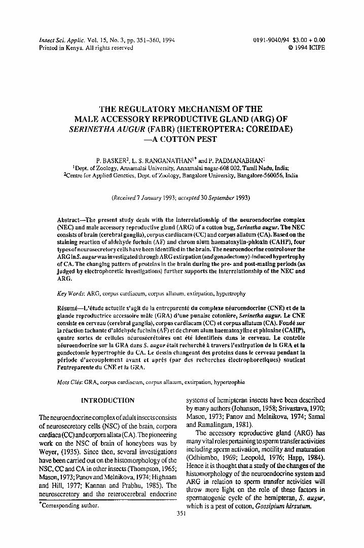
Accessory reproductive gland. The accessoryreproductive gland (ARG) is situated around thecommon ejaculatory duct which lies at the posteromedian end of the abdominal cavity below the junctionof the two vasa deferentia (Fig. 4). The gland consistsof a single layer of columnar epithelium (Fig. 5). Atthe posterior region, towards the inner side of thegland, on either side of the common ejaculatory duct,a group of squamous epithelial cells are present (Fig.6). The mode of secretion appears to be holocrine inthe squamous epithelial cells (Fig. 5) and apocrine inthe peripheral columnar epithelial cells (Fig. 5).
0.5 mmFig. 3. Frontal section showing the histomorphology ofthe corpus allatum. Note the syncytial appearance andclosely packed cells. CA—corpus allatum; Pc—Packedcells
Histology
The corpus allatum of the adult male bug of 5.augur is a small ovoid structure. Since the constituentcells are closely packed, it is difficult to make out thecellular boundaries. The nuclei are placedeccentrically, with eosinophilic cytoplasm. Thechromatin granules are distributed homogeneouslyand stain intensely with haematoxylin. The volumeof the CA was 144,237 jjm3 in the pre-mating period.However, it increased to 186,624 |im3 during thepost-mating period (an increase of about 25%).
Changes in the neurosecretory cells of brain duringpost-mating period
The neurosecretory materials in the neurosecretorycells (of A and B types) were reduced after matingand as such the cells appeared to be vacuolated.
Fig. 4. Photographic picture showing gross morphologyof the male reproductive system of Serinetha augur
Regulatory mechanism of ARG 355
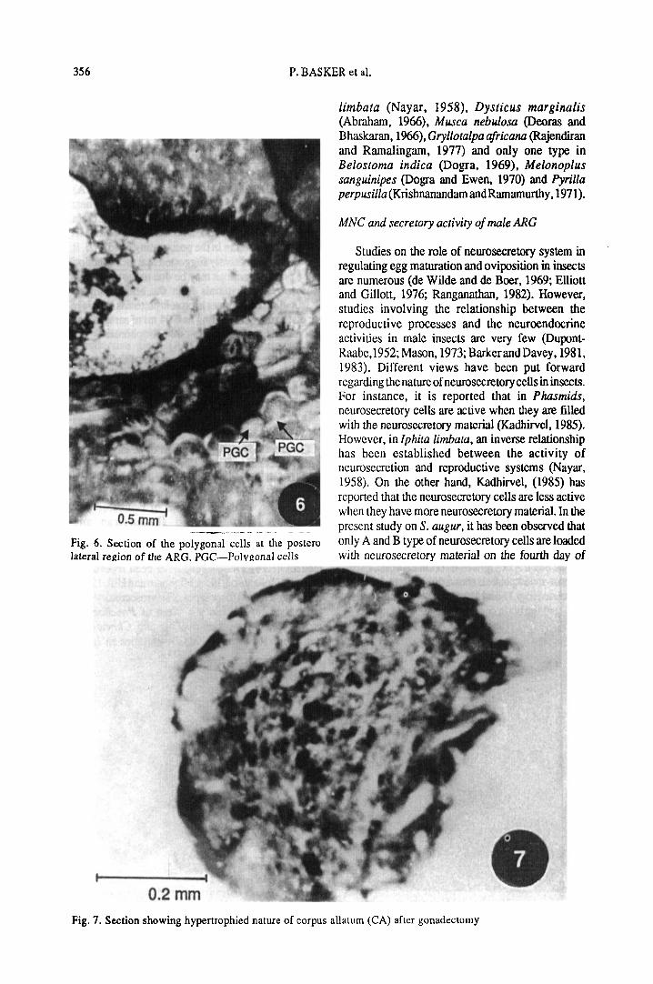
Changes in CA after gonadectomy
The gonadectomy of the male adult bugs did notaffect their reproductiveactivity. The gonadectomisedinsects mated on the eighth day just as did unoperatedmales. However, this operation resulted in increaseof the CA by 9% when compared to that found in apre-mating insect (Fig. 7).
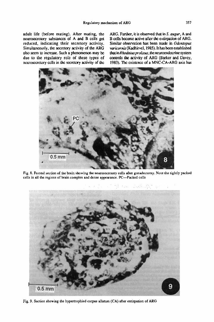
Effect of gonadectomy on the NSC of brain
Gonadectomy resulted in reduction of the volumeof neurosecretory cells. The mean volume of 234,15um3 was reduced to 158.65 pm3 (Fig. 8).
Effect of extirpation of ARG on CA and NSC
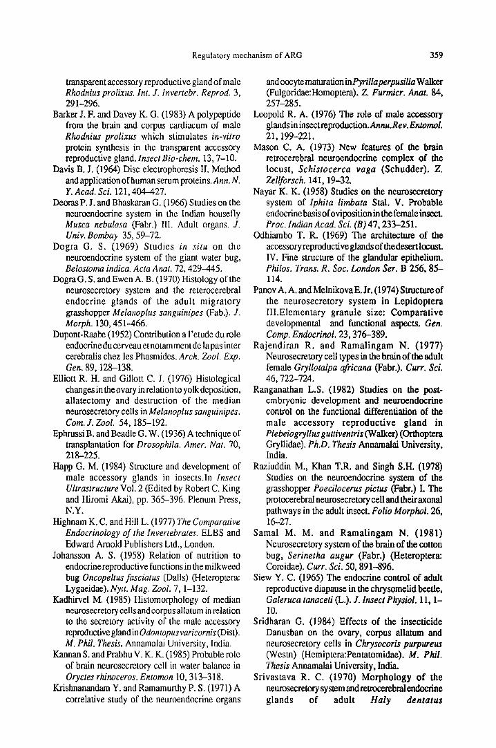
In the absenceof ARG, the male insects attemptedto mate on the eighth day. However, the matingbehaviour such as caressing and chasing were absentin the experimental insects. The CA becamehypertrophied and the neurosecretory cells A and Bwere larger and vacuolated (Figs 9 and 10).
Effect of sham operation
Sham operations of gonadectomy and ARGextirpation did not cause any noticeable change in thebehaviour or activities of the CA, NSC or ARG.
Electrophoretic studies on the brain complex
The electropherograms of brain complex in pre-and post- mating insects showed quantitative changesof protein (Figs 11 and 12). There were four bands ofprotein in the brain complex of pre mated insects (I,II, III and IV). Those bands found near the originwere designated as slow-moving protein (SMP) and
those found towards the marker front, as fast movingprotein (FMP). In the pre-mating insects, four bands(I, II, III and IV) were found to be placed at more orless equal distance from the origin towards the marker.Hence bands I and II were considered as SMP and IIIand IV as FMP. In the post-mating individuals, on theother hand, there were three bands (I, II and IIA)coinciding with the SMP of the pre-mating ones.These results indicate a possible shift in the proteinfractions, i.e., a reduction in both the SMP and FMPwhen the animals are in the post-mating condition. Itis, therefore, tentatively suggested that the reductionin the protein fractions may be due to the release ofneurosecretory materials from the brain complexafter mating. Differences in protein pattern is certainlynot due to unequal loading as 0.04 ml of sample wasused uniformly in each run of electrophoresis.
DISCUSSION
Median neurosecretory cell types
The neurosecretory cells in the brain of insectsare identified on the basis of their staining affinities.In the present study on the brain of S. augur, fourtypes of median neurosecretory cells (MNC) namelyA, B, C and D were recognised on the basis of theiraffinity to AF and CAHP. This finding agrees withobservations where many types of neurosecretorycells have been reported (Rajendiran andRamalingam, 1977; Sridharan, 1984). In Oncopeltusfasciatus and Schistocerca gregaria four types ofmedian neurosecretory cells have been reported inthe brain (Johansson, 1958; Highnam and Hill, 1977).On the other hand three types of neurosecretory cellshave been reported in the brain of Poeciloceruspictus (Raziuddin et al., 1978) and Chrysocorispurpwreus (Sridharan, 1984), two types in Iphita
Fig. 5. Section of the glandular epithelium of the peripheral region of the ARG. GE—Glandular epithelium;Lumen; SP—Secretory product
356 P. BASKER et al.
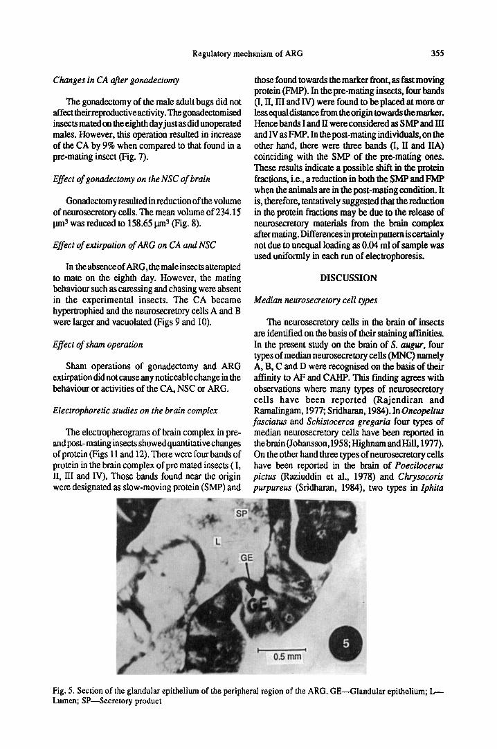
d0.5 mm1 ! * JFig. 6. Section of the polygonal cells at the posterolateral region of the ARC PGC—Polygonal cells
limbata (Nayar, 1958), Dysticus marginalis(Abraham, 1966), Musca nebulosa (Deoras andBhaskaran, 1966), Gryllotalpa africana (Rajendiranand Ramalingam, 1977) and only one type inBelostoma indica (Dogra, 1969), Melonoplussanguinipes (Dogra and Ewen, 1970) and Pyrilla/>erpnsz7/a(Krishnanandarn andRamamurthy, 1971).
MNC and secretory activity of male ARG
Studies on the role of neurosecretory system inregulating egg maturation and oviposition in insectsare numerous (de Wilde and de Boer, 1969; Elliottand Gillott, 1976; Ranganathan, 1982). However,studies involving the relationship between thereproductive processes and the neuroendocrineactivities in male insects are very few (Dupont-Raabe, 1952; Mason, 1973;BarkerandDavey, 1981,1983). Different views have been put forwardregarding the nature of neurosecretory cells in insects.For instance, it is reported that in Phasmids,neurosecretory cells are active when they are filledwith the neurosecretory material (Kadhirvel, 1985).However, in Iphita limbata, an inverse relationshiphas been established between the activity ofneurosecretion and reproductive systems (Nayar,1958). On the other hand, Kadhirvel, (1985) hasreported that the neurosecretory cells are less activewhen they have more neurosecretory material. In thepresent study on S. augur, it has been observed thatonly A and B type of neurosecretory cells are loadedwith neurosecretory material on the fourth day of
0.2 mmFig. 7. Section showing hypertrophied nature of corpus allatum (CA) after gonadectomy
Regulatory mechanism of ARG 357
adult life (before mating). After mating, theneurosecretory substances of A and B cells getreduced, indicating their secretory activity.Simultaneously, the secretory activity of the ARGalso seem to increase. Such a phenomenon may bedue to the regulatory role of these types ofneurosecretory cells in the secretory activity of the
ARG. Further, it is observed that in S. augur, A andB cells become active after the extirpation of ARG.Similar observation has been made in Odontopusvaricornis (Kadhirvel, 1985). It has been establishedthalinRhodnius prolixus, the neuroendocrine systemcontrols the activity of ARG (Barker and Davey,1983). The existence of a MNC-CA-ARG axis has
Fig. 8. Frontal section of the brain showing the neurosecretory cells after gonadectomy. Note the tightly packedcells in all the regions of brain complex and dense appearance. PC—Packed cells
0.5 mm
Fig. 9. Section showing the hypertrophied corpus allatum (CA) after extirpation of ARG
o
358 P. B ASKER et al.
Fig. 10. Frontal section of the brain showing the vacuolated appearance of the median neurosecretory cells
been shown in Plebiogryllus guttiventris(Ranganathan, 1982). It is known that in Oncopeltusfasciatus, MNC have direct control over the secretoryactivity of ARG (Johansson, 1958).
The neuroendocrine control of ARG in S. augurwas further confirmed through the extirpationexperiment of ARG and its effect on theneurosecretory cells (NSC) and C A. In the absence ofARG, the feed back to CA/NSC could have been lostand might have resulted in hypertrophy of CA andvacuolisation of NSC. Electrophoretic studies on theproteins of the brain complex revealed differentpatterns in the brain complex during the pre- andpost-mating cycles and with the secretory activity ofthe ARG. This observation further supports the viewthat the NSC in the brain complex have a regulatoryrole in the ARG. This is only a correlation of changesrather than a casual relation.
Acknowledgement—We are grateful to the Professorand Head, Department of Zoology, AnnamalaiUniversity, for providing facilities to carry out thisstudy. We also express our sincere thanks to Dr. S.Krishnan, Reader in Zoology, Bangalore Universityfor his valuable suggestions and reading thismanuscript
REFERENCES
Abraham A. (1966) Neurosecretory activity in thebrain of the water beetle Dysticus marginalis.Ada Anat. 65,435-446.
Barker J. F. and Davey K. G. (1981) Neuroendocrineregulation of protein accumulation by the
Regulatory mechanism of ARG 359
transparent accessory reproductive gland of maleRhodnius prolixus. Int. J. Invertebr. Reprod. 3,291-296.
Barker J. F. and Davey K. G. (1983) A polypeptidefrom the brain and corpus cardiacum of maleRhodnius prolixus which stimulates in-vitroprotein synthesis in the transparent accessoryreproductive gland. Insect Bio-chem. 13,7-10.
Davis B. J. (1964) Disc electrophoresis II. Methodand application of human serum proteins. Ann. N.Y.Acad.Sci. 121,404-427.
Deoras P. J. and Bhaskaran G. (1966) Studies on theneuroendocrine system in the Indian houseflyMusca nebulosa (Fabr.) III. Adult organs. J.Univ. Bombay 35,59-72.
Dogra G. S. (1969) Studies in situ on theneuroendocrine system of the giant, water bug,Belostoma indica. Ada Anat. 72,429-445.
Dogra G. S. andEwen A. B. (1970) Histology of theneurosecretory system and the reterocerebralendocrine glands of the adult migratorygrasshopper Melanoplus sanguinipes (Fab.). / .Morph. 130,451-466.
Dupont-Raabe (1952) Contribution a l'etude du roleendocrine du cerveau et notamment de la pas intercerebralis chez les Phasmidcs. Arch. Zool. Exp.Gen. 89,128-138.
Elliott R. H. and Gillott C. J. (1976) Hislologicalchanges in the ovary in relation to yolk deposition,allatectomy and destruction of the medianneurosecretory cells in Melanoplus sanguinipes.Com. J. Zool. 54,185-192.
Ephrussi B. and Beadle G. W. (1936) A technique oftransplantation for Drosophila. Amer. Nat. 70,218-225.
Happ G. M. (1984) Structure and development ofmale accessory glands in insects.In InsectUltrastructure Vol. 2 (Edited by Robert C. Kingand Hiromi Akai), pp. 365-396. Plenum Press,N.Y.
Highnam K. C. and Hill L. (1977) The ComparativeEndocrinology of the Invertebrates. ELBS andEdward Arnold Publishers Ltd., London.
Johansson A. S. (1958) Relation of nutrition toendocrine reproductive functions in the milkweedbug Oncopeltus fasciatus (Dalls) (Heteroptera:Lygaeidae). Nytt. Mag. Zool. 7, 1-132.
Kadhirvel M. (1985) Histomorphology of medianneurosecretory cells and corpus allatutn in relationto the secretory activity of the male accessoryreproductive gland in Odontopus varicornis (Dist).M. Phil. Thesis. Annamalai University, India.
Kannan S. and Prabhu V. K. K. (1985) Probable roleof brain neurosecretory cell in water balance inOryctes rhinoceros. Entomon 10, 313-318.
Krishnanandam Y. and Ramamurthy P. S. (1971) Acorrelative study of the neuroendocrine organs
and oocy te maturation in Pyrillaperpusilla Walker(Fulgoridae:Homoptera). Z. Furmicr. Anat. 84,257-285.
Leopold R. A. (1976) The role of male accessoryglands in insectreproduction. Annu.Rev. Entomol.21,199-221.
Mason C. A. (1973) New features of the brainretrocerebral neuroendocrine complex of thelocust, Schistocerca vaga (Schudder). Z.Zellforsch. 141,19-32.
Nayar K. K. (1958) Studies on the neurosecretorysystem of Iphita limbata Stal. V. Probableendocrinebasis of oviposition in the female insectProc. Indian Acad. Sci. (B) 47,233-251.
Odhiambo T. R. (1969) The architecture of theaccessory reproductive glands of thedesertlocust.IV. Fine structure of the glandular epithelium.Philos. Trans. R. Soc. London Ser. B 256, 85-114.
Panov A. A. and MelnikovaE. Jr. (1974) Structure ofthe neurosecretory system in LepidopteraIII.Elementary granule size: Comparativedevelopmental and functional aspects. Gen.Comp. Endocrinol. 23,376-389.
Rajendiran R. and Ramalingam N. (1977)Neurosecretory cell types in the brain of the adultfemale Gryllotalpa ajricana (Fabr.). Curr. Sci.46,722-724.
Ranganathan L.S. (1982) Studies on the post-embryonic development and neuroendocrinecontrol on the functional differentiation of themale accessory reproductive gland inPlebeiogryllus guttiventris (Walker) (OrthopteraGryllidae). PhD. Thesis Annamalai University,India.
Raziuddin M., Khan T.R. and Singh S.H. (1978)Studies on the neuroendocrine system of thegrasshopper Poecilocerus pictus (Fabr.) I. Theprotocerebral neurosecretory cell and their axonalpathways in the adult insect. Folio Morphol. 26,16-27.
Samal M. M. and Ramalingam N. (1981)Neurosecretory system of the brain of the cottonbug, Serinetha augur (Fabr.) (Heteroptera:Coreidae). Curr. Sci. 50,891-896.
Siew Y. C. (1965) The endocrine control of adultreproductive diapause in the chrysomelid beetle,Galeruca tanaceti (L.). J. Insect Physiol. 11,1-10.
Sridharan G. (1984) Effects of the insecticideDanusban on the ovary, corpus allatum andneurosecretory cells in Chrysocoris purpweus(Westn) (Hemiptera:Pentatomidae). M. Phil.Thesis Annamalai University, India.
Srivastava R. C. (1970) Morphology of theneurosecretory system and retrocerebral endocrineglands of adult Haly dentatus
360 P. BASKER et al.
(Heteroptera:Pentatomidae). Ann. Entomol. Soc.Amer. 63,1372-1376.
Tembhare D. B. and Thakare T. K. (1976) Thecephalic neuroendocrine system of the Dragonfly Orthetramchrysis (Selys). Odontaiologica 5,355-370.
Thompson M. (1965) The neurosecretory system ofthe adult Calliphora erythrocephala II —
Histology of the brain and some related structures.Z. Zellforsch. 67,693-717.
de Wilde J. and de Boer J.A. (1969) Humoral andnervous pathways in photoperiodic induction ofdiapause in Leptinotarsa decemlineata. J. InsectPhysiol. 15,661-675.