HEPATITIS B VIRUS 97 (j Factors that modify the risk for HCC associated with HBV Male HBsAg carriers are more likely to develop HeC than female carriers (Anthony, 1984; Coursaget et aL., 1987), and there is some evidence that establishment of the carrier state prenataIIy or early in life is associated with a higher OR for HCC than establishment of a similar state in adulthood (Larouzé et al., 1976; London, 1981; Hsieh et al., 1992). Several factors other than HBV have been evaluated as causaIIy associated with HCC. ln particular, aflatoxins, drinking of alcoholic beverages and oral contraceptives have been determined to be human carcinogens (lARC, 1987, 1988, 1993). Whether these factors modify the effect of HBV in the causation of HCC is inconcIusive. The modifyng effect of concurrent infection with HCV on the action ofHBV is discussed in the monograph on HCV (p. 165). 2.4.2 Cholangiocarcinoma Two case-control studies found no association between HBsAg seropositivity and the occurrence of cholangiocarcinoma. Parkin et al. (1991) conducted a case-control study in north-east Thailand involving 103 cases and 103 hospital controls matched for sex, age and hospital; patients with tobacco- and alcohol-related disease and other liver disease were excluded. No association was found with HBsAg seropositivity (OR, 1.0; 95% CI, 0.4-2.7). ln Taiwan, China, Chen and Sung (1978) reported that of seven cases one (14%) was HBsAg seropositive, giving a similar rate to that seen among 729 controls (15%). 2.4.3 Other cancers The relationship between HBsAg seroprevalence (as measured by reverse passive haemagglutination) and the occurrence of oral and uterine cervcal cancer was examined in one study (Vijayakumar et al., 1984). The subjects analysed were 350 oral cancer patients (232 men, 118 women), 150 cervcal carcinoma patients and 100 healthy con trois (50 men, 50 women); aIl were 40-60 years old and had no history of jaundice. Significant differences (p .c 0.001) were found for aIl sex-specific comparisons between cases and controls: the seroprevalence of HBsAg was Il % in male and 12% in female oral cancer cases, 13% in cervcal cancer cases and 4% in both male and female controls. (No data were provided on social class, nor was it clear whether the carrier state preceded treatment of the disease.) 3. Studies of Cancer in Experimental AnimaIs 3.1 Primates 3.1.1 Infection with HBV (a) Chimpanzee Chimpanzees (Pan troglodytes) have been used for manyyears to test for the presence of pathogens in biological products derived from human serum. Chimpanzees inoculated with HBV (Barker et al., 1975) or cloned HBV DNA (Sureau et al., 1988) ~xpress HBV antigens in
Transcript
HEPATITIS B VIRUS 97
(j Factors that modify the risk for HCC associated with HBV
Male HBsAg carriers are more likely to develop HeC than female carriers (Anthony,1984; Coursaget et aL., 1987), and there is some evidence that establishment of the carrierstate prenataIIy or early in life is associated with a higher OR for HCC than establishment ofa similar state in adulthood (Larouzé et al., 1976; London, 1981; Hsieh et al., 1992).
Several factors other than HBV have been evaluated as causaIIy associated with HCC. lnparticular, aflatoxins, drinking of alcoholic beverages and oral contraceptives have beendetermined to be human carcinogens (lARC, 1987, 1988, 1993). Whether these factorsmodify the effect of HBV in the causation of HCC is inconcIusive. The modifyng effect ofconcurrent infection with HCV on the action ofHBV is discussed in the monograph on HCV(p. 165).
2.4.2 Cholangiocarcinoma
Two case-control studies found no association between HBsAg seropositivity and theoccurrence of cholangiocarcinoma. Parkin et al. (1991) conducted a case-control study innorth-east Thailand involving 103 cases and 103 hospital controls matched for sex, age andhospital; patients with tobacco- and alcohol-related disease and other liver disease wereexcluded. No association was found with HBsAg seropositivity (OR, 1.0; 95% CI, 0.4-2.7). lnTaiwan, China, Chen and Sung (1978) reported that of seven cases one (14%) was HBsAgseropositive, giving a similar rate to that seen among 729 controls (15%).
2.4.3 Other cancers
The relationship between HBsAg seroprevalence (as measured by reverse passivehaemagglutination) and the occurrence of oral and uterine cervcal cancer was examined inone study (Vijayakumar et al., 1984). The subjects analysed were 350 oral cancer patients(232 men, 118 women), 150 cervcal carcinoma patients and 100 healthy con trois (50 men,50 women); aIl were 40-60 years old and had no history of jaundice. Significant differences(p .c 0.001) were found for aIl sex-specific comparisons between cases and controls: theseroprevalence of HBsAg was Il % in male and 12% in female oral cancer cases, 13% incervcal cancer cases and 4% in both male and female controls. (No data were provided onsocial class, nor was it clear whether the carrier state preceded treatment of the disease.)
3. Studies of Cancer in Experimental AnimaIs
3.1 Primates
3.1.1 Infection with HBV
(a) Chimpanzee
Chimpanzees (Pan troglodytes) have been used for manyyears to test for the presence ofpathogens in biological products derived from human serum. Chimpanzees inoculated withHBV (Barker et al., 1975) or cloned HBV DNA (Sureau et al., 1988) ~xpress HBV antigens in
kajo
Rectangle
98 IARC MONOGRAHS VOLUME 59
liver and blood and can develop a carrier state. Chimpanzees chronicaIly infected with HBVcan develop a mild chronic hepatitis resembling chronic persistent hepatitis in humanpatients infected with HBY: The extent of inflammation in chronicaIly infected chimpanzeesappears to be mil der than that seen in human patients, and chronic active hepatitis (Shouvalet al., 1980) and cirrhosis have apparently not been reported in HBV-infected chimpanzees.HCC has not been seen in chimpanzees infected with HBY, except in one brief report of theoccurrence of a liver tumour in a 15-year-old male (Muchmore et aL., 1990). This animal hadbeen under surveilance since 1978 after developing seropositivity for anti-HBs andanti-HBc (HBsAg seronegativity) two years after inoculation of human serum thought to beinfectious for non-A, non-B hepatitis. The chimpanzee's serum did not transmit non-A,non-B hepatitis to another susceptible animal (details not presented). HCC was found10 years later during investigation of liver disease associated with elevated serum alanineaminotransferase and gamma glutamyl transpeptidase. The HCC was composed of neo-plastic hepatocyes arranged in trabeculae and plates, and the surrounding non-neoplasticliver was infiltrated by amyloid. The authors reported that hybridization showed free HBVgenomes in the liver but no HBV sequences in tumour ceIIs (data not presented). (Limiteddetails were reported, and there was limited evidence that HBV was causally involved.)
(No other report of HCC developing in chimpanzees with HBV in serum was availableto the Working Group. The Group noted the limited reporting of studies on chimpanzeesobserved for many years after infection with HBY: Little published evidence is available tosuggest that HBV-infected chimpanzees develop progressive liver disease.)
(b) Monkey
Five monkeys (three male and one female rhesus and one female cyomolgus), rangingin age from less than one month to 19 months, were inoculated intravenously with a singledose of2 ml of a pool offive human sera each containing HBsAg titres ranging from 1:640 to1:2560. Between 22 and 26 months later, three monkeys (two male and one female rhesus)were given a second inoculation of a single human serum with a complement fixing titre of1:1280, containing 'abundant HBV particIes'; aIl animais were kiIIed three years after thefirst inoculation. Another group of 12 monkeys (six rhesus and six cyomolgus) (sex and ageunspecified) served as uninoculated controls. Ali monkeys were HBV seronegative beforeinitiation of the study, and ail survved up to three years. None of the monkeys wasseropositive for HBsAg three to four weeks after inoculation, but HBV core particles wereoccasionally observed in hepatocyes by electron microscopy. Gross and histologicalexamination of the animais at the end of the study showed no tumour in the livers of thoseinoculated with HBY, but there was mild persistent hepatitis in the livers of three monkeys.No IIver tumour was observed in uninoculated controls (Gyorkey et al., 1977). (There was noevidence that the monkeys were infected with HBY, and the observation period after thesecond inoculation was brief.)
Seven of 10 monkeys (Macaca assamensis; ni ne males and one female, 6-12 months ofage) were inoculated with serum from patients seropositive for HBsAg, anti":HBs, anti-HBc,anti- HBe, HBV DN A or Dane particIes (viral titres not stated), and three served as con troIs.Liver biopsy samples were taken to establish histopathological evidence of hepatitis andhepatic neoplasia and were analysed for the presence of HBsAg, HBcAg, HBeAg, anti- HBs,
HEPATITIS B VIRUS 99
anti-HBc, anti-HBe and HBV DNA and also for alanine aminotransferase four to six timesover a period of 2.5 years. Alanine aminotransferase levels were elevated after inoculationand were still persistently high in five of seven animais 137 weeks after inoculation. TwoanimaIs died of other causes during the course of the study. HBsAg, anti-HBs, anti-HBc andHBV DNA were found in sera of aIl seven treated monkeys. Histopathological changesconsistent with hepatitis, including hepatocellular degeneration, necrosis, inflammatory cellinfiltration, bile-duct proliferation and fibroplasia, were seen. Liver lesions in sorne animaIsprogressed to cirrhosis, and one of them developed a mucin-producing, weII-differentiatedtumour described as an HCC (Ge et aL., 1991). (The reporting was limited, and the statementthat the HCC secreted mucin and arose from bile ducts was noted.)
3.1.2 Infection with HBVwith concomitant administration of chemical carcinogens
Monkey
ln a study described in section 3.1.1 (Gyorkey et al., 1977), three groups of animais wereused. Nine monkeys in group 1 (four male and two female rhesus, one male and two femalecyomolgus), ranging in age from less than one to 20 months, were given intraperitonealinjections of20 mg/kg bw N-nitrosodiethylamine (NDEA) (vehicle unspecified) twce a weekfor two years. Six of the monkeys (two male and one female rhesus, one male and two female
cyomolgus) were given a single intravenous injection of2 ml of serum containing HBV (seesection 3.1.1) 25-33 months after the NDEA injections were started, and three were kiledthree to six months after inoculation and the remaining three Il months after inoculation.The three animaIs that were not inoculated were kiIIed three years after the start of theexperiment. A second group of Il monkeys ranging in age from one to 20 months (four maleand five female rhesus, one male and one female cyomolgus) were given a singleintravenous injection of 2 ml of the pooled HBV serum (see section 3.1.1), followed onemonth later by the same NDEA treatment as animais in group 1. Five of the Il monkeys ingroup 2 (three male and two female rhesus) received a second inoculation of the individualHBV serum (see section 3.1.1) approximately two years after the initial inoculation and werekiled one year later. Animais in group 3 (seven monkeys aged 1-21 months: one male andthree female rhesus, two male and one female cyomolgus) were given intraperitonealinjections of 20 mg/kg bw NDEA twce a week for two weeks. One month after the firstinjection of NDEA, each monkey received a single intravenous injection of 2 ml of thepooled HBV serum, foIIowed two weeks later by re-institution of the twce-weekly NDEAtreatment, which was continued for two years. Three of the seven animaIs received a furtherinoculation with the individual HBV serum about 21 months after the first HBV inoculation.AlI survving animais were kiled three years after the start of the experiment. The livers of aIlanimaIs in group 1 had large invasive HCCs with central haemorrhagic necrosis; sorneanimais also had cirrhosis. Six had metastases to the lung. The incidence and time of onset ofliver tUmours in monkeys given injections of NDEA in combination with HBV were notdifferent from those in animaIs that received NDEA alone. Ali monkeys In group 2 thatsurvved to the end of the experiment developed cirrhosis and invasive multifocal HCC; sixdeveloped metastases to the lung. Reinoculation with HBV of monkeys in group 3 failed toaffect tumour outcome. (No evidence of viral infection was observed, and the study was
100 IAC MONOGRAHS VOLUME 59
inadequately designed to allow demonstration of an enhancing effect of HBV on hepato-carcinogenicity. )
3.2 Transgenic mice
3.2.1 With no concomitant administration of chemical carcinogens
The expression of various HBV gene sequences in livers of transgenic mice has beenexamined in several studies (for reviews, see Chisari, 1991; Slagle et al., 1992). Only thosetransgenic models in which hepatic neoplasia was the end-point are included in this section(for a discussion of gene expression and mechanisms of tumour induction, see also section4.3.2).
Male mice of three transgenic lineages producing the HBV large surface antigen wereback-crossed with normal C57BI/6J females (see Table 7). Mice from the first generation(36 animais from lineage 50-4, 12 from lineage 45-2, eight from lineage 45-3 and fournon-transgenic controls) were observed for two years for the development ofliver injury (asindicated by increased levels of serum glutamic and pyruvic transaminases) and neoplasms(determined by abdominal palpation and serum levels of ~-fetoprotein). Variable expressionof large surface antigen (as measured by western blot of total liver protein) was correlatedwith lineage, being highest in lineage 50-4, medium in lineage 45-2 and lowest in lineage45-3. Lineages that expressed the highest levels of large surface antigen and filamentousprotein in hepatocyes had the highest level of liver injury, and liver tumours developed inanimais that had hepatocellular injury. Tumours were first detectable in lineages 50-4 and45-2 at 9-12 months after the onset of injury, and virtuaIIy all mice of the 50-4 lineage withpre-existing chronic liver-cell injury developed HCC by 18-21 months of age. One or twolarge tumours (1.0-2.0 cm) usually predominated, and numerous smaller tumours werescattered throughout the livers. Thirty-eight animais (25 males and 13 females) (lineageunspecified) with palpable abdominal masses were examined histologically at necropsy.Males developed more palpable liver tumours and displayed more HCCs (18/25) than didfemales (4/13). Adenomas predominated (5/7) in younger males (10-15 months) andcarcinomas (13/18) in older males (16-21 months). Ali tumours occurred concurrentlywithhepatoceIIular injury, characterized by ground-glass hepatocyes, necrosis and inflam-mation. Neither metastases nor fibrosis or cirrhosis were observed (Chisari et al., 1989). (TheWorking Group noted that the terms 'hepatoma' and 'hepatocellular carcinoma' appear tohave been used interchangeably.)
A group of 59 male and female transgenic mice (Iineage 50-4; see Table 7) wereexamined for abdominal masses every four months for 24 months. Selected animais werekiIed at monthly intervals from 1 to 23 months, and nine control nontransgenic animaIs werekiIed at 3, 11, 18 and 24 months (exact number of animaIs killed at each time point notspecified). LIver sections were examined histologicaIIy for the presence of hepatocellularadenomas and carcinomas. Livers of nontransgenic mice were normal histologically at ailtime points. Starting at two months, mice progressively developed liver injury and
inflammation, including hepatoceIIular necrosis, Kupffer-cell hyperplasia and mono-nuclear-cell infiltration, with concurrent preneoplastic lesions which appeared by sevenmonths of age. Preneoplastic lesions consisted of hepatoceIIular dysplasia and foci of altered
HEPATITIS B VIRUS 101
hepatocyes, which progressively developed into larger compressive nodular masses.
Seventy-five adenomas, characterized by masses of neoplastic hepatocyes which com-pressed adjacent parenchyma, occurred in 18 mice from eight months and peaked inincidence around the 17th month of the study. HCC (29 in aIl) had occurred in aU survvingtransgenic mice by 20 months of age (Dunsford et al., 1990).
Table 7. Transgenic mice that express hepatitis B surface antigen as the major product
Transgenic mice containing the entire coding region of the HBx gene, incIuding the Xpromoter, the principal RNA start sites, transcriptional enhancer and polyadenylation site,were created by microinjecting embryos from outbred CD1 mice (see Table 7). Six transgenicmice, each with at least one intact, stably expressed copy of the X gene, were identified bySouthern blot analysis, and three animais with a high level of expression were bred intopermanent lines (Iineages CIL, H9 and El) (strain and sex of crosses and total numbers oftransgenic and nontransgenic offspring from aIl three lineages unspecified). Livers wereexamined histologically at various times. At four months, preneoplastic lesions consisting ofmultifocal are as of altered hepatocyes were observed in progeny from aIl three lines oftransgenic mice but not in nontransgenic littermates. Neoplastic nodules (sizes unspecified),which occurred by 8-10 months of age, compressed surrounding hepatocyes and accumu-lated high levels of HBx protein. The authors reported that fewer than 10% of control maleCDi mice develop hepatic neoplasms during an average lifespan of 24 months (no datashown). HCCs were observed in 19/21 males and 12/20 females of line Cl1, in 8/10 malesand 4/6 females of line H9 and in the Ei line (details not given). Most males of the CIL linedied with HCC between 11 and 15 months of age, and most females between 17 and 21months of age. There was no difference in the incidence of liver tumours in male and femalemice. Liver damage, determined by concentration of serum alanine aminotransferase, wasnot observed, and the levels were consistently within normal range (Kim et al., 1991).(Detailed data on liver lesions in nontransgenic littermates were not provided.)
102 IAC MONOGRAPHS VOLUME 59
ln another study of a transgenic lineage expressing the X gene driven by the æ-1-anti-trysin promoter, mice did not exhibit liver disease or tumour development. This lineageexhibited an early but transient expression of the HBx protein (Lee et al., 1990).
3.2.2 With concomitant administration of known chemical carcinogens
The transgenic mouse strain E36 was derived from founder (C57Bl/6 x SJL/J)Fi micecontaining aIl of the HBV genome except for the core gene, allowing expression of HBsAgunder control of the HBV promoter and enhancer sequences (see Table 7). Two hundred andfour transgenic and nontransgenic mice (Fi hybrids resuiting from crosses of males of thetransgenic strain E36 with C3H/He females) were allocated to three treatment groups.AnimaIs of group 1 (23 HBV-seropositive males, 21 HBV-seropositive females, 19
HBV-seronegative males and 22 HBV-seronegative females stiII alive at 30 weeks) weretreated at seven days of age by oral gavage with a single dose of 10 mg/kg bw NDEA in 0.9%saline solution. Animais of group 2 (12 HBV-seropositive and 22 HBV-seronegative malesstil alive at 30 weeks) were treated with a single dose of 150 mg/kg bw para-dimethyl-aminoazobenzene (DAB) in corn oil by gavage at seven days of age. Group 3 consisted of52 untreated con trois (15 HBV-seropositive males, 11 HBV-seropositive females, 14 HBV-seronegative males and 12 HBV-seronegative females still alive at 30 weeks). Survvors at 30weeks were 92% of those treated with NDEA and 98% of those given DAB. AnimaIs werekiIIed at 30 weeks of age and examined both grossly and microscopically for the presence ofIIver tumours. No tumour was observed in the 52 control animaIs. Liver nodules:; 220 ¡.m indiameter were counted, and those 5 mm in diameter were classified as either adenomas orcarcinomas by histological criteria. ln NDEA-treated male groups, the total number ofnodules per cubic centimetre of liver was about the same for HBV-seropositive andHBV-seronegative animaIs, but larger nodules (:; 330 ¡.m diameter) occurred at about twcethe frequency in HBV-seropositive mice as compared with HBV-seronegative mice
(p ~ 0.05, Wilcoxon test). The frequency of nodules in the DAB-treated group was muchlower th an that in NDEA-treated animaIs. The frequency of nodules per cubic centimetre ofliver was 1.5-2 times higher in transgenic th an in nontransgenic animais, but the increase wassignificant only for nodules , 110 ¡.m. The incidence of hepatocellular adenomas andcarcinomas was higher in HBV-seropositive (18/56) than in HBV-seronegative (14/63)
animais treated with either NDEA or DAB; this difference was not significant (Dragani et al.,1990).
Six groups of 10 female transgenic mice that produce the HBV large surface antigen(Iineage 50-4; see Table 7) and of 10 nontransgenic littermates were treated as follows.Group 1 served as untreated controls; group 2 received five monthly intraperitonealinjections of 0.25 ¡.g/g bw aflatoxin Bi as a suspension in tricaprylin beginning at three or fourmonths of age; group 3 received a single intraperitoneal injection of 0.25 ¡.g/g bw aflatoxinBi suspended in tricaprylin at three or four months of age; group 4 received three weeklyintraperitoneal injections of2.0 ¡.g/g bw aflatoxin Bi suspended in tricaprylin at four monthsof age; group 5 received a single intraperitoneal injection of 50 ¡.g/g bw NDEA dissolved insterile saline at four or five months of age; and group 6 received 0.1 % phenobarbital inpowdered diet beginning at six months of age for one year. The study was terminated whenthe animaIs were 15 months of age. Survval rates were approximately 90%, except for
HEPATITIS B VIRUS103
group 6 in which survval was about 50%. Liver nodules and tumour masses were observedgrossly post mortem and by histological examination. Nodules were classified by size intothree categories: 0.1-1.9 mm, 2-4.9 mm and:; 4.9 mm in diameter. Adenomas and HCCswere distinguished histologicaIIy. No gross or histological lesions were seen in the livers ofnontransgenic control mice, whereas the livers of control transgenic mice contained multiplenodules of different sizes. Livers of transgenic mice treated with aflatoxin Bi had 15-23nodules (0.1-1.9 mm in diameter) per liver, as compared with 0.1-0.2 nodules of the samesize per liver in nontransgenic aflatoxin B1-treated mice and five nodules per liver intransgenic control mice not treated with aflatoxin Bi. Similar results were obtained for theincidence of larger nodules. Afatoxin B1-treated transgenic mice had 6.2-8.8 nodules(2.0-4.9 mm in diameter) per liver, whereas untreated transgenic mice had an average of3.7nodules per liver and aflatoxin Bi-treated nontransgenic mice had 0-0.1. Adenomas andHCCs were seen only in transgenic mice treated with aflatoxin Bi or NDEA. ln the threeaflatoxin-treated groups (2, 3 and 4), a total of20 adenomas and two HCCs were observed in26 transgenic mice and none in 27 nontransgenic mice. ln the NDEA-treated group (5), ni neadenomas and two HCCs were seen in eight transgenic mice and none in ni ne nontransgenicmice examined. Livers of transgenic mice fed phenobarbital showed increased nodularity butno adenoma or HCC; however, survval was poor (SeII et al., 1991).
3.3 Woodchucks (Marmota monax)
3.3.1 Hepatocellular carcinoma in woodchucks naturally infected with woodchuck hepatitisviruS
The first non-human hepadnavirus was identified in woodchucks (Marmota monax) in aseries of studies that began at the Philadelphia (USA) Zoo (for a review, see Paronetto &Tennant, 1990).
ln the initial report, which appeared as an abstract (Snyder, 1968), a group of 50woodchucks (42 males and eight females), trapped in the wild in the vicinity of Philadelphiawhen about five months of age, were held in captivity one or two per cage on tap-water and astandard feed. After about 72 months in captivity, 30 animais had died. HCC were observedin nine (six males and three females). ln one of the ni ne animais, metastatic nodules werefound in retroperitoneal fat. The author concluded that dietary carcinogens were probablynoi responsible, since other captive animais in the Philadephia Zoo fed on the same diet hadnot developed liver tumours; he proposed that a viral agent was involved in the etiology ofliver cancer in woodchucks.
Ten years later, Summers et al. (1978) reported that post-mortem examination of 102woodchucks that had been caught in the wild and kept at the Philadelphia Zoo for 18 yearshad revealed 23 HCCs (22.5%), which appeared at a mean age of 59 months. Three animaIshad acute hepatitis. About 15% of serum samples taken from captive woodchucks werefound to contain DNA polymerase-containing particles in amounts comparable with thosefound in some human sera positive for HBsAg. Detailed investigations were carried out onthree animais, two of which had died with HCC and one of which had died with a normalliver: Sera from the two animaIs with HCC, but not that from the control animal, haddetectable levels of DNA polymerase-containing particles. When the particles were
104 IAC MONOGRAHS VOLUME 59
characterized and compared with particIes from an HBV-infected human by caesiumchloride equilibrium sedimentation, electron microscopy and electrophoresis, the particlesfrom the woodchucks were found to be similar, but not identicaL. DNA of similar size andphysical structure was found in sera and liver samples from the two animaIs with HCC. Theauthors concluded that the particles represented a distinct virus, which they caIled'woodchuck hepatitis virus', which is phylogeneticaIIy related to HB\!
ln a review, Summers (1981) reported that aIl 16 woodchucks in the colony at thePhiladelphia Zoo that developed HCC also had chronic active hepatitis of varyng severityand had been persistently infected with WH from an early age, when they were obtainedfrom the wild. No HCC had developed in groups of animais with anti-WHs and no marker ofviral infection.
Seventy-three woodchucks from Pennsylvania and Delaware which had been trapped asyearlings or as adults and observed for at least one month in a colony established at theNational Institute of Allergy and Infectious Diseases (NIAID) (Mitamura et al., 1982) werestudied by Popper et al. (1981). Thirty-three selected animais, including aIl six animaIs thathad developed HCC (criteria for selection of the remaining 27 animaIs not described), werestudied in detaiL. The six animaIs with HCC were aIl seropositive for WHsAg, WH DNAand WH DNA polymerase. Of the remaining 27 animaIs, four were seropositive for aIlthree markers and four for anti-WHs, three were seronegative for aIl markers and 16 wereseropositive for one marker only or gave inconsistent or discrepant results. The authorspointed out that cirrhosis did not occur in animaIs with HCC. Furthermore, inflammationwas generaIly characterized as mild, and chronic active hepatitis was seen in only two animaIswith HCC. The authors also noted the direct transition to HCC from neoplastic nodules inthese woodchucks.
Mitamura et aL. (1982) extended the observations on the NIAID colony ofwoodchucksand analysed markers of WHV infection among 62 animaIs that had died of various causes.Death from HCC occurred in 11 of 13 (85%) chronic carriers of ~ in two of 33 (6%)
animaIs with anti-WHs and no evidence of viral replication, and in none of 16 animais with noviral marker.
Of 113 woodchucks that had been trapped in different areas of Pennsylvania, Marylandand Delaware and kept in a colony at the New Bolton Center at the University ofPennsylvania, eight developed HCC between 44 and 88 weeks of captivity (Mil man et al.,1984). Seven of the animaIs were seropositive for WHsAg at the time of capture; one animalthat was seronegative at that time converted to WHsAg seropositivity after 33 weeks ofcaptivity.
Nineteen WHsAg-seropositive woodchucks that had been trapped in Pennsylvania andMaryland were kept for up to two years at Cornell University, New York (Roth et al., 1985),and the livers of 16 animaIs were examined. HCC was found in 13, aIl ofwhich had chronicactive or persistent hepatitis. Metastases to the lung were observed in one animaL. Among149 WHsAg-seronegative woodchucks trapped in New York State and kept in captivity forfour weeks or more, a single case of HeC was observed, although five had acute hepatitis.
HEPATITIS B VIRUS105
3.3.2 Hepatocellular carcinoma in woodchucks experimentally infected with woodchuck hepa-titis virus
(a) Infection with woodchuck hepatitis virusA breeding colony of woodchucks consisting of the offspring of female woodchucks
trapped in New York State and shown to be free of present or past WHV infection wasestablished at CorneII University, New York (Popper et al., 1987). Newborn animais wereinoculated with 105.5_106.5 50% infectious doses of WH one day after birth. Adult
woodchucks with no evidence of active or past WH infection, maintained in the NIAIDwoodchuck colony, were inoculated with 105.8 50% infectious doses. AnimaIs were kept ontap-water and aflatoxin-free laboratory chow. A total of eight woodchucks, six infected atbirth and two as adults, developed chronic infection, as indicated by the presence of WHsAgfor one year or longer. AlI eight animaIs subsequently developed HCC 17-36 months afterinfection; no HCC was observed in 19 animaIs with virological markers of past infection or in15 uninfected controls foIIowed for 18-57 months. Mild hepatitis, characterized bylymphocyic infiltra tes, was seen in the portal tracts of woodchucks infected as adults ornewborns. ln animaIs infected as adults, the portal inflammation regressed with time and theIIver assumed the appearance of control livers. ln woodchucks with HCC, the portal tractinflammation was more extensive, occasionally resembling that seen in human chronic activehepatitis. Furthermore, inflammation appeared to be most severe in the immediate vicinityof the HCC. Cirrhosis was not seen.
Two groups of 43 woodchucks were inoculated with infectious serum at birth or at eightweeks of age. Thirteen of those inoculated at birth (32%) became chronic carriers, 28animaIs cleared the infection and two died within six months after birth. Afer three years, Il
of the chronic carriers and two of the animais with past infection had developed HCC. Ofthose inoculated at eight weeks of age, 23 developed acute WHV infection; three becamechronic carriers (13%), while 20 animaIs recovered from the infection. Two of the threechronic carriers and eight of the 20 animaIs with past infection were followed for three years.Both chronic carriers but none of the eight woodchucks with past infection developed HCC.None of 46 uninfected, laboratory-born woodchucks foIIowed for three years or moredeveloped HCC (Tennant et al., 1988).
Gerin et al. (1989) extended the analysis of HeC occurrence in experimentally infectedwoodchucks maintained at CorneII University: HCC developed in 61/63 chronic carriers(97%), 11/63 (17%) animais with past infection and in none of 108 concurrent, uninfectedcontrols. FoIIow-up was for at least three years; the sex of the animal did not influence theoccurrence of HCC. Ali three pair-wise comparisons between the three groups weresignificant at p -( 0.001 by Fisher's exact test.
(b) Infection with woodchuck hepatitis virus in combination with ajlatoxin Biln a study described in an extended abstract (Tennant et al., 1990), 52 woodchucks (sex
unspecified) were inoculated subcutaneously with WH (5 x 10650% infectious doses) at1-3 days of age. A group of 27 of these animaIs received no further treatment; 25 inoculatedand 29 uninoculated animais subsequently received aflatoxin Bi in the diet (0.25-1.0 iig/kg)from three months of age for six months or comparable cumulative doses of aflatoxin Bi in
106 IARC MONOGRAHS VOLUME 59
dimethyl sulfoxide solution by intraperitoneal injection (125 J,g/kg bw, three times weekly)beginning at 1-4 months of age for 3-4 months. Twenty-three animais served as untreated
con trois. WHV-specific serological tests (unspecified) indicated that the rate of chronicinfection (73%) at one year of age in the group given aflatoxin Bi and WH was similar tothat of those infected with WHV alone (70%). Survval rates were 60% for woodchucksinfected with WHV and given aflatoxin Bi and 72% for those that received aflatoxin Bialone; no death occurred among animaIs infected with WH al one. Histological analysis oflivers from aflatoxin Bi-treated woodchucks revealed lesions consistent with hepatotoxicitydue to that compound. Thirty-six months after initiation of the study, 6 of the 15 survvinganimais (40%) given aflatoxin Bi and WHV had HCC, in contrast to 21 of the 27 animaIs(78%) inoculated only with WHV During the same period, 2 of 21 woodchucks that weretreated with aflatoxin Bi al one and survved more than one year developed HCC, while noneof 22 untreated controls had hepatic tumours. (The high dose of aflatoxin Bi compromisedthe interpretation of the results of this study by reducing the survval of the animaIs.)
3.3.3 Hepatocel/ular carcinoma in woodchucks experimental/y infected with ground squirrelhepatitis virus
Seeger et al. (1991) reported experiments in which woodchucks were infected withBeechey ground squirrel (Spermophilus beecheyi) hepatitis virus (GSHV) or WH Three-day-old woodchucks from the breeding colony at Corne il University were inoculatedsubcutaneously with serum from infected woodchucks or from infected ground squirrels. Of29 woodchucks infected with GSH\~ 17 (59%) became chronic carriers; of 36 woodchucksinoculated with WH~ 27 (75%) became chronic carriers. Sixte en ofthese were selected forcomparison with the 17 chronic carriers of GSHV Two years after experimental infection,7 of the 16 WHV-infected but none of the 17 GSHV-infected woodchucks had liver masses(detected by ultrasound imaging), ail of which were verified histologically as HCC.Histological examination of aIl animaIs after 26 months revealed neoplastic lesions in twoGSHV-infected woodchucks. At 51 months after infection, ail 16 WH carriers haddeveloped one to five HCCs each (total, 41), and 6 of the 14 GSHV carriers that weretumour-free at 26 months and that survived laparotomy at that time developed one to fourHCCs (total, 14). The median time to diagnosis of HCC in WHV-infected woodchucks was32 months; the projected median time to diagnosis of HCC in GSHV-infected woodchuckswas 55 months. The extent of non-neoplastic liver disease and chronic inflammation did notdiffer according to the virus inoculated.
3.4 Ground squirrels, ducks and other species
3.4.1 Beechey ground squirrels
GSHV infecting Beechey ground squirrels was discovered in 1979 in northernCalifornia, USA, as a result of a search for a virus similar to HBV in animaIs related towoodchucks (Marion et al., 1980). The biology, genetic structure, gene products and viralreplication of GSHV have been reviewed recently (Marion, 1991). The virus was originallydetected in sera taken from apparently healthy animaIs. To date, the only known location ofthis virus is on the San Francisco Peninsula, although the virus that putatively infects
HEPATITIS B VIRUS107
Richardson ground squirrels (Spermophilus richardsonii) (see section 3.4.2) (and viruses thatpossibly infect other ground squirrel species) may be a variant of the Beechey squirrel virus.
ln an experiment described in a series of reports (Marion et al., 1983, 1986, 1987),Beechey ground squirrels, estimated to be one to two years of age, were trapped live atvarious locations on the San Francisco Peninsula between 1980 and 1984. Animais were heldindividuaIIy in quarantine for one month, during which time their serum was tested for(i) surface antigen (GSHsAg), by a commercial solid-phase radioimmunoassay for cross-reacting HBsAg; (ii) anti-GSHs, by a virus-specific sol id-phase radioimmunoassay; and(iii) virion-associated DNA polymerase activity, as a measure of virus load (Marion et aL.,1983). AnimaIs with serum GSHsAg and DNA polymerase activity were housed in a roomseparate from GSHsAg-seronegative animais (Marion et al., 1986). Marion et al. (1987)reported that 24/103 ground squirrels examined at necropsy had tumours at various sites; aIltumour-bearing squirrels were 4.5-8 years of age and had been in captivity for a minimum of2.4 years. Among animaIs under 4.5 years, no tumour of any kind was observed in19 persistent carriers of GSHV 22 seropositive for anti-GSHs or 19 with no serologicalmarker of GSHV Of the tumours observed in older squirrels, Il were found in 17 GSHVcarriers, eight in Il squirrels seropositive for anti-GSHs and five in 15 GSHV marker-freesquirrels. The predominant tye of tumour observed in squirrels over 4.5 years of age wasHCC, which was detected in 10/17 persistent GSHV carriers and in 3/11 squirrelsseropositive for anti-GSHs, but in none of 15 GSHV marker-free squirrels in the same agerange, resulting in a highly significant association between HCC and the GSHV carrier state
(p = 0.0005, Fisher's exact test) and a weaker association with seropositivity for anti-GSHs.Development of HCC in carrier squirrels may be related either to age or to the length of thecarrier state, as aH animais appeared to have become carriers before 1.5 years of age. HCCwas seen at necropsy in six of ni ne carrier squírrels (67%) over six years of age but in onlythree of nine carriers aged four to six years and none of 17 carrier squirrels less than fouryears of age. AlI HCCs except one were of the sa me histological tye: a trabecular, highlydifferentiated liver carcinoma; the only non-trabecular HCC was seen in one of the threesquirrels seropositive for anti-GSHs and was of the meduHary tye and less differentiated.The diameters of the major tumours were generaIly larger in squirrels that were older whenthe HCC was detected. Single nodules of HCC were commoner in the younger squirrels,while older squirrels usuaIIy had more than one nodule. Four of the five oldest squirrels withHCC also had metastases to or adhesions of the tumour in the spleen. While viral DNA wasintegrated into the host DNA of some of the HCCs examined, the majority of those fromsquirrels with GSHV markers did not have detectable integrated viral DNA. Chronic activehepatitis and cirrhosis were not seen (Marion et al., 1986, 1987).
ln a further assessment of the development of HCC in the squirrel colonies after ni neyears of observation (Marion & Cu lien, 1992), 18 cases (45% of aIl neoplasms) wereobserved in the study population of 24 GSHV-infected, 20 anti-GSHs-seropositive and 26GSHV marker-free ground squirrels over four years of age. Eleven of the liver tumours wereseen in carrier animaIs, five in anti-GSHs-seropositive squirrels and two in GSHVmarker-free animais. The association of HCC with the GSHV carrier state was significant(p = 0.0016). As in WH-infected woodchucks, the incidence of HCC in animaIs that hadrecovered from infection was relatively high (20%). Anti-GSHs-seropositive squirrels that
108 !AC MONOGRAHS VOLUME 59
developed tumours experienced only a brief period of viraemia. No sex difference was noted.The average age of carrier squirrels at the time of detection of HCC was 6.5 years.
3.4.2 Richardson ground squirrels
HCC has been observed in Richardson ground squirrels from the southern half of theCanadian province of Alberta. The hepadnavirus thought to be associated with thesetumours has not been characterized geneticaIIy or biologically, nor has it been transmittedexperimentally to other animais. ln a study by Minuk et al. (1986), animaIs were trapped andkept in captivity for less than one month. Two of 25 adult squirrels but no ne of 15 juveniles
had HCC at necropsy (Table 8). Anti-GSHs was found in 7 of the 25 adult animais, and theserum of one animal reacted positivelywhen tested with a commercial radioimmunoassayforHBsAg known to detect GSHsAg. Serum was not tested for the presence of virions.Anti-GSHs seropositivity was assayed with a commercial radioimmunoassay for anti- HBs.Of the animaIs in which HCC was found, one had GSHsAg reactivity in the serum, while theother was seropositive for anti-GSHs. No viral DNA was detectable in the HCC of theseropositive animal or in the DNA of adjacent liver tissue. (The assay to detect anti-GSHswas unspecific and insensitive. i
Table 8. Studies of hepatocellular carcinoma (HCC) in Richardson grouDd squirrelstrapped or born in captivity iD Alberta, Canada, accordiDg to age at necropsy
Age at No. of Loction GSHsAg- Anti-GSHs- Liver tumours Referencenecropsy animaIs seropositive seropositive
Adult 25 South of Calgary 1 7 2 HCC Minuk et al.Juvenile 15 South of Calgary 1 4 0 (1986)
1- ~3 years 562 Picture Butte ND ND 0 Tennant et aL.3-4 months 56° Picture Butte ND ND 0 (1991)14-17 months 54° Picture Butte ND ND 31 with nodules15 months 36 Cochrane ND ND 1 HCC, 4 with
nodules~ 3 years 5 Edmonton 0
J 10/12
2 HCC, 2 withnodules
~ 3 years 7b Picture Butte 0 4 HCC
GSHsAg, ground squirrel hepatitis surface antigen; ND, not determinedllom in captivitybDams of 54 bom in captivity
ln a study by Tennant et aL. (1991), several groups of Richardson ground squirrels wereexamined for the presence of masses in the liver at necropsy. The majority, collected atPicture Butte, Alberta, Canada, and not maintained in captivity, were not tested for hepadna-virus markers or examined histologically. None of 618 squirrels ranging in age from three tofour months to three years or more had evidence of liver cancer (Table 8). Squirrels held incaptivity for 14 months or longer for various experiments were also examined for HeC atnecropsy; nodules or histological evidence ofHCC were detected in sorne animais (Table 8).
HEPATITIS B VIRUS109
HCCs were found in squirrels trapped at Picture Butte only after the animais had beenmaintained in captivity. Hepadnavirus markers were assayed in the sera of the five squirrelstrapped near Edmonton and the seven from Picture Butte. None cross-reacted with HBsAg,but most had evidence of anti-GSHs. Viral DNA was detected in two of four HCCs; noanti-GSHc was detectable in any sample using an assay which readily detects this antibody.Non-neoplastic lesions in the Iivers of animaIs kept for three years in captivity included mildto moderate portal inflammation, with somewhat more severe inflammation adjacent totumours. ln the livers of six of seven animaIs with moderate portal inflammation, focalhepatoceIIular necrosis and inflammation were seen. (Limited data are available to supporthepadnavirus infection per se.)
3.4.3 Ducks
Observations of liver tumours in domestic ducks (Anas domesticus) were first describedin China by Wang et al. (1980), which led to the discovery of duck hepatitis B virus (DHBV)(Mason et al., 1980). The biology, genetic structure, gene products and viral replication ofDHBV have been reviewed (Schödel et al., 1991).
Studies of the oncogenic potential of DHBV are of three tyes: (i) assessment of livertumours and markers of DHBV in ducks coIIected on farms or free-ranging in communities;(ii) prospective studies of the development of HCC in ducks of known DHBV status andhistory; and (iii) experimental studies of the joint effects of DHBV infection and aflatoxin Biexposure in the development of HCC in ducks.
(a) Liver tumours and markers of duck hepatitis B virus in ducks collected on farms and infree-ranging flocks.
Afer the initial discovery of DHBV in ducks with hepatitis and liver tumours in theChinese Province of Qidong, several studies were carried out to determine whether thepresence of HCC and hepatitis in domestic ducks was linked to current or past replication ofDHBV in the same animais (see Table 9).
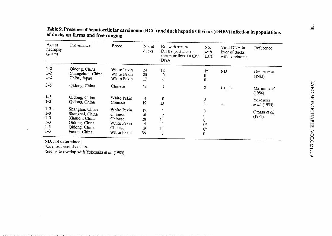
DHBV was found in 70/195 ducks from three of five locations in China but in none of17 ducks from Chiba, Japan (Table 9); HCC was found only in four ducks from Qidong, andevidence of present or past DHBV infection was seen in three of them. Moderate to severehepatitis was observed in both DHBV-seropositive and -seronegative ducks from Qidong,where there are known to be relatively high levels of aflatoxin Bi, a known cause of liverdisease in ducks (IARC, 1993). Moderate hepatitis consisted of mild portal inflammation,with rare necrosis of hepatocyes. Severe hepatitis was associated with dense chronic inflam-mation of portal tracts, which extended into adjacent parenchyma and was accompanied byfocal necrosis of hepatocyes. Severe hepatitis was sometimes accompanied by septalfibrosis, focal areas of parenchymal colIapse and regenerative nodules; cirrhosis was seen inone duck with HCC (Marion et al., 1984). Three of the four HCCs were observed in Chineseducks and none in white Pekin ducks. OveraII, while there was concomitant presence ofDHBV and HCC in sorne ducks from Qidong, the two have not been firmly linked, nor hasthe simultaneous presence of DHBV replication and liver inflammation been assocIated ¡nthese ducks.
,.¡-0Table 9. Presence ofhepatocellular carcinoma (HeC) and duck hepatitis B virus (DHBV) infection in populations
of ducks on farms and free-ranging
Age at Provenance Breed No. of No. with serum No. Viral DNA in Referencenecropsy ducks DHBV particles or with liver of ducks(years) serum or lIver DHBV HCC with carcinoma
DNA
1-2 Qidong, China White Peki 24 12 ia ND Omata et al.1-2 Changchun, China White Peki 20 0 0 (1983)1-2 Chiba, Japan White Peki 17 0 0 -)-3-5 Qidong, China Chinese 14 7 2 1+,1- Marion et al.f3
(1984)~1-3 Qidong, China White Peki 4 0 0 Yokosuka 01-3 Qidong, China Chinese 19 13 1 + et al. (1985) Z01-3 Shanghai, China White Peki 17 1 0 Omata et al. a1-3 Shanghai, China Chinese 10 7 0 (1987) ~1-3 Ximen, China Chinese 28 14 0 "'1-3 Qidong, China White Pekin 4 1 Ob =Çj1-3 Qidong, China Chinese 19 15 Ob ~1-3 Funan, China White Peki 36 0 0 0BND, not determined~aCirhosis was also seen.t'bSeems to overlap with Yokosuka et al. (1985)Vi\D
HEPATITIS B VIRUS111
(b) Prospective studies of hepatocellular carcinoma in ducks of known duck hepatitis Bviral status
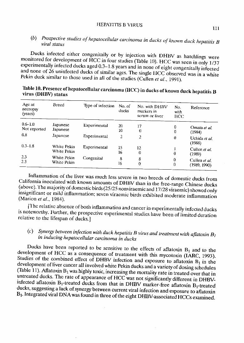
Ducks infec.ted either congenitaIly or by injection with DHBV as hatchlings weremonitored for development of HCC in four studies (Table 10). HCC was seen in only 1/37experimentaIly infected ducks aged 0.3-1.8 years and in no ne of eight congenitally infectedand no ne of 26 uninfected ducks of similar ages. The single HCC observed was in a whitePekin duck similar to those used in aIl of the studies (Cullen et al., 1991).
Table 10. Presence ofhepatocellularcarcinoma (HCC) in ducks ofknown duck hepatitis Bvirus (DHBV) status
Age at B reed Type of infection No. of No. with DHBV No. Referencenecropsy ducks markers in with(years) serum or liver HCC
0.6-1.0 Japanese Experimental 20 17 0 Omata et al.Not reported Japanese 10 0 0 (1984)0.8 Japanese Experimental 2 2 0 Uchida et aL.
(1988)0.3-1.8 White Pekin Experimental 15 12 1 Cullen et al.White Pekin 16 0 0 (1989)2.3 White Pekin Congenital 8 8 0 Cullen et al.2.3 White Pekin 16 0 0 (1989, 199)
Inflammation of the liver was much less severe in two breeds of domestic ducks fromCalifornia inoculated with known amounts of DHBV than in the free-range Chinese ducks(above). The majority of domestic birds (25/25 nonviraemic and 17/28 viraemic) showed onlyinsignificant or mild inflammation; seven viraemic birds exhibited moderate inflammation(Marion et al., 1984).
(The relative absence of both inflammation and cancer in experimentaIly infected ducksis noteworthy. Further, the prospective experimental studies have been of limited durationrelative to the lifespan of ducks.)
(c) Synergy between infection with duck hepatitis B virus and treatment with ajlatoxin B1
in inducing hepatocellular carcinoma in ducks
Ducks have been reported to be sensitive to the effects of aflatoxin Bi and to thedevelopment of HCC as a consequence of treatment with this mycotoxin (lARe, 1993).Studies of the combined effect of DHBV infection and exposure to aflatoxin Bi in thedevelopment of liver cancer ail involved white Pekin ducks and a variety of dosing schedules(Table Il). Afatoxin Bi was highly toxic, increasing the mortality rate in treated over that inuntreated ducks. The rate of appearance of HCC was not significantly different in DHBV-infected aflatoxin Bi-treated ducks from that in DHBV marker-free aflatoxin Bi-treatedducks, suggesting a lack of synergy between current viral infection and exposure to aflatoxinBi. Integrated viral DNA was found in three ofthe eight DHBV-associated HCCs examined.
Table 11. Development ofhepatocellular carcinoma (HeC) in ducks with and without duck hepatitis B virus (DHBV) -infection treated with aflatoxin Bi (AFBi) -NTreatment Age at No. of Effective No. with DHBV No. with HCC Integrated Reference
necropsy ducks n umber markers in serum viral DNAin carcinoma
Experimentally infected at hatch; AFBi soon after inoculationUchida et al. (1988)
(The existence of species-specific hepadnaviruses closely related to HB~ which produceHCC in two species (woodchuck and Beechey ground squirrels), strengthens the plausibilityof the conclusion that HBV is carcinogenic.)
3.4.4 Other species
The DNA of a hepadnavirus that infects herons (HHBV) has been cloned andcharacterized genetically, but it has not been characterized biologically nor has infectionwith the virus been associated with the development of liver cancer (Sprengel et al., 1988).
Evidence that a hepadnavirus infects tree squirrels (Sciurus carolinensis pennsylvanicus)was reported from studies of their livers, but viraemia has never been described in treesquirrels, and the virus remains uncharacterized both genetically and biologically (Feitelsonet al., 1986a,b). Liver cancer has not been observed in tree squirrels with evidence ofhepadnaviral infection.
4. Other Relevant Data
4.1 Pathology
The pathology of infection by HBV involves an acute phase, which may be recognizedclinicaIIy as acute viral hepatitis, and then a long chronic phase with development of chronicactive hepatitis and often cirrhosis. HCC may evolve during some phase of chronic hepatitis,usually after cirrhosis has supervened but in some cases without cirrhosis. The majority ofHCCs associated with HBV appear to arise in a dinically silent way, with few symptoms andno evidence of chronic hepatitis until the carcinoma is in a late stage.
4.1.1 Acute hepatitis
The histological features of acute viral hepatitis are highly variable, and hepatitis A, B,C, D and E viruses cannot be distinguished. They are also highly variable for the same agentin patients of different ages and immune status. ln the neonate and other immuno-compromised individu aIs, the histological changes are usually very mild with no significanthepatocellular cyopathic change and no significant hepatoceIIular hydropic sweIIng, acido-philic necrosis or cholestasis. The tyical histological changes in an adult with symptomatic,icteric acute viral hepatitis include: (i) portal expansion with lymphoid hyperplasia (as occursin many systemic viral infections); (ii) lobular inflammatory reaction with proliferation ofsinusoidal lining cells; and (iii) marked hepatocellular changes, including hydropic change(especially in the perivenular areas), acidophilic necrosis and hepatocellular dropout. ln themost severe cases of acute viral hepatitis, extensive hepatocellular necrosis occurs (calledsubmassive or massive acute hepatic necrosis), and this is usually fataL. Such severe viralreactions are rare in the very young. ln young people with severe viral hepatitis and necrosis,regeneration of hepatocyes is rapid, and recovery occurs with no chronic sequelae (Peters,1975). There are few histopathological data on the transition of acute viral to chronIchepatitis in large series of patients.