39
The Relevance of Dose for Low-Energy Beta Emitters Dudley T Goodhead EU Scientific Seminar Emerging Issues on Tritium and Low Energy Beta Emitters Luxembourg, 13 November 2007 DTG 9.11.07

The Relevance of Dosefor
Low-Energy Beta Emitters
Dudley T Goodhead
EU Scientific SeminarEmerging Issues on Tritium and Low Energy Beta Emitters
Luxembourg, 13 November 2007
DTG9.11.07
OUTLINE
Introductory comments on dose, radiation quality and RBE
ICRP system
Some issues for this symposium
Beta-decay of radionuclidesLow-energy beta emitters
Unusual features of low-energy beta emitters
A few additional comments
Conclusions and recommendations
©DTG12.11.07

Introductory comments on ‘Dose’ (and radiation quality)Absorbed Dose
• Physical quantity, precisely defined, no changeable parameters• Absorbed dose is the quotient of de by dm, where de is the mean
energy imparted to matter of mass dm.• Absorbed dose = Deposited Energy ÷ Mass• Units: joule per kilogram = gray (Gy)
• Independent of type (quality) of ionizing radiation• Approximately proportional to the average density of ionizations
in the mass (volume) of interest
BUT biological effectiveness of a given absorbed dose dependson many additional factors, including:
• Type of radiation (i.e. radiation quality)• Dose rate, dose fractionation• Particular biological system, effect and level of interest
D = de/dm
©DTG23.11.07
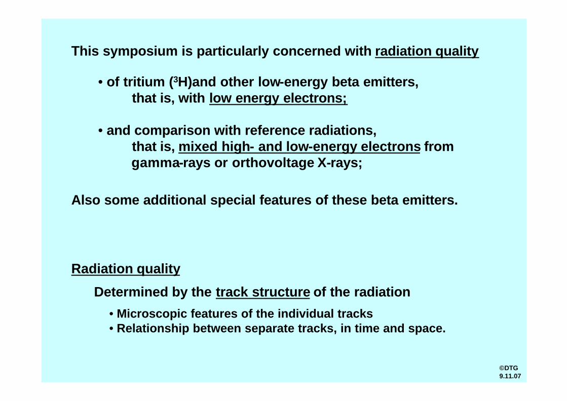
• of tritium (3H)and other low-energy beta emitters,that is, with low energy electrons;
• and comparison with reference radiations,that is, mixed high- and low-energy electrons fromgamma-rays or orthovoltage X-rays;
This symposium is particularly concerned with radiation quality
Also some additional special features of these beta emitters.
Radiation quality
Determined by the track structure of the radiation
• Microscopic features of the individual tracks• Relationship between separate tracks, in time and space.
©DTG9.11.07
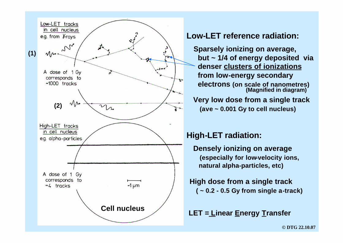
Low-LET reference radiation:
Sparsely ionizing on average,but ~ 1/4 of energy deposited via denser clusters of ionizationsfrom low-energy secondary electrons (on scale of nanometres)
Very low dose from a single track(ave ~ 0.001 Gy to cell nucleus)
(1)
(2)
High-LET radiation:
Densely ionizing on average(especially for low-velocity ions, natural alpha-particles, etc)
High dose from a single track( ~ 0.2 - 0.5 Gy from single a-track)
LET = Linear Energy TransferCell nucleus
(Magnified in diagram)
© DTG 22.10.07

DNA
electron(1)
(2)
Alpha-particle
Clustered ionizations fromlow-energy electron
Delta-ray electron
All radiation tracks are highly structured on the scale of DNA
Single ionization
Opposing trends: Alpha-particle has-- low probability of hitting DNA
(few tracks per Gy)-- high probability of damage when
it does hit.
© DTG21.8.03
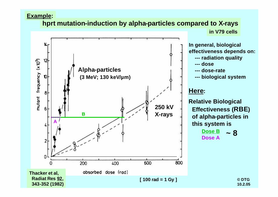
hprt mutation-induction by alpha-particles compared to X-rays
Alpha-particles(3 MeV; 130 keV/µm)
250 kVX-rays
Relative Biological Effectiveness (RBE)of alpha-particles in this system is
Here:
[ 100 rad = 1 Gy ]Thacker et al,Radiat Res 92,343-352 (1982)
in V79 cells
B
A
Dose BDose A
~ 8
In general, biological effectiveness depends on:
--- radiation quality--- dose--- dose-rate--- biological system
© DTG10.2.05
Example:
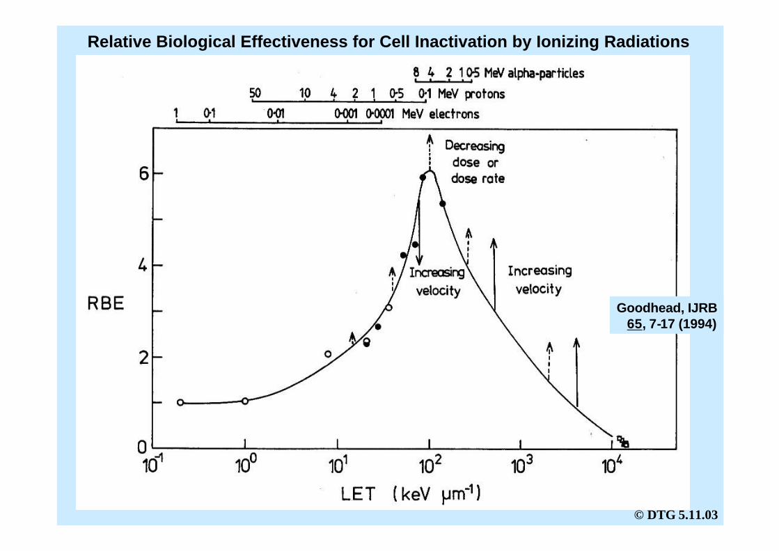
Relative Biological Effectiveness for Cell Inactivation by Ionizing Radiations
Goodhead, IJRB65, 7-17 (1994)
© DTG 5.11.03
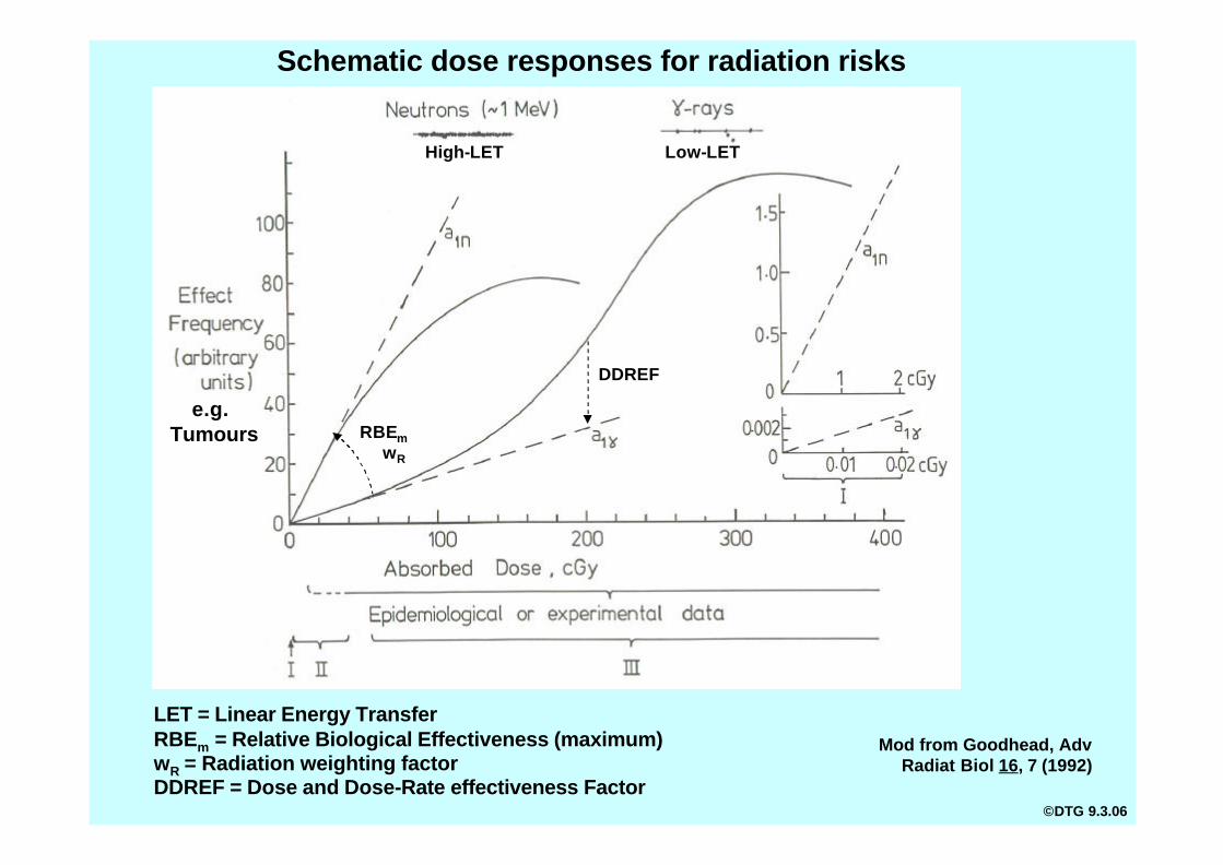
Mod from Goodhead, Adv Radiat Biol 16, 7 (1992)
RBEmwR
DDREF
Schematic dose responses for radiation risks
Low-LET
LET = Linear Energy TransferRBEm = Relative Biological Effectiveness (maximum)wR = Radiation weighting factorDDREF = Dose and Dose-Rate effectiveness Factor
e.g.Tumours
High-LET
©DTG 9.3.06
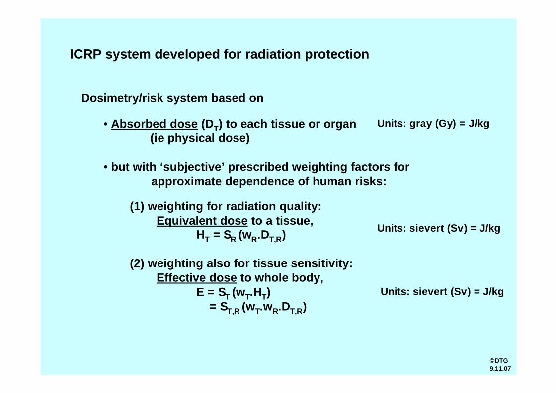
ICRP system developed for radiation protection
Dosimetry/risk system based on
• Absorbed dose (DT) to each tissue or organ(ie physical dose)
• but with ‘subjective’ prescribed weighting factors for approximate dependence of human risks:
(1) weighting for radiation quality:Equivalent dose to a tissue,
HT = SR (wR.DT,R)
(2) weighting also for tissue sensitivity:Effective dose to whole body,
E = ST (wT.HT)= ST,R (wT.wR.DT,R)
Units: sievert (Sv) = J/kg
Units: sievert (Sv) = J/kg
Units: gray (Gy) = J/kg
©DTG9.11.07
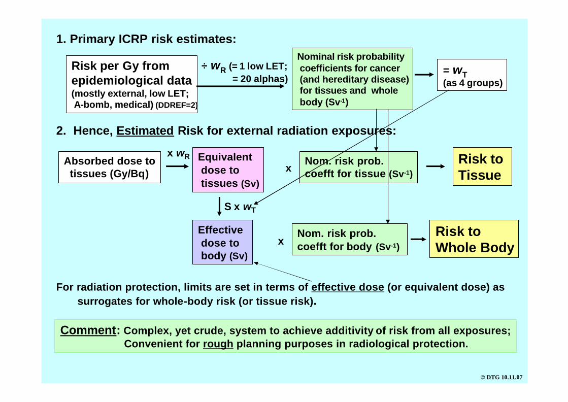
Risk per Gy fromepidemiological data(mostly external, low LET;A-bomb, medical)
÷ wR (= 1 low LET;= 20 alphas)
Nominal risk probabilitycoefficients for cancer(and hereditary disease)for tissues and wholebody (Sv-1)
= wT(as 4 groups)
1. Primary ICRP risk estimates:
2. Hence, Estimated Risk for external radiation exposures:
Absorbed dose totissues (Gy/Bq)
x wR Equivalentdose to tissues (Sv)
Effective dose tobody (Sv)
S x wT
Nom. risk prob.coefft for tissue (Sv-1)
Risk to Tissue
Nom. risk prob.coefft for body (Sv-1)
Risk toWhole Body
Comment: Complex, yet crude, system to achieve additivity of risk from all exposures;Convenient for rough planning purposes in radiological protection.
x
x
© DTG 10.11.07
(DDREF=2)
For radiation protection, limits are set in terms of effective dose (or equivalent dose) assurrogates for whole-body risk (or tissue risk).

Epi data(mostly external, low LET;A-bomb, medical)
x wR (=1 low LET;= 20 alphas)
Nominal risk probabilitycoefficients for cancer(and hereditary) (Sv-1)
= wT(as 4 groups)
1. Primary ICRP risk estimates:
2. ICRP Dose Coefficients for internal radionuclides:Biokinetic models
(intake ? tissues)+Dosimetric models
(decays ? absorbeddose)
Absorbed dose totissues (Gy/Bq)
x wREquivalentdose to tissues (Sv/Bq)
Effective dose tobody (Sv/Bq)
S x wT
3. Hence, Estimated Risk from internal radionuclide exposure:
Estimated intake (Bq)(ingestion, inhalation,absorption)
Tissue dosecoefft (Sv/Bq)
Nom. risk prob.coefft for tissue (Sv-1)
Risk to Tissue
Body dosecoefft (Sv/Bq)
Nom. risk prob.coefft for body (Sv-1)
Risk toWhole Body
Comment: Complex, yet crude, system to achieve additivity of risk from all exposures;Convenient for rough planning purposes in radiological protection.
x x
x x
© DTG10.11.07
For radiation protection, limits are set in terms of effective dose (or equivalent dose) assurrogates for whole-body risk (or tissue risk)
(i.e.Dose per unit intake)

Hence, effective dose is used
• as primary quantity for dose-limits in radiation protection--- for prospective dose assessment, optimization and for demonstrating
compliance
• as surrogate for risk (within the broad approximations of the ICRP system)
• for simple additivity of doses (and implied risks) from low-dose exposurescenarios, including • non-uniform irradiation of body or tissues
• mixed radiation qualities• internal and external radiation sources• any temporal distributions of dose
(i.e. dose-rate and dose fractionations)
Effective dose is not suitable for
• more accurate retrospective assessments of individual doses and risks
• use in epidemiological studies
• probability of causation in exposed individuals
[ICRP draft recommendations, Jan 2007]©DTG12.11.07
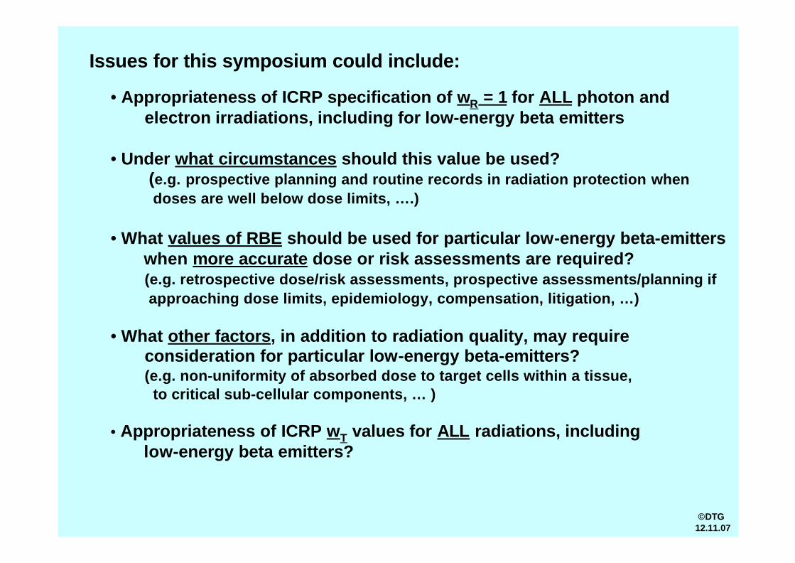
Issues for this symposium could include:
• Appropriateness of ICRP specification of wR = 1 for ALL photon and electron irradiations, including for low-energy beta emitters
• Under what circumstances should this value be used? (e.g. prospective planning and routine records in radiation protection when doses are well below dose limits, ….)
• What values of RBE should be used for particular low-energy beta-emitters when more accurate dose or risk assessments are required?(e.g. retrospective dose/risk assessments, prospective assessments/planning ifapproaching dose limits, epidemiology, compensation, litigation, …)
• What other factors, in addition to radiation quality, may requireconsideration for particular low-energy beta-emitters?(e.g. non-uniformity of absorbed dose to target cells within a tissue,
to critical sub-cellular components, … )
• Appropriateness of ICRP wT values for ALL radiations, including low-energy beta emitters?
©DTG12.11.07
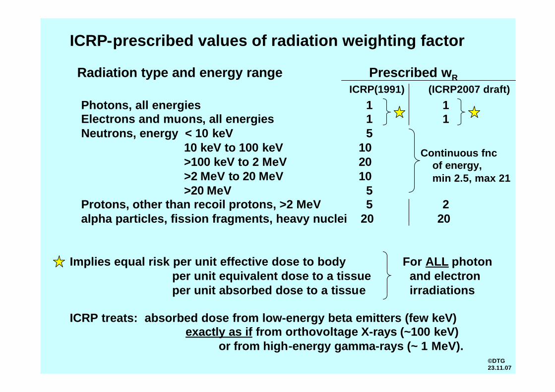
ICRP-prescribed values of radiation weighting factor
Implies equal risk per unit effective dose to bodyper unit equivalent dose to a tissueper unit absorbed dose to a tissue
For ALL photon and electronirradiations
ICRP treats: absorbed dose from low-energy beta emitters (few keV)exactly as if from orthovoltage X-rays (~100 keV)
or from high-energy gamma-rays (~ 1 MeV).
Radiation type and energy range Prescribed wR
Photons, all energies 1 1Electrons and muons, all energies 1 1Neutrons, energy < 10 keV 5
10 keV to 100 keV 10>100 keV to 2 MeV 20>2 MeV to 20 MeV 10>20 MeV 5
Protons, other than recoil protons, >2 MeV 5 2alpha particles, fission fragments, heavy nuclei 20 20
Continuous fncof energy,min 2.5, max 21
ICRP(1991) (ICRP2007 draft)
©DTG23.11.07

Beta decay of radionuclides:
Electron emission (ß- decay):
AX AY + 0e + 0nu
Positron emission (ß+ decay):
AX AY + 0e + 0nu
_Z Z+1 -1 0
Z Z-1 +1 0
H He + e + nu3 3 - _
1 2
Tritium betaspectrum
Electron
Tritium ß- decay:Emaxmax = 18.6 = 18.6 keVkeV
Eave = 5.7 keV
©DTG23.11.07
[where nu is neutrino]

Some relevant low-energy beta- -emitting radionuclides:
3H 3He 18.6 5.7 ~7 ~0.56 12.3 y1 2
Electron energy (keV) Electron range (µm)Half-life
14C 14N 157 ~290 5730 y6 7
35S 35Cl 167 ~320 87 d
106Ru 106Rh 39.4 ~28 574 d
Compare:90
Sr 90
Y 546 ~1950 29 y38 39
131I
131Xe 971 ~4200 8 d53 54
137Cs
137Ba 1176 ~5200 30 y
55 56
210Pb 210Bi 63.5 ~64 22 y
Max Average Max Average
16 17
44 45
82 83 (ß,a)
ß- -decay
(ß)
(+gamma)
(+gamma)
©DTG 23.11.07

Unusual features of low-energy beta-emitters:
1) Increased average ionization density (LET)
2) Short electron tracks
3) Non-uniformity of dose
4) Cell (or nucleus) hit frequencies per unit dose (numbers of tracks)
5) Nuclear transmutations
6) Isotopic mass differences
7) Molecular forms
[8) Positron annihilation for ß+-emitters]
Most of these features are not incorporated into conventional radiation protection dosimetry.
©DTG8.11.07
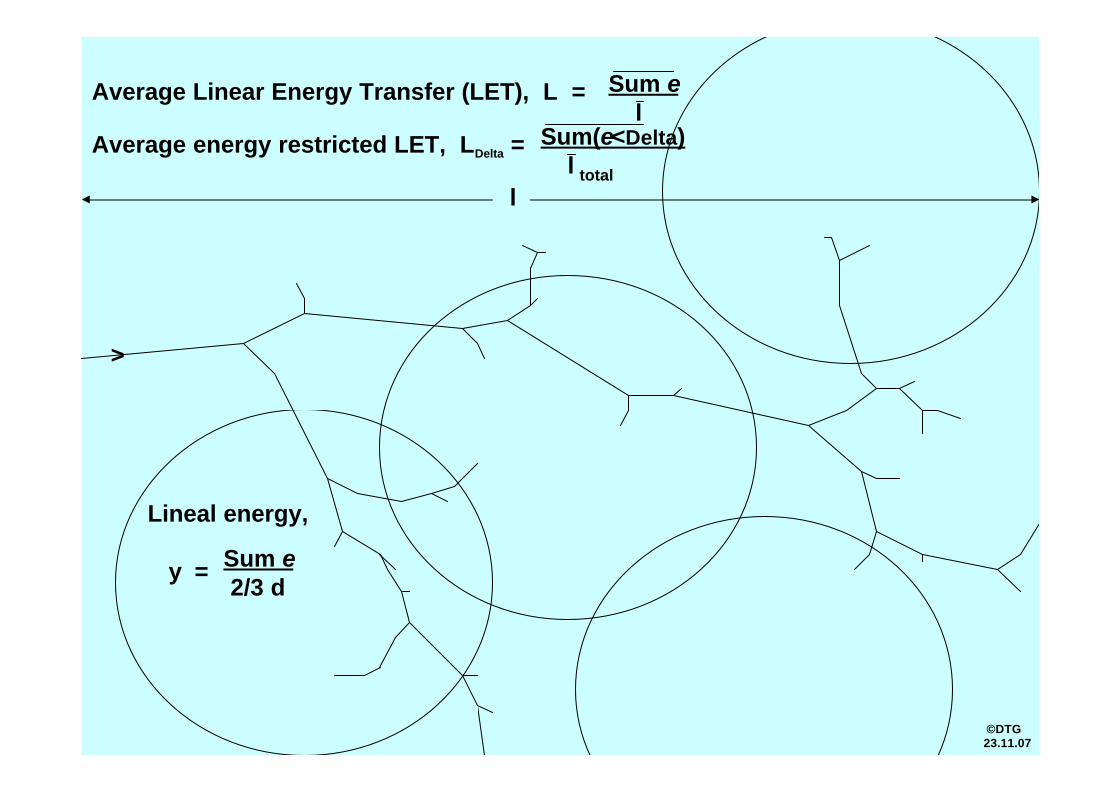
Average Linear Energy Transfer (LET), L = Sum el
l
>
Average energy restricted LET, LDelta = Sum(e<Delta)l total
Lineal energy,
y = Sum e2/3 d
©DTG23.11.07

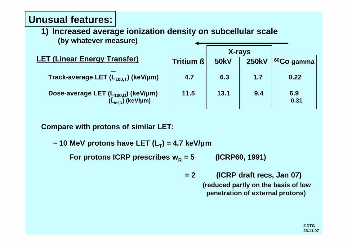
1) Increased average ionization density on subcellular scale(by whatever measure)
LET (Linear Energy Transfer) (keV/µm)
Track-average LET (L100,T) 4.7 6.3 1.7 0.22
Dose-average LET (L100,D) 11.5 13.1 9.4 6.9[LInf,D] [0.31]
Tritium ß 50kV 250kV 60Co gamma
Lineal energy (keV/µm)
Frequency-mean (yF) 1.4 ~1.7 1.0 0.28Dose-mean (yD) 2.1 ~2.6 2.1 0.62
Sitediameterd = 5 µm
Frequency-mean (yF) 3.1 2.2 1.2 0.37Dose-mean (yD) 5.2 5.0 3.7 1.6
Frequency-mean (yF) 4.0 - - -Dose-mean (yD) 9.2 - 8.1 4.3
d = 1 µm
d = 0.1 nm
Frequency-mean (yF) 7.8 6.9 6.1 -Dose-mean (yD) 18.0 17.7 17.0 12.6
d = 0.01 nm
Unusual features:
X-rays
Frequency-mean (yF) 4.1 2.6 1.4 0.52Dose-mean (yD) 7.3 5.4 4.7 2.3
d = 0.5 µm
65kV 200kV
40kV 250 kV
[L8 ,T] [~12]
©DTG 23.11.07
1) Increased average ionization density on subcellular scale(by whatever measure)
LET (Linear Energy Transfer)
Track-average LET (L100,T) (keV/µm) 4.7 6.3 1.7 0.22
Dose-average LET (L100,D) (keV/µm) 11.5 13.1 9.4 6.9(LInf,D) (keV/µm) 0.31
Tritium ß 50kV 250kV 60Co gamma
Unusual features:
X-rays
Compare with protons of similar LET:
~ 10 MeV protons have LET (LT) = 4.7 keV/µm
For protons ICRP prescribes wR = 5 (ICRP60, 1991)
= 2 (ICRP draft recs, Jan 07) (reduced partly on the basis of low
penetration of external protons)
©DTG23.11.07

Two low-energy-electron tracks(Typical of secondary e’s from X-, gamma-rays)
1 keVelectron
0.5 keV electron
DNA [ Nikjoo, Charlton, GoodheadAdv Space Res 14,161(1994) ]
© DTG 21.8.03

nn n
h n
n
h
n
n
n
n
n
n
n n
n
n
hh
h
h
h
h
hh
h
h
h
h
h
hh
h hh
h
h
h
hh h
h
hh
h
h
h
h
h
h
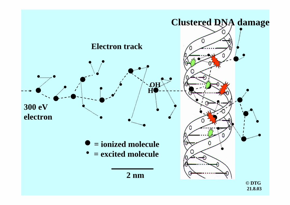
Electron track
2 nm
300 eVelectron
n = ionized moleculeh = excited molecule
OHH
Clustered DNA damage
© DTG21.8.03

2) Short ranges of electrons (beta-particles)
Ranges of tritium beta-particles:
Average 0.56 µmMaximum ~ 7 µm
Compare with:
Typical cell diameters ~ 7 µm to 30µmTypical cell nucleus diameters ~ 6 µm to 15 µmChromatin fibre diameter ~ 0.030 µmDNA diameter ~ 0.0024 µm
Short range • does not mask increased LET of these electrons on scale of
DNA and chromatin;• limits ability of single track to damage two distant targets on cellular scale;• can lead to non-uniformity of dose when emitters are inhogeneously
distributed.
Unusual features:
Hence:
©DTG12.11.07
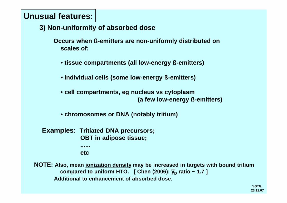
3) Non-uniformity of absorbed dose
Unusual features:
Occurs when ß-emitters are non-uniformly distributed on scales of:
• tissue compartments (all low-energy ß-emitters)
• individual cells (some low-energy ß-emitters)
• cell compartments, eg nucleus vs cytoplasm (a few low-energy ß-emitters)
• chromosomes or DNA (notably tritium)
Examples: Tritiated DNA precursors;OBT in adipose tissue;......etc
NOTE: Also, mean ionization density may be increased in targets with bound tritiumcompared to uniform HTO. [ Chen (2006): yD ratio ~ 1.7 ]
Additional to enhancement of absorbed dose.©DTG
23.11.07

Unusual features:
4) Cell (or nucleus) hit frequencies per unit dose• Larger mean energy deposition by single 3H ß than from single
track from Co gamma;
• Hence, fewer hits from tritium than from Co gamma-rays (for equal average absorbed dose to tissue);
• i.e. Fewer cells (or nuclei) are hit by 3H, but they are hit harder.
• Any consequences?
zF (mGy) 4.6 1.1
Hit frequency =1/zF (mGy-1) 0.2 0.9
zF 1.3 0.4
Hit frequency =1/zF (mGy-1) 0.8 2.5
For sphere d = 7 µm
For sphere d = 12 µm
3H
where zF = mean specific energy
(Thresholds, Dose rate)
©DTG23.11.07
Cogamma
Unusual features:
5) Nuclear transmutation
• Molecular changes result from transmutation of ß-emitting radionuclide
• Conversion of 3H to 3He loses its chemical binding in molecule(e.g. deprotonation in a DNA base, potentially mutagenic?
disruption of hydrogen bonding in DNA)
• Conversion of 14C to 14N in DNA base (potentially mutagenic?)
• Conversion of 35S to 35Cl alters the biomolecule
©DTG9.11.07

Unusual features:
6) Isotopic mass difference ratio compared to stable isotope
• Affects physico-chemical properties
• Mass difference is very large for 3H compared to normal 1H,by ratio of 3
(e.g. affect chemical reaction rates for uptake and clearance;differential diffusion;
‘buried tritium’:differential binding of water in hydration shell of DNA – enrichment factor 2?differential binding in proteins, other macromolecules -- ” ” 1.4?
• Ratios are very small for most other ß-emitters
©DTG9.11.07
Unusual features:
7) Molecular forms
• Different molecular compounds of ß-emitters can influence uptakeratios, retention times and other biokinetic parameters
• Notable forms for 3H include:-- tritiated water-- organically bound tritium (OBT) – exchangable
-- non-exchangable-- DNA precursors
©DTG8.11.07

Unusual features:
8) Positron annihilation (ß+ emitters)
e+ + e- 2 gamma (High energies, >0.5 MeV each)
• Delocalizes energy of ß+ -emitters
©DTG23.11.07

Unusual features of low-energy beta-emitters:
1) Increased average ionization density (LET)
2) Short electron tracks
3) Non-uniformity of dose
4) Cell (or nucleus) hit frequencies per unit dose (numbers of tracks)
5) Nuclear transmutations
6) Isotopic mass differences
7) Molecular forms
[8) Positron annihilation for ß+-emitters]
• Most of these features are not incorporated into conventional radiation protection dosimetry.
• They may be incorporated in various ways into experimentalmeasurements of RBE
©DTG8.11.07

A few additional comments
©DTG8.11.07
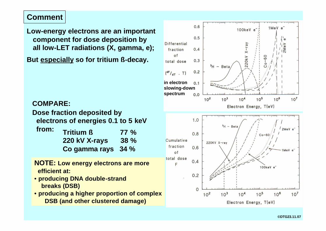
Dose fraction deposited by electrons of energies 0.1 to 5 keVfrom: Tritium ß 77 %
220 kV X-rays 38 %Co gamma rays 34 %
Low-energy electrons are an importantcomponent for dose deposition by all low-LET radiations (X, gamma, e);
in electron slowing-downspectrum
But especially so for tritium ß-decay.
COMPARE:
NOTE: Low energy electrons are more efficient at:
• producing DNA double-strand breaks (DSB)
• producing a higher proportion of complexDSB (and other clustered damage)
Comment
©DTG23.11.07
nn n
h n
n
h
n
n
n
n
n
n
n n
n
n
hh
h
h
h
h
hh
h
h
h
h
h
hh
h hh
h
h
h
hh h
h
hh
h
h
h
h
h
h
Electron track
2 nm
300 eVelectron
n = ionized moleculeh = excited molecule
OHH
Clustered DNA damage
© DTG21.8.03
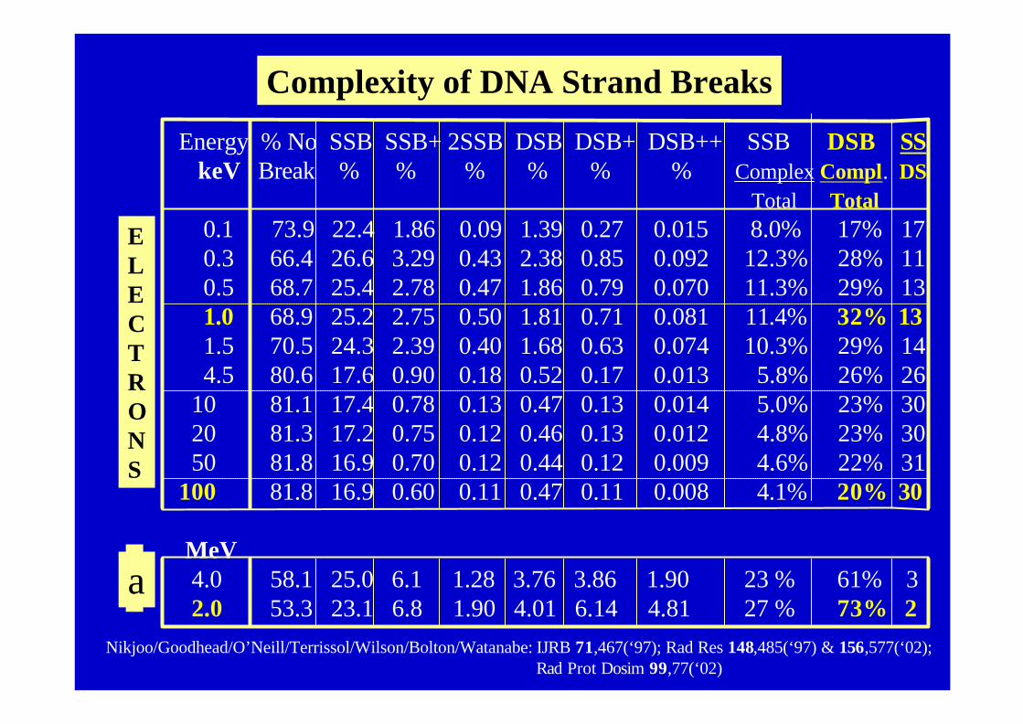
Energy % No SSB SSB+ 2SSB DSB DSB+ DSB++ SSB DSB SSkeV Break % % % % % % Complex Compl. DS
Total Total0.1 73.9 22.4 1.86 0.09 1.39 0.27 0.015 8.0% 17% 170.3 66.4 26.6 3.29 0.43 2.38 0.85 0.092 12.3% 28% 110.5 68.7 25.4 2.78 0.47 1.86 0.79 0.070 11.3% 29% 131.0 68.9 25.2 2.75 0.50 1.81 0.71 0.081 11.4% 32% 131.5 70.5 24.3 2.39 0.40 1.68 0.63 0.074 10.3% 29% 144.5 80.6 17.6 0.90 0.18 0.52 0.17 0.013 5.8% 26% 26
10 81.1 17.4 0.78 0.13 0.47 0.13 0.014 5.0% 23% 3020 81.3 17.2 0.75 0.12 0.46 0.13 0.012 4.8% 23% 3050 81.8 16.9 0.70 0.12 0.44 0.12 0.009 4.6% 22% 31
100 81.8 16.9 0.60 0.11 0.47 0.11 0.008 4.1% 20% 30
MeV4.0 58.1 25.0 6.1 1.28 3.76 3.86 1.90 23 % 61% 32.0 53.3 23.1 6.8 1.90 4.01 6.14 4.81 27 % 73% 2
ELECTRONS
a
Complexity of DNA Strand Breaks
Nikjoo/Goodhead/O’Neill/Terrissol/Wilson/Bolton/Watanabe: IJRB 71,467(‘97); Rad Res 148,485(‘97) & 156,577(‘02); Rad Prot Dosim 99,77(‘02)
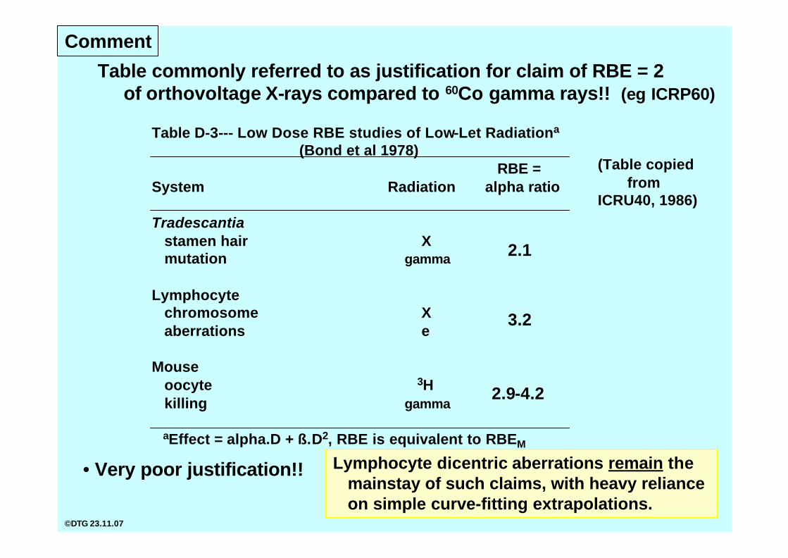
Table D-3--- Low Dose RBE studies of Low-Let Radiationa
(Bond et al 1978)RBE =
System Radiation alpha ratio
Tradescantiastamen hair X mutation gamma
Lymphocytechromosome Xaberrations e
Mouseoocyte 3Hkilling gamma
aEffect = alpha.D + ß.D2, RBE is equivalent to RBEM
2.1
3.2
2.9-4.2
Comment
Table commonly referred to as justification for claim of RBE = 2of orthovoltage X-rays compared to 60Co gamma rays!! (eg ICRP60)
• Very poor justification!!
(Table copied from
ICRU40, 1986)
Lymphocyte dicentric aberrations remain themainstay of such claims, with heavy reliance on simple curve-fitting extrapolations.
©DTG 23.11.07
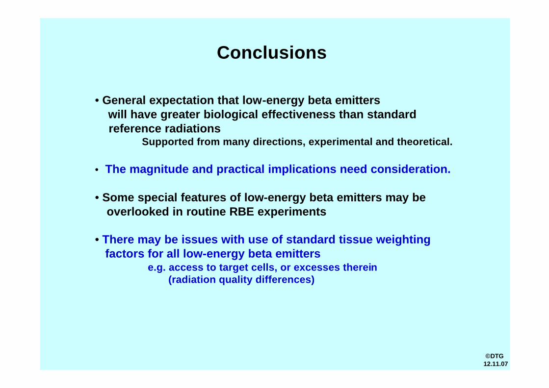
Conclusions
• General expectation that low-energy beta emitterswill have greater biological effectiveness than standard reference radiations
Supported from many directions, experimental and theoretical.
• The magnitude and practical implications need consideration.
• Some special features of low-energy beta emitters may beoverlooked in routine RBE experiments
• There may be issues with use of standard tissue weighting factors for all low-energy beta emitters
e.g. access to target cells, or excesses therein(radiation quality differences)
©DTG12.11.07
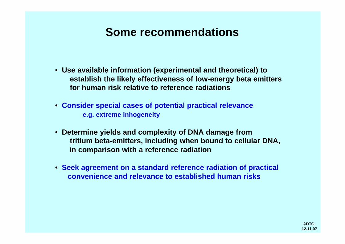
Some recommendations
• Use available information (experimental and theoretical) to establish the likely effectiveness of low-energy beta emittersfor human risk relative to reference radiations
• Consider special cases of potential practical relevancee.g. extreme inhogeneity
• Determine yields and complexity of DNA damage fromtritium beta-emitters, including when bound to cellular DNA, in comparison with a reference radiation
• Seek agreement on a standard reference radiation of practical convenience and relevance to established human risks
©DTG12.11.07
THE END


![URANIUM - National Film Board of Canada1].pdf · alpha emitters are the least harmful while gamma emitters are more dangerous than beta emitters. Inside the body, however, alpha emitters](https://static.documents.pub/doc/80x56/604a60e06cb0dd2c8f04d503/uranium-national-film-board-of-1pdf-alpha-emitters-are-the-least-harmful-while.jpg)


