108
The Reproductive System Chapter 17
| Date post: | 01-Jan-2016 |
| Category: |
Documents |
| Upload: | carla-bradley |
| View: | 20 times |
| Download: | 1 times |
The Reproductive SystemChapter 17

Introduction
• The Reproductive System seeks to ensure the survival of the species.
• Is not essential to life of the animal but for perpetuation of the species.
• Generally requires a second animal of the opposite sex in order to carry out its functions.
• Complete reproductive system is all the structures both male and femal required for reproduction.

Fertilization
• Fertilization occurs when the spermatazoon penetrates the cytoplasm of the ovum– Once egg is fertlized, must be provided with
hospitable environment in order to develop.– In order for this to occur both male and female
systems must be in sync.

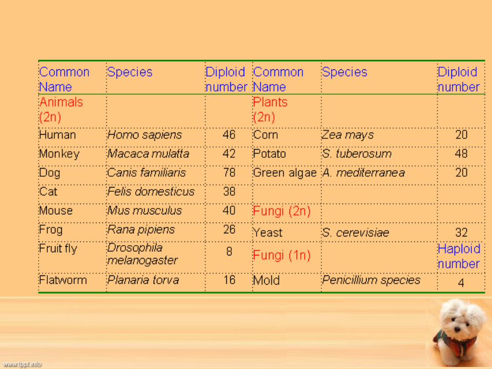
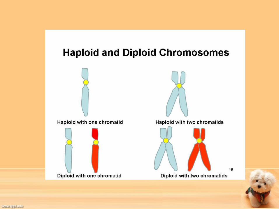
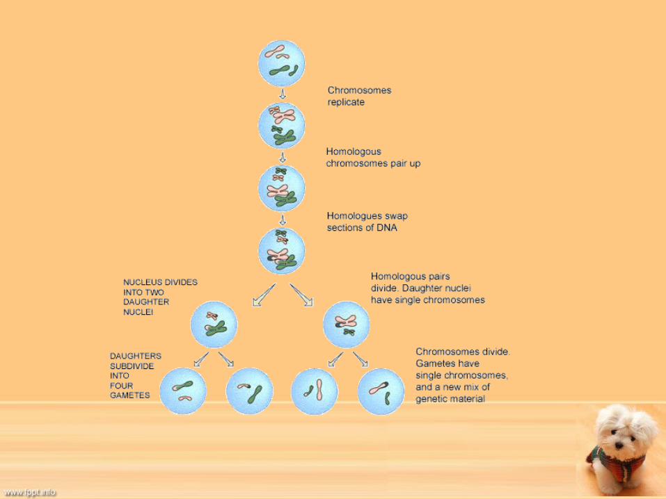
Meiosis• Unique process of cell division• Is a reduction division of reproductive cells so that a
chromosome number goes from a diploid number to a haploid number.
• It ensures that each animal’s genetic make up is unique.
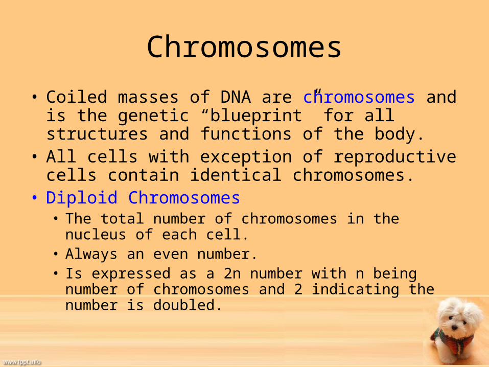
Chromosomes• Coiled masses of DNA are chromosomes and is the
genetic “blueprint” for all structures and functions of the body.
• All cells with exception of reproductive cells contain identical chromosomes.
• Diploid Chromosomes• The total number of chromosomes in the nucleus of each
cell.• Always an even number. • Is expressed as a 2n number with n being number of
chromosomes and 2 indicating the number is doubled.

Chromosomes Continued
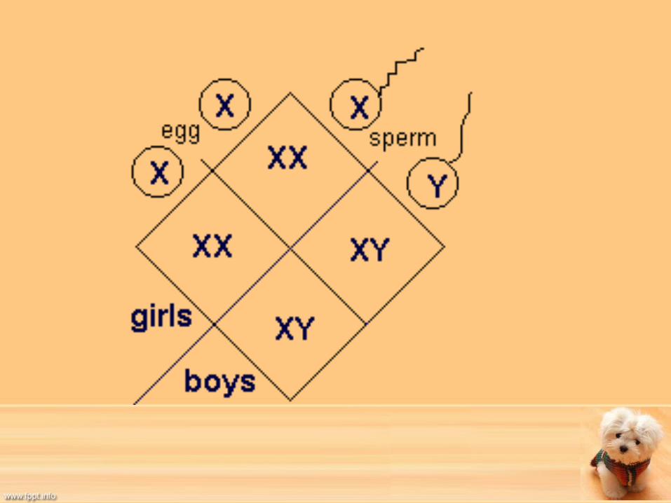
• Sex Chromosomes– Determine the gender of the animal.– Designated as X or Y.– XX=Female– XY= Male– YY combination is not possible since all males
produce an XY and females only produce XX.– Full diploid number may be expressed as 2n, XX or
2n, XY.
Chromosomes Continued
• Haploid Chromosome Number– Haploid is half of the diploid number.– Why are reproductive cells haploid in nature?– Is abbreviated as n, X or n, Y depending on sex of
the chromosome present. – Haploid number is a result of meiosis.• Reduction Division
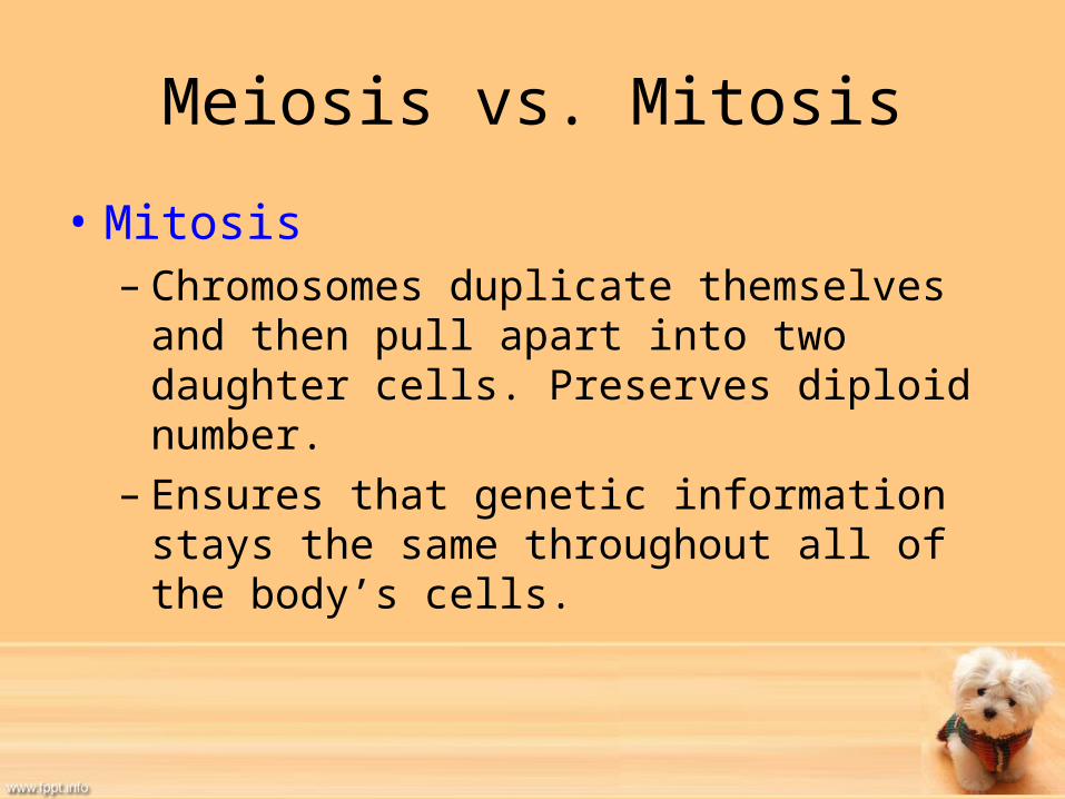
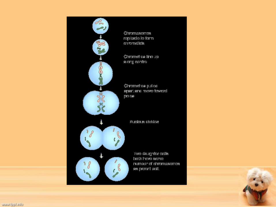
Meiosis vs. Mitosis
• Mitosis– Chromosomes duplicate themselves and then pull
apart into two daughter cells. Preserves diploid number.
– Ensures that genetic information stays the same throughout all of the body’s cells.
Meiosis vs. Mitosis
• Meiosis– Do not produce copy before daughter cells are
pulled apart.– Therefore half of total chromosomes go to each
daughter cell.– This makes process entirely random, resulting in
unique offspring.
Spermatogenesis
• Process of producing spermatozoa- the male sex cells.– Spermatozoa are produced continuously.– Designed to produce large numbers of
spermatozoa.– Occurs in seminiferous tubules of the testes.

Spermatogenesis• Begin with cell called primary spermatocyte.
– Primary spermatocyte has normal diploid number.– Divides by meiosis into two secondary spermatocytes.
• Now are haploid in number and are pushed to tubule lumen.– Secondary spermatocytes divide by mitosis into four spermatids.
• Why mitosis at this point?• 2 will have X markers and two will have Y markers
– So it is the sperm that dictate gender of offspring. – Why are there more females than males produced?
• In center of tubular lumen at this point.• Do not undergo any more cell divisions, but will grow tails and be
converted to spermatozoa• Once mature, will be transported to epididymis for storage before
ejaculation.


Oogenesis• Ova- female sex cells, are produced in the follicles of
the ovaries through process known as oogenesis. • Have fixed number of oocytes-precursor cells to ova,
soon after birth. – Oocyte has diploid number.– Do not become active until start ovarian cycle.
• Once activated will divide by meiosis.– Division produces a secondary oocyte and a polar body.– Then another mitotic division takes place to become ovum and 3
polar bodies. » Polar bodies never mature into ovum. Just are where excess
chromosomes are stored. – Each cycle produces one or more mature ova.
• Why just one or few ova are produced?
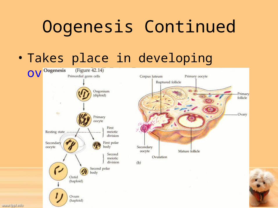
Oogenesis Continued
• Takes place in developing ovarian follicle.
Male Reproductive System
• Functions are:– 1. Produce male sex hormones– 2. Develop male reproductive cells• What are these cells called?
– 3. Deliver reproductive cells to female
Male Reproductive Organs• Many and complex, but include:
– Testes– Scrotum– Spermatic Cord– Seminiferous Tubules– Ducts– Epididymis– Vas Deferens– Urethra– Penis– Prostate gland– Bulbourethral glands– Seminal vesicles


Testes• Are male gonads- organs where male reproductive cells are
formed. • Usually housed in scrotum.
- Size and exact location may vary by animal- Outside abdomen
- Why?• Have two main functions:
– Spermatogenesis• Previously discussed.• Where does this take place?
– Hormone production• Takes place in interstitial cells between the seminiferous tubules. • Also may be called androgens.
– What is main hormone/androgen that is produced?


Testes Development and Location
• Located in abdominal cavity before birth near kidneys.
• Attached to scrotal area by gubernaculum- connective band of tissue.
• As embryo grows, gubernaculum does not so in a way, testes are “pulled” toward scrotum.
• Eventually are pulled through inguinal rings into scrotum.
• What we call testicles “descending”.
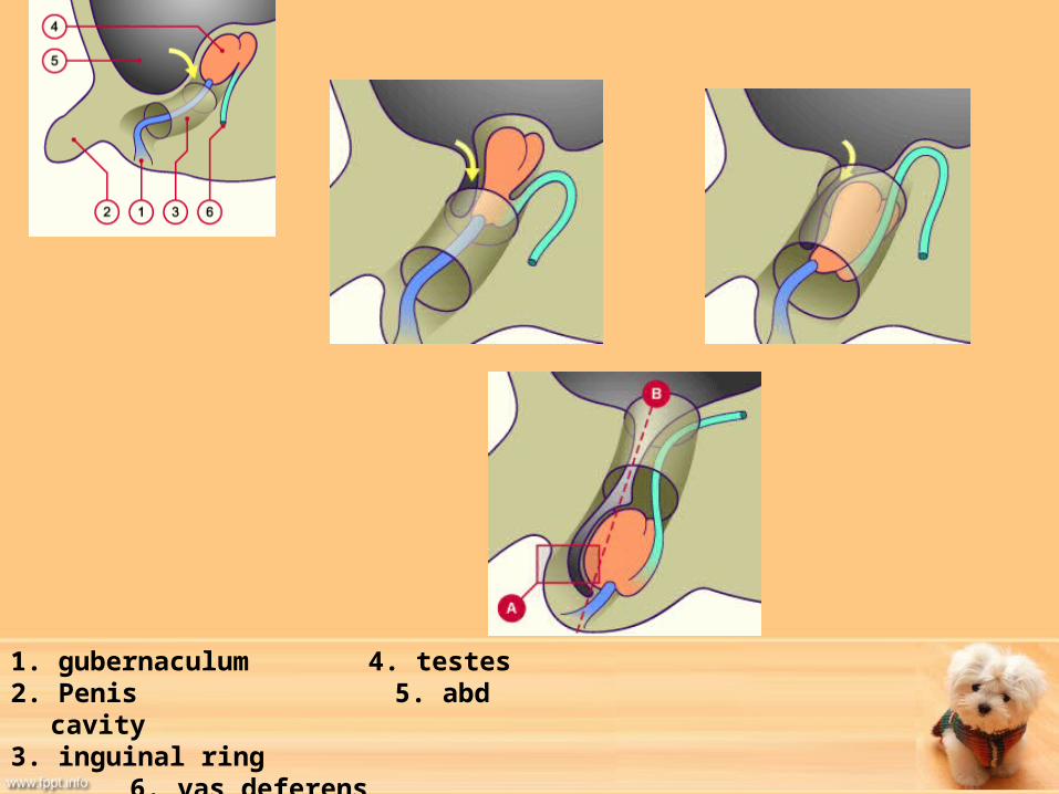
1. gubernaculum 4. testes2. Penis 5. abd cavity3. inguinal ring 6. vas
deferens
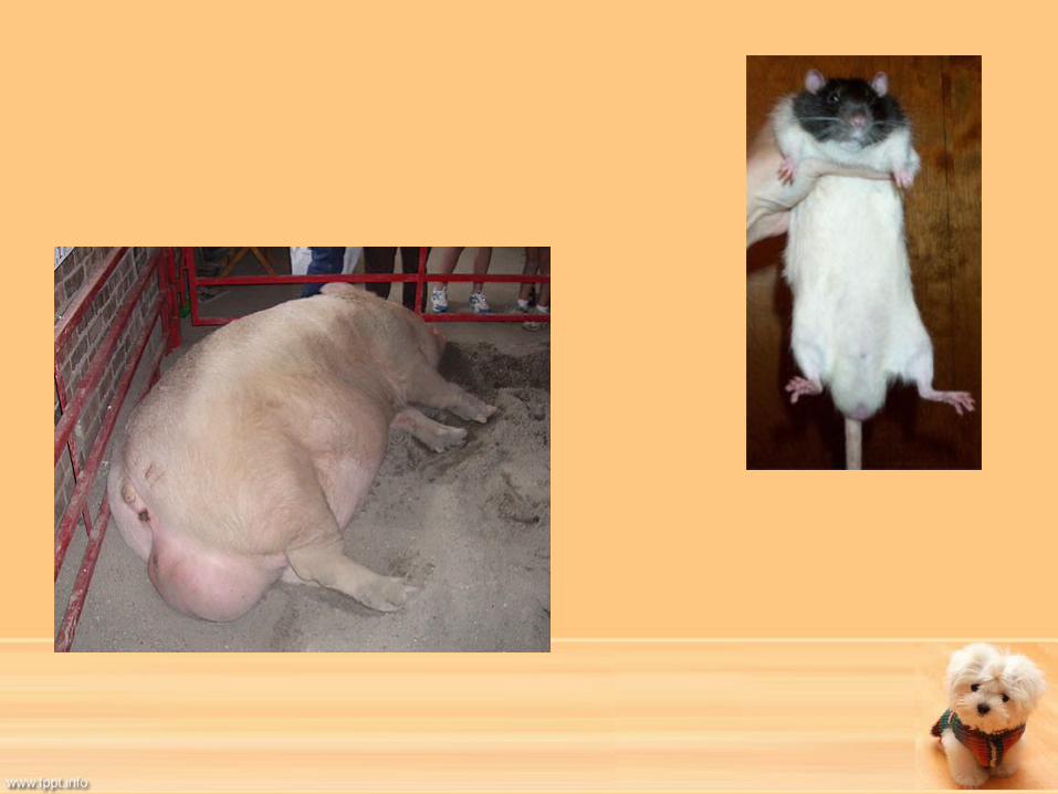
Undescended Testicles
• Generally all testicles should have descended to scrotum by 16 weeks of age.
• If testicles do not descend, then these male dogs are termed cryptorchid or have a retained testicle in the abdomen or inguinal area.
• May be able to still breed, but should be surgically removed as may cause testicular cancer.
• Is a genetic trait that may be passed down to future generations.

Testosterone
• Main male androgen that is produced in testes.
• Responsible for:– Development of secondary sex characteristics• Which are?
– Male libido• Which is?
– Anabolic effect on body• Which does what?
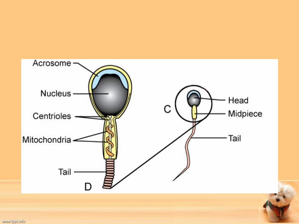
Spermatozoa
• Long thin cells composed of:– Head
• Contains nucleus• Covered by acrosome- caplike structure
– Acrosome has digestive enzymes that helps spermatozoa penetrate ovum
– Midpiece• Has large amount of mitochondria arranged in spiral pattern.
– Tail• Muscle-like fibers that help with propulsion once activated.
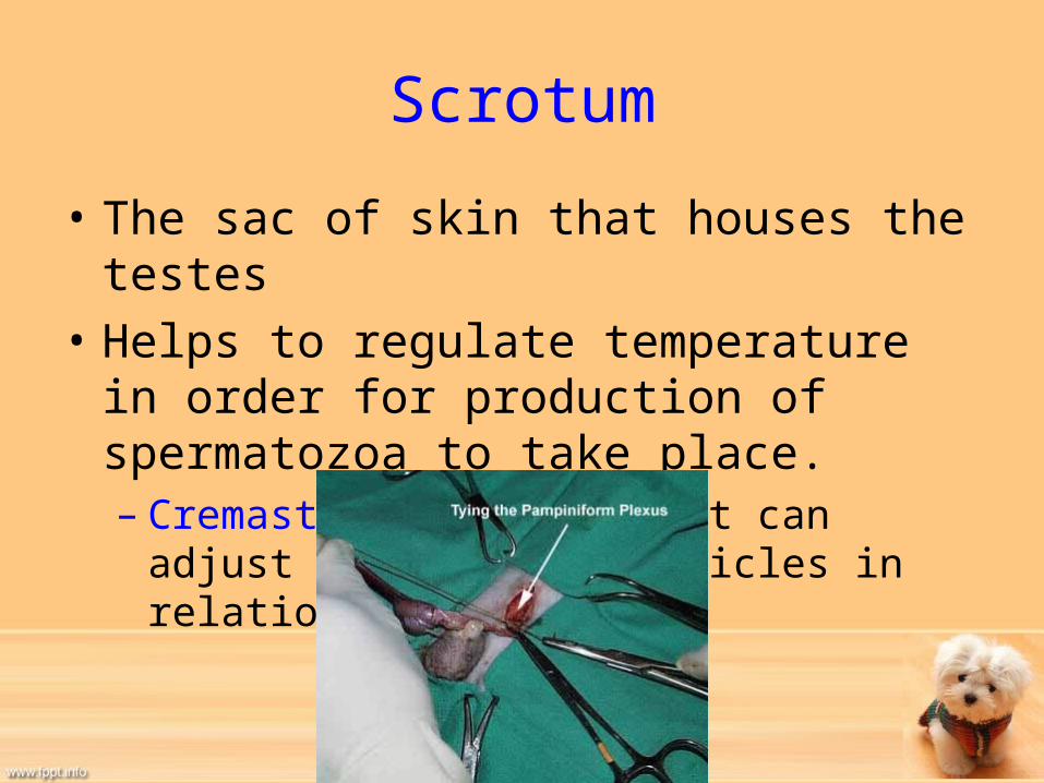
Scrotum
• The sac of skin that houses the testes• Helps to regulate temperature in order for
production of spermatozoa to take place.– Cremaster muscle is what can adjust position of
testicles in relation to body.

Spermatic Cord
• Link the testes with the rest of the body.• Contain blood vessels, nerves, lymphatic vessels, and
vas deferens.– Only one artery (testicular artery) carries blood down to
the testis. – Artery is surrounded by pampiniform plexus-network of
veins.• This network helps keep temperature lower than rest of body. • As blood passes down from body, it is cooled by blood returning
from the testis in the pampiniform plexus.• Also blood in pampiniform plexus is warmed by the blood in the
testicular artery as it goes back to body.


Structure of the Testes
• Composed of two layers of vaginal tunics– Surround testes in scrotum and spermatic cord. • Inner layer is visceral vaginal tunic or proper vaginal
tunic.– Derived from visceral layer of peritoneum that coat the testes
as they develop in the abdomen.– Thin and transparent to the naked eye.
• Outer parietal vaginal tunic or common vaginal tunic.– Derived from the parietal layer of peritoneum that lines
abdominal cavity.– Forms fibrous sac around each testis and spermatic cord.


Tunics importance
• Important during an orchiectomy- a castration. – Incision is made through outer parietal tunic and
testis is everted through tunic to expose blood vessels and spermatic cord.
– Blood vessels are ligated-tied off and testis is removed.
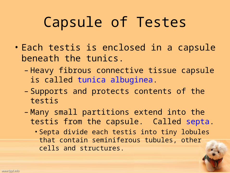
Capsule of Testes
• Each testis is enclosed in a capsule beneath the tunics. – Heavy fibrous connective tissue capsule is called
tunica albuginea. – Supports and protects contents of the testis– Many small partitions extend into the testis from
the capsule. Called septa. • Septa divide each testis into tiny lobules that contain
seminiferous tubules, other cells and structures.

Seminiferous Tubules
• Where spermatogenesis takes place.– Produce spermatozoa through meiosis.
• Long tubules that are U-shaped and attached at both ends to rete testis- a complex system of ducts.
• Intersitial cells are located between seminiferous tubules. – Interstitial cells are what produce androgens under
influence of Luteinizing Hormone (LH) from the anterior pituitary gland. • In male LH may also be referred to as Interstitial cell-stimulating
hormone (ICSH).
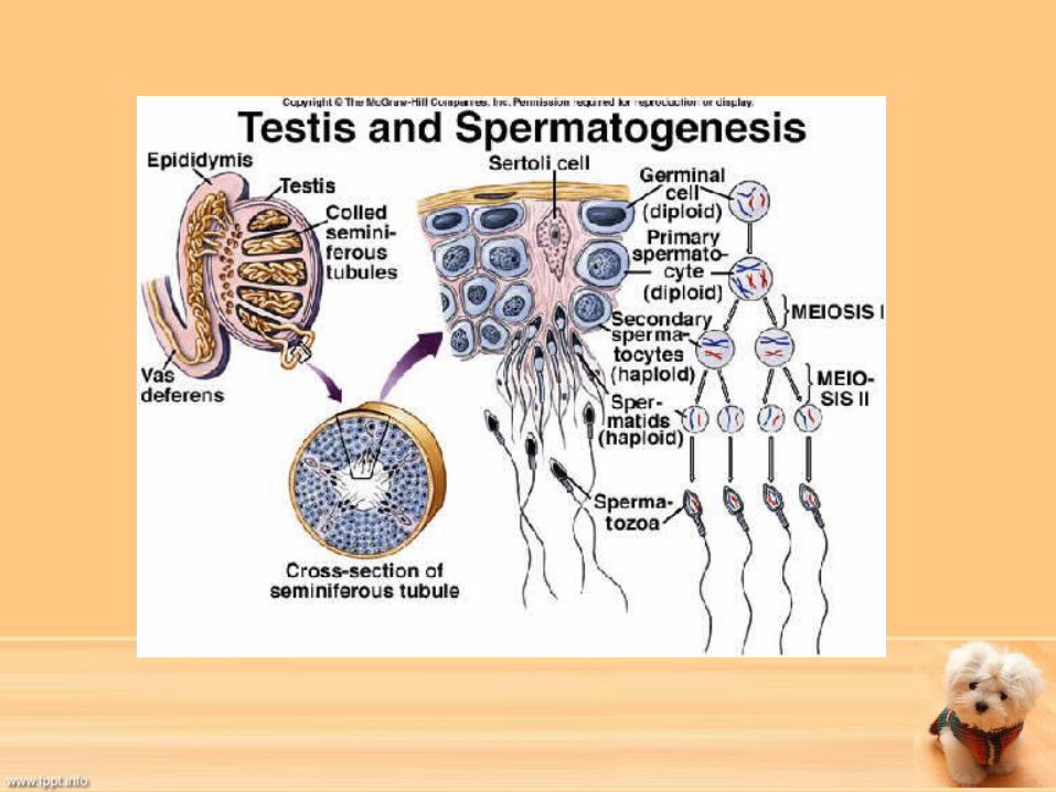
Seminiferous Tubules Continued
• Once spermatids are undergoing changes, they are attached to Sertoli cells- large “nurse” cells.– Sertoli cells help to shield spermatozoa from
body’s immune system.– If not shielded, proteins on surface of
spermatozoa would stimulate immune system to produce antibodies.

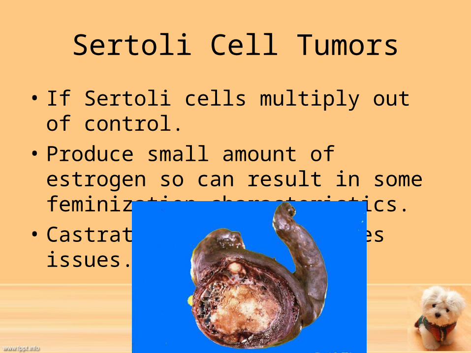
Sertoli Cell Tumors
• If Sertoli cells multiply out of control.• Produce small amount of estrogen so can
result in some feminization characteristics. • Castration usually resolves issues.

Duct System
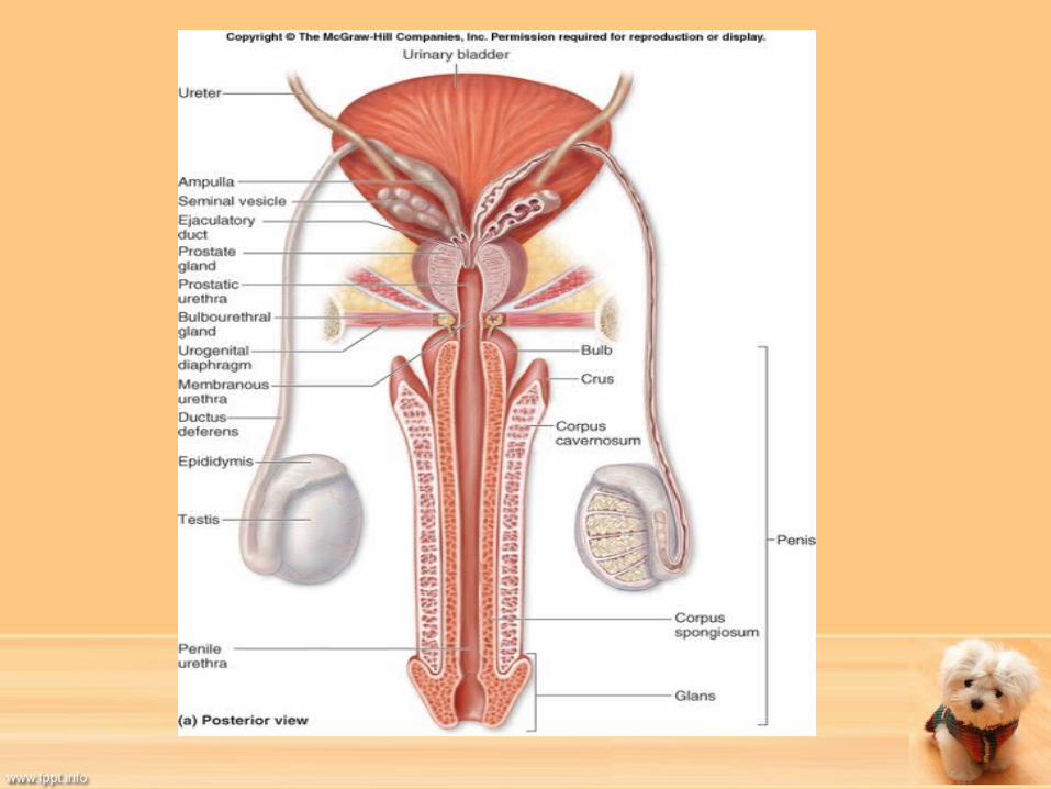
• Once spermatozoa have completed development, they are trasnported via rete testis to the efferent ducts of the testes to epididymis.
Epididymis• Flat, ribbon-like structure that lies along surface of
testis. – If stretched out would be 20 feet long
• Connects efferent ducts of testis with the vas deferens.
• Made up of 3 regions:– Head
• Where spermatozoa enter from efferent ducts– Body
• Main portion that lies next to testis– Tail
• Continues on to become vas deferens
Epididymis continued
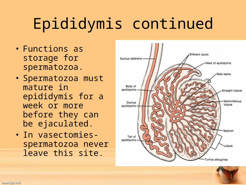
• Functions as storage for spermatozoa.
• Spermatozoa must mature in epididymis for a week or more before they can be ejaculated.
• In vasectomies- spermatozoa never leave this site.
Vas Deferens
• When ejaculation occurs, this muscular tube connects the epididymis with the urethra.– Must propel spermatozoa quickly at time of ejaculation.
• Also may be referred to as the ductus deferens. • In abdomen, separates from spermatic cord and
loops back to connect with urethra.– At this point, may enlarge and contain glands to contribute
to semen. This enlargement is called the ampulla.

Vasectomy
• Removal or clamping of a section of vas deferens to prevent spermatozoa from reaching urethra.
• Rest of seminal fluid is still present.• May be used to create teaser bulls or animals
that are considered endangered.• Can be done in a way so that it is reversible.

Urethra
• Carries both urine and semen.– Urine is blocked temporarily during ejaculation.
• Has pelvic portion and penile portion. – Pelvic portion is entry point of vas deferens and
accessory reproductive glands.– Penile portion runs the length of the penis.
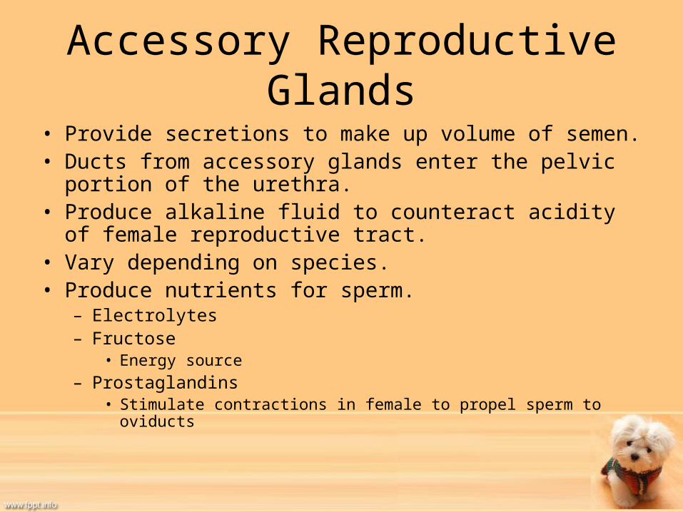
Accessory Reproductive Glands• Provide secretions to make up volume of semen.• Ducts from accessory glands enter the pelvic portion of the
urethra.• Produce alkaline fluid to counteract acidity of female
reproductive tract.• Vary depending on species.• Produce nutrients for sperm.
– Electrolytes– Fructose
• Energy source– Prostaglandins
• Stimulate contractions in female to propel sperm to oviducts

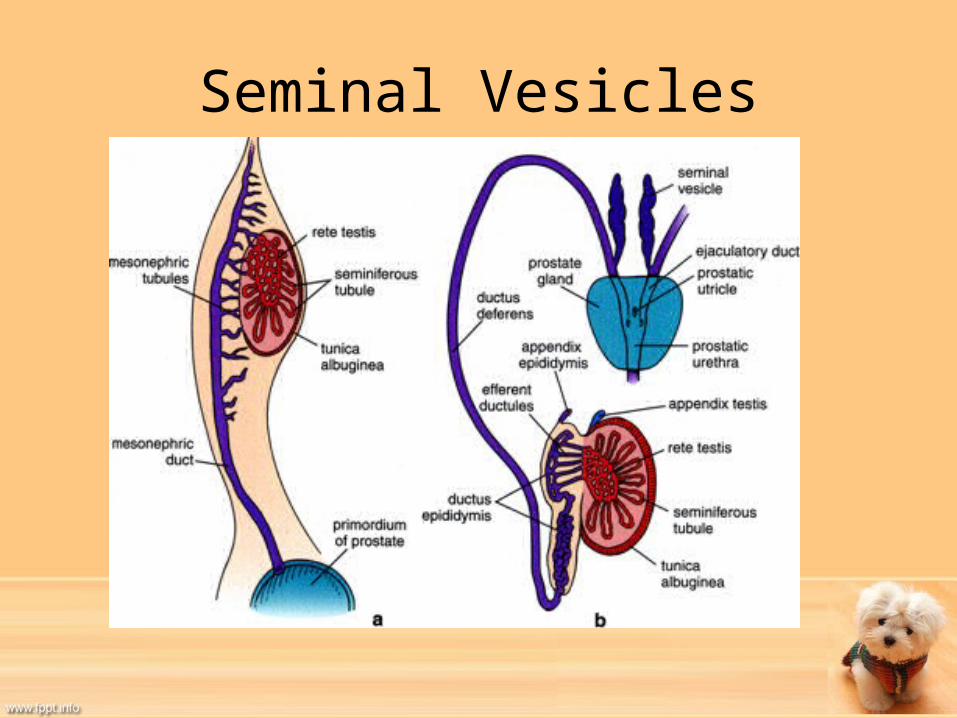
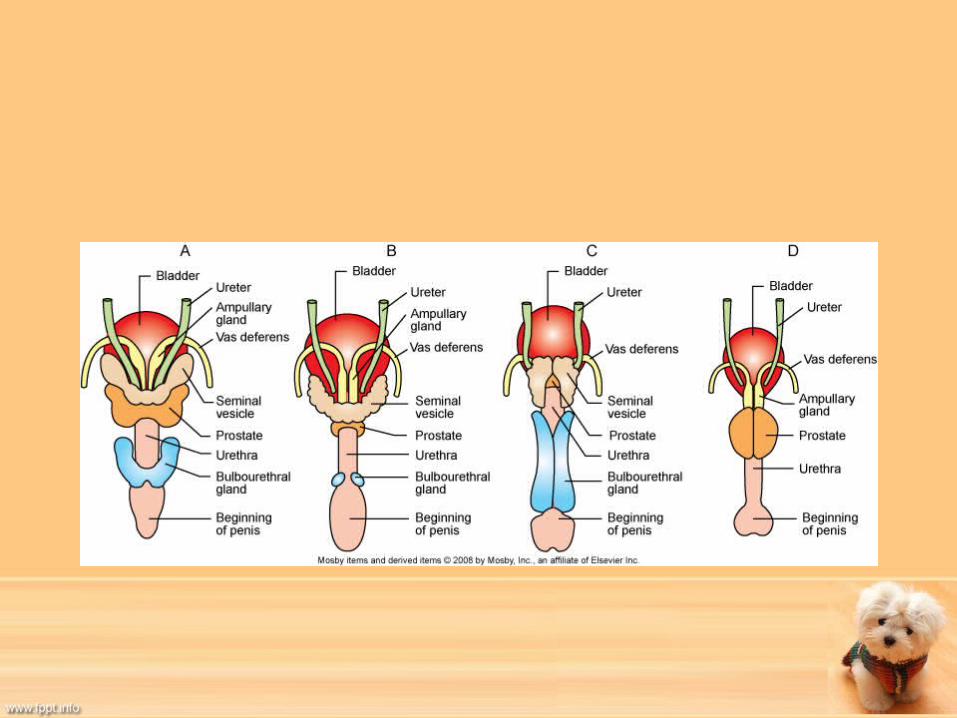
Accessory Glands Continued• Seminal Vesicles
– May be called vesicular ducts and enter pelvic urethra in same area as vas deferens.
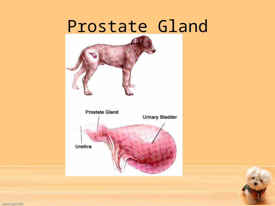
– Absent in dogs and cats.• Prostate Gland
– Completely surrounds urethra and has multiple ducts to carry secretions into urethra.
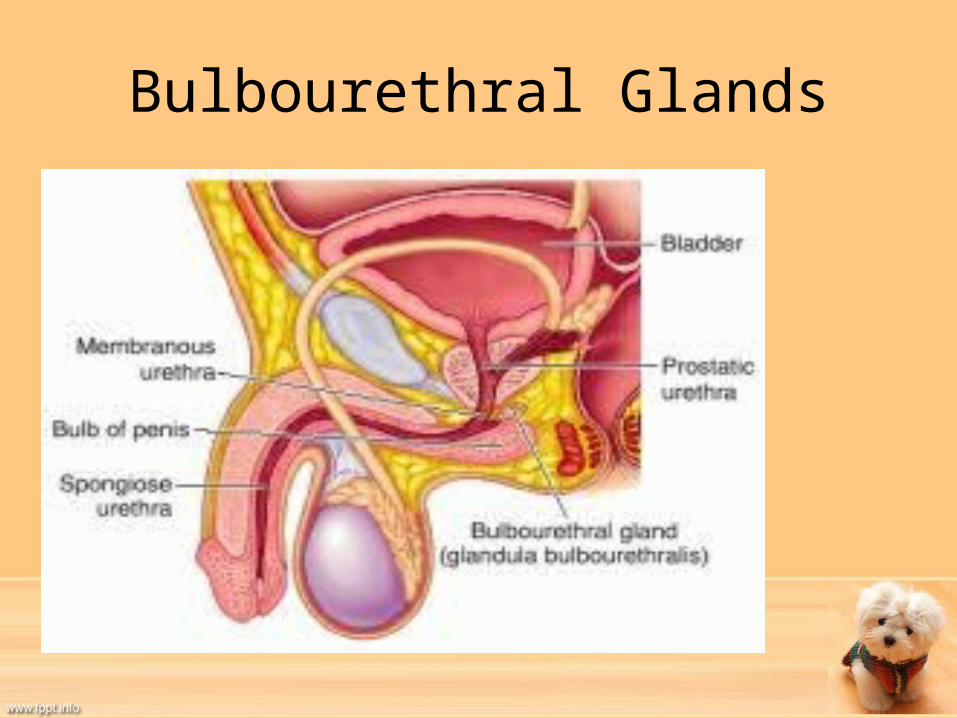
– Only accessory reproductive gland of dogs.• Bulbourethral Glands
– Also known as Cowper’s glands– Ducts enter urethra near caudal border of pelvis– Secrete mucus containing fluid just before ejaculation that helps to
lubricate urethra for passage of semen.– Absent in the dog.
Seminal Vesicles
Prostate Gland
Prostate Problems
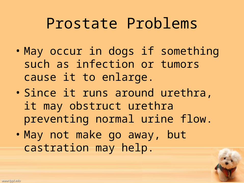
• May occur in dogs if something such as infection or tumors cause it to enlarge.
• Since it runs around urethra, it may obstruct urethra preventing normal urine flow.
• May not make go away, but castration may help.
Bulbourethral Glands

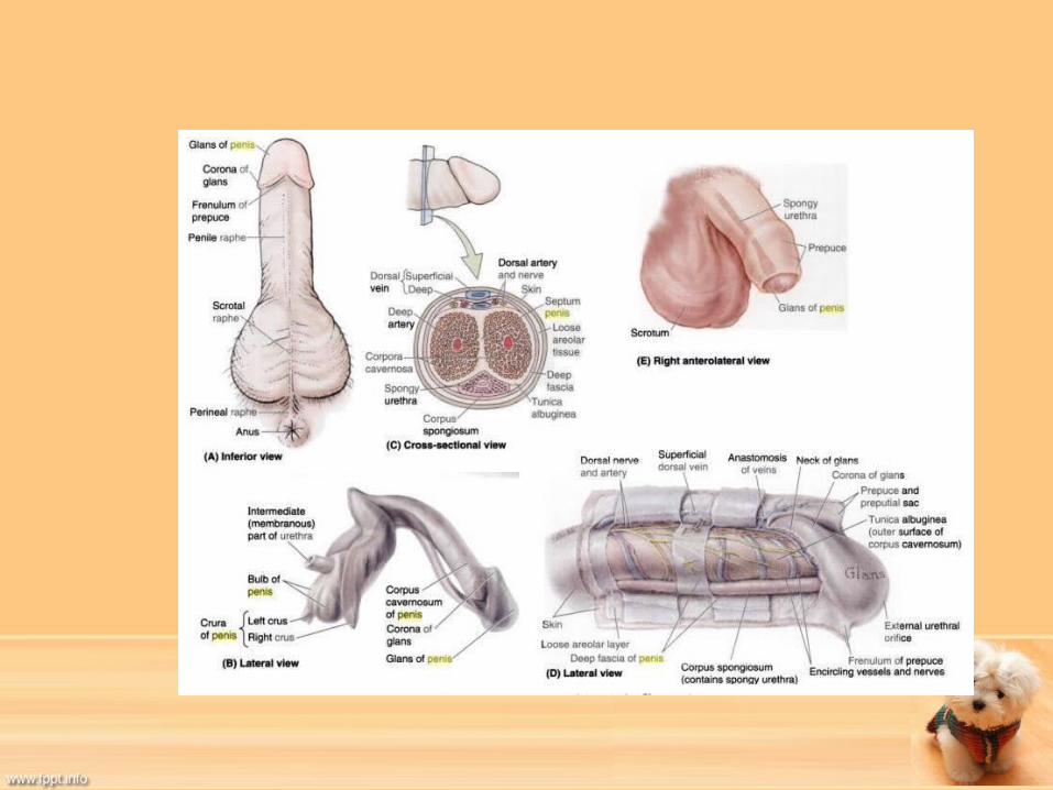
Penis
• Male breeding organ• Composed of muscle, erectile tissue and
connective tissue.• Urethra runs down the center.• Has large blood supply and many sensory
nerve endings.• When aroused and stimulated, erectile tissue
becomes engorged with blood and penis will enlarge and stiffen to allow for copulation.
Penis Continued• Three parts:
– Roots• Attach to the brim of the penis• Composed of crura or two bands of connective tissue covered by
ischiocavernous muscles. – The Body
• Largest part of penis• Made up of two bundles of erectile tissue
– Made up of blood filled spaces called sinuses– Two erectile tissues are smaller corpus cavernosum urethrae (corpus
spongiosum) and corpus cavernosum penis• When sinuses become engorged with blood, erection results.
– Glans• Tip or free end of penis• Cats have spines covering glans• Has rich supply of sensory nerve endings.

Penis Continued
• Prepuce– Skin sheath that encloses penis when it is not
erect.– Outer portion is normal skin, inner portion is
moist, smooth mucous membrane.
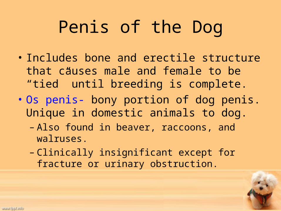
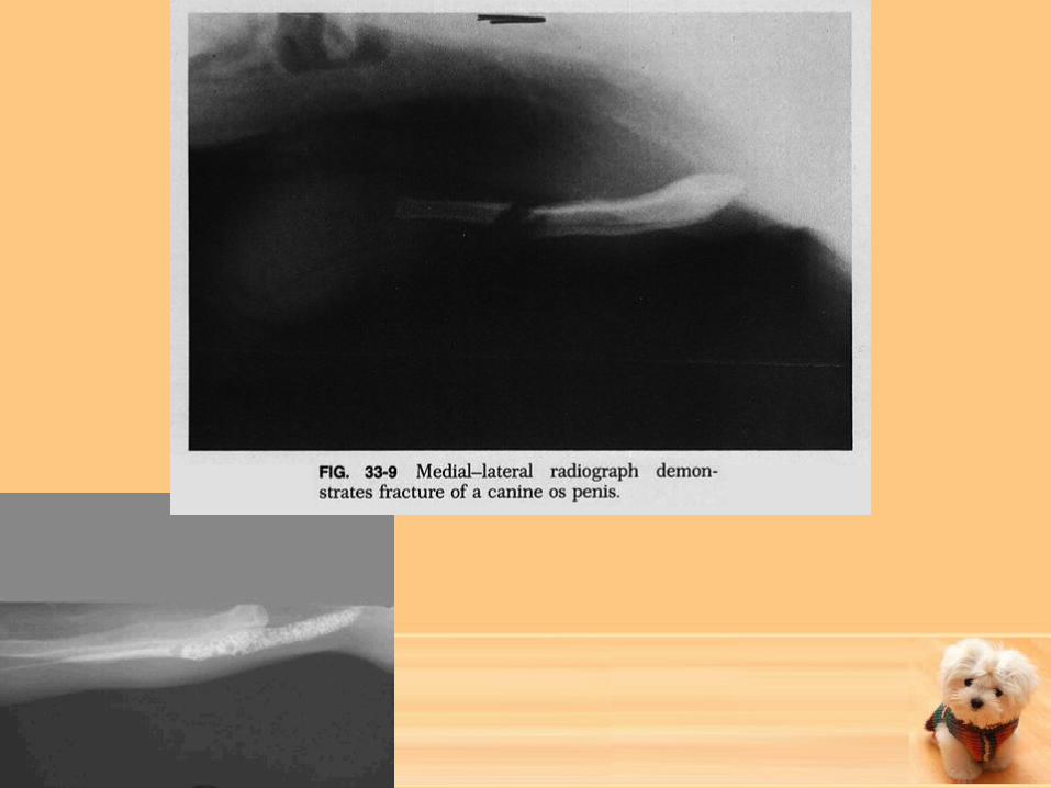
Penis of the Dog
• Includes bone and erectile structure that causes male and female to be “tied” until breeding is complete.
• Os penis- bony portion of dog penis. Unique in domestic animals to dog.– Also found in beaver, raccoons, and walruses.– Clinically insignificant except for fracture or
urinary obstruction.

Penis of the Dog continued
• Has enlargement toward rear of the glans called bulb of the glans.– Made of erectile tissue from corpus cavernosum
urethrae.– Bulb becomes enlarged during breeding slowly
and does not become fully enlarged until after ejaculation has taken place.
– Is clamped in place by contractions of female– Once male is done, will dismount but will stay tied
to female for 15 -20 minutes.
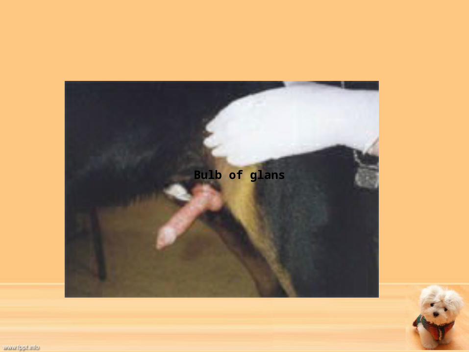
Bulb of glans

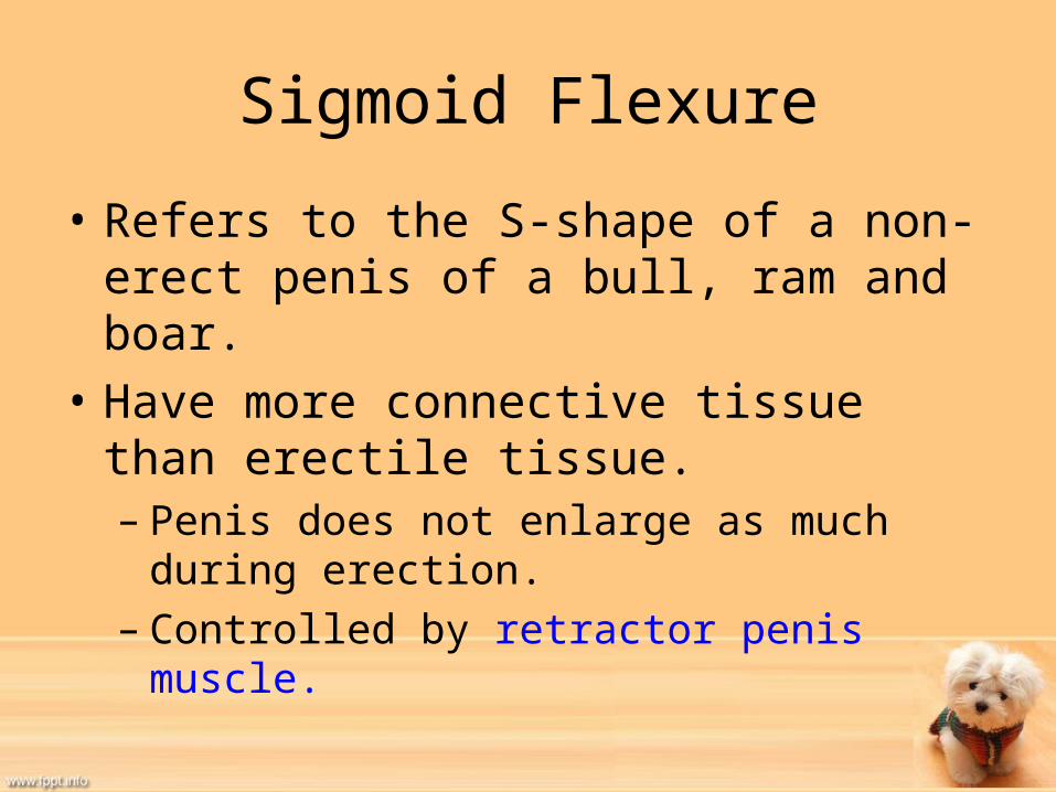
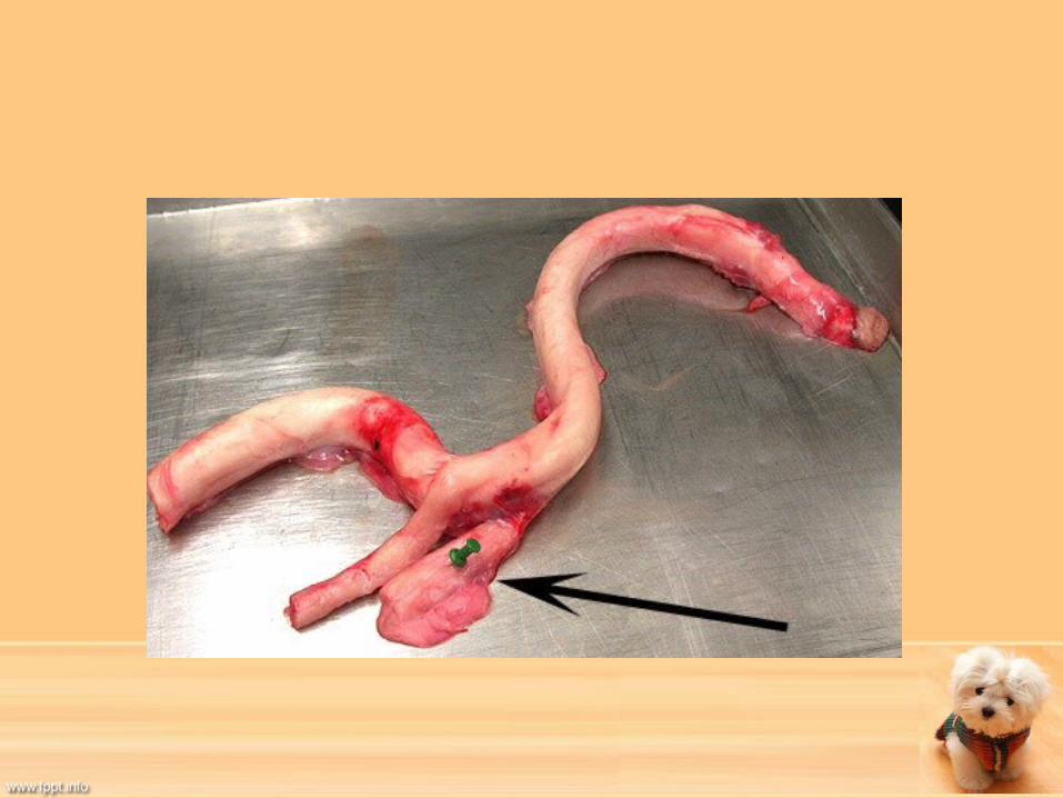
Sigmoid Flexure
• Refers to the S-shape of a non-erect penis of a bull, ram and boar.
• Have more connective tissue than erectile tissue.– Penis does not enlarge as much during erection.– Controlled by retractor penis muscle.
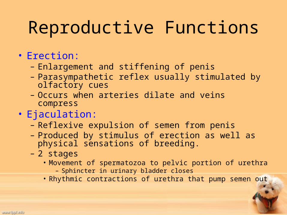
Reproductive Functions• Erection:– Enlargement and stiffening of penis– Parasympathetic reflex usually stimulated by olfactory
cues– Occurs when arteries dilate and veins compress
• Ejaculation:– Reflexive expulsion of semen from penis– Produced by stimulus of erection as well as physical
sensations of breeding.– 2 stages
• Movement of spermatozoa to pelvic portion of urethra– Sphincter in urinary bladder closes
• Rhythmic contractions of urethra that pump semen out
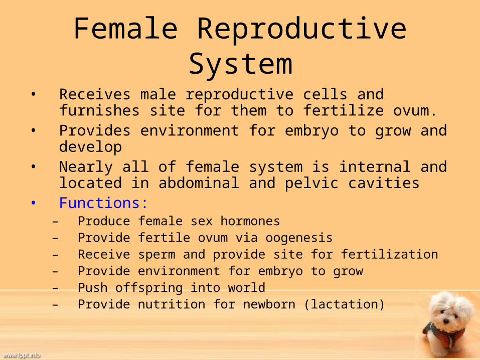
Female Reproductive System• Receives male reproductive cells and furnishes site for
them to fertilize ovum.• Provides environment for embryo to grow and develop• Nearly all of female system is internal and located in
abdominal and pelvic cavities• Functions:
– Produce female sex hormones – Provide fertile ovum via oogenesis – Receive sperm and provide site for fertilization– Provide environment for embryo to grow– Push offspring into world– Provide nutrition for newborn (lactation)

Female Reproductive System

Ligaments of Female Reproductive System
• Broad Ligaments- sheets of peritoneum by which the ovaries, oviducts and uterus hang.
• Contain blood vessels, nerves and fat. – Each section is named by what they support• Mesovarium-supports ovary• Mesosalpinx- supports oviduct• Mesometrium- supports uterus
Ligaments continued
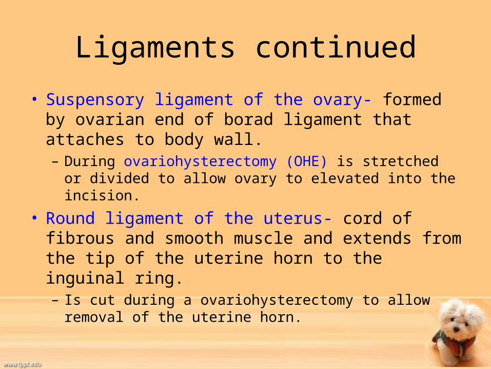
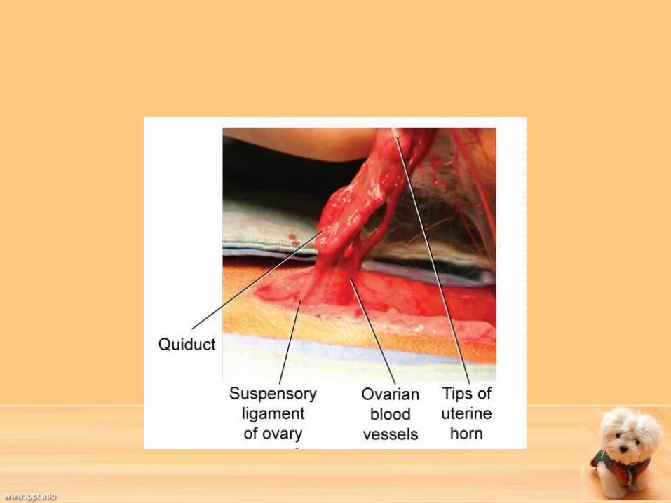
• Suspensory ligament of the ovary- formed by ovarian end of borad ligament that attaches to body wall.– During ovariohysterectomy (OHE) is stretched or divided
to allow ovary to elevated into the incision.
• Round ligament of the uterus- cord of fibrous and smooth muscle and extends from the tip of the uterine horn to the inguinal ring.– Is cut during a ovariohysterectomy to allow removal of the
uterine horn.

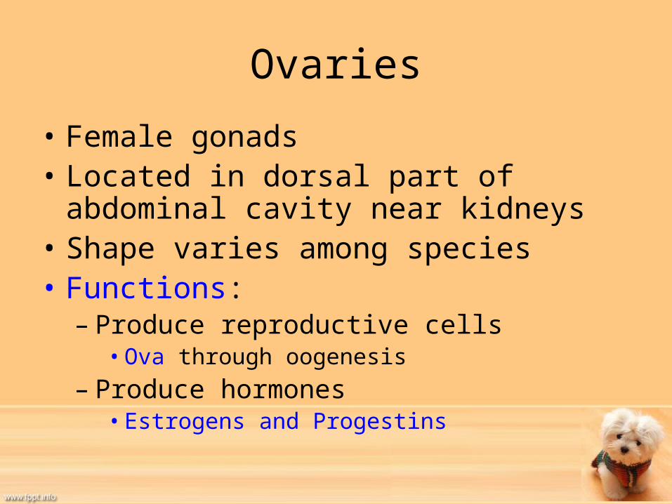
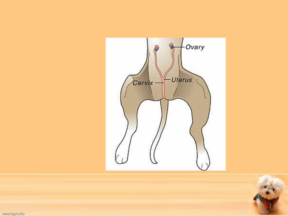
Ovaries
• Female gonads• Located in dorsal part of abdominal cavity
near kidneys• Shape varies among species• Functions:– Produce reproductive cells• Ova through oogenesis
– Produce hormones• Estrogens and Progestins
Ovaries continued
• Are not constantly produced, animal is born with predetermined set of oocytes.
• Estrogens:– Produced by the cells of the developing ovarian follicles
and are responsible for physical and behavioral changes
• Progestins:– Produced by corpus luteum- develops from empty follicle
after ovulation and help prepare the uterus for implantation of fertilized ovum.
– Necessary for maintenance of pregnancy.


Ovarian Cycle
• Ova production requires complex cycle of events that occur in a cyclical manner.
• Number of follicles produced is dependant on species.– Uniparous-normally give birth to one offspring at a time.– Multiparous-give birth to litters due to multiple ova
production per cycle. • Two hormones influence ovarian cycle (from anterior
pituitary gland):– 1. Follicle Stimulating Hormone (FSH)– 2. Luteinizing Hormone (LH)
Ovulation
• Includes:– The development of an ovum within the follicle,– Ovums release from the follicle, – Formation of the corpus luteum, – Degeneration of unripened follicles and corpus
luteum
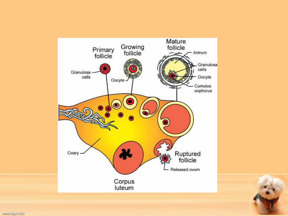

Ovarian Cycle
• Begins with primary follicle stage.– Immature oocytes reside in this stage until they
become activated to mature.– Consists of oocyte surrounded by follicular cells.– When FSH is released, stimulates some primary
follicles to begin development.• This process is called follicular recruitment or follicular
activation.
Ovarian Cycle Continued
• Next, follicle is now a growing follicle– Follicular cells become thickened and cuboidal and start to
multiply.– Multiple layers of follicular cells form around developing
oocyte.– Follicular cells then are called granulosa cells
• Granulosa cells multiply and follicle grows in size• Help to produce estrogen hormones that prepare animal for
breeding and pregnancy• Fluid filled spaces between cells eventually join together to form
the antrum
Ovarian Cycle Continued
• Once follicle has reached maximum size, is called mature follicle.– also called graafian follicle and vesicular ovarian follicle– Oocyte sits on granulosa cells inside mature follicle known
as cumulus oophorus and is surrounded by granulosa cells called corona radiata.
– When LH rises, ovulation takes place and ova is released into oviduct.
– Mature cell that is released is called an ovum.


Estrous Cycle
• Spermatozoa are constantly produced so testosterone is fairly constant in male.
• In domestic animals, breeding takes place only when chances for pregnancy are the greatest.
• This is known as estrus or heat• Estrous cycle is beginning of one heat period to
beginning of the next.– Controlled by FSH andLH– Different species have different estrous cycle patterns.
Estrous Cycle Intervals
• Polyestrous: animals that cycle continuously throughout the year if they are not pregnant– cattle and swine
• Seasonally polyestrous: animals with seasonal variations in estrous cycles– horse, sheep, cat
• Diestrous: animals with two cycles per year, usually spring and fall – dog
• Monoestrous: animals with one cycle per year – fox and mink
Estrous Cycle Stages• Proestrus
– Period of follicular development in ovary– Linings of oviduct, uterus and vagina all thicken– Vaginal epithelial lining begins to cornify to protect against breeding trauma
• Estrus– Heat period– Estrogen levels are at their peak– Signal readiness to breed by female to male
• Metestrus– Period after ovulation– Corpus Luteum is developing– Inhibits follicular development in the ovary
• Diestrus– Luteal stage when corpus luteum is of maximum size– Corpus luteum is either retained if pregnancy has ocurred or degenerates if it has not
• Anestrus (in some species)– Temporary ovarian inactivity– Seen in seasonally polyestrous, diestrous, and monoestrous animals
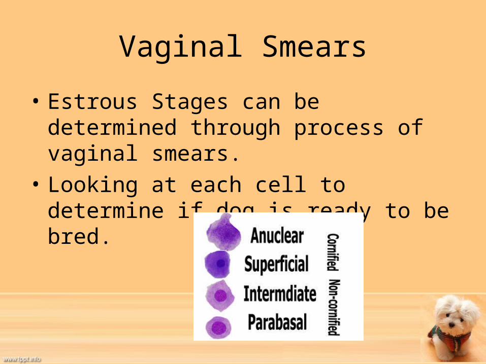
Vaginal Smears
• Estrous Stages can be determined through process of vaginal smears.
• Looking at each cell to determine if dog is ready to be bred.

Induced Ovulators
• Most species must ovulate prior to being bred, however some species must be bred for ovulation to occur.
• Are called induced ovulators– Ferrets, rabbits, cats
Ovulation• Surface of follicle weakens and ruptures and ovum is
released from fluid.• Empty follicle fills with blood and is called the corpus
hemorrhagicum.• High LH stimulates granulosa cells to form solid
structure inside corpus hemorrhagicum and becomes corpus luteum. – Corpus luteum produces progestins necessary for
pregnancy– If ovum is not fertlized, the corpus luteum will degenerate.
• Follicular atresia is the degeneration of follicles that do not develop into ovum.


Oviducts• Also known as fallopian tubes or uterine tubes• Generally are site of fertilization
– Then guide fertilized ovum to uterus• Small convoluted tubes that extend from tips of uterine
horns.– Lined with cilia and smooth muscle fibers
• Guide ova from ovary to uterus• Are not directly attached to ovaries and “catch” ova in
infundibulum.– Surrounds ovary area where follicles are formed and fimbrae- muscle
like fingers, help to position infundibulum in correct spot. – If ova is lost in abdominal cavity, it generally disintegrates.
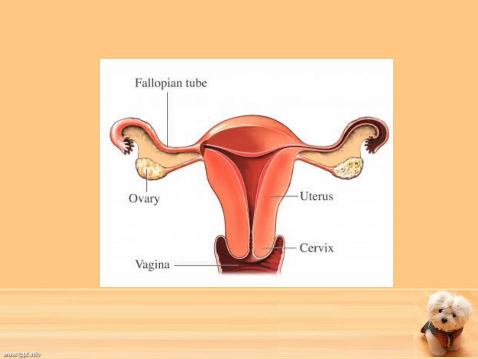
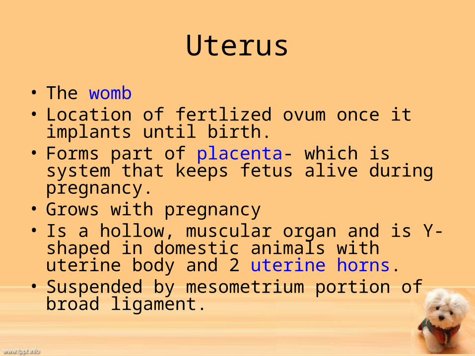
Uterus• The womb• Location of fertlized ovum once it implants until
birth.• Forms part of placenta- which is system that keeps
fetus alive during pregnancy. • Grows with pregnancy• Is a hollow, muscular organ and is Y-shaped in
domestic animals with uterine body and 2 uterine horns.
• Suspended by mesometrium portion of broad ligament.


Uterus Continued
• Wall of uterus is composed of:– Endometrium• Inner layer• Composed of simple columnar epithelium and glands
that secrete mucus and other substances– Myometrium• Thickest portion made up of smooth muscle
– Perimetrium• Outermost layer• Covered by visceral layer of peritoneum


Cervix
• Muscular valve that seals uterus from outside.• Smooth muscle sphincter located between
uterus and vagina.• Is closed except for estrus and parturition.– Opens to admit spermatozoa during breeding– At birth, fetus is pushed against cervix until it
dilates or opens.
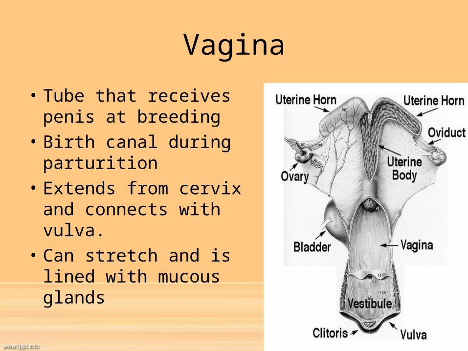
Vagina
• Tube that receives penis at breeding
• Birth canal during parturition
• Extends from cervix and connects with vulva.
• Can stretch and is lined with mucous glands
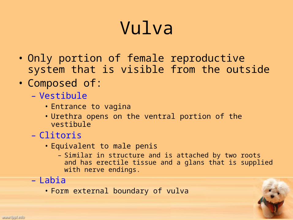
Vulva
• Only portion of female reproductive system that is visible from the outside
• Composed of:– Vestibule
• Entrance to vagina• Urethra opens on the ventral portion of the vestibule
– Clitoris• Equivalent to male penis
– Similar in structure and is attached by two roots and has erectile tissue and a glans that is supplied with nerve endings.
– Labia• Form external boundary of vulva


Ovariohysterectomy (OHE)
• “Spay” • Includes removal of the ovaries, oviducts, and
uterus.• Major Abdominal Surgery.

Pyometra
• Occurs after cervix has opened for breeding.
• Bacteria enters uterine area and cervix closes
• Creates a closed space for infection.
• Usually is a surgical emergency and requires complete ovariohysterectomy.

False Pregnancy
• Also called pseudocyesis or pseudopregnancy• Affected animals may act or look pregnant• Is an exaggerated diestrous period• Usually resolves spontaneously or through the
use of hormones.


