67
The respiratory System Prepared by Mickelder Kercy, MD - Instructor
| Date post: | 15-Jul-2015 |
| Category: |
Health & Medicine |
| Upload: | mickelder-kercy |
| View: | 530 times |
| Download: | 0 times |

The respiratory
System
Prepared by Mickelder Kercy, MD - Instructor

Upper Respiratory Tract : Nose, Paranasal
Sinuses, Pharynx(throat), Larynx(voice box)
and Trachea(Windpipe)
Lower Respiratory Tract : Bronchi, Bronchioles,
Alveolar Ducts, Alveolar Sacs
Organs of the Respiratory System

Divided into the 2 nares/nostrils by the septum
Septum has on each lateral wall a superior,
middle and inferior concha
The mucous membrane lines the conchae
The Nose


Drains fluid from the frontal sinuses, ethmoidal sinuses, sphenoidal sinuses and the maxillary sinuses
Drains fluid from the eyes through the nasolacrimal duct
Serves as a passageway for air (nares)
Warms and moistens inhaled air (conchae)
Traps dust, pollen and other foreign matter with hair-like projections (cilia) and mucous (mucous membrane)
Functions of the Nose

Air-filled spaces located within the bones of the
skull and face
Four sets of paired sinuses exist: Frontal,
Ethmoid, Sphenoid and Maxillary
Paranasal Sinuses


Lightens the weight of the head
Humidifies and heats inhaled air
Assist in the making of sounds for speaking and
singing by increasing resonance
Serves as a crumple zone to protect vital
structures in the event of facial trauma
Functions of the Paranasal Sinuses

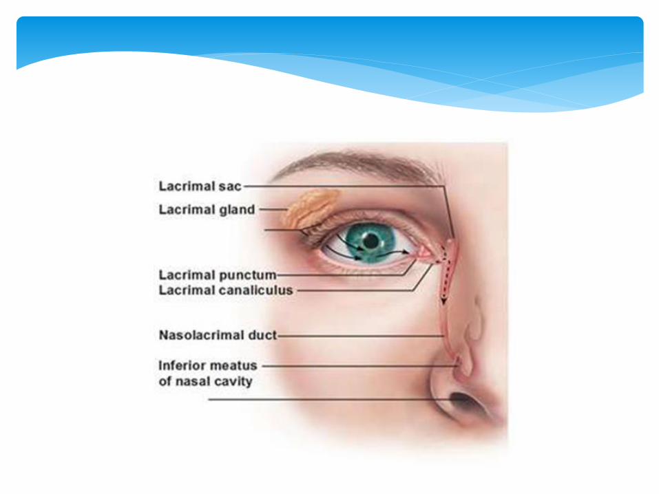
Lacrimal System
Lacrimal gland (secretes the tears)
Excretory ducts (drain the tears to the surface of
the eye)
Lacrimal canaliculi, the lacrimal sac, and the
nasolacrimal duct (drain the tears to the nares)


Connects the base of the skull to the trachea and
esophagus
Divided into the Nasopharynx, oropharynx and
laryngopharynx
Contains the pharyngeal, palatine & lingual
tonsils/adenoids and the uvula
Pharynx


Carries air from the nose and mouth toward the larynx
Pushes food toward the esophagus by swallowing reflex
Helps form sounds with the larynx, tongue and soft
palate
Enables equilibration of pressure differences between
the throat and the middle ear during quick changes of
altitude, diving or in disorders of the middle ear through
the Eustachian tubes
Prevents some microbes and foreign particles to enter
the lungs by the tonsils/adenoids catching them
Functions of the Pharynx

Consists of the thyroid cartilage (adam’s apple), the epiglottic cartilage (epiglottis), cricoid cartilage, three paired cartilages (arytenoid, corniculate, and cuneiform) besides the muscles and ligaments etc..
Contains the false vocal cords (minimal role in phonation) located above the true vocal cords (major role in phonation)
Larynx


Prevents food from entering the glottis (opening
between the vocal cords) when pulled upwards
while the epiglottis closes up the glottis
Production of vocal sounds when air passes
through the vocal cords
Functions of the Larynx


Links the larynx to the right and left bronchi
Passageway for the air entering the lungs
Trachea


Two main branches of the trachea that extend
into the lungs
Right bronchus is wider, shorter, and more
vertical
Left bronchus is longer with a smaller caliber
They each divide into many small bronchi that
subdivide into the smaller bronchioles
Bronchi & Bronchioles


The bronchi conduct the air into the bronchioles
The bronchioles conduct the air into the alveolar
ducts
Functions of the Bronchi & Bronchioles

Mucus is a thin layer of sputum (phlegm) lining
the inside of the breathing tubes (trachea,
bronchi and bronchioles) to prevent irritants
from getting into the lungs
Cilia are little hairs lining the breathing tubes
moving the mucus from the lungs upward
toward the throat to the epiglottis that opens up
before the mucus is swallowed into the
esophagus
Cilia and Mucus

Composed of microscopic alveoli/alveolar sacs
(subdivision of the alveolar ducts)
Right lung has an upper, middle and lower lobe
Left lung has an upper and lower lobe
Covered by the visceral (internal) and parietal
(external) pleurae (pleural membranes)
Lungs


Gas exchange by bringing oxygen (O2) into the
body and removing carbon dioxide (CO2) from
the body
Functions of the Lungs

Produce serous fluid that cause the pleurae to
adhere closely to one another, holding the lungs
to the thoracic wall and allowing the pleural
membranes to move easily against each other
during breathing
Functions of the Pleurae


Ventilation is a process of inhalation (air moving down to the alveoli) and exhalation (air moving out of the alveoli)
- During inhalation the diaphragm contracts
and flattens → elevation of the ribs → ↑ size of the thorax
(- pressure) as the air moves down to the alveoli
- During exhalation the diaphragm relaxes →
thorax returns to its resting size and shape (+ pressure) as
the air moves out of the alveoli
- During forceful expiration the internal and
external intercostal and abdominal muscles are involved
Mechanism of Breathing


Tidal Volume : Volume of air entering or leaving
the lungs during a single breath
Inspiratory Reserve Volume : Volume of air that
can be inhaled after a forceful inspiration
Expiratory Reserve Volume : Volume of air that
can be expelled after a forceful expiration
Residual Volume : Volume of air remaining in
the lungs after a maximal/forceful expiration
Respiratory Volumes and Capacity

Asphyxia : Suffocation resulting from the lack
of oxygen. ABG is a test used to detect the level
of oxygen and carbon dioxide in the blood
Asthma : Chronic inflammatory disease of the
bronchi caused by allergens or other irritating
substances.
Common Disorders associated with the Respiratory System


COPD : - Chronic Bronchitis (Chronic inflammation of the mucous membrane in the bronchial passages causing the membrane and the mucus to thicken making it hard to breathe (SOB) because the air cannot pass through easily. It leads to SOB). Acute (lasting less than 6 weeks). Chronic (recurring for more than a year). Mostly viral. Tobacco, Coal miners, grain handlers, metal holders, dust etc…


- Emphysema (Destruction of the lung tissue around the bronchioles causing these airways to collapse preventing the air to come out during exhalation. The chest wall muscles have to work harder to expel the air. There is also damage of the walls between many of the air sacs. As a result, the air sacs lose their shape and become floppy causing a reduction in the amount of gas exchange in the lungs. Tobacco, alpha-1 antitrypsin, air pollution, heredity etc..


Pulse Oxymeter

Spirometer

Common Cold : Viral infection of the upper respiratory tract. Rhinovirus. Spread by droplets through sneezing, coughing, and contacts and fomites
Hay Fever : Seasonal allergic rhinitis or pollinosis. Mucous membrane of the nose and the eyes become inflamed
Influenza : Flu caused by viruses
Legionnaire’s Disease : Pneumonia caused by the Legionella bacteria

Lung Cancer : Leading cause of deaths in both
women and men in the USA. Smoking


Pertussis : Whooping cough (Persistent and
severe cough) caused by the bacteria Bordetella
Pertussis


Pleurisy: Pleuritis or inflammation of the
pleurae


Pneumonia : Inflammation of the lungs caused
by bacteria/viruses/chemical irritants


Pneumothorax : Air accumulation in the pleurae
compressing the lungs (collapsing)


Pulmonary Embolism : Blood clot in the lungs
coming from smaller vessels in the legs, pelvis
arm or heart. The clot obstructs partially or
completely the blood flow to a section of the
lung causing a pulmonary infarct


Severe Acute Respiratory syndrome : Mild to
moderate respiratory illness caused by
coronavirus
Sinusitis : Inflammation of mucous membranes
of the sinuses

Pulmonary Tuberculosis : Pneumonia caused by
the bacteria Mycobacterium tuberculosis


Pleural Effusion : Excessive liquid accumulating
in the space between the visceral and parietal
pleural membranes. CHF, Pneumonia, RF,
Cancer etc…


The End


