26
The Respiratory System Chapter 15
| Date post: | 30-Dec-2015 |
| Category: |
Documents |
| Upload: | barnaby-webb |
| View: | 215 times |
| Download: | 0 times |

The Respiratory System
Chapter 15

Human Anatomy, 3rd editionPrentice Hall, © 2001
Introduction• Responsible for the exchange of gases between the
body and the external environment.– Cells need a supply of O2 and to eliminate CO2
– 3 basic processes• Breathing• External respiration
• Internal respiration
• Two systems supply O2 & eliminate CO2
– Respiratory system
– Cardiovascular system
Human Anatomy, 3rd editionPrentice Hall, © 2001
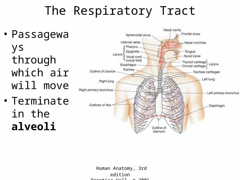
The Respiratory Tract
• Passageways through which air will move
• Terminate in the alveoli

Human Anatomy, 3rd editionPrentice Hall, © 2001
The Respiratory Tract
• Upper respiratory system– Superior to the
larynx– Functions – intake,
moistening, filtering, sensing
• Lower respiratory system– Larynx and below– Functions – sound
production, transport of air, gas exchange

Human Anatomy, 3rd editionPrentice Hall, © 2001
The Respiratory Epithelium
• Lines the upper respiratory system
• Pseudostratified ciliated columnar epithelium with goblet cells – Entraps & removes
dust – Moistens incoming air– Olfactory sensation
• Capillaries warm incoming air
• The common cold

Human Anatomy, 3rd editionPrentice Hall, © 2001
Surface View of the Epithelium
Human Anatomy, 3rd editionPrentice Hall, © 2001
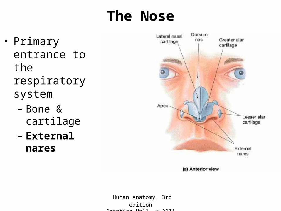
The Nose
• Primary entrance to the respiratory system– Bone & cartilage
– External nares

Human Anatomy, 3rd editionPrentice Hall, © 2001
The Nasal Cavity• Nasal septum
– Divides into left and right sides
• Hard & soft palate form the floor
• Nasal conchae cause turbulence through nasal cavity
• Internal nares lead to the pharynx

Human Anatomy, 3rd editionPrentice Hall, © 2001
Paranasal Sinuses
• Cavities in cranial bones
• Functions– Lighten skull bones
– Produce mucus
– Resonate during sound production

Human Anatomy, 3rd editionPrentice Hall, © 2001
The Pharynx
• Area from the internal nares to the larynx
• Shared between the respiratory and digestive systems

Human Anatomy, 3rd editionPrentice Hall, © 2001
The Larynx• Connects the pharynx
to the trachea• Lined by mucus-
producing columnar epithelium
• Functions– Produces sound
• Vocal folds
– Keeps food from entering the airways
• Vestibular folds• Epiglottis

Human Anatomy, 3rd editionPrentice Hall, © 2001
The Larynx
• Formed by 9 cartilages– Thyroid cartilage
• Adam’s apple
– Epiglottis• Closes glottis

Human Anatomy, 3rd editionPrentice Hall, © 2001
The Larynx

Voice Production• Ligaments lie under
mucous membrane• Muscles attached to
the cartilages– Control vocal
ligament tension
• Sound production– Air flowing over
vocal folds (through the glottis) produces sound waves
• Resonating chambershttp://www.entdocsonline.com/images/larynx.jpg

Human Anatomy, 3rd editionPrentice Hall, © 2001
The Trachea• Extends from larynx
to primary bronchi• The “windpipe”• Structure
– Incomplete cartilaginous rings
• Lined by mucus-producing epithelium
• Function – passageway for air

Human Anatomy, 3rd editionPrentice Hall, © 2001
Bronchi, Bronchioles, & Alveoli• Trachea divides into
• Primary bronchii divide into
• Secondary bronchi divide into
• Tertiary bronchi divide into
• Bronchioles divide into• Terminal bronchioles
divide into
• Respiratory bronchioles divide into
• Alveolar ducts end in • Alveoli

Human Anatomy, 3rd editionPrentice Hall, © 2001
The Respiratory Tree

Human Anatomy, 3rd editionPrentice Hall, © 2001
Changes as the Respiratory Tree Branches
• As branching of bronchi becomes more extensive– Rings of cartilage
become plates– Smooth muscle
increases– Columnar epithelium
becomes cuboidal, then squamous (in alveoli)
• Asthma

Human Anatomy, 3rd editionPrentice Hall, © 2001
The Alveoli
• Site of gas exchange with the blood
• Closely associated with capillaries
• Wall of the alveolus + wall of capillary– Respiratory
membrane
• Gas exchange occurs by diffusion

Human Anatomy, 3rd editionPrentice Hall, © 2001
Alveoli

Human Anatomy, 3rd editionPrentice Hall, © 2001
The Alveoli• Alveolar epithelium
contains:– Alveolar Type 1 cells
• Gas exchange • Emphysema
– Alveolar Type 2 cells• Secrete surfactant • Respiratory
Distress Syndrome
• Macrophages

Human Anatomy, 3rd editionPrentice Hall, © 2001
The Lungs
• Soft, spongy, cone-shaped
• Right lung– 3 lobes
• Left lung– 2 lobes

Human Anatomy, 3rd editionPrentice Hall, © 2001
Blood Supply to the Lungs
– Pulmonary arteries, pulmonary veins

Human Anatomy, 3rd editionPrentice Hall, © 2001
Breathing• Lungs are prevented from collapsing
– Pressure in the pleural cavity is always slightly lower than the pressure in lungs

Human Anatomy, 3rd editionPrentice Hall, © 2001
Inhaling is Active Work
• Diaphragm• Intercostals contract• Other muscles in
thorax contract• Thoracic cage expands• Lung volume
increases• Pressure in lungs
drops below atmospheric pressure
• Air rushes in

Human Anatomy, 3rd editionPrentice Hall, © 2001
Exhaling is Passive
• Diaphragm relaxes• Intercostal muscles
relax• Thoracic cage drops• Lung volume
decreases• Pressure in lungs
increases above atmospheric pressure
• Air rushes out


