67
The RESPIRATORY System
The RESPIRATORY System
Respiration
• The exchange of gases between the atmosphere, blood, and cells
• Pulmonary Ventilation - the exchange of air between the atmosphere and lungs
• External (Pulmonary) Respiration - gas exchange between the lungs and blood
• Internal (Tissue) Respiration - gas exchange between the blood and cells
Functions of the Respiratory System
• Provides structures and mechanisms for gas exchange
– Intake of O2
– Elimination of CO2
• Helps maintains body’s pH
• Sense of smell
• Speech and sound production
Organs of the Respiratory System
• Nose
• Pharynx
• Larynx
• Trachea
• Bronchi
• Lungs

Respiratory System
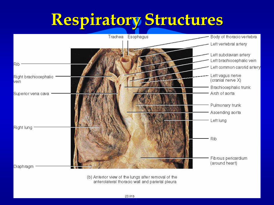
Respiratory Structures
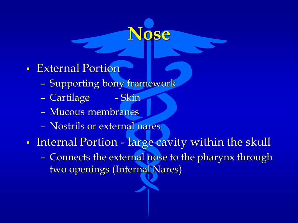
Nose
• External Portion
– Supporting bony framework
– Cartilage - Skin
– Mucous membranes
– Nostrils or external nares
• Internal Portion - large cavity within the skull
– Connects the external nose to the pharynx through two openings (Internal Nares)

External Nose Structures

Nose
• Nasal Cavity - large cavity that contains both the external and internal nose cavities
• divided into the right and left sides by the NASAL SEPTUM
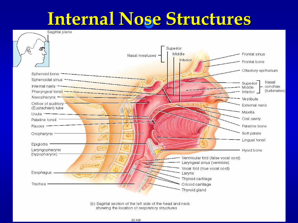
Internal Nose Structures

Functions of the Nose
• Warming, moisturizing and filtering incoming air
• Smell (reception of olfactory stimulus)
• Resonating chamber for speech

Pharynx
• Funnel-shaped tube about 13 cm long
• Extends from the internal nares down to the cricoid cartilage of the larynx
• Walls composed of skeletal muscle lined with a mucous membrane
• Divided into three areas– Nasopharynx
– Oropharynx
– Laryngopharynx

Regions of the Pharynx

Functions of the Pharynx
• Passageway for food and air
• Resonating chamber for speech

Larynx (Voice Box)
• A short passageway that connects the pharynx with the trachea
• Walls of the larynx is composed of 9 pieces if cartilage– Three single pieces of cartilage
• epiglottic cartilage (Epiglottis)
• thyroid cartilage (Adam’s Apple)
• cricoid cartilage (attaches the Larynx to the Trachea)
– Three paired pieces of cartilage• arytenoid - corniculate - cuneiform

Larynx Structures
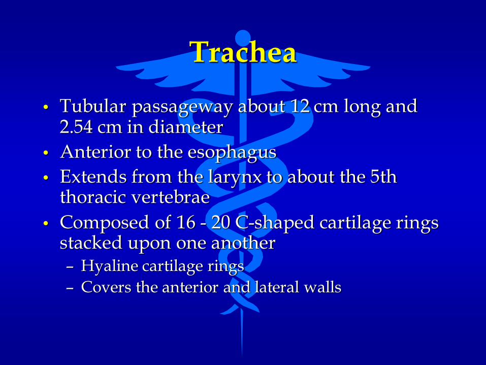
Trachea
• Tubular passageway about 12 cm long and 2.54 cm in diameter
• Anterior to the esophagus
• Extends from the larynx to about the 5th thoracic vertebrae
• Composed of 16 - 20 C-shaped cartilage rings stacked upon one another– Hyaline cartilage rings
– Covers the anterior and lateral walls

Trachea and Esophagus

Trachea
• Non-cartilaginous posterior softer portion of the trachea allows for expansion of the esophagus during swallowing
• Lined with ciliated epithelium
• The point where the trachea bifurcates is called the carina– About the 5th thoracic vertebrae

Bronchi
• Tubes that branch off of the trachea at the carina and extend into the lungs
• Left Primary Bronchus (Left Mainstem Bronchus)
• Right Primary Bronchus (Right Mainstem Bronchus)
– Shorter and more vertical
– Swallowed objects more likely to lodge in the right primary bronchus than the left

Bronchi
• Also composed of cartilaginous rings
• Continue branching as they enter the lungs into a structure called the bronchial tree– Trachea
-Mainstem (Primary) Bronchi - Secondary (Lobar) Bronchi
- Segmental (Tertiary) Bronchi - Terminal Bronchioles
- Respiratory Bronchioles - Alveolar Ducts

Bronchial Tree
Bronchioles
• Bronchioles are smaller air passages which branch from the bronchi. Bronchioles are small, muscular tubes with a narrow diameter. Changes in the size of the bronchioles help direct the flow of air to various parts of the lungs.
Alveolar Ducts
• Alveolar ducts are enlarged chambers found at the end of the bronchioles. These very fine passageways end at the alveolar sacs and connect to the alveoli.

Alveoli
• A cup shaped out pouching of epithelial tissue
• Place where external respiration occurs (gas exchange between the lungs and the blood)
• Lungs contain 300 - 500 million alveoli– Surface area of about 750 sq. ft.
– The size of a Tennis Court
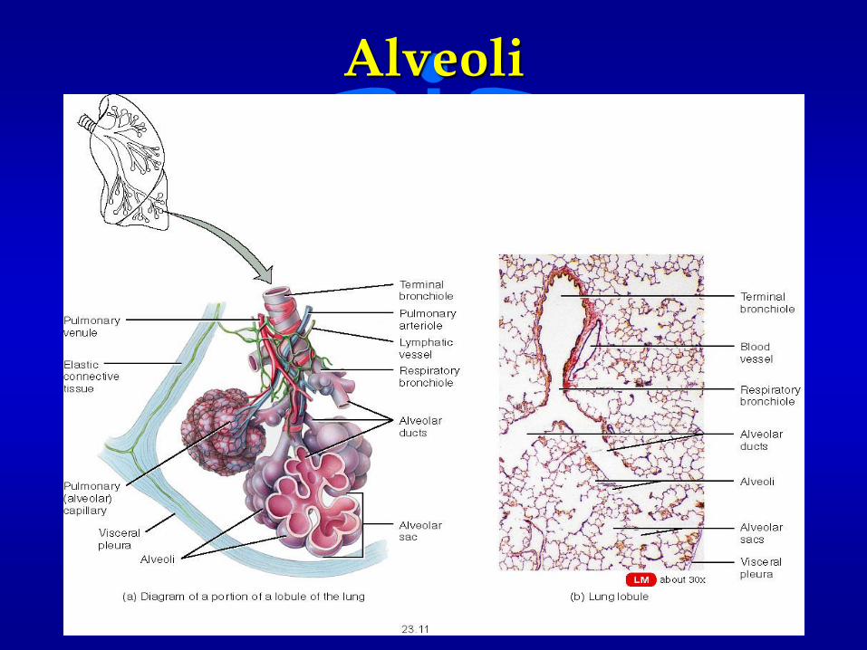
Alveoli
Pharynx
• Funnel-shaped tube about 13 cm long
• Extends from the internal nares down to the cricoid cartilage of the larynx
• Walls composed of skeletal muscle lined with a mucous membrane
• Divided into three areas– Nasopharynx
– Oropharynx
– Laryngopharynx

Structures of the Larynx
• Epiglottis
• Glottis
• Hyoid Bone
• Thyroid Cartilage
• Cricoid Cartilage
• True and False Vocal Cords

Epiglottis
• Large leaf-shaped piece of cartilage lying on top of the larynx
– The stem of the epiglottic cartilage is attached to the thyroid cartilage
• Leaf portion of the cartilage is unattached and acts like a trap door covering the opening to the trachea which is called the glottis.
– Dependent upon breathing or swallowing

Epiglottis

Glottis
• The opening from the pharynx to the larynx that contains the vocal cords
– Vocal Cords - mucous membrane folds that extend across the glottis in two layers
• upper layer or folds - false vocal folds
• lower layer or folds - true vocal folds
• Sounds originate from vibration of these true vocal cords

Glottis and Vocal Cords
Hyoid Bone
• Is located in the neck between the lower jaw and the larynx.
• It does not articulate with any other bones.
• It serves as the posterior attachment for the tongue and helps in swallowing.
Thyroid Cartilage
• The thyroid cartilage is the largest cartilage of the larynx.
• It give the larynx its characteristic triangular shape.
• Nicknamed the Adam’s Apple because of its enlarged size due to the influence of testosterone.
Cricoid Cartilage
• The most inferior cartilage of the larynx.
• Used as a landmark to perform a tracheotomy.

True Vocal Cords
• The most inferior of the horizontal folds in the larynx.
• Contain elastic fiber which vibrate to create sound.
False Vocal Cords
• The most superior of the folds in the larynx.
• Help the glottis close during swallowing to prevent food from entering the lower respiratory passages.

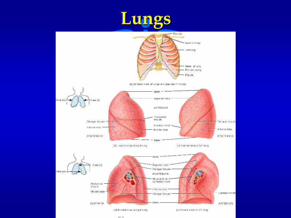
Lungs
• Paired - cone shaped organs that occupy most of the thoracic cavity
• Separated from each other by the heart and other structures of the mediastinum
• Surrounded by a double layered serous membrane called the pleural membrane

Features of the Lungs
• Apex - the pointed, superior portion of the lungs.
• Base – the broad, inferior surface of the lungs which rests on the diaphragm.
Lungs
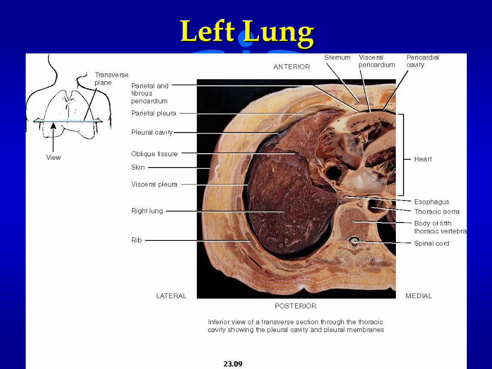
Left Lung
Pleural Membrane
• Parietal Pleura - outer layer of the pleural membrane
– Attached to the thoracic wall
• Visceral Pleura - inner layer of the pleural membrane
– Attached to the lungs themselves
• Between the parietal pleura and the visceral pleura is a potential space called the pleural cavity
– Contains pleural (serous) fluid (reduces friction)

Gross Anatomy of the Lungs
• Extend from the diaphragm to an area about 2.54 cm above the clavicles on both sides of the thoracic cavity
• Base
• Apex
• Hilus
• Lobes

Lobes and Fissures
• Each lung is divided into lobes by one or more fissures
• There are three lobes in the right lung and two lobes in the left lung.
Pleural Cavity
• The mediastinum divides the thoracic cavity into two pleural cavities, each of which contains one lung.

Gas Exchange
• Gas exchange occurs in the alveoli due to the difference in the partial pressures of oxygen and carbon dioxide in the capillary blood and the alveoli.
• Since the concentration of oxygen is greater in the alveoli, it diffuses into the capillary blood.
• Since the level of carbon dioxide is higher in the capillary blood than in the alveoli, carbon dioxide diffuses out of the blood and into the alveoli.
Lung Parameters
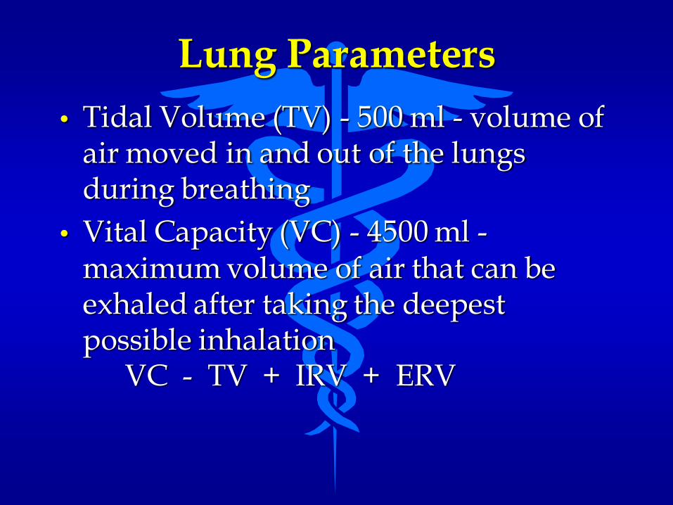
• Tidal Volume (TV) - 500 ml - volume of air moved in and out of the lungs during breathing
• Vital Capacity (VC) - 4500 ml -maximum volume of air that can be exhaled after taking the deepest possible inhalation
VC - TV + IRV + ERV
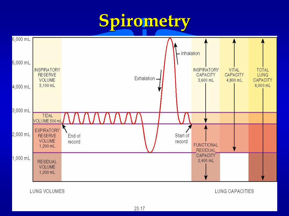
Spirometry
Physiology of Ventilation
• Ventilation - the process of inhaling and exhaling air in and out of the lungs
• Pulmonary Ventilation - the process by which air flows between the lungs and the external environment
• Due to a change in pressure between the atmosphere and the air in the lungs
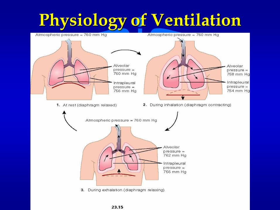
Physiology of Ventilation

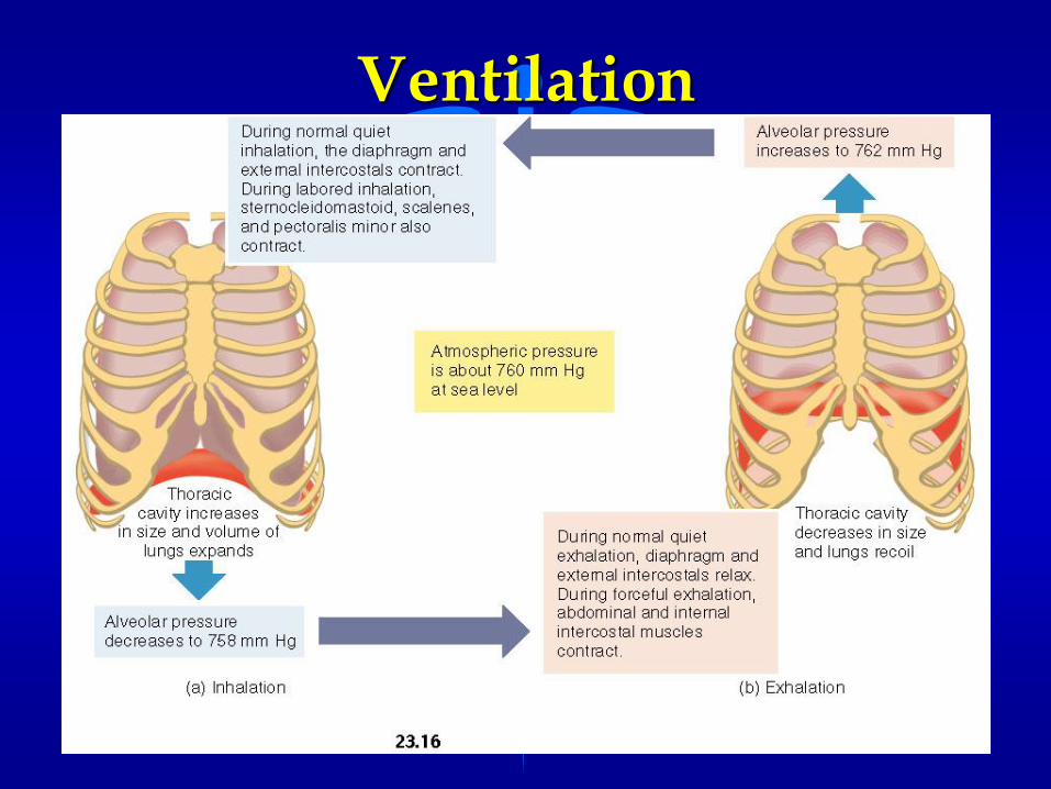
Inspiration (Inhalation)
• Bringing air into the lungs from the external environment
• The lungs themselves contain no muscles and thus depend upon the relationship with the muscles of the walls of the thoracic cavity to alter lung volumes
Ventilation

Muscles of Ventilation
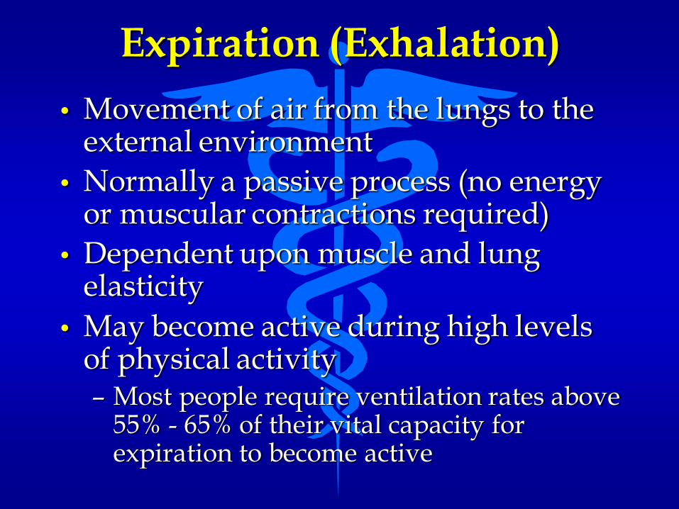
Expiration (Exhalation)
• Movement of air from the lungs to the external environment
• Normally a passive process (no energy or muscular contractions required)
• Dependent upon muscle and lung elasticity
• May become active during high levels of physical activity– Most people require ventilation rates above
55% - 65% of their vital capacity for expiration to become active

Physiology of Respiration
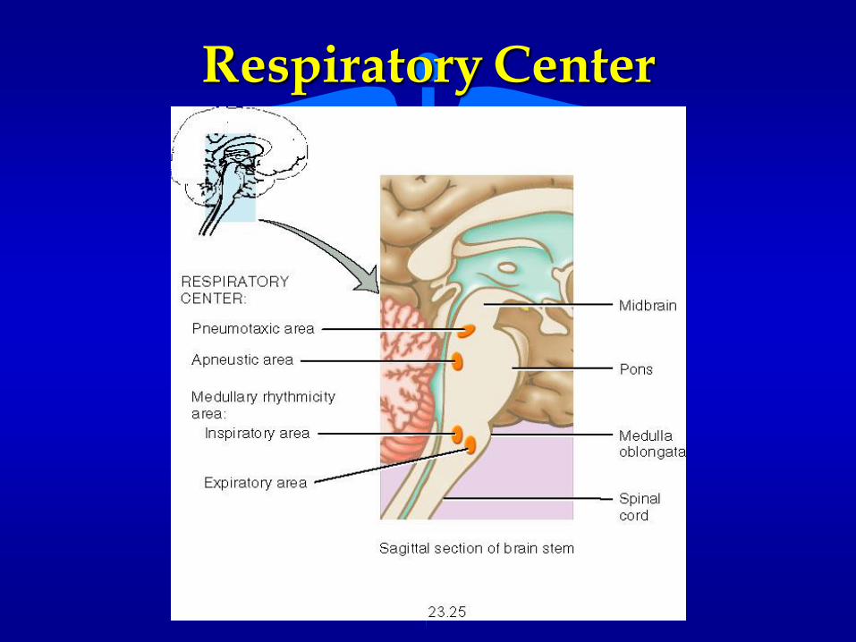
Respiratory Center

Chemical Stimuli
• Determines how fast and how deeply an individual breathes
• Very sensitive to the levels of CO2 and H+ ion concentration in the blood
• Monitored by chemoreceptors at:
– carotid arteries, aorta, and the medulla oblongata.
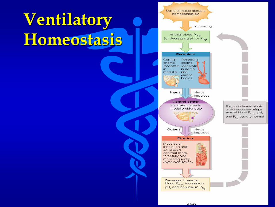
VentilatoryHomeostasis
RESPIRATORY DISORDERS AND HOMEOSTATIC IMBALANCES
Bronchiogenic Carcinoma(Lung Cancer)
• Most fatal cancer in the U.S.
• Highly metastatic
• Usually linked with cigarette smoking
• Starts in the walls of the bronchi due to irritation of the bronchiole epithelium
• Common irritants include smoking, pollution, dust particles
• 20 times more prevalent in smokers than non- smokers

Emphysema
• “Blown up or full of air”
• A condition where the alveolar walls lose their elasticity and remain filled with air during expiration
• Alveoli become damaged and eventually merge together to form large air sacs with reduced overall volume
• Patients often develop a barrel chest
• Generally caused by cigarettes, pollution, industrial dust particles
Influenza
•Caused by one of many viruses
• Antibiotics cannot help
• Medications used to treat the symptoms– sneezing - coughing
– congestion - rhinorrhea
• May result in rhinitis: inflammation of the nasal mucosa
• Commonly known as the flu

Pneumonia
• Acute infection or inflammation of the alveoli of the lungs
• Most common infectious cause of death in the U.S.
• Alveolar sacs fill with fluid and dead white blood cells reducing the amount of functional surface area of the lungs
• Most commonly caused by bacterium
– Streptococcus pneumoniae
• Affects those in poor health or compromised immune system
Sudden Infant Death Syndrome (SIDS)
• 10,000 infant deaths per year in the U.S.
• Cause is not known but thought to be caused by an infectious agent or compressed carotid artery
• Most deaths occur in the fall or winter
• Over 50% of SIDS death children had an upper respiratory infection within the past two weeks
• May also be caused by improper positioning for sleeping in the crib
Tuberculosis (Tb)
• Caused by a bacterium– Mycobacterium tuberculosis
• An infectious communicable disease that destroys the lung tissue and pleura
• Replaced by fibrous connective tissue called tubercles
• Disease is spread by inhalation of the bacterium


