24
The Respiratory system • Pulmonary ventilation – Chp 16 • Respiration
| Date post: | 20-Dec-2015 |
| Category: |
Documents |
| View: | 225 times |
| Download: | 0 times |
The Respiratory system
• Pulmonary ventilation – Chp 16
• Respiration

Outline
• Overview of the respiratory system
• Anatomy
• Forces for pulmonary ventilation
• Factors affecting pulmonary ventilation
• Clinical significance of respiratory volumes and air flows

Outline
• Overview of the respiratory system
• Anatomy
• Forces for pulmonary ventilation
• Factors affecting pulmonary ventilation
• Clinical significance of respiratory volumes and air flows
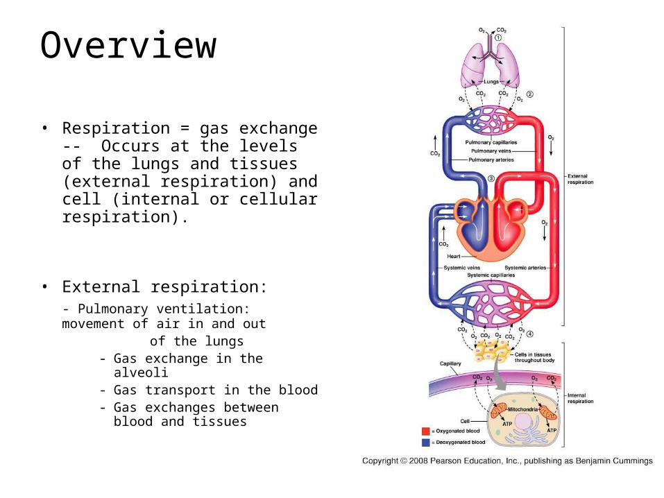
Overview
• Respiration = gas exchange -- Occurs at the levels of the lungs and tissues (external respiration) and cell (internal or cellular respiration).
• External respiration: - Pulmonary ventilation:
movement of air in and out of the lungs
- Gas exchange in the alveoli- Gas transport in the blood- Gas exchanges between
blood and tissues

Outline
• Overview of the respiratory system
• Anatomy
• Forces for pulmonary ventilation
• Factors affecting pulmonary ventilation
• Clinical significance of respiratory volumes and air flows

Airways

Airways
• Upper airways: - nose to pharynx
• Lower airways:- Conducting airway:
larynx bronchioles
- Respiratory airway:alveoli
Due to the wall structure of the airway: one cell layer (SSE) allows for gas exchange

Conducting airways
• Presence of cartilage in the wall, from larynx to small bronchi prevents airway collapse.
• Goblet cells secreted mucus. Ciliated cells help move the mucus out of the airway.
• Presence of smooth muscle fibers in the bronchioles (but no cartilage)
• Volume of the conducting airway: 150 ml

Respiratory airway: Alveoli
• Alveolar wall is formed by simple squamous epithelium = type I cells (SSE) gas exchange
• Respiratory membrane: membrane separating alveolus from blood capillary.
• Large surface area from the numerous alveoli better gas exchange
• Presence of elastic fibers between alveoli

Blood supply to the lungs

Alveolar structure
• Type I cells gas exchange
• Type II cells secrete surfactant (lipoproteins) decrease surface tension allowing for easier alveoli inflation
• Surfactants start to be secreted by the 7th month of pregnancy risk of lung disease in premature babies
• Presence of macrophages in alveoli

Structure of the thoracic cavity

The pleura
• Formed by 2 layers: the parietal and visceral pleura
• Roles: - prevents friction of the lungs against the rib cage (due to the thin layer of liquid present in the pleural space)- maintains lung expansion: due to the negative pressure within the pleural space
What is negative pressure? What is its importance?

Pleura and negative pressure
• Pneumothorax: lung collapse due to air entering in the pleural cavity
• (not to be confused with atelectasy alveoli collapse)

Outline
• Overview of the respiratory system
• Anatomy
• Forces for pulmonary ventilation
• Factors affecting pulmonary ventilation
• Clinical significance of respiratory volumes and air flows

Mechanics of breathing
• Boyle’s law: The pressure of a gas in a closed container is inversely proportional to the volume of the container.
• Air flow in the lungs is driven by the differences in pressure between the atmosphere and the alveoli
• P.atm is constant changes in P.alv drive ventilation

Inspiration and expiration
• Inspiration: chest wall expands due to muscle contraction (diaphragm and/or other muscles)
Pressure in alveoli ↓ air moves toward alveoli
• Expiration: passive process muscle relax chest wall return to resting state alveoli become compressed ↑ alveolar pressure move moves out

Ventilation

Outline
• Overview of the respiratory system
• Anatomy
• Forces for pulmonary ventilation
• Factors affecting pulmonary ventilation
• Clinical significance of respiratory volumes and air flows

Factors affecting pulmonary ventilation
• 1- Lung compliance: ease with which lungs can be stretched- Compliance is a measure of the elasticity of lung tissue and the alveolar surface tension
• 2- Airway resistance: to changes in airway radius (↓radius ↑resistance)
Pathology
• lung disease resulting in stiffness of tissue
• no or ↓ surfactant
• Asthma• Airway obstruction• COPD

Outline
• Overview of the respiratory system
• Anatomy
• Forces for pulmonary ventilation
• Forces affecting pulmonary ventilation
• Clinical significance of respiratory volumes and air flows

Lung volumes
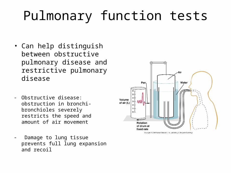
Pulmonary function tests
• Can help distinguish between obstructive pulmonary disease and restrictive pulmonary disease
- Obstructive disease: obstruction in bronchi-bronchioles severely restricts the speed and amount of air movement
- Damage to lung tissue prevents full lung expansion and recoil

Anatomical dead space
• Anatomical dead space: space within the conductive airway, about 150 ml.
• What will happen to a person who has a tidal volume of 150 ml due to lung disease?
• What can be done to help the person?


