40
THE RESPONSE OF SYNTHETIC 4- HYDROXYBENZOIC ACID ON Kv1.4 POTASSIUM CHANNEL SUBUNIT EXPRESSED IN Xenopus laevis OOCYTES FATIN HILYANI MOHAMAD UNIVERSITI SAINS MALAYSIA 2016

THE RESPONSE OF SYNTHETIC 4-
HYDROXYBENZOIC ACID ON Kv1.4 POTASSIUM
CHANNEL SUBUNIT EXPRESSED IN Xenopus laevis
OOCYTES
FATIN HILYANI MOHAMAD
UNIVERSITI SAINS MALAYSIA
2016
THE RESPONSE OF SYNTHETIC 4-HYDROXYBENZOIC ACID ON Kv1.4
POTASSIUM CHANNEL SUBUNIT EXPRESSED IN Xenopus laevis OOCYTES
By
FATIN HILYANI MOHAMAD
Thesis submitted in partial fulfillment of the requirement
for the degree of
Master of Neuroscience
JUNE 2016
TINDAK BALAS SINTETIK 4-HYDROXYBENZOIK ASID KE ATAS SALURAN
KALIUM Kv1.4 YANG DI EKSPRESIKAN KE DALAM OOSIT Xenopus laevis.
oleh
FATIN HILYANI MOHAMAD
Tesis diserahkan untuk memenuhi sebahagian keperluan bagi
Ijazah Sarjana Neurosains
JUN 2016
ACKNOWLEDGEMENT
For,
All lecturers and supervisors; Dr Jingli Zhang, Prof. Dato’ Dr Jafri Malin Abdullah,
Assoc. Prof. Dr Muzaimi Mustapha, Prof. Dr Shaharum Shamsuddin (PPSK) and Dr
Wan Amir Nizam Wan Ahmad (PPSK).
Special thanks to Prof. Dr Robert Bahring (University of Hamburg, Germany) who
kindly donated DNA of Kv1.4 potassium channel for the DAAD Electrophysiology
Workshop in Universiti Sains Malaysia (USM) in June 2009 and gave permission to
subsequently use it for the Department of Neurosciences, School of Medical Sciences,
Universiti Sains Malaysia for future experiments.
Friends whom I owe much gratitude and respect; Jia Hui Wong, Mazira Ghazali
(Neuro Dept), Sui Mei Kee, Nuraza Othman (Neuro Lab) and all Integrated
Neuroscience (INP) and Neuroscience Department family,
Mohamad and Wan Naimah, the parent who have no idea what I am doing, but is still
proud of me anyway,
Thank you,
Fatin H. Mohamad, (11th July, 2016)

ii
TABLE OF CONTENTS
PAGE
ACKNOWLEDGEMENTS i TABLE OF CONTENTS ii LIST OF TABLES v LIST OF FLOWCHART vi LIST OF FIGURES vii LIST OF ABBREVIATIONS AND ACRONYMS viii ABSTRAK x ABSTRACT xii CHAPTER 1: INTRODUCTION 1.1 Background of Study 1
1.2 Rationale of Study 5 1.3 Experiment Group 6
1.4 Data Collection 6 CHAPTER 2: LITERATURE REVIEW 2.1 The Regulation of Membrane Potential 7 2.2 Voltage-Gated Potassium Channel 10 2.3 Kv1.4 Channel 15 2.4 4-Hydroxybenzoic acid (4-hba) 22 2.5 Xenopus laevis Oocytes 26 2.6 Two Electrode Voltage Clamp 30 2.7 An Overview of Kv1.4 Associated Diseases 32

iii
2.8 Hypothesis 36 2.9 Objective 2.9.1 General Objective 37 2.9.2 Specific Objective 37 CHAPTER 3: METHODOLOGY 3.1 Ethical Approval 38 3.2 Preparation of Lysogeny broth (LB) for cDNA 40 3.3 Competence Cells Preparation 40 3.4 In-vitro Transcription 43 3.5 Preparation of Xenopus laevis 44 3.5.1 Surgery 45 3.6 Injection of cRNAs 47 3.7 Potassium Kv1.4 Channel Current Recording 48 3.8 Preparing Solutions for Perfusion System 3.8.1 5M 4-Hydroxybenzoic acid 50 3.8.2 2.5mM 4-Hydroxybenzoic acid 50 3.8.3 1.0mM 4-Hydroxybenzoic acid 51 3.8.4 100µM 4-Hydroxybenzoic acid 51 3.8.5 10µM 4-Hydroxybenzoic acid 51 3.8.6 1µM 4-Hydroxybenzoic acid 51 3.8.7 Tetraethylammonium (TEA) 52 3.8.8 4-Aminopyridine (4-AP) 52 3.9 The Perfusion System 52 3.10 The Analysis of Kv1.4 Channel Activity 54 CHAPTER 4: RESULTS 4.1 The Current Reading of 1µM 4-hydroxybenzoic acid 56 4.2 The Current Reading of 10µM 4-hydroxybenzoic acid 59 4.3 The Current Reading of 100µM 4-hydroxybenzoic acid 61

iv
4.4 The Current Reading of 1mM 4-hydroxybenzoic acid 64 4.5 The Current Reading of 2.5mM 4-hydroxybenzoic acid 66 4.6 The Normalized Ratio Decrease 69 4.7 The Normalized Ratio for Potassium Channel Blockers 72 4.8 The Effective Time Response of 4-hydroxybenzoic acid on Kv1.4 75 4.9 Summary of the Effects of 4-Hba Concentrations 78 CHAPTER 5: DISCUSSION 5.1 The Response of 4-Hydroxybenzoic acid on Kv1.4 80 5.2 The Dose Response Curve 83 5.3 The Response of Potassium Channel Blockers 85 5.4 The Effective Time Response of 4-hydroxybenzoic acid 86 5.5 Limitations and Future Prospects of the Study 87 CHAPTER 6: CONCLUSION 89 REFERENCES 90 APPENDICES 102

v
LIST OF TABLES
PAGE
Table 2.1
Kv1.4 potassium channel nomenclature and details
15
Table 2.2 Anti-oxidation and Pro-oxidation of Phenols 26
Table 4.1 Mean, Standard Error and P Values of Kv1.4 Ratio
69
Table 4.2 Mean, Standard Error and P Values of Blockers Ratio
73
Table 4.3 Mean and Standard Error of Effective Time Ratio
76

vi
LIST OF FLOWCHART
PAGE
Flowchart 3.1 Methodology process applied in the study
39

vii
LIST OF FIGURES
PAGE
Figure 2.1 Changes of Membrane Potential by Ionic Movements 9
Figure 2.2 3 Different Structures of Voltage-Gated Potassium Channels 10
Figure 2.3 Structure of the 6 Transmembrane Kv Channel and Its Family 12
Figure 2.4 Comparison of Therapeutic Intervention of K Channel Enhancement and Inhibition
14
Figure 2.5 Family of Kv1 Shaker and Its Inactivation 17
Figure 2.6 The Permeation and Allosteric Inactivation Mechanisms of Kv1.4
19
Figure 2.7 Effects of [K] and pH on C-type Inactivation 22
Figure 2.8 Structural Comparison of 3-hba and 4-hba 24
Figure 2.9 Xenopus laevis and Its Extracted Oocytes
28
Figure 2.10 Two-electrode Voltage Clamp Machine
31
Figure 3.1 Surgery of Xenopus laevis for Oocytes Extraction
46
Figure 3.2 Selected Viable Oocytes and cRNA Injection into Oocytes
48
Figure 4.1 Readings of 0.1% DMSO, K Blocker and 1µM 4-hba
56
Figure 4.2 Readings of 0.1% DMSO, K Blocker and 10µM 4-hba
59
Figure 4.3 Readings of 0.1% DMSO, K Blocker and 100µM 4-hba
62
Figure 4.4 Readings of 0.1% DMSO, K Blocker and 1mM 4-hba
64
Figure 4.5 Readings of 0.1% DMSO, K Blocker and 2.5mM 4-hba
67
Figure 4.6 Bar Graph and Dose Response Curve of 4-hba on Kv1.4 71
Figure 4.7 Bar Graph and Dose Response Curve of K Blockers on Kv1.4 74
Figure 4.8 Dose Response Curves of Time Response of 4-hba on Kv1.4 77
viii
LIST OF ABBREVIATIONS AND ACRONYMS
3-Hba 3-hydroxybenzoic acid
3,4-DHba 3,4-dihydroxybenzoic acid
4-AP 4 – aminopyridine
4-Hba 4-hydroxybenzoic acid
AEDs Anti epileptic drugs
BADs Benzoic acid Derivatives
Ca2+
Calcium ion
cDNA Competence DNA
Cl-
Chloride ion
cRNA Competence RNA
DMSO Dimethyl sulfoxide
DNA Deoxyribonucleic acid
GA Gallic acid
IC50
Inhibitory concentration (50%)
[K]o
Extracellular potassium ion concentration
K+ Potassium ion
Kv Voltage-gated potassium channel
mM mili molar
ms mili second
mV mili volt
Na+
Sodium ion
ND96 Frog’s Ringer or buffer solution

ix
OR-2 Buffer solution to wash away oocytes follicles
RNA Ribonucleic acid
RPM
Rotation per minute
SA Salicylic acid
TEA Tetraethylammonium
TEVC Two-electrode voltage clamp
µA micro ampere
µM micro molar
X.laevis Xenopus laevis
x
TINDAK BALAS SINTETIK 4-HYDROXYBENZOIK ASID KE ATAS
SALURAN KALIUM Kv1.4 YANG DI EKSPRESIKAN KE DALAM OOSIT
Xenopus laevis
ABSTRAK
Kajian yang mendalam sedang dijalankan ke atas produk semulajadi terutamanya
pokok-pokok herba yang telah lama dipraktikkan di dalam perubatan tradisional
seperti Cina dan Ayurveda untuk merawat penyakit saraf seperti sawan dan sakit
kepala. 4-hydroxybenzoik asid adalah fenol tidak flavonoid yang boleh ditemui dari
pucuk Dendrocalamus asper (buluh), buah-buahan (strawberi dan epal) dan bunga-
bungaan. Di dalam kajian ini, tindak balas 4-hydroxybenzoik asid diuji ke atas
saluran kalium Kv1.4 yang telah diekspresikan ke dalam oosit Xenopus laevis
sebagai model sistem. Kv1.4 adalah saluran kalium dari keluarga Shaker yang pantas
dinyahaktifkan melalui dua mekanisma; jenis N yang pantas dan jenis C yang
perlahan. Ianya memainkan peranan penting dalam repolarisasi, hyperpolarisasi dan
mengembalikan potensi membran melalui pengawalan pergerakan K+ menyeberangi
luar membran sel. cRNA Kv1.4 yang telah disediakan dalam kerja molecular
disuntik ke dalam oosit sihat yang telah diambil melalui pembedahan X.laevis di
bahagian abdomen bawah. Arus dihasilkan daripada K ions dikesan oleh voltan
apitan dua-elektrod-mikro (TEVC), dengan potensi kawalan dari -80mV dan
peningkatan 20mV sehingga +80mV. Bacaaan dari rawatan oleh 0.1% DMSO,
konsentrasi 4-Hba dan penghalang saluran kalium diambil pada +60mV. Analisis
dijalankan menggunakan perisian pClamp diikuti t-test pelajar. Nisbah amplitud

xi
akhir / puncak adalah merupakan indeks aktiviti saluran Kv1.4 dengan n ≥ 6
(bilangan oosit yang diuiji). Nisbah yang rendah menunjukkan potensi membran
yang rendah (repolarisasi) dan penambahan nyahaktif saluran Kv1.4. Pengurangan
nisbah dari 5 konsentrasi yang berbeza (1µM, 10µM, 100µM, 1mM dan 2.5mM)
dibandingkan dengan 0.1% DMSO sebagai kawalan. Kesemua konsentrasi
menunjukkan keputusan signifikasi statistik dengan p < 0.05 kecuali untuk 100µM.
Peningkatan arus konsentrasi yang dinormalisasikan melalui perbandingan dengan
penghalang saluran kalium (TEA dan 4-AP) menunjukkan signifikasi statistik bagi
kesemua konsentrasi. Kajian ini juga menunjukkan tempoh masa yang diambil oleh
setiap konsentrasi untuk mempengaruhi nyahaktiviti Kv1.4 didapati tidak
memainkan apa-apa peranan penting. Kesimpulannya, 4-hydroxybenzoik asid
dikenal pasti dapat menambah baik kesan nyahaktiviti Kv1.4 dalam mengurangkan
atau merepolarisasikan potensi membran supaya ledakan saraf yang tidak normal
dapat dihalang. Ini dapat dilihat melalui perbandingan dengan DMSO dan juga
penghalang saluran kalium. IC50 didapati sedikit tinggi dari 10µM dan konsentrasi
yang lebih tinggi (100µM, 1mM dan 2.5mM) menujukkan kesan sampingan toksik.
Oleh itu, konsentrasi yang terbaik dari kajian ini adalah 10µM dengan curaman Hill
(slope) 0.1799.

xii
THE RESPONSE OF SYNTHETIC 4-HYDROXYBENZOIC ACID ON Kv1.4
POTASSIUM CHANNEL SUBUNIT EXPRESSED IN Xenopus laevis
OOCYTES
ABSTRACT
Extensive researches are being made on natural products especially herbs and plants
that have long been practiced in traditional medicines such as Chinese and
Ayurvedic that have been used to treat neuronal disorders such as convulsive,
dizziness and headaches. 4-hydroxybenzoic acid is a non-flavonoid phenol found
abundantly in Dendrocalamus asper shoots (bamboo), fruits (strawberries and
apples) and flowers. In this study, the response of synthetic 4-hydroxybenzoic acid
was tested on Kv1.4 potassium channel that was expressed in Xenopus laevis oocytes
as the model system. Kv1.4 is a rapidly inactivating Shaker-related member of the
voltage-gated potassium channels with two inactivation mechanisms; the fast N-type
and slow C-type. It plays vital roles in repolarization, hyperpolarization and
signaling the restoration of resting membrane potential through the regulation of the
movement of K+ across the cellular membrane. cRNA of Kv1.4 prepared during
molecular work was injected into viable oocytes that was extracted through surgery
at the lower abdomen of X.laevis. The current produced from K ions were detected
by the two-microelectrode voltage clamp (TEVC) method, holding potential starting
from -80mV with 20mV step-up until +80mV. Readings of treatments with 0.1%
DMSO, 4-Hba concentrations and K channel blockers were taken at +60mV. The
ratio of tail / peak amplitude is the index of the activity of the Kv1.4 channels with n

xiii
≥ 6 (number of oocytes tested). Lower ratio signifies lower membrane potential and
enhancement of Kv1.4 channel inactivation. The decreases of the ratios of 5
different concentrations (1µM, 10µM, 100µM, 1mM and 2.5mM) were compared
with 0.1% DMSO as the control. All concentration showed statistically significant
results with p < 0.05 except for 100µM. The normalized current of the 4-hba
concentrations were compared with potassium channel blockers (TEA and 4-AP) and
all groups showed statistically significant results. This study also showed that time
taken for each concentration to affect Kv1.4 does not play any significant roles. In
conclusion, 4-hydroxybenzoic acid was found to be able to enhance the inactivation
of Kv1.4 by lowering the membrane potential so that the abnormal neuronal firing
can be inhibited which can be seen through comparison of DMSO and potassium
channel blockers. With IC50 slightly higher than 10µM, increasing concentrations
(100µM, 1mM and 2.5mM) had shown to exhibit toxicity effects. The best
concentration from this study is 10µM with Hill slope of 0.1799.
1
CHAPTER 1
INTRODUCTION
1.1 Background of Study
Following the regulation of membrane potential by the movements of
inhibitory ions such as K+ and Cl- and excitatory ions such as Na+ and Ca2+,
depolarization or more positive membrane potential could results in neuronal
firing whereas repolarization and hyperpolarization or more negative
membrane potential contribute to neuronal inhibition (Purves, et al. 2012).
Any irregularities or disruption to this mechanism would interfere with the
normal neuron action and inactivation which could result in many neuronal
disorders.
Based on previous studies and researches, potassium ions have been
identified as the most diverse ion channels that can be found in almost every
part of the human body encoded by more than 70 genes classified into 12
subfamilies (Kv1 – 12) (D’Adamo, et al. 2013). Nevertheless, limited studies
have been done on A-type transient current producing with double
inactivation mechanisms (N-type and C-type), Kv1.4 channel (Jefferys,
J.G.R. 2010; Chen H. et al. 2013; Oliva, et al. 2005). Kv1.4 channel can be
found abundantly in Schaffer collateral axons and part of the molecular layer
2
of the dentate gyrus. It also formed heteromerization with Kv1.1 and Kvβ1.1
subunits in the mossy fiber boutons that synapse with the pyramidal neurons
in CA3. Mutations from this heteromeric formation can contribute to the
episodic ataxia type 1 (EA1) seizure (D’Adamo, et al. 2013).
Kv1.4 is responsible in regulating the amplitude of back-propagating action
potentials of the neuron through its double inactivation mechanisms which
could result in non-conductance of K+ and repolarization (Rasmusson, et al.
1998). Therefore, the enhancement Kv1.4 inactivation will help to overcome
the abnormal high frequency of action potential which underlies many
neuronal disorders such as convulsive, fits, epilepsy and even stroke (Wulff,
et al. 2009). The double inactivations are hypothesized to occur through the
permeation and allosteric mechanisms which are influenced by many factors
such as pH, oxidation and extracellular [K] (Claydon, et al. 2004; Xu, et al.
2001).
In this study, the efficacy of synthetic 4-Hydroxybenzoic acid (4-Hba) in
promoting and enhancing potassium Kv1.4 channel inactivation was tested by
expressing the channel via cRNA injection in the model system of Xenopus
laevis oocytes. 4-Hba is a non-flavonoid phenolic compound (Khadem and
Marles, 2010) that can be found in many natural products such as
Dendrocalamus asper and Veronica peregrina L. (Kim, et al. 2014).
Extraction of Dendrocalamus asper shoots by our collaborator Universiti
Malaysia Terengganu (UMT), found abundance of 4-hydroxybenzaldehyde.
3
However, as the compound is in excess of valence electrons for bonding, it
can be easily oxidized to 4-hydroxybenzoic acid (Dobhal, et al. 2010) which
is a more stable structure. In addition, based on previous study by Bilal
(2015), preliminary screening of the effects of 4-hba, palmitic acid and lauric
acid found that 4-hba can enhance the inhibitory current of GABA (A)
channels which were expressed in X.laevis oocytes unlike palmitic and lauric
acids. Nevertheless, the effect of this compound on Kv1.4 channels which
also helps to lower the membrane potential has never been tested, making it
as the objective of this study.
Every cell functions and regulates itself by the movement of ions, proteins
and molecules across the cellular membrane that separates the intracellular
from the extracellular environment. These movements of ions with charges
across a barrier lead to electrical potential difference or membrane potential
that can be detected by electrodes. This is called the electrophysiology study
techniques (Bierwirtz and Schwarz, 2014). Two-electrode voltage clamp
(TEVC) technique used in this experiment allows the measurement of ions
flow across the oocytes membrane by injecting two microelectrodes, one for
detecting the voltage and another for current injection. The voltage is
clamped at -80mV and any current and membrane potential changes read by
the electrodes are compared to calculate the differences which can vary due
to additional Kv1.4 expression of cRNA in the oocytes.

4
The extraction of Xenopus laevis oocytes were carried out by surgery.
Incision less than 1cm were made on the lower abdomen of the anesthetized
selected frog and all the lobes of oocytes were pulled out using forceps. A
normal female frog usually has more than 5 lobes of oocytes that produce
hundreds of them. The incised muscle and skin were sutured again so that the
frog can live for another oocytes extraction on the opposite side of the
abdomen if necessary (at least 3 months apart). These oocytes will be
individually screened and selected for bigger oocytes with clear separation of
yellowish ‘vegetal’ and dark brown ‘animal’ poles (Sigel and Minier, 2005).
Then, the oocytes were incubated for a night before cRNA injection of the
Kv1.4 using the micropipette can be done.
The voltage reading of the injected oocytes under 0.1% DMSO was
compared with five different concentrations of 4-Hba (1µM, 10µM, 100µM,
1mM and 2.5mM). At least 6 different viable oocytes were used for each
concentration. The solutions were controlled by the perfusion system whilst
the oocytes were impaled by the double electrodes. The voltage is hold at -
80mV, with step-up of 20mV until +80mV. Recordings were taken at +60mV
which is the potential at which potassium channel is activated. The oocytes
are bathed in the neutral ND96 (1 min) before 0.1% DMSO (1 min) followed
by 4-Hba (5 mins) and finally the potassium channel blockers (12.5 mM TEA
and 5mM 4-AP) (5 mins). In total, there were 12 voltage readings taken for
every voltage-clamping of each oocytes.

5
In conclusion, this study investigates the response of 4-Hba effect on Kv1.4
potassium channels inactivations which are responsible in controlling the
repolarization and restoring the resting membrane potential of the neurons.
These channels are expressed in Xenopus laevis oocytes that act as model
system so that the electrophysiological changes elicited can be read by the
two-electrode voltage clamp technique. Lowering of membrane potential
shows enhancement action of 4-Hba on the inactivation mechanisms of
Kv1.4, resulting in prolonged repolarization which is the targeted action to
overcome abnormal continuous neuronal firing.
1.2 Rationale of Study
In general, this study aims to investigate the response of synthetic 4-Hba
which can be found abundantly in natural products. This is to find additional
and new potential compounds that are able to enhance repolarization of
membrane potential as a targeted mechanism for abnormal continuous
neuronal firing (convulsant, fits) treatment through natural resources such as
herbs and plants that are less toxic and with lower prolong effects. Although
there are many previous studies investigating potential compounds that can
enhance repolarization, there have been none on 4-Hba based on our findings
and researches. In addition, there are also fewer studies on the inactivation
response of Kv1.4 channels against natural compound and its importance in
affecting membrane potential and inhibiting the neuronal excitability. If this
research provides positive answers, it could offer additional discovery on the
6
mechanism and importance of Kv1.4 channel along with proving the
practicality of natural compounds in treating disorders and deregulations that
cannot be answered by drugs.
1.3 EXPERIMENT GROUP
There are 4 channels filled with different solutions for every recording. The
first and second channels are constant but the third and fourth channels are
manipulated depending on the concentrations of 4-hydroxybenzoic acid
groups.
1) First channel = 50ml ND96 solution
2) Second channel = 50µl DMSO + 50ml ND96
3) Third channel = experimental group (50µl of 1µM or 10µM or 100µM or
1mM or 2.5mM 4-hydroxybenzoic acid + 50ml ND96)
4) Fourth channel = negative control group (50µl of 1µM or 10µM or
100µM or 1mM or 2.5mM 4-hydroxybenzoic acid + 50µl 12.5mM TEA +
50µl 5mM 4-AP + 50ml ND96)
1.4 DATA COLLECTION
The current obtained from recordings were saved in the computer and
analyzed with p-Clamp10 (Axon Instruments, USA) software and statistically
tested with student t-test with SigmaPlot12 (Systat Inc, USA) software and
Prism6 (GraphPad Software, USA).
7
CHAPTER 2
LITERATURE REVIEW
2.1 The Regulation of Membrane Potential
Movement of ions across cellular membrane creates potential difference or
electrical gradient due to difference of ionic positive and negative charges
between extracellular and intracellular membrane. This current-like potential
difference is called membrane potential and it can be detected using
microelectrodes reading. There are basically four important and influential ions
that can regulate the membrane potential. These are the Na+, K+, Cl- and Ca2+.
However, the firing, inhibition and resting of the neurons are majorly affected
by the influx and efflux of Na+ and K+.
The membrane is at resting phase (-70 to - 60 mV) at (0) based on Fig. 2.1.
During this phase, Na-K ATPase pump channel is opened allowing the influx
of 2 potassium ions and efflux of 3 sodium ions across the cellular membrane.
Therefore, the concentration of K+ is higher inside and Na+ is higher outside.
According to Purves, et al. (2012), intracellular membrane potential is also
aided by protein anions and is balanced by extracellular Cl- concentration.
When a signal or stimulus is received, it causes the opening of sodium
channels, allowing the ions to enter the cellular membrane and causing the
8
membrane potential to be more positive resulting in the depolarization phase
(1). Action potential is triggered when the depolarization overshoots and
reaches the peak (2 and 3).
Overshooting also triggered the activation of potassium ion channels, which
will allow the efflux of K+ across cellular membrane into the extracellular
environment. However, when the membrane potential reaches the peak (3), it
signals the closing of the sodium ion channels. Continuous efflux of K+ and
blocking of Na+ influx results in decreasing membrane potential (4) which will
lead to repolarization. Repolarization is important in inhibiting the neuronal
firing and initiating membrane permeability restoration. During this phase, the
potassium ion channels start to become inactivated, causing the non-conduction
of K+. Prolonged inactivation and delayed closing of the potassium ion channel
eventually lead to undershooting where the membrane potential becomes too
low. This is the hyperpolarization stage (5). Ultimately the potassium ion
channel will close and the movement of K+ is blocked signaling for the
restoration of resting membrane potential. In general, whilst Na+ is mainly
responsible in bringing the intracellular membrane potential towards positivity
(or increasing the potential), Cl- and K+ are of the opposite (lowering
membrane potential) (Purves, et al. 2012).

9
Figure 2.1: The changes of the membrane potential (mV) due to the movement of the ions across cellular membrane. Where 0 = resting membrane potential, 1 = depolarization, 2 = overshooting, 3 = peak, 4 = repolarization and 5 = undershooting or hyperpolarization. From: ‘Neuronal Action Potential’, Physiology Web, Accessed 6th June 2016 http://www.physiologyweb.com/lecture_notes/neuronal_action_potential/neuronal_action_potential.html
The regulation of membrane potential and the ion channels are extremely
important in sustaining normal functioning cells and physiological systems.
Abnormal and irregular control of the membrane potential has been revealed to
be the main cause of many disorders and diseases such as convulsive, epilepsy,
dizziness, depression and even stroke (Wulff, et al. 2009). These disorders
occur due to abnormalities and mutations of the ionic channels and are usually
referred as ‘channelopathies’.

10
2.2 Voltage-Gated Potassium Channel
Since the past decades, researches have been targeting ion channels as
treatment for many diseases and clinical abnormalities (Camargos, et al. 2011).
One of these channels is potassium channels which can be classified into 3
structural families according to its amino acid sequence and pore-containing
subunit; i) six transmembrane voltage-gated one-pore with S1 – S6 ii) two-
transmembrane one-pore inward rectifier K+ and iii) four transmembrane two-
pore (Shieh, et al. 2000). Figure 2.2 shows the structural difference between
these 3 potassium channels.
Figure 2.2: Representation of the 3 structural differences of K+ channels based on the amino acids and pore subunits arrangements. The 6 transmembrane voltage-gated one pore channel (A) is the most abundant K+ channels in the human body mostly found around active cells such as cardiac, skeletal and neurons. From: “Potassium Channels: Molecular Defects, Diseases and Therapeutic Opportunities” by Shieh, et al. (2000), Pharmacological Reviews, 52: 557 – 593.

11
Voltage dependant or voltage-gated potassium channels are vital in
determining the resting membrane potential and membrane excitability of the
neurons (Jugloff, et al. 2000). It also plays important roles in inhibiting action
potential, potassium channel, neurotransmitter-mediated signaling, regulating
Ca2+ homeostasis and cell survival (D’Adamo, et al. 2013).
More than 40 voltage-gated potassium channels have been identified and
classified into 12 sub-families so far (Kv1 – Kv12). Most of these channels are
found in almost every cells of the human body encoded by more than 70 genes
which makes voltage-gated potassium channels as the most diverse ion
channels. Kv channels appear as either homomeric or heteromeric within
groups Kv1 (delayed-rectifier and A-current), Kv2 (delayed rectifier), Kv3
(high-voltage-activated, fast kinetics), Kv4 (somatodendritic A-current) and
Kv7 (M-current). Kv1 – 4 was discovered in Drosophila and cloning
identification of Kv1 Shaker (Sh) was followed by Kv2 Shab (Sb), Kv3 Shaw
(Sw) and Kv4 Shal (Sl) (Judge and Bever, 2006). Figure 2.3 shows the subunits
of the Kv family with its IUPAC names and structural component.

12
Figure 2.3: Structure of the 6 transmembrane of voltage-gated K+ channels and its subunits according to International Union of Pharmacology. From: “Neuronal and Cardiovascular Potassium Channels as Therapeutic Drug Targets: Promise and Pitfalls” by Humphries and Dart (2015), Journal of Biomolecular Screening, 1 – 19, DOI: 10.1177/1087057115601677.
As mentioned by Shieh, et al. (2000), the ability of the channels to transport K+
across the cellular membrane is dependent on 3 factors i) permeation pathway
which allows K+ to cross the cellular membrane ii) selectivity filter that
recognize K+ and iii) gating mechanism that changes between open and closed
state. Hence, even a small structural difference between the channels can
influence the regulation of ionic movement and transport.

13
According to Ping Li et al. (2013), there are 6 transmembrane segments of
voltage-gated potassium or Kv channels that are grouped into voltage-sensor
domain or VSD (S1 – S4), a pore domain (S5 – P – S6) and a re-entrant P loop
region (Chanda and Bezanilla, 2008). Pore domain or the selectivity filter is
responsible in gating or opening and closing the channels. These changes will
be detected by VSD in response to membrane potential changes or K+
permeability (Lee, J.H. et al. 2009). S4 is called the ‘voltage sensor’ which
possesses positively charged arginine residues and is able to influence
membrane voltage to exert forces on the gating of the pore (Mann, 2011).
Thus, the opening of the pore is regulated by the movement of the voltage
sensor by monitoring the ionic current flow (Rodriguez-Menchaca, et al. 2012).
It has been reported in a few studies, X-ray structures of S1 – S4 voltage-
sensing domains interact with lipids when embedded in the membrane and this
helps to reset the sensor to its activated state after depolarization (Long, S.B.,
et al. 2007; Jiang, Y. et al. 2003; Milescu, M. et al. 2009; Ramu, Y. et al. 2006
and Xu, Y. et al. 2008). Abnormality within the regulation of activation and
inactivation of voltage-gated potassium channel due to factors such as
mutations could manifest into neuronal disorders.
As shown in Figure 2.4, increased action potential frequency can theoretically
be corrected by enhancing K channels so that the positive peak amplitudes can
be lowered through efflux of K+. Prolonged depolarization or delayed
repolarization phase causes higher membrane potential which can results in
CNS hyperexcitability and also epilepsy. However, CNS depression and

14
cognition disorders could develop if the membrane potential is too low or
prolonged repolarization occurs. This abnormality can be reduced by inhibiting
the activation of K channels so that the efflux of K+ can be prevented and the
membrane potential can be increased.
Figure 2.4: The comparison of normal neuronal action potential with depression and epileptic brain waves. These abnormalities are reversed by the action of K channel inhibitors and activators which help to stabilize the membrane potential suited to the disorders. From “Theoretical Effects of Kv Channel Inhibitors and Activators on Pathologically Altered Neuronal Activity” byWulff et al., (2009), Nature Reviews Drug Discovery, 8, 982 – 1001, doi: 10.1038/nrd2983.
15
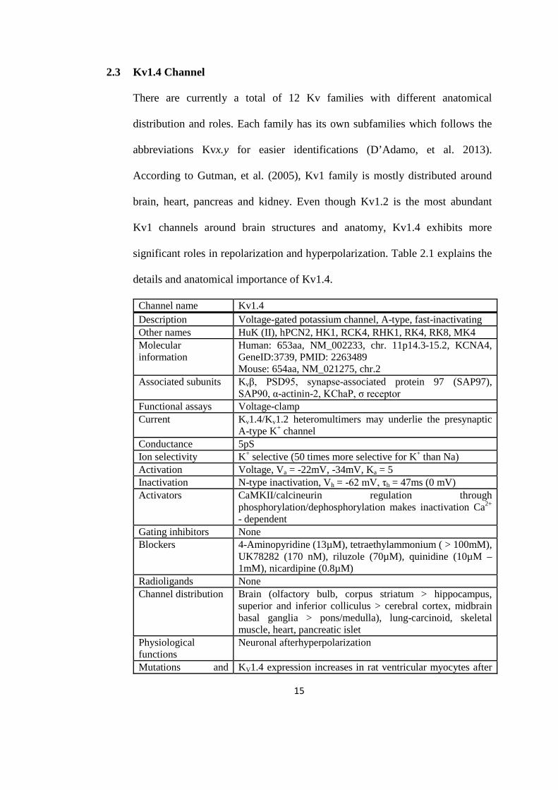
2.3 Kv1.4 Channel
There are currently a total of 12 Kv families with different anatomical
distribution and roles. Each family has its own subfamilies which follows the
abbreviations Kvx.y for easier identifications (D’Adamo, et al. 2013).
According to Gutman, et al. (2005), Kv1 family is mostly distributed around
brain, heart, pancreas and kidney. Even though Kv1.2 is the most abundant
Kv1 channels around brain structures and anatomy, Kv1.4 exhibits more
significant roles in repolarization and hyperpolarization. Table 2.1 explains the
details and anatomical importance of Kv1.4.
Channel name Kv1.4 Description Voltage-gated potassium channel, A-type, fast-inactivating Other names HuK (II), hPCN2, HK1, RCK4, RHK1, RK4, RK8, MK4 Molecular information
Human: 653aa, NM_002233, chr. 11p14.3-15.2, KCNA4, GeneID:3739, PMID: 2263489 Mouse: 654aa, NM_021275, chr.2
Associated subunits Kvβ, PSD95, synapse-associated protein 97 (SAP97), SAP90, α-actinin-2, KChaP, σ receptor
Functional assays Voltage-clamp Current Kv1.4/Kv1.2 heteromultimers may underlie the presynaptic
A-type K+ channel Conductance 5pS Ion selectivity K+ selective (50 times more selective for K+ than Na) Activation Voltage, Va = -22mV, -34mV, Ka = 5 Inactivation N-type inactivation, Vh = -62 mV, τh = 47ms (0 mV) Activators CaMKII/calcineurin regulation through
phosphorylation/dephosphorylation makes inactivation Ca2+ - dependent
Gating inhibitors None Blockers 4-Aminopyridine (13µM), tetraethylammonium ( > 100mM),
UK78282 (170 nM), riluzole (70µM), quinidine (10µM – 1mM), nicardipine (0.8µM)
Radioligands None Channel distribution Brain (olfactory bulb, corpus striatum > hippocampus,
superior and inferior colliculus > cerebral cortex, midbrain basal ganglia > pons/medulla), lung-carcinoid, skeletal muscle, heart, pancreatic islet
Physiological functions
Neuronal afterhyperpolarization
Mutations and KV1.4 expression increases in rat ventricular myocytes after

16
pathophysiology myocardial infarction and induction of diabetes Pharmacological significance
Not established
Comments Can coassemble with other KV1 family members in heteromultimers but not with members of other KV families; intronless coding region; mouse KV1.4 mRNA contains an internal ribosome entry site in its 5’-noncoding region and may be translated by cap-independent mechanisms, mammalian Shaker-related family.
Table 2.1: The nomenclature, molecular relationship and details of Kv1.4 channel as of 2005. From: Table 5 of ‘International Union of Pharmacology. LIII. Nomenclature and Molecular Relationships of Voltage-Gated Potassium Channels’ by Gutman, et al. (2005), Pharmacological Reviews, 57: 473 – 508.
Current produced by potassium channel is the IA which allows action potential
to reach dendrites (Jefferys, J.G.R. 2010). Generally there are two classes
generated by Kv currents, the dominant sustained K-current (IK,V) and the fast
inactivating transient A-current (IK,A) which is elicited by Kv1.4 (Chen, H. et
al. 2013). It is estimated that the molecular weight of Kv1.4 is 73 211 and it
shares similar membrane topology along with moderate amino acid sequences
as Kv1.1. Its rapid inactivating characteristic influence the lowering of
membrane potential after action potential and helps to halt the neuronal
excitability faster compared to other channels (Figure 2.5). This channel also
plays a crucial role in repolarization of cardiac myocytes along with Kv4.2 and
Kv4.3 as the molecular bases (Rasmusson, et al. 1998).

17
Figure 2.5: Shaker-related family of Kv1 channels. The phylogenetic tree of the gene family with IUPHAR and HGNC names shown with localization of the chromomes. Currents produced by each Kv1 families showing comparison of the rapid inactivation rate of Kv1.4 with others. Currents amplitudes in µA. Adapted from Heinemann, et al. 1996; Tian, et al. 2002; Finol-Urdaneta, et al. 2006. From: “Distinctive Role of Kv1.1 Subunit in the Biology and Functions of Low Threshold K+ Channels with Implications for Neurological Disease” by Ovsepian, et al. (2016), Pharmacology and Therapeutics, 159, 93 – 101.
Kv1.4 plays major roles in many physiological processes including the quantal
release of neurotransmitters, neuronal excitation, cardiac action potential,
muscle contraction, hormonal secretion, transporting electrolytes for epithelial,
cell volume and cell proliferation in neuronal and non-neuronal cells (Lee, J.H.
et al. 2009). Shaker K channels are structurally designed with two types of
inactivation; the fast N-type and slow C-type inactivation mechanisms (Oliva,
et al. 2005; Gonzales-Perez, et al. 2008).

18
The N-type inactivation is rapid and characterized by the NH2 terminal that
blocks the current flow of the channel intracelullarly by the ‘ball’ linked to a
‘chain’ domain structure as proposed by Armstrong and Bezanilla in 1972
(Lee, J.H. et al. 2009). The exact molecular composition of the ball is roughly
composed of 20 amino acids in the amino-terminal followed by 40 more
residues which constitutes the chain (Cai, et al. 2007). The ball is made of
hydrophobic residues and positive charges which is vital to push the ball
towards the pore during depolarization thus, initiating inactivation. Binding of
the ‘ball and chain’ to the pore is voltage insensitive and initiates occlusion of
the permeation pathway (Figure 2.6A) and conformational changes (allosteric
mechanism) (Figure 2.6B) (Bett and Rasmusson, 2004).
The permeation pathway occurs by blocking of the pore by the ‘ball’ during N-
type inactivation and prevents the movement of K+ across the cellular
membrane (Figure 2.6A). Hence, direct effects of the fast inactivation can be
seen immediately. The exact mechanisms of conformational changes are the
focus of many ongoing studies involving K channels. It occurs due to the
coupling of N-type to C-type. Unlike the N-type, the molecular and structural
basis of C-type is still debatable. Yet, it is stipulated to involve the selectivity
filter, extracellular conformational changes and permeant ions and intracellular
pore closure (Bett and Rasmusson, 2004), intracellular quinidine binding
(Wang, et al. 2003), intracellular osmotic pressure (Jiang, et al. 2003),
mutations on the extracellular face of the mouth of the pore and mutations on
the intracellular side of the pore (Figure2.6B). The inactivation of N-type also
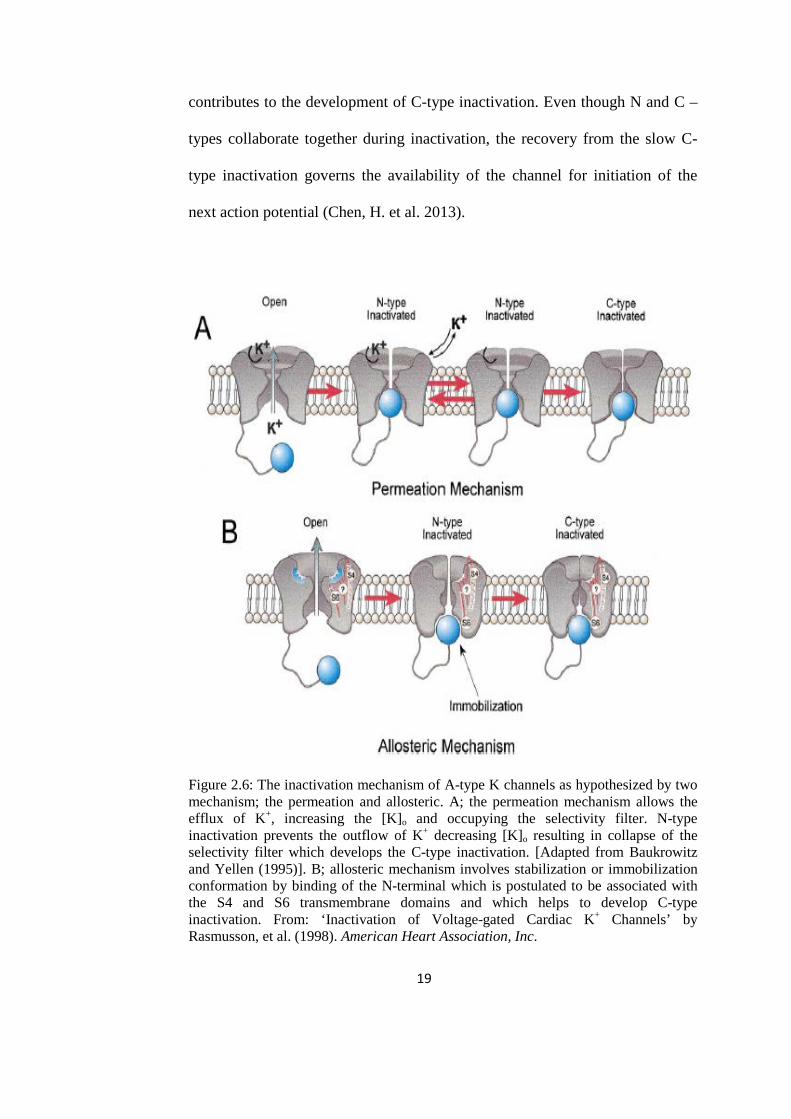
19
contributes to the development of C-type inactivation. Even though N and C –
types collaborate together during inactivation, the recovery from the slow C-
type inactivation governs the availability of the channel for initiation of the
next action potential (Chen, H. et al. 2013).
Figure 2.6: The inactivation mechanism of A-type K channels as hypothesized by two mechanism; the permeation and allosteric. A; the permeation mechanism allows the efflux of K+, increasing the [K]o and occupying the selectivity filter. N-type inactivation prevents the outflow of K+ decreasing [K]o resulting in collapse of the selectivity filter which develops the C-type inactivation. [Adapted from Baukrowitz and Yellen (1995)]. B; allosteric mechanism involves stabilization or immobilization conformation by binding of the N-terminal which is postulated to be associated with the S4 and S6 transmembrane domains and which helps to develop C-type inactivation. From: ‘Inactivation of Voltage-gated Cardiac K+ Channels’ by Rasmusson, et al. (1998). American Heart Association, Inc.

20
The activation, inactivation and closing of K+ channels are influenced and
regulated by many factors. Activation of the channels allows conductance of
K+ across the cellular membrane, whether inward or outwardly rectifying
(depending on the type of K+ channels) and is mostly involved during
subthreshold depolarization, whilst inactivation mostly occurs during
depolarization and results in a state of opened channels but with no
conductance of the K+. Inactivation contributes to repolarization and
hyperpolarization which also help in channels recovery (Bahring, et al. 2012).
Closed channels blocked the channel gating preventing total flow of K+ and
initiates the restoration of resting membrane potential (Antz and Fakler, 1998).
In 1966, McAllister and Noble proved that extracellular K+ concentration can
activates potassium channel and increases the inwardly rectifying cardiac K+
current. This effect has since been discovered to be applicable to almost all
potassium channels (both inward and outward rectifying currents) (Baukrowitz
and Yellen, 1995). Increased efflux of K+ through the open channel results in
accumulation of extracellular [K]o in the selectivity filter through a modulatory
site, which enhances the activation of K channels and increasing the K current
(Figure 2.7A). Rapid N-type inactivation causes the occlusion of the pore
through the ‘ball and chain’ permeation mechanism preventing efflux of K+
and empties the selectivity filter. The selectivity filter has been proven to
collapse with low extracellular [K] which will signal the C-type inactivation.
Thus, the inactivation of C-type is also modulated and initiated by the
inactivation of N-type (Hoshi and Armstrong, 2013; Claydon, et al. 2004;

21
Lopez-Barneo, et al. 1993). The exact modulation of independent C-type
inactivation is also hypothesized to involve S4 and S6 residue which will help
in signaling the pore occlusion. The occurrence of double inactivations (N-type
and C-type) result in prolonged repolarization and lowering of the membrane
potential.
In a study carried by Claydon, et al. (2004) on the activation of Kv1.4 channels
by extracellular charges, found that the channel activation and inactivation are
also influenced by pH changes. It is postulated that acidic environment releases
H+ with positive charges which may interfere and compete with occupancy of
K+ on the selectivity filter. As the filter is low of K+, it will collapse and thus
signaling the development of C-type inactivation. Therefore, a lower pH or
acidic environment enhances Kv1.4 inactivation and could also contribute to
prolong repolarization (Figure2.7B). Similar study by Li, et al. (2002) also
showed the same conclusions.

22
Figure 2.7: A; current reading by lower [K]o 3mM is much lower as compared with higher 9mM [K]o showing the inactivation of Kv1.4 channels is enhanced at lower K+ concentration as the selectivity filter is emptied and collapsed. B; a lower pH of 6.5 (acidic) results in much lower current reading compared to higher pH 7.4. Thus, the inactivation of Kv1.4 can also be influenced by an acidic environment. From: ‘K+ Activation of Kir3.1/3.4 and Kv1.4 K+ Channels is Regulated by Extracellular Charges’ by Claydon, et al. (2004). Biophysical Journal, 2407 – 2418.
2.4 4-Hydroxybenzoic acid (4-Hba)
In light of the increasing demands of natural products constituents, more
compounds and extraction of plants and herbs have been carried out. Some of
the herbs are well-known and are still used and practiced especially among
Asian and African communities. They are reported to have antiepileptic effects
and proven to be effective to treat convulsions by direct or indirect
pharmacological mechanisms (Zhu, et al. 2014; Ekstein and Schachter, 2010).
4-hba can be found in many plants and fruits such as Dendrocalamus asper
(bamboo), Veronica peregrina (flower) strawberries, apples, mulberries
(Juurlink, et al. 2014), Daucus carota (carrots), Elaeis guineensis (oil palm),
B

23
Vitis vinifera (grapes), Fagara macrophylla (east african satinwood),
Xanthophyllum rubescens (yellow leaf tree), and many more (Manuja, et al.
2013). Due to the abundance of hydroxybenzoic acids in many famously
consumed foods, further studies on its effective mechanism has been carried on
such as on cardiovascular system (Juurlink, et al. 2014), root membrane
potential of tobacco plants (Mucciarelli, et al. 2000), mediated lifespan
extension on Caenorhabditis elegans (Kim, et al. 2013), cucumber seed
germination (Crisan, et al. 2007) and cucumber root membrane potential
(Camusso, et al. 2008). However, there are not much researches that has been
carried out on the effects of 4-hba on the membrane potential of animal models
or even terrestrial organisms.
In the extraction of Dendrocalamus asper shoots by Universiti Malaysia
Terengganu (UMT) in 2014 found 5 major compounds namely 4-
hydroxybenzaldehyde, palmitic acid, lauric acid and another two impure major
palmitic acid with minor fatty acid attached. However, 4-hydroxybenzaldehyde
is easily oxidized into 4-hydroxybenzoic acid due to its excess valence
electrons and is less stable (Dobhal, et al. 2010), making it less suitable for
further test. Preliminary electrophysiological studies on the enhancement effect
of these compounds (synthetic) on GABA (A) receptor found that 4-
hydroxybenzoic acid can positively modulates GABA (A) current unlike
palmitic and lauric acids which fail to increase the current amplitude of GABA
(A). As GABA (A) is inhibiting, its enhancement could potentially reduce the
irregularly high membrane potential spikes seen in neuronal disorders such as

24
epilepsy (Bilal, 2015). Nevertheless, there are no studies carried out on the
action of synthetic 4-hba on potassium channels, which also help in lowering
the membrane potential.
According to Japan’s report (by Ishikawa Kazuhide) for SIDS Initial
Assessment for 9th SIAM (France, 1999) on 4-hba, this compound is mostly
used as intermediate for pesticide, antiseptics and pharmaceuticals. However,
recent studies show that 4-hba is currently being added as potential food
additives, as paints and coatings and for personal care products (National
Center for Biotechnology Information, 2016). It is also reported to have
antifungal, antialgal, antimutagenic, antisickling, extrogenic activity and used
as trapping agent on hydroxyl radical generation using cerebral ischemia and
reperfusion (Manuja, et al. 2013). With molecular weigh 138.12074 g/mol, it is
able to pass through blood vessels, blood brain barriers and also cerebrospinal
fluid (CSF). It has a pKa of 4.58 which is a low acid as compared to
hydrochloric acid with pKa of -10 (acidity increases with more negative value)
but it is more acidic than amines such as lysine with pKa more than +10.
4-Hba is a phenolic compound from benzoic acid derivatives (BADs) along
with salicylic acid, gallic acid and vanilic acid (Camusso, et al. 2007).
Phenolics compounds exist mostly as secondary metabolites in plant tissues
that play important roles as antioxidants that can decrease oxidative stress
induced tissue damage from chronic diseases and possess anticancer activities
(Khadem and Marles, 2010). 4-Hba is part of the non-flavonoids group of


