The Role of B Cells in lpr/lpr-induced Autoimmunity By Mark J. Shlomchik,*~ Michael P. Madaio, S Donghui Ni,* Mary Trounstein,II and Dennis Huszarll From *Fox Chase Cancer Center, Philadelphia, Pennsylvania 19111; the ~Department of Laboratory Medicine and Section of Immunobiology, Yale University School of medicin6 New Haven, Connecticut 06510; SPenn Centerfor the Molecular Studies of Renal Disease, The Renal, Electrolyte, and Hypertension Division, Department of Medicine, University of Pennsylvania School of Medicine, Philadelphia,Pennsylvania 19104; and IIGenPharm International, Ina, Mountain View, California 94043 Summary The primary roles of T cells and B cells in the initiation of systemic autoimmunity are unclear. To investigate the role of B ceils, we crossed the "Jh knockout" mutation onto the autoimmune Ipr/Ipr background. Animals homozygous for both traits were obtained. As expected, these animals lack B cells. These animals also show no signs of autoimmune kidney destruction nor vasculitis, in spite of carrying the lpr/Ipr mutation. In contrast, lpr/Ipr littermates that had B cells had severe nephritis and vasculitis, as well as autoantibodies. These results demonstrate a primary role for B cells and/or (auto)antibodies in initiating several types of autoimmune-mediatedtissue destruction. The implications of this finding for models and therapy of autoimmunity are discussed. S Ystemic autoimmune diseases are the result of complex interactions among T cells, B cells, and target tissues. How- ever, it has been difficult to distinguish the contributions of each cell type in both the initiation of the autoimmune re- sponse and the induction of specific pathologic lesions. This is in large part because the dependence of B cell activation on T cells and, reciprocally, the ability of B cells to act as potent APC for T cells. Evidence of direct roles of T cells and B cells (or secreted Ig) in human lupus has been largely circumstantial. Examples include the presence of T cells in lesions or the presence of autoantibody at sites of inflammation (1-3). Animal models, on the other hand, have provided direct evidence for a role of both cell types (4-8). Intrinsic T cell defects have been dearly shown in Ipr (9), BXSB, and NZB-based (10) models. Furthermore, in autoimmune MRL mice homozygous for the Ipr mutation, thymectomy (11), anti-Thyl (12), or anti- CD4 (13) antibody treatment ameliorates lymphoprolifera- tion and delaYs autoimmune-mediated inflammation. T cells have also been observed within lesions of affected organs (14), most notably within the renal interstitium, surrounding vessels, and, to a lesser extent, glomeruli of MRL-Ipr/lpr mice, coincident with the development of nephritis (8, 15). There is also evidence of a primary role for autoantibodies and B cells in autoimmune pathogenesis. For example, some but not all anti-DNA antibodies cause nephritis upon injec- tion into normal mice (6, 16, 17), whereas certain RF cryoglobulins (particularly of the IgG3 subclass) cause vas- culitis and nephritis upon injection (18, 19). The role of B cells in initiating autoimmunity, perhaps by priming or ex- panding autoreactive T cells, is less clear. Using immuniza- tion of normal mice with cross-reactive variants of self-proteins, Lin et al. and Mamula et al. (20, 21) have suggested a key role for B cells in initiating T cell autoreactivity followed by a cascade of additional B cell autoreactivity. B cells from both Ipr/Ipr and NZB mice are intrinsically prone to autoan- tibody secretion (22-25). However, genetic studies have sepa- rated the secretion of certain autoantibodies from autoim- mune manifestations, indicating that the role of B cells may be complex (26, 27). Experiments using strategies to inactivate B cells have sug- gested their importance in autoimmunity. NZB.xid mice, which have developmentally arrested B cells owing to the xid defect, have a milder form of disease than their NZB coun- terparts (10). Similarly, C57Bl/6-1pr/lpr mice, treated with anti-IgM from birth, had few B cells and minimal glomeru- lonephritis; systemic vasculitis was unaffected (28). However, because anti-IgM treatment may have effects aside from reduc- tion orb cells, genetic approaches would be helpful to deter- mine whether Ig, B cells, or both are required for disease expression. For this purpose, the lpr/Ipr model of autoimmunity (29, 30) is ideal because fulminant autoimmunity and accumula- tion of abnormal T cells (31, 32) are controlled by a single recessive gene. Genetic studies are further facilitated by the recent discovery that Ipr is a mutation in theJ~s gene (33). 1295 J. Exp. Med. The Kockefeller University Press 0022-1007/94/10/1295/12 $2.00 Volume 180 October 1994 1295-1306
Transcript
The Role of B Cells in lpr/lpr-induced Autoimmunity By Mark J. Shlomchik,*~ Michael P. Madaio, S Donghui Ni,* Mary Trounstein, II and Dennis Huszarll
From *Fox Chase Cancer Center, Philadelphia, Pennsylvania 19111; the ~Department of Laboratory Medicine and Section of Immunobiology, Yale University School of medicin6 New Haven, Connecticut 06510; SPenn Center for the Molecular Studies of Renal Disease, The Renal, Electrolyte, and Hypertension Division, Department of Medicine, University of Pennsylvania School of Medicine, Philadelphia, Pennsylvania 19104; and IIGenPharm International, Ina, Mountain View, California 94043
Summary The primary roles of T cells and B cells in the initiation of systemic autoimmunity are unclear. To investigate the role of B ceils, we crossed the "Jh knockout" mutation onto the autoimmune Ipr/Ipr background. Animals homozygous for both traits were obtained. As expected, these animals lack B cells. These animals also show no signs of autoimmune kidney destruction nor vasculitis, in spite of carrying the lpr/Ipr mutation. In contrast, lpr/Ipr littermates that had B cells had severe nephritis and vasculitis, as well as autoantibodies. These results demonstrate a primary role for B cells and/or (auto)antibodies in initiating several types of autoimmune-mediated tissue destruction. The implications of this finding for models and therapy of autoimmunity are discussed.
S Ystemic autoimmune diseases are the result of complex interactions among T cells, B cells, and target tissues. How-
ever, it has been difficult to distinguish the contributions of each cell type in both the initiation of the autoimmune re- sponse and the induction of specific pathologic lesions. This is in large part because the dependence of B cell activation on T cells and, reciprocally, the ability of B cells to act as potent APC for T cells.
Evidence of direct roles of T cells and B cells (or secreted Ig) in human lupus has been largely circumstantial. Examples include the presence of T cells in lesions or the presence of autoantibody at sites of inflammation (1-3). Animal models, on the other hand, have provided direct evidence for a role of both cell types (4-8). Intrinsic T cell defects have been dearly shown in Ipr (9), BXSB, and NZB-based (10) models. Furthermore, in autoimmune MRL mice homozygous for the Ipr mutation, thymectomy (11), anti-Thyl (12), or anti- CD4 (13) antibody treatment ameliorates lymphoprolifera- tion and delaYs autoimmune-mediated inflammation. T cells have also been observed within lesions of affected organs (14), most notably within the renal interstitium, surrounding vessels, and, to a lesser extent, glomeruli of MRL-Ipr/lpr mice, coincident with the development of nephritis (8, 15).
There is also evidence of a primary role for autoantibodies and B cells in autoimmune pathogenesis. For example, some but not all anti-DNA antibodies cause nephritis upon injec- tion into normal mice (6, 16, 17), whereas certain RF cryoglobulins (particularly of the IgG3 subclass) cause vas-
culitis and nephritis upon injection (18, 19). The role of B cells in initiating autoimmunity, perhaps by priming or ex- panding autoreactive T cells, is less clear. Using immuniza- tion of normal mice with cross-reactive variants of self-proteins, Lin et al. and Mamula et al. (20, 21) have suggested a key role for B cells in initiating T cell autoreactivity followed by a cascade of additional B cell autoreactivity. B cells from both Ipr/Ipr and NZB mice are intrinsically prone to autoan- tibody secretion (22-25). However, genetic studies have sepa- rated the secretion of certain autoantibodies from autoim- mune manifestations, indicating that the role of B cells may be complex (26, 27).
Experiments using strategies to inactivate B cells have sug- gested their importance in autoimmunity. NZB.xid mice, which have developmentally arrested B cells owing to the xid defect, have a milder form of disease than their NZB coun- terparts (10). Similarly, C57Bl/6-1pr/lpr mice, treated with anti-IgM from birth, had few B cells and minimal glomeru- lonephritis; systemic vasculitis was unaffected (28). However, because anti-IgM treatment may have effects aside from reduc- tion orb cells, genetic approaches would be helpful to deter- mine whether Ig, B cells, or both are required for disease expression.
For this purpose, the lpr/Ipr model of autoimmunity (29, 30) is ideal because fulminant autoimmunity and accumula- tion of abnormal T cells (31, 32) are controlled by a single recessive gene. Genetic studies are further facilitated by the recent discovery that Ipr is a mutation in theJ~s gene (33).
1295 J. Exp. Med. �9 The Kockefeller University Press �9 0022-1007/94/10/1295/12 $2.00 Volume 180 October 1994 1295-1306
The fact that the wild-typefas gene product transduces a signal for apoptosis (33, 34) has important implications for Ipr/lpr autoimmunity. However, how a global J~s defect causes au- toimmunity remains unclear, in part becausefas is expressed in many cell types and its regulation is activation dependent (35, 36). Thus, further work on the cellular contributions to autoimmunity in Ipr/lpr mice is required.
To examine the role of B cells in lpr-induced autoimmu- nity, in the current studies we have used a novel genetic approach whereby Ipr/Ipr mice were deprived of B cells from birth without exogenous manipulation. We found that these lpr/Ipr mice that lacked B cells failed to develop nephritis or vasculitis. Thus B cells and/or antibody play an important role in the initiation of a wide variety of autoimmune mani- festations in lpr/1pr mice.
Materials and Methods
Mice Jh knockout mice were produced as described (37) and had back-
ground genes of 129/Sv and C57B1/6 origin. Mice carrying a single mutant Jh allele were intercrossed at an early stage of propagation, and homozygous mice were identified by PCR assay (see below). These mice were maintained usually by brother-sister rantings. Henceforth, the mutated Jh allele will be referred to as JhD. F1 mice were produced from two separate matings of these homozy- gousJhD mice with MR.L l lpr/lpr mice. The MR.blprAlor mice were bred at Fox Chase Cancer Center (Phihdelphia, PA) from an original breeding pair obtained from The Jackson Laboratory (Bar Harbor, ME). Three separate 1:2 crosses were set up at the same time. These mice were analyzed at 4.5-6 mo of age, as indicated in the tables and figures.
Genetic Testing A dual-primer pair PCR assay was used to type for heavy-chain
locus genotype. One pair of primers was specific for the Neo gene. These primers were Neo 5' (5'CCTIGCGCAGCTG'IGCTCGAC- GTTG 3') and Neo 3' (5'GCCGCATIGCATCAGCCATGATC, GA 3'). A second pair of primers amplified a region of the locus deleted by the gene targeting Jh 5' ( 5 ' G G A C C A ~ A G G T C A C - TCAGG 3') andJh 3' (5' GAGGAGACGGTGACCGTGGTCCCT- GC 3'). Tail DNA preparation and PCR were carried out essen- tially as described (38). PCR buffer contained 2.5 mM MgCh. The following thermal cycler temperature program was used: 94~ l-rain initial denaturation; and then 30 cycles of 94~ 30 s/65~ 30 s/72~ 30 s, followed by 2 min at 72~ (MJ Research, Water- town, MA). Mice positive in both assays were considered hetero- zygotes, whereas mice positive in only one assay were considered homozygotes. FACS ~ analysis agreed with genotyping in all cases tested. A Southern blot assay was used to detect the lpr mutation, using the 180-bp Xba-RI fragment spanning the 5' end of thetis eDNA (33) as a probe. As expected, homozygous mutant mice had a smaller EcoRI fragment than wild-type mice, whereas bet- erozygotes had both bands at about half the homozygote intensity. Lymphoproliferation occurred in all mice typed as lpr/Ipr with this assay with the exception of a single mouse (see Results). This mouse also lacked double negative T ceils. Retyping by Southern blot clearly showed this mouse to be a heterozygote.
FACS | Cell Preparation. Bone marrow (BM) x from a single femur was
harvested from the first cohort of mice by fushing the marrow cavities with staining medium (RPMI 1640 deficient in biotin and lacking phenol red, supplemented with 2.5% FCS and 0.04% so- dium azide). Spleens, thymi, and inguinal LN were harvested from all mice, and single-cell suspensions were prepared. After washing, ~5-20 x 10 s cells were included in staining reactions, which were performed as described (39). LN were weighed in both experiments. Spleens were weighed in the second experiment only; the large size of the spleens from the older Ipr/llor mice precluded disruption of entire spleens via our standard procedure. Therefore, a few rep- resentative portions were severed and disrupted.
Staining. The following mAbs were used in this study: 331.3 (40; anti-IgM), RA3-6B2 (41; anti-B220), 30H12 (42; anti-Thyl.2), and 53.7 (42; anti-Lyl.1). GIO.5 (43; anti-CD4) and 53.6 (42; anti- CDS) were prepared and kindly provided by N. Reutsch in the laboratory of M. Bosma (Fox Chase Cancer Center). 50AA12 (anti- CD3; PE conjugated) was purchased from PharMingen (San Diego, CA) and provided by N. Reutsch. Multicolor FACS | analysis was performed on a FACstar Plus (Becton Dickinson & Co., Mountain View, CA) equipped with dual hsers. 50,000 ungated events were collected. In all plots, dead cells were excluded from analysis by staining with propidium iodide. IL-rcentages given of cells in var- ious FACS | gates are of live cells.
Kidney Analysis Histology. Kidney sections from each animal were analyzed by
direct immunofluorescence and light microscopy, as previously de- scribed (6, 44). For direct immunofluorescence, fluoresceinated subclass-specific antisera were used, and the intensity of fluores- cence was graded on a scale from 0 to 3 + for the presence and quantity of immune deposits (45). For fight microscopy, one en- tire kidney from each animal was fixed in 10% formalin and em- bedded in paraffin. Multiple 4-/~m sections through the center of the longitudinal axis of each whole kidney were obtained, and they were stained with hematoxylin and eosin.
Microscopic Evaluation. The sections were evaluated by one of us (M. P. Madaio) without knowledge of the donor mouse geno- type. The severity of disease in each compartment (glomerular, in- terstitial, vascular) was graded on semiquantitative scoring of bi- opsy features (0-3 +) according to previously described methods of Austin et al. (46), used in the analysis of human lupus nephritis. For purposes of grading in this study, 5-6-too-old MRDlpr/~vr mice with severe disease and 2-too-old normal CBA/J mice were used for comparison (defined as 3 + and 0, respectively). Morphologic analysis involved assessment of the following: (a) for glomerulone- phritis: glomerular hyperceLlnlarity (including glomerulm cell proliferation, leukocyte exudation), karyorrhexis and fibrinoid necrosis, luminal occulusion, and cellular crescents; (b) for intersti- tial nephritis: infiltration of mononudear cells, loss of normal ar- chitecture, and tubular necrosis; and (c) for vasculitis: perivascular infiltrates, intimal hyperplasia, and hmiual occuhision.
ELISA~ for Ig and RF ELISA assays were performed as previously described (38). Stan-
dard titrations were done on each plate, and concentrations were
t Abbreviations used in thispaper: BM, bone marrow; DN, double negative for CD4 and CD8; FL, fluorescein; SP, single positive for CD4 or CDS.
1296 The Role of B Cells in Ipr/Ipr-induced Autoimmunity
Table I. Analysis of Lymph Nodes
4.5-mo-old mice 5.5-6-mo-old mice
Genotype" CD4 § / Total cell CD4 § / Total cell DN CD8- count LN DN CDS- count LN
lpr Jh n T ceils* T ceils ( X 107) $ weight n T ceils T ceils ( X 107) weight
Numbers are percentages of live lymphocytes, rounded to the nearest whole number. When multiple mice have been analyzed in a group, the SEM is given in parentheses. " Genotype was determined by Southern blot for Ipr and PCR for Jh loci. +, wild-type allele; 1, Ipr mutant allele; - , inactivated Jh locus allele. * DN T cell values are percentages of total T ceUs (as determined by CD3 or Thyl expression in multicolor staining) to fadlitate direct comparison of mice with and without B ceils. S Total number of nucleated cells recovered from two inguinal LN.
estimated by comparison to curves fitted to standard values by use of a four-parameter curve fit on semilog plots.
Resul ts
Analysis ofF2 Progeny and Experimental Design. F2 mice provided all possible genotypic combinations. The genetic background of these mice was half MILL and half from the Jh background, which was itself a mixture of 129/Sv and C57B1/6. Individual background genes segregate indepen-
dently of Jh and Ipr types in such crosses and thus should not induce a bias between the groups, except those genes closely linked to the Jh and lpr loci themselves.
From 3 F1 males crossed to 9 F1 females, 81 F2 progeny were genotyped. The distribution of the nine possible geno- types (not shown) does not differ from the expected frequency (p -- 0.86, X 2, eight degrees of freedom). Two cohorts of mice, each Containing three mice of the lpr/lprJhDIJhD geno- type along with randomly chosen individuals of other geno- types, were analyzed, the first at 4.5 mo and the second at
Table 2. Analysis of Spleens
Genotype" 4.5-mo-old mice 5.5-6-mo-old mice
DN B220§ B220+/ Total cell count DN B220+/ B220+/ Spleen lpr Jh n T cells* Thyl + Thyl - ( x 107)s n T ceils. Thyl + Thyl - weight
Numbers are percentages of live lymphocytes, rounded to the nearest whole number. When multiple mice have been analyzed in a group, the SEM is given in parentheses. " Genotype was determined by Southern blot for lpr and PCK for Jh loci. +, wild-type allele; 1, lpr mutant allele; - , inactivated Jh locus allele. * DN T cell values are percentages of total T cells (as determined by CD3 or Thyl expression in multicolor staining) to facilitate direct comparison of mice with and without B cens. s Total number of nucleated cells recovered from the whole spleen. This was determined only for the 4.5-mo-old mice. Spleen weight was deter- mined for the second cohort.
1297 Shlomchik et al.
5.5-6 mo of age. In the first cohort, 12 mice were analyzed for autoantibodies and renal disease; nine were also subjects of FACS | analysis. In the second cohort, 14 mice were ana- lyzed, 12 by FACS | as well. The data from the two cohorts are presented separately in the tables.
Peripheral Lymphoid Organs. Lymphadenopathy was ob- served in LN of all lpr/Ipr mice (Table 1). Among the younger cohort of mice, lymphadenopathy (both weight and cell number) was similar regardless of whether mice lacked B cells. However, in the older cohort, lymphadenopathy was more severe among mice that had B cells. Spleens of the 4.5-mo- old Ipr/Ipr mice that had B cells contained r more cells on average than mice heterozygous for Ipr or + /+ mice (Table 2). Spleen weight averaged 15-fold higher in the older (5.5-6 mo) Ipr/Ipr mice with B cells. Thus, for splenomegaly, a differ- ence was already apparent in the younger cohort, but it be-
came more pronounced in the older cohort, whereas a differ- ence in LN hyperplasia was apparent only among the older cohort of B cell-containing Ipr/lpr mice. That lymphoaccumu- lation fails to progress with age in Ipr/lpr mice lacking B cells suggests a role for B cells in augmenting the accumulation of Ipr/Ipr T cells.
Phenotypes of Peripheral Lymphoid Cells. lpr/lpr mice >4 mo are known to harbor large numbers of T cells with unusual phenotypes. The majority of these are Thyl+/CD3a"ll/ B220a" /CD4- /CD8- , termed double negative (DN) T cells (32). Lymphoaccumulation also includes an increase in the absolute numbers of CD4 + and CD8 § single positive (SP) cells, although to a lesser degree. Since B cells are re- quired for efficient priming of T cells during immunization (47, 48), we investigated by FACS | whether the absence of B cells would affect T cell accumulation. As shown in Fig.
co
0 10 !
= ~
e- < 1
. 1 ,
lO0
Probabl I I ' (y
A I "~';T" I B
C
E
20 0.4
PrcC)Obl [ t ' ty
20 0.4
58 0.5
4.4 38
- - r - - ~ 10 100 .I
Anti-CD4
D
PrObabt I r t y 5~
p r~bob l [ f l y 5,{
PrObabt i IX)'
I 10 100
Figure 1. FACS analysis of CD4 and CD8 e~- pression in CD3 + LN cells of 4.5-mo-old mice. Single cell suspensions of inguinal LN were stained with fluorescein (FL)-anti-CDS, PE-anti- CD3, and allophyencyanin anti-CD4, followed by multicolor FACS ~ analysis. Shown are 5% probability plots from representative animals. Only live cells (unstained with propidium iodide) with forward and side scatter characteristics of lympliocytes that were positive for CD3 are shown. Genotypes of the animals are as follows: (A and B) Ipr/Ipr JhD/JhD; (C and D) lpr/lpr +/+; (E) +/+ JhD/JhD; and (F) +/+ +/+. Percentages of total CD3 § cells in quadrants are given in each panel. Note the increased frequency of CD4-/CD8 - T cells in Ipr mice. Animal-to- animal variability in DN T cell frequency is typ- ical at this early stage of disease.
1298 The Role of B Cells in lpr/lpr-induced Autoimmunity
1 and Tables 1 and 2, absence of B cells had no effect on the proportions of these cells in LN or spleen. Whether defined as Thyl + or CD3 + cells, there were no differences in the proportions of DN, CD4 +, or CD8 + SP T cell subsets. In addition, there were similar frequencies of cells that were dull for Thyl, CD3, and B220 (Fig. 2 and Tables 1 and 2). Thus the presence of B cells does not affect the proportional na- ture of lymphoproliferation caused by the homozygous Ipr mutation. However, because the number of accumulated cells was greater in Ipr/lpr mice that had B cells, the absolute numbers of both DN and SP T cells were greater in such mice than in Ipr/lpr mice lacking B cells. This was particu- larly evident in the oldest cohort of mice tested.
We also performed staining with anti-B220 and anti-IgM to enumerate B cells. As revealed by bright anti-B220 staining and (in the second cohort) anti-IgM staining, mature B cells were present only in those mice genotyped as heterozygous or wild type at theJh locus (Fig. 2 and Tables 1 and 2). Some Ipr/Ipr mice with a wild-type Jh allele had a relatively small proportion of splenic B cells, presumably due to extensive T cell accumulation. However, in all such Ipr/Ipr mice, the absolute number of splenic B cells was increased.
Two curiosities were noted in the FACS ~ experiments. First, in some Ipr/IprJhD/JhD mice, a small population of B220 a~ Thyl - cells was found in LN (data not shown). Three-color staining with anti-Thyl, anti-IgM, and anti-B220 staining in the second experiment showed that these cells were slgM-. Thus, these cells may have reflected seeding of the
LN by pre-B cells, which do develop in JhD mice (37, our unpublished data). However, similar cells have also been reported in previous studies of lpr/Ipr mice; it was speculated that such cells are in the T lineage (49). This latter conclu- sion is consistent with the observation in separate staining reactions that nearly all lymphoid cells from the same LN were CD3 + (data not shown). Further muhicolor staining will be required to define the lineage of these cells.
Second, we noted in a single + /+ + /+ mouse a popula- tion of cells staining brightly for CD3 and B220 (Fig. 2 D). These B220+/CD3 + cells also stained with anti-IgM but not anti-Thyl. Thus, they probably represent B cells. The reason for the bright CD3 staining is unclear; it does not represent reagent interaction, as most B220 + cells were negative for CD3. It is possible that a subpopulation of B cells expressed high levels of FcR, which bound the anti-CD3 antibody. Unfortunately, because of the experimental design, it was not possible to repeat the staining with different re- agents and controls. Since cells falling in the B220b~ig h' CD3 + area were IgM + Thyl - in Fig. 2 D but IgM- Thyl + in Fig. 2, A and B, we gated on the B220 a~ CD3 a~n popu- lation characteristic of DN T cells to quantitate comparable cell populations in each mouse.
Central Lymphoid Organs. Single-cell suspensions derived from thymi and BM were examined by FACS | Similar quan- tities of thymic cells were recovered from all mice. lpr/lF mice with B cells had higher proportions of CD4- /CD8- thymocytes than lpr/Ipr mice that lacked B ceils (Fig. 3 and
Q 0
<
10C
10
.1
100
10
Jh knockout Pr~ t t t r y Ss
A. 25%
Ppc~3ob t 1 I t [ 5Jr
C. 7%
.1 1 10 100
Jh wild-type PrObob I 11 t y 5~
B, 16%
Ppc/oob r 1 i t ~ 5X
D. 3%
T
.1 1 10 100
A n t i - B 2 2 0 , r
Ipr/Ipr
+I+
Fig,~ 2. F^cs �9 analysis of B220 and CD3 expression on splenocytes of 5.5-too-old mice. Cells were stained with FL-anti-B220 and PE- anti-CD3. Gating and plots are as in Fig. t. The Ipr andJh genotypes of the animals are as follows: (A) lpr/lpr JhD/JhD; (B) Ipr/lpr JhD/ + ; (c) +/+ JhD/JhD; and (D) +/+ +/+. Note the virtual absence of B220b~ht/CD3 - B cells (lower right box in each panel) in the JhD/ JhD animals (A and C). The upper left bo~ in each panel shows a popu- lation of B220a~/CD3 a~u cells. These cells are rare in +/+ mice (C and D) but are common in Ipr/Ipr mice (A and B), and accumulate in similar proportions regardless of the Jh genotype (compare A and B). The population of cells brighter for both CD3 and B220 seen in A and B likely comprises an additional ab- normal population of T cells. See text for details. The 3% of B220 + cells in A and C do not express IgM (not shown) and thus resemble pre-B cells.
1299 Shlomchik et al.
,~176 I, 10
A
Prc~obl l l l ) " 5X
~ i i O .1 = ~ PrcJ~abfl mty 5 g
r 50 33
100. 5 10
10
B[
.1
D
�9 1 1 10 100 .1 1 10 100
PrC~obt l ixy 5g
Pr -~ob l | I t y 5X
13 1 7 2 4 13
Anti-CD4
Figure 3. FACS analysis of CD4 and CD8 expression among Thyl + thymocytes of 4.5-mo-old mic~ Single-cell suspensions were stained with biotin anti-Thyl, FL- anti-CD8, and allophycocyanin- anti-CD4, followed by ~ Paxl- avidin. Gating and plots are as in Fig. 1. Percentages of total Thyl + cells are given for each quadrant. In all thymi, >96% of live lymphocytes were Thyl+. The Ipr and Jh genotypes of the animals are as follows: (A) Ipr/lpr JhD/JhD; (B) lpr/Ipr +/+; (C) +/+ JhD/JhD; and (D) +/+ +/+. Note the large fraction of DN Thyl + cells in the Ipr/lpr +/+ animal (B) not present in any of the other genotypes. The thymi of the mice used for A and B yielded similar numbers of total ceils.
Table 3). The proportion of D N thymocytes in Ipr/Ipr mice with B cells was also higher than in + / + littermates with or without B cells. The nature of these excess D N thymo- cytes, which have been previously observed in Ipr/Ipr mice,
is somewhat uncertain (49-51). They may represent D N ab- normal T cells, which were also found in larger numbers in peripheral lymphoid organs of the older lpr/Ipr mice that had B cells. It is unlikely that these are B cells since thymic profiles
Numbers are percentages of live lymphocytes, rounded to the nearest whole number. When multiple mice have been analyzed in a group, the SEM is given in parentheses. Percentages are of T lymphocytes as determined by gating on Thyl as a third color. * Genotype was determined by Southern blot for lpr and PCR for Jh loci. +, wild-type allele; 1, lpr mutant allele; - , inactivated Jh locus allele.
1300 The Role of B Cells in Ipr/llJr-induced Autoimmunity
were gated for the expression of Thyl (which included usu- ally >95% of live calls). The significance of the B cell de- pendence ofDN thymic T cells is discussed below. BM staining for B220 and anti-IgM was performed on the first cohort of mice and revealed no differences in the proportions ofpre- B (i.e., B220'~/IgM -) or B (B220*/IgM +) phenotype cells between Ipr/Ipr and heterozygous or +/+ mice (data not shown). As expected, mice homozyguus for the JhD muta- tion lacked mature (B220t~ h=) or immature (13220 'ha) slgM + B cells, although most had detectable pre-B cells (data not shown).
AutoantibodyProduction. Serum RF activity as well as serum IgG1 and K levels were measured in all mice (Fig. 4). As ex- pected, mice that lacked B cells lacked serum RF and Ig. lpr/llrr animals with B cells had markedly devated Ig and RF levels compared with mice with B calls but homozygous for Ipr. Elevations of RF (two to five orders of magnitude compared with non-lpr controls) were proportionally much greater than general hypergammaglobulinemia (about one order of mag- nitude of elevation; see Fig. 4), consistent with antigen- sdective autoantibody production in Ipr/Ipr mice (52). Thus,
Figure 4. Serum Ig and R.F levels. Levels of serum ~ (top), K-positive anti-IgG2a (middle) and IgG1 (bottom) were determined. The different age cohorts are represented separately (10q and right columns). Each point represents a single mouse. Mice are divided by Ipr or Jh genotype, which is given at the bottom of the figure. I/l, Ipr/Ipr; or + /+ , wild type; - / - , homozygous Jh deletion; + / + , homozygous wild type; + / - , hereto- zygotes.
age-related lpr/ltrr-controlled autoantibody production resem- bling that of MRL-lpr/Ipr mice had ensued among the co- horts of F2 mice.
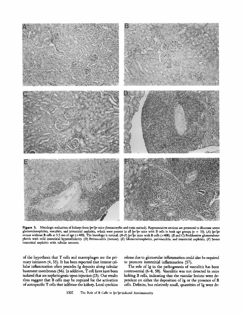
Nephritis and Vasculitis. Renal pathology was interpreted without knowledge of mouse genotypes. The mice could be readily distinguished into two groups with strikingly different morphologies: a group with normal architecture and no im- mune deposits and a group with severe glomerulonephritis, interstitial nephritis, vasculitis, and both glomerular and in- terstitial immune deposits. Representative examples are il- lustrated in Fig. 5. Details of the pathology are described in the legends. Severe nephritis always segregated with the presence of both the Ipr/lpr genotype and B cells, and this was observed for all animals in both cohorts (a total of 10 lpr/Ipr mice with B cells). By contrast, none of the six Ipr/lpr mice that lacked B cells and circulating Ig developed nephritis; their histology was indistinguishable from the four +/+ lit- termates that had B cells and with normal control CBA/J mice.
Discusaion
To further elucidate the role of B cells in MRL-lpr/lpr mice, a series ofF2 mice were created from crosses oflpr/lpr homo- zygotes and mice homozygous for a mutation 0hD) that prevents development of mature B cells. Strikingly, in the absence of B cells, lpr/Ipr mice developed neither glomerulo- nephritis, vasculitis, nor interstitial nephritis. In contrast, all of the lpr/lpr littermate mice with B cells developed severe nephritis and autoantibodies. These results indicate that B cells and/or autoantibodies play a primary role in the initia- tion of lupus nephritis in this genetic background. Severe nephritis was present in B cell-containing lpr//pr mice at 4 mo of age, a time when lymphoaccumulation was equivalent in mice lacking B cells and animals with B cells. Thus, suppres- sion of nephritis was not the result of decreased lymphoac- cumulation. For each form of nephritis observed in B cell-sufficient mice, it is formally possible that the role of B cells is as (auto)antibody-forming cell precursors, as APC in causing T cell activation, or both. However, likely roles of B cells for each form of nephritis are suggested by previous work on pathogenesis.
The requirement of B calls for induction of glomerulone- phritis substantiates previous work suggesting a role of im- mune deposits in glomerulonephritis (1, 5, 6, 16-18, 45) and indicates that immune deposit formation is likely a pivotal event in the initiation of the glomerular lesion. Furthermore, although other systemic and local cellular and cytokine per- turbations are present in these animals (53, 54), they are by themselves insufficient to induce even microscopic disease. In this regard, it will be of particular interest to evaluate cytokine levels within the glomeruli of mice lacking B ceils to determine the influence of B calls and Ig deposition on cytokine disregulation reported to occur before the onset of overt disease (54).
Absence of interstitial nephritis in Ipr/lpr mice lacking B cells is also interesting and surprising, particularly because
1301 Shlomchik et al.
Figure 5. Histologic evaluation of kidneys from Ipr/Ipr mice (hematoxylin and eosin stained). R~presentative sections are presented to illustrate severe glomerulonephritis, vasculitis, and interstitial nephritis, which were present in all lpr/lpr mice with B cells in both age groups (n z 10). (A) lpr/lpr mouse without B cells at 5.5 mo of age (x 400). The histology is normal. (B-F) Ipr/Ipr mice with B calls (x 400). (B and C) Proliferative glomerulone- phritis with mild interstitial hypercellularity. (D) Perivasculitis (intense). (E) Glomerulonephritis, perivasculitis, and interstitial nephritis. (F) Severe interstitial nephritis with tubular necrosis.
of the hypothesis that T cells and macrophages are the pri- mary initiators (4, 55). It has been reported that intense cel- lular inflammation often precedes Ig deposits along tubular basement membranes (56). In addition, T cell lines have been isolated that are nephritogenic upon injection (15). Our results thus suggest that B cells may be required for the activation of autospecific T cells that infiltrate the kidney. Local cytokine
release due to glomerular inflammation could also be required to promote interstitial inflammation (57).
The role of Ig in the pathogenesis of vasculitis has been controversial (6-8, 58). Vasculitis was not detected in mice lacking B cells, indicating that the vascular lesions were de- pendent on either the deposition of Ig or the presence of B cells. Definite, but relatively small, quantities of Ig were de-
1302 The Role of B Cells in lpr/Ipr-induced Autoimmunity
tected within the vessel walls of diseased mice (when glo- merular immune deposits were abundant; data not shown). This observation is consistent with Berden et al. (58), who found Ig within inflammed vessels but not within normal vessels of the same MRL-Ipr/Ipr mice. However, Moyer et al. (8) reported that perivascular lymphocytes and macrophages were present before vascular immune deposits; antibody depo- sition coincided with the development of necrotizing vascu- litis in older MRL-Ipr/Ipr mice. These workers emphasized the cell-mediated nature of Ipr/lpr vasculitis. However, they did find B cells in the outer zone of the inflammatory infiltrate. Cerny et al. (28) observed that anti-IgM treatment of C57BL/ 6-1pr/Ipr mice ameliorated nephritis, but did not affect the vasculitis, suggesting, in contrast to our results, that B cells/Ig are not required for the development of vasculitis. It is difficult to reconcile the differences observed between JhD mice and anti-IgM treatment. The presence of low levels of B cells, heterologous rabbit antiserum, and presumably, immune com- plems in the anti-IgM-treated animals may have been sufficient to allow for the development of vasculitis. These potential artifacts were not a problem in the JhD mice. Interestingly, previous work suggests that the role of B cells in vasculitis may differ from that in glomerulonephritis. Genetic (59) and passive autoantibody injection studies (18) have suggested that vaseulitis and glomerulonephritis can occur and be induced separately, implying that they arise by distinct mechanisms. However, further work will be required to distinguish the underlying mechanisms.
Aside from its effect on nephritis, it is interesting that a lack of B cells was associated with reduced accumulation of T cells, which was prominent in the older cohort of mice. It has been proposed by Huang et al. (60) that activation of CD4 + and/or CD8 + SP cells is required for accumula- tion of DN T cells. Based on the present data, we propose that B cell interaction is an important pathway for such acti- vation. Accumulation of both DN and SP T cells was re- duced in mice lacking B cells, suggesting that B cells either interact equally with these subsets to promote their accumu- lation or else interact with a precursor that contributes equally to these T cell subsets. T cell accumulation is not, however, completely abrogated in the absence of B cells, especially in young animals. This suggests that other cell types, perhaps dendritic cells or macrophages, are also competent to acti- vate precursors of accumulating T cells. The role of B cells in promoting further accumulation would become more ap- parent as macrophages and dendritic cells become more lim- iting amid the accumulating T cells in mice lacking B cells. The requirement for B cells to induce maximal lymphoac- cumulation further suggests that theJhs defect prevents an apoptotic event, which would normally occur only after T cell interaction (either stimulatory or tolerogenic) with an APC. Whether the Jhs deficiency is also required in the B cell in order to promote T cell accumulation could be tested via reconstitution of B cells in this model system.
In contrast to the periphery, DN T cells were dispropor- tionately overrepresented in the thymi of Ipr/Ipr mice that had B cells. This curious observation could be explained by either of two mechanisms. First, DN T cells may be gener-
1303 Shlomchik et al.
ated in the periphery and home to the thymus. The differ- ence between B cell-sufticient Ipr/lpr mice and those lacking B cells would then be attributed to a difference in the overall number of DN T cells, which in the mice lacking B cells would be two small to permit an "overflow" into the thymus. However, young lpr/lpr mice lacking B cells have as much lymphadenopathy as the B cell-sufficient mice, yet have a lower proportion of thymic DN T cells. Alternatively, B cells may directly affect the generation of some DN T cells in the thymus. This is plausible since small numbers of B cells are present in thymus (61). As in the periphery, a lack of B cells required for DN T cell expansion would limit DN T cell generation in the thymus of mice lacking B cells.
An assumption inherent in our conclusions is that the phenotypes of homozygous JhD-mutant mice are the result of the lack of B cells. An alternative interpretation is that a genetic locus linked to the JhD mutation on chromosome 12, rather than the mutation itself, is responsible for our findings. A candidate for such a locus, mapped by Watson et al. (62) to chromosome 12, modifies Ipr/lpr renal disease in the MRL background. Although this locus is linked to the Jh locus (most likely ",,40 cM away), it is unlikely that i t - ra ther than the Jh knockout itself-is responsible for the present observations. The locus described by these workers is a "modifying locus" which, along with another unlinked locus, has an effect on the "renal index" that accounts for only "~50% of the variance in renal disease. By contrast, the difference in nephritis in our studies was dramatic: disease was either severe or absent. Thus it is unlikely that this locus accounts solely for our observations. The observation that Ipr/Ipr mice with either one (i.e., JhD/+) or two (i.e., + /+) copies of the MILL-derived chromosome 12 had equally se- vere disease also supports this conclusion.
Very recently, Jevnikar et al. (63) have shown that MRL- Ipr/Ipr mice that cannot express class II MHC molecules do not get nephritis. The authors considered that nephritis was abrogated either because of blocking of autoaggressive CD4 + T cell generation within the thymus or lack of T cell activation within the kidney in the absence of class II expres- sion on renal cells. Class II knockout Ipr/Ipr mice lack CD4 + T cells, autoantibodies and MHC expression on B cells and tissues, complicating interpretation of why these mice do not get nephritis. Mice lacking B cells, on the other hand, have CD4 + T cells and can express MHC class II in tissues, yet still do not get nephritis. In light of our data, we suggest that in class II-deficient mice, suboptimal B cell activation because of lack of class II expression may also play a role in preventing nephritis in these mice. Indeed, the MRL-Ipr/Ipr class II-deficient mice lacked serum autoantibodies (63). In addition, we would predict that, after restoration of CD4 § T cells in these mice through cell transfer, disease would not ensue because of the inability to activate B cells. In any case, it is of great interest that in both class II-deficient lpr/Ipr mice and JhD/JhD Ipr/Ipr mice, B cells are directly affected by the genetic alteration, and nephritis is prevented.
Our work underscores the importance of B cells in gener- ating autoimmune-mediated tissue damage. The results in- dicate that B cells are critical for multiple components of
autoimmune-mediated inflammation, including those previ- ously thought to be primarily mediated by humoral autoim- munity (glomerulonephritis), cdlular autoimmunity (inter- stitial nephritis) or both (vasculitis). These data demonstrate that B cells could be an important target for therapy of sys- temic autoimmunity. Elimination of B cells or B cell subsets would have distinct advantages over removal of Ig alone (as in plasmapheresis). Pathogenic autoantibodies would be re-
moved more efficiently than in plasmapheresis; moreover, B cell suppression would also ameliorate immune-mediated events requiring cell-cell interaction, such as interstitial ne- phritis and vasculitis, which would be unaffected by removal of Ig alone. A better understanding of the role of B cells and antibody in inducing autoimmune pathology will be neces- sary to design appropriate B cell-directed therapy.
Much of this work was performed in the laboratory of Dr. Martin Weigert, whom we thank for con- tinuous support, encouragement, and intellectual contribution. We thank N. Reutsch and the Bosma laboratory for generously providing reagents. We thank R. R. Hardy for providing superb FACS | facili- ties and S. Shinton for expert technical help with the FACS | We appreciate the help of S. Litwin with statistics. We acknowledge the technical help of T. Witman. We thank I. N. Crispe, C. Janeway, and L. Huang for critical review of the manuscript at various stages.
This work was supported by National Institutes of Health grants GM-20964-19 to M. Weigert, George M. O'Brien Kidney and Urological Research Center grant DK-45191, DK-33694 and AI-27915 to M. P. Madaio, and 1K43-A132268-01 to D. Huszar. M. Weigert and M. P. Madaio are both recipients of a Sheryl M. Hirsch Award from the Lupus Foundation of Philadelphia. M. J. Shlomchik was a Warner- Lambert Life Sciences Research Fellow.
Address correspondence to Mark J. Shlomchik, Department of Laboratory Medicine, Yale University School of Medicine, 333 Cedar St., Box 208035, New Haven, CT 06520-8035.
Received for publication 11 April 1994 and in revised form 15June 1994.
1. Winfield, J.B., I. Faiferman, and D. I ~ e r . 1977. Avidity of anti-DNA antibodies in serum and IgG glomerular eha~ from patients with systemic lupus ery~ematosus: association of high avidity anti-native DNA antibody with glomerulonephritis. J. Clin. Invest. 59:90.
2. Couser, W.G., D.J. Salant, M.P. Madaio, S. Adler, and G.C. Groggel. 1982. Factors influencing glomerular and tubuloin- terstitial patterns of injury in SLE. A m . f Kidney Dis. 2(Suppl. 1):126.
3. Foster, M.H., B. Cizman, and M.P. Madaio. 1993. Nephrito- genic autoantibodies in systemic lupus erythematosus: im- munochemical properties, mechanisms of immune deposition, and genetic origins. La/2 Invest. 69:494.
4. Kelley, V.R., G.C. Diaz, A.M. Jevnikar, and G.G. Singer. 1993. Renal tubular epithelial and T cell interactions in autoimmune renal disease. Kidney Int. 39(Suppl.):S108.
5. Dixon, F.J., M.B.A. Oldstone, and G. Tonietti. 1971. Patho- genesis of immune complex glomerulonephritis of New 7.ealand mice. J. Exit Med. 134:65.
6. Vlahakos, D.V., M.H. Foster, S. Adams, M. Katz, A.A. Ucci, K.J. Barrett, S.K. Datta, and M.P. Madaio. 1992. Anti-DNA antibodies form immune deposits at distinct glomemlar and vascular sites. Kidney Int. 41:1690.
7. Jabs, D.A., and R.A. Prendergast. 1987. Reactive lymphocytes in lacrimal gland and vasculitic renal lesions of autoimmune MRL/Ipr mice express L3T4. f Exit Med. 166:1198.
8. Moyer, C.F., J.D. Strandberg, and C.L. Reinisch. 1987. Sys- temic mononuclear-cell vasculitis in MRL/Mp-lpr/lpr mice.
A histologic and immunocytochemical analysis. Ara.J. Pathol. 127:229.
9. Katagiri, T., P.L. Cohen, and R.A. Eisenberg. 1988. The lpr gene causes an intrinsic T cell abnormality that is required for hyperproliferation. J. Exit bled. 167:741.
10. Tautog, J.D., E.S. Raveche, P.A. Smathers, L.H. Glimcher, D.P. Huston, C.T. Hansen, and A.D. Steinberg. 1981. T cell abnor- malities in NZB mice occur independently of autoantibody production, f Exit Med. 153:221.
11. Steinberg, A.D., J.B.R.oths, E.D. Murphy, R.T. Steinberg, and E.S. Raveche. 1980. Effects of thymectomy or androgen ad- ministration upon the autoimmune disease of MRL/Mp-lpr/Ipr mice. f Iramunol. 125:871.
12. Wofsky, D., J.A. Ledbetter, P.L. Hendler, and W.E. Seaman. 1985. Treatment of murine lupus with monoclonal anti-T cell antibody, f Iramunol. 134:852.
13. Santoro, T.J., J.p. Portanova, and B.L. Kotzin. 1988. The con- tribution of L3T4 + T cells to lymphoproliferation and au- toantibody production in MRL-lpr/lpr mice. J. Exit Med. 167:1713.
14. Rajagopalan, S., T. Zordan, G.C. Tsokos, and S.K. Datta. 1990. Pathogenic anti-DNA autoantibody-inducing T helper cell lines from patients with active lupus nephritis: isolation of CD4- 8- T helper cell lines that express the 3'/~ T-cell antigen receptor. Proa Natl. Acad. Sci. USA. 87:7020.
15. Gallo, C.D., A.M. Jevnikar, D.C. Brennan, S. Florqnin, S.A. pacheco, and V.K. Kelley. 1992. Autoreactive kidney-infiltrating T-cell clones in murine lupus nephritis. Kidney Int. 42:851.
1304 The Role of B Cells in Ipr/lpr-induced Autoimmunity
16. Dang, H., and K.J. Harbeck. 1984. The in vivo and in vitro glomerular deposition of isolated anti-double-stranded-DNA antibodies in NZB/W mice. Clin. ImmunoL ImmunoI~thoL30:265.
17. Pankewycz, O.G., P. Migliorini, and M. Madaio. 1987. Foly- reactive autoantibodies are nephritogenic in murine lupus nephritis, f lmmunol. 139:3287.
18. Raininger, L., T. Berney, T. Shibata, F. Spertini, Ik. Merino, and S. Izui. 1990. Cryoglobulinemia induced by a mutine IgG3 rheumatoid factor: skin vasculitis and glomerulonephritis arise from distinct pathogenic mechanisms. Pro~ Natl. Acad. Sci. USA. 87:10038.
19. Berney, T., T. Fulpius, T. Shibata, L. Reininger, J. Van Snick, H. Shall, M. Weigert, A. Marshak-Rothstein, and S. Izui. 1992. Selective pathogenicity of murine rheumatoid factors of the cryoprecipitable IgG3 subchss. Int. Immunol. 4:93.
20. Lin, R..-H., M.J. Mamula, J.A. Hardin, and C.A. Janeway, Jr. 1991. Induction of autoreactive B cells allows priming of autoreactive T cells, f Ext~ Med. 173:1433.
21. Mamuh, M.J., K.-H. Lin, C.A. Janeway, Jr., andJ.A. Hardin. 1992. Breaking T cell tolerance with foreign and self co-im- munogens: a study of autoimmune B and T cell epitopes of cytochrome c. f lramunol. 149:789.
22. Herron, L.K., ILL. Coffman, and B.L. Kotzin. 1988. Enhanced response of autoantibody-secreting B cells from young NZB/ NZW mice to T-cell-derived differentiation signals. Clin. Im- munol. Immunopathol. 46:314.
23. Klinman, D.M., and A.D. Steinberg. 1986. Proliferation of anti-DNA producing NZB B cells in a non-antoimmune envi- ronment. J. Immunol. 137:69.
24. Sobel, E.S., T. Katagiri, K. Katagiri, S.C. Morris, EL. Cohen, and R.A. Eisenberg. 1991. An intrinsic B cell defect is required for the production of autoantibodies in the lpr model of mu- line systemic autoimmunity. J. Extx Med. 173:1441.
25. Nemazee, D., C. Guiet, K. Buerki, and A. Marshak-Rothstein. 1991. B lymphocytes from the autoimmune-prone mouse strain MLP/lpr manifest an intrinsic defect in tetraparental MRL/lpr DBA/2 chimeras. J. Immunol. 147:2536.
26. Raveche, E.S., E.A. Novotny, C.T. Hansen, J.H. Tjio, and A.D. Steinberg. 1981. Genetic studies in NZB mice, V. Recombinant inbred lines demonstrate that separate genes control autoim- mune phenotype. J. Exlx Med. 153:1187.
27. Eastcott, J.W., K.S. Schwartz, and S.K. Datta. 1983. Genetic analysis of the inheritance of B cell hyperactivity in relation to the development of autoantibodies and glomerulonephritis in NZB x SWK crosses, f Imrnunol. 131:2232.
28. Cerny, A., M. Kimoto, A.W. Hugin, R. Merino, and S. Izui. 1989. Anti-IgM treatment of C57BL/6-1pr/lpr mice: deple- tion of B cells reduces lpr gene-induced lymphoproliferation and mononuclear cell vasculitis. Clin. ExF Iraraunol. 77:124.
29. Andrews, B.S., R.A. Eisenberg, A.N. Theofilopoulos, S. Izui, C.B. Wilson, p.J. McConahey, E.D. Murphy, J.R R_oths, and F.J. Dixon. 1978. Spontaneous murine lupus-like syndromes. Clinical and immunopathological manifestations in several strains, f ExF Med. 148:1198.
30. Izui, S., V.E. Kelley, K. Masuda, H. Yoshida, J.B. R_oths, and E.D. Murphy. 1984. Induction of various autoantibodies by mutant gene Iprin several strains ofmice.f Iraraunol. 133:227.
31. Wofsky, D., K.R. Hardy, and W. Seaman. 1984. The prolifer- ating cells in autoimmune MRL/lpr mice lack L3T4, an an- tigen on "helper" T ceils that is involved in the response to class II major histocompatibility antigens.J. ImmunoL 132:2686.
and H.C.I. Morse. 1986. Phenotypic, functional, and molec- ular genetic comparisons of the abnormal lymphoid cells of C3H-lpr/lpr and C3H-gld/gld mice. f lrnmunol. 136:4075.
33. Watanabe-Fukunaga, K., C.I. Brannan, N.G. Copeland, N.A. Jenkins, and S. Nagata. 1992. Lymphoprolifaration disorder in mice explained by defects of Fas antigen that mediates apop- toffs. Nature (Lond.). 356:314.
34. Itoh, N., S. Yonehara, A. Ishii, M. Yonehara, S.-I. Mizushima, A. Hase, Y. Seto, and S. Nagata. 1991. The polypeptide en- coded by the cDNA for human cell surface antigen fas can mediate apoptosis. Cell. 66:233.
35. Watanabe-Fukunaga, K., C.I. Brannan, N. Itoh, S. Yonehara, N.G. Copeland, N.A. Jenkins, and S. Nagata. 1992. The cDNA structure, expression, and chromosomal assignment of the mouse Fas antigen. J. lmmunol. 148:1274.
36. Drappa, J., N. Brot, and K.B. Elkon. 1993. The Fas protein is expressed at high levels on CD4 + CD8 § thymocytes and activated mouse mature lymphocytes in normal mice but not in the lupus-prone strain, MILL lpr/lpr. Proc. Natl. Acad. Sci. USA. 90:10340.
37. Chen, J., M. Trounstine, F.W. Alt, F. Young, C. Kurahara, J.F. Loring, and D. Huszar. 1993. Immunoglobulin gene rear- rangement in B cell deficient mice generated by targeted dele- tion of the JH locus. Int. Immunol. 5:647.
38. Shlomchik, M.J., D. Zharhary, S. Camper, T. Saunders, and M. Weigert. 1993. A rheumatoid factor transgenic mouse model of autoantibody regulation. Int. Imrnunol. 5:1329.
39. Hardy, R.R. 1986. Purification and coupling of fluorescent proteins for use in flow cytometry. In Handbook of Experi- mental Immunology. D.M. Weir, editor. Blackwell Scientific Publishers, Edinburgh. 31.
40. Kincade, P.W., G. Lee, and T. Watnabe. 1981. Monoclonal rat antibodies to murine IgM determinants.f Imraunol. Methods. 42:17.
41. Coffman, R.L., and I.L. Weissman. 1981. A monoclonal anti- body that recognizes B cells and B cell precursors in mice. J. Exlx Med. 153:269.
42. Ledbetter, J.A., and L.A. Herzenberg. 1979. Xenogeneic mono- clonal antibodies to mouse lymphoid differentiation antigens. Immunol. R~. 47:63.
43. Dialynas, D.P., Z.S. Quan, K.A. WaLl, A. Pierres, J. Quintans, M.R. Loken, M.R. Pierres, and F.W. Fitch. 1983. Character- ization of the mutine T cell surface molecule, designated L3T4, identified by monoclonal antibody GK1.5.J. Iramunol. 131:2445.
44. Vlahakos, D., M.H. Foster, A.A. Ucci, K.J. Barrett, S.K. Datta, and M.P. Madaio. 1992. Murine monodonal anti-DNA anti- bodies penetrate cells, bind to nuclei, and induce glomerular proliferation and proteinuria in vivo.J. Am. So~ Nephrol. 2:1345.
45. Madaio, M.P., J. Carlson, J. Cataldo, A. Ucci, P. Migliorini, and O. Pankewycz. 1987. Murine monoclonal anti-DNA an- tibodies bind directly to glomerular antigens and form immune deposits, f Irnraunol. 138:2883.
46. Austin, H.A.I., L.K. Muenz, K.M. Joyce, P.T. Antonovych, and J.E. Balow. 1984. Diffuse proliferative hpus nephritis: identification of specific pathologic features affecting renal out- come. Kidne 7 Int. 25:689.
47. Run, Y., andJ. Sprent. 1987. T cell priming in vivo: a major role for B cells in presenting antigen to T cells in lymph nodes. f Immunol. 138:2848.
48. Janeway, C.A., Jr., J. Ron, and M.E. Katz. 1987. The B cell is the initiating antigen-presenting cell in peripheral lymph nodes, f Imraunol. 138:1051.
1305 Shlomchik et al.
49. Budd, L.C., M. Schreyer, G.C. Miescher, and H.R. MacDonald. 1987. T cell lineages in the thymus of lpr/Ipr mice: evidence for parallel pathways of normal and abnormal T cell develop- ment. J. lmmunol. 139:2200.
50. Matsuzaki, Y., C. Pannetien, O. Kanagawa, G. Gachelin, and H. Nakanchi. 1992. Evidence for the existence of two parallel differentiation pathways in the thymus of MRL lpr/lpr mice. j. lmmunol. 149:1069.
51. Zhou, T., H. Bluethmann, J. Eldridge, K. Berry, and J.D. Mountz. 1993. Origin of CD4- CDS- B220" T cells in MR.blpr/Ipr mice. Clues from a T cell receptor beta trans- genic mouse. J. Immunol. 150:3651.
52. Shlomchik, M.J., A. Marshak-Rothstein, C.B. Wolfowicz, T.L. Rothstein, and M.G. Weigert. 1987. The role of clonal selec- tion and somatic mutation in autoimmunity. Nature (Lond.). 328:805.
53. Levine, J., D. Hartwell, and D.I. Belier. 1991. Imbalanced cytokine production by macrophages from autoimmune-prone mice. lmraunol. Lett. 30:183.
54. Bloom, R.D., S. Florquin, G.G. Singer, D.C. Brennan, and V.R. Kelley. 1993. Colony stimulating factor-1 in the induc- tion of lupus nephritis. Kidney Int. 43:1000.
55. Kelley, V.R., R.D. Bloom, M.A. Yui, C. Martin, and D. Price. 1994. Pivotal role of colony stimulating factor-1 in lupus nephritis. Kidney Int. 45(Suppl.):S83.
56. Hatloran, P.F., J. Urmson, V. Ramassar, C. Laskin, and P. Au- tenried. 1988. Increased class I and class II MHC products and
mRNA in kidneys of MR.blpr/lpr mice during autoimmune nephritis and inhibition by cyclosporine.J. Immunol. 141:2303.
57. Yee, J., G.S. Kuncio, and E.G. Neilson. 1991. Tubulointersti- tial nephritis following glomerulonephritis. Semin. Nephrol. 11:361.
58. Berden, J.H., L. Hang, P.J. McConahey, and F.J. Dixon. 1983. Analysis of vascular lesions in routine SLE. I. Association with serologic abnormalities. J. Iramunol. 130:1699.
59. Nose, M., M. Nishimura, and M. Kyogoku. 1989. Analysis of granulomatous arteritis in MRL/Mp autoimmune disease mice bearing lymphoproliferative genes. Am.J. Pathol. 135:271.
60. Huang, L., K. Sye, and I.N. Crispe. 1994. Proliferation and apoptosis of B220 + CD4- CD8- TCKci/3 ;"<~'~ T ceils in the liver of normal adult mice: implication for lpr pathogen- esis. Int. Immunol. 6:533.
61. Miyama-Inaba, M., S.-I. Kuma, K. Inaba, H. Ogata, H. Iwai, K. Uasumizu, S. Muramatsu, R.M. Steinman, and S. Ikehara. 1988. Unusual phenotype of B cells in the thymus of normal mice. J. Exp. Med. 168:811.
62. Watson, M.L., J.K. Rao, G.S. Gilkeson, P. Ruiz, E.M. Eicher, D.S. Pisetsky, A. Matsuzawa, J.M. Rochelle, and M.F. Seldin. 1992. Genetic analysis of MLK-lpr mice: relationship of the Fas apoptosis gene to disease manifestations and renal dis- ease-modifying loci. J. Ext~ Med. 176:1645.
63. Jevnikar, A.M., M.J. Grusby, and L.H. Glimcher. 1994. Preven- tion of nephritis in major histocompatibility complex class II-deficient MRDIpr mice. J. Ext~ Med. 179:1137.
1306 The Role of B Cells in lpr/lpr-induced Autoimmunity